
geal anatomy with the acoustic structures of register of ...
21
SYRINGEAL MECHANICS REASSESSED: EVIDENCE FROM STREPTOPELIA ASBOT S. GAUNT, SANDRA L. L. GAUNT, AND RICHARDM. CASV.¾ • Department of Zoology, 1735 Neil Avenue, The Ohio StateUniversity, Columbus, Ohio 43210 USA ABSTi•ACT.--Avian phonations are generally reportedto consist of an harmonicseries or sounds with no overtones (pure tones). Neither of these kinds of sounds is produced by freely oscillating, edge-clamped membranes suchas the tympanic membranes of a syrinx. A possible alternative mechanism, at leastfor pure tones,is a whistle, i.e. a regular per- turbationin the flowing air column, possibly interacting with the membranes. The cooof a Ring Dove is sucha whistledsong.It shows strong modulation of amplitude(AM) but not of frequency (FM). We have examined air flow, internal pressures, and electrical activityin syringeal and body-wallmuscles during phonation by Ring Doves.Flow patterns and tracheal pressures showthat the nares are closed duringcooing; expired air is forced into the inflatable esoph- agus. This inflation appears to be of little acoustic significance. Pulsatile activity in the abdominal muscles instillsa strong AM that confers a warbledquality to part of the coo. More subtle AM is effectedby oscillations of the lateral tympanic membranes. Beat fre- quencies also produce AM-like patterns in the wave envelope. Contraction of the syringeal muscles shapes the syrinxinto a vocalconfiguration but appears to have little modulatory effect. The structure of a dove's syrinx suggests that whistles could be made by any of several techniques that differ in the interactions among syringeal components. Received 22 May 1981, accepted 18 November 1981. THE year 1968 provided two major attempts to assess syringealfunction by linking syrin- geal anatomywith the acoustic structures of avian phonations. Stein analogized the avian vocal system with a radio transmitter, while Greenewalt provided an extensive analysis based on detailed and meticulous, electronic dissections of a wide variety of calls and songs. Together, these studies formed the Greene- walt-Stein model of syringeal action. That model, now much elaborated, remains the ba- sis of our present understanding. In its entirety, the model is extensive,intri- cate,and subtle.Of its many arguments, those pertinent to this paper include: 1. Sound is producedby the vibration of sy- ringeal membranes; exactly which mem- brane(s) depends on the nature of the syr- inx. 2. Neither amplitude(AM) nor frequency (FM) is modulated primarily by resonance phe- • Present address: 29 Highway 52, Erie, Colorado 80516 USA. nomena of the kind exploitedby the human voice and various wind instruments. Tra- cheal resonances may help determine the register of some calls of some species (Myers 1917, Sutherland and McChesney 1965, Hersh 1966) but are not used for active mod- ulation. (Throughout this paper the term "modulation" refers to a change, although, strictly speaking, even a constant tone is modulated by whatever phenomena deter- mine its parameters.) With few exceptions, modulations are source generated. To some extentthe valid- ity of this argument dependson what is meant by "source." Greenewalt evidently wished to restrict the term to the vibrating membranes. We prefer to considerthe syr- inx in its entirety as the source. Modula- tions are effecteddirectly by changingthe position and/or tension of the membranes or indirectly by the interactionof membrane oscillationwith other syringeal structures. Hence, modulation derives from adjust- ments of syringeal configuration, presum- ably engenderedby the action of the sy- ringeal muscles. Much literature implies 474 The Auk 99:474-494. July1982
Transcript of geal anatomy with the acoustic structures of register of ...

SYRINGEAL MECHANICS REASSESSED: EVIDENCE FROM STREPTOPELIA
ASBOT S. GAUNT, SANDRA L. L. GAUNT, AND RICHARD M. CASV.¾ • Department of Zoology, 1735 Neil Avenue, The Ohio State University,
Columbus, Ohio 43210 USA
ABSTi•ACT.--Avian phonations are generally reported to consist of an harmonic series or sounds with no overtones (pure tones). Neither of these kinds of sounds is produced by freely oscillating, edge-clamped membranes such as the tympanic membranes of a syrinx. A possible alternative mechanism, at least for pure tones, is a whistle, i.e. a regular per- turbation in the flowing air column, possibly interacting with the membranes. The coo of a Ring Dove is such a whistled song. It shows strong modulation of amplitude (AM) but not of frequency (FM).
We have examined air flow, internal pressures, and electrical activity in syringeal and body-wall muscles during phonation by Ring Doves. Flow patterns and tracheal pressures show that the nares are closed during cooing; expired air is forced into the inflatable esoph- agus. This inflation appears to be of little acoustic significance. Pulsatile activity in the abdominal muscles instills a strong AM that confers a warbled quality to part of the coo. More subtle AM is effected by oscillations of the lateral tympanic membranes. Beat fre- quencies also produce AM-like patterns in the wave envelope. Contraction of the syringeal muscles shapes the syrinx into a vocal configuration but appears to have little modulatory effect. The structure of a dove's syrinx suggests that whistles could be made by any of several techniques that differ in the interactions among syringeal components. Received 22 May 1981, accepted 18 November 1981.
THE year 1968 provided two major attempts to assess syringeal function by linking syrin- geal anatomy with the acoustic structures of avian phonations. Stein analogized the avian vocal system with a radio transmitter, while Greenewalt provided an extensive analysis based on detailed and meticulous, electronic
dissections of a wide variety of calls and songs. Together, these studies formed the Greene- walt-Stein model of syringeal action. That model, now much elaborated, remains the ba- sis of our present understanding.
In its entirety, the model is extensive, intri- cate, and subtle. Of its many arguments, those pertinent to this paper include:
1. Sound is produced by the vibration of sy- ringeal membranes; exactly which mem- brane(s) depends on the nature of the syr- inx.
2. Neither amplitude (AM) nor frequency (FM) is modulated primarily by resonance phe-
• Present address: 29 Highway 52, Erie, Colorado 80516 USA.
nomena of the kind exploited by the human voice and various wind instruments. Tra-
cheal resonances may help determine the register of some calls of some species (Myers 1917, Sutherland and McChesney 1965, Hersh 1966) but are not used for active mod- ulation. (Throughout this paper the term "modulation" refers to a change, although, strictly speaking, even a constant tone is modulated by whatever phenomena deter- mine its parameters.) With few exceptions, modulations are source generated. To some extent the valid- ity of this argument depends on what is meant by "source." Greenewalt evidently wished to restrict the term to the vibrating membranes. We prefer to consider the syr- inx in its entirety as the source. Modula- tions are effected directly by changing the position and/or tension of the membranes or indirectly by the interaction of membrane oscillation with other syringeal structures. Hence, modulation derives from adjust- ments of syringeal configuration, presum- ably engendered by the action of the sy- ringeal muscles. Much literature implies
474 The Auk 99: 474-494. July 1982

JvL¾ 1982] Syringeal Mechanics 475
that such changes are produced solely by the activity of the syringeal musculature. As Stein emphasized, however, a carrier fre- quency could be modulated by an oscilla- tion of any of several components of the vo- cal system. Those oscillators could be driven by the same air flow responsible for the vi- bration of the vocal membranes but might be independent of that vibration.
4. Frequency and amplitude are often linked, directly at lower frequencies, inversely in the higher range.
Most studies since 1968 have confirmed the
major elements of this model while extending or elaborating details (see Dtirrwang 1974 for an extensive review). A series of experiments in the last decade, however, suggests need of at least one, major revision. Electromyographic studies of phonation in chickens (Youngren et al. 1974, Peek et al. 1975, Gaunt and Gaunt 1977) and in ducks (Lockner and Youngren 1976) indicate that the musculature of the ab- dominal wall is responsible for major changes in loudness (related to amplitude). Further, the interactions of syringeal muscles with each other and with the abdominal muscles may be far more complex than had been previously supposed.
Ducks and chickens show no essential dif-
ferences in the patterns of muscular activity. We, therefore, assume that the sequence of electromyographic events established in those studies may fairly be taken as representing a basic pattern, and we may now address another series of questions. Among these is whether or not procedural differences might be discovered between forms with limited and
forms with varied repertoires. A comparison of the Columbiformes with the Psittaciformes
is ideal for such a question. These orders are widely regarded as closely related (e.g. Sibley and Ahlquist 1972). Both have extremely sim- ple syringes that have been well described, with models of their actions postulated (War- ner 1972, Nottebohm 1976). Doves are noto- rious for their limited, stereotyped repertoires; parrots are famed for the variety of their calls and their ability to mimic. The calls of doves are essentially musical; those of parrots may be either musical or harsh. We had intended that
the investigation of syringeal mechanisms in Streptopelia reported here would serve simply
as the first step in a comparative study, but the results of our studies have led us further, to a reconsideration of the entire mechanism of
syringeal action.
METHODS AtqV MATERIAI, S
Birds.--Our experimental animals were the do- mestic Streptopelia risoria. Birds were from colonies maintained by the Department of Zoology at The Ohio State University and the Conservatory Aviary, Pittsburgh. We made no attempt to select birds of a given age or sex, but chose the noisiest in the avail- able pool. Hence, most of the birds used were males, but a few aggressive females were included (either sex will call when isolated).
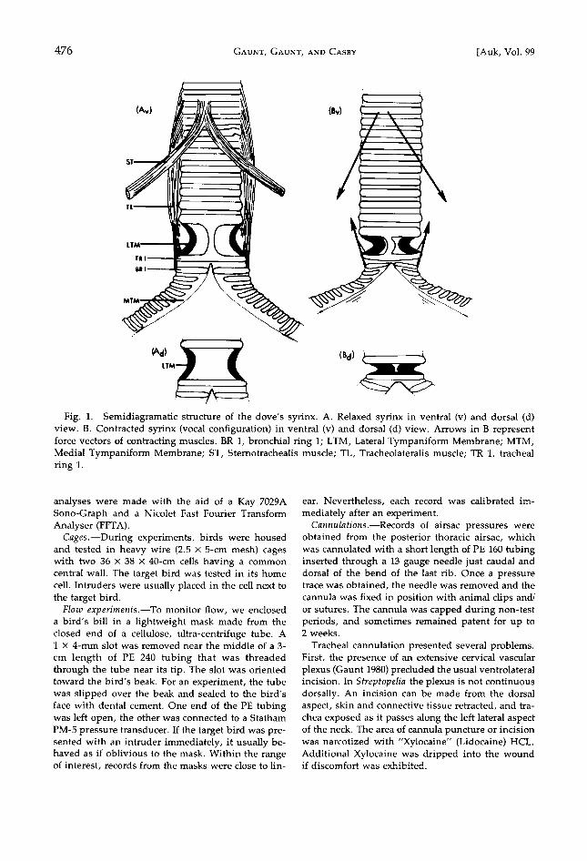
Anatomy.--The syrinx of doves has been described in great detail elsewhere (Warner 1972). We present here only those facts necessary to understand our discussion.
A dove's syrinx has two sets of membranes (Fig. 1A). The heavy, connective tissue, Lateral Tympanic Membranes (LTM; terminology after King 1979), are located in the lateral walls of the trachea immediately cranial to the tracheobronchial junction. The ex- tremely thin Medial Tympanic Membranes (MTM) are located in the cranio-medial walls of the bronchi.
The syrinx lacks a pessulus. The configuration of the syrinx is determined by
the action of two pairs of muscles. The tracheolater- alis (TL) extends along the side of the trachea from just caudal of the interclavicular membrane (tracheal ring 13) to its tendinous insertion directly onto the LTM. Our dissections revealed that some tendi-
nous slips extend across the LTM's onto the first tra- cheal bar.
The sternotrachealis muscles (ST) originate from the medial surfaces of the sternum, but, unlike the condition in most other birds, in doves they do not insert separately on the lateral aspect of the trachea. Rather, they insert together, slightly to the right of the ventral midline at about tracheal rings 10 and 11.
Sound analysis.--Sound was analyzed with both oscillographic and spectrographic techniques. The wave form was observed directly with and, if de- sired, photographed from a storage oscilloscope (Tektronix 5111). Some calls were replayed from magnetic tapes at speeds slow enough to drop their frequencies into the response range of a pen-and-ink chart recorder (Gould 2400). Such slowed paper rec- ords did not differ significantly from oscilloscope tracings of the same call taken in real time with a fast beam sweep-speed. These displays are referred to herein as oscillographic. Most records were made at either 9.7 or 19 cm/s. From consistent performers, records were made at 38, 76, and 152 cm/s. Playback was possible at 2.5, 9.7, and 19 cm/s. Spectrographic
476 GAUNT, GAVNT, ANY CASEY [Auk, Vol. 99
TL
Fig. 1. Semidiagramatic structure of the dove's syrinx. A. Relaxed syrinx in ventral (v) and dorsal (d) view. B. Contracted syrinx (vocal configuration) in ventral (v) and dorsal (d) view. Arrows in B represent force vectors of contracting muscles. BR 1, bronchial ring 1; LTM, Lateral Tympaniform Membrane; MTM, Medial Tympaniform Membrane; ST, Sternotrachealis muscle; TL, Tracheolateralis muscle; TR 1, tracheal ring 1.
analyses were made with the aid of a Kay 7029A Sono~Graph and a Nicolet Fast Fourier Transform Analyser (FFTA).
Cages.--During experiments, birds were housed and tested in heavy wire (2.5 x 5-cm mesh) cages with two 36 x 38 x 40-cm cells having a common central wall. The target bird was tested in its home cell. Intruders were usually placed in the cell next to the target bird.
Flow experirnents.--To monitor flow, we enclosed a bird's bill in a lightweight mask made from the closed end of a cellulose, ultra-centrifuge tube. A 1 x 4-mm slot was removed near the middle of a 3-
cm length of PE 240 tubing that was threaded through the tube near its tip. The slot was oriented toward the bird's beak. For an experiment, the tube was slipped over the beak and sealed to the bird's face with dental cement. One end of the PE tubing was left open, the other was connected to a Statham PM-5 pressure transducer. If the target bird was pre- sented with an intruder immediately, it usually be- haved as if oblivious to the mask. Within the range of interest, records from the masks were close to lin-
ear. Nevertheless, each record was calibrated im- mediately after an experiment.
Cannulations.--gecords of airsac pressures were obtained from the posterior thoracic airsac, which was cannulated with a short length of PE 160 tubing inserted through a 13 gauge needle just caudal and dorsal of the bend of the last rib. Once a pressure trace was obtained, the needle was removed and the cannula was fixed in position with animal clips and/ or sutures. The cannula was capped during non-test periods, and sometimes remained patent for up to 2 weeks.
Tracheal cannulation presented several problems. First, the presence of an extensive cervical vascular plexus (Gaunt 1980) precluded the usual ventrolateral incision. In Streptopelia the plexus is not continuous dorsally. An incision can be made from the dorsal aspect, skin and connective tissue retracted, and tra- chea exposed as it passes along the left lateral aspect of the neck. The area of cannula puncture or incision was narcotized with "Xylocaine" (Lidocaine) HCL. Additional Xylocaine was dripped into the wound if discomfort was exhibited.

Juicy 1982] Syringeal Mechanics 477
Second, our usual tracheal cannulation, which scarcely troubled several species of medium-sized oscines, induced severe respiratory distress in doves. We therefore adopted a new cannula consist- ing of a short length of PE 90 or 100 with a flange melted onto one end. This cannula was implanted by fitting the cut end over a hypodermic needle that had been threaded through the trachea beginning as far craniad and ending as far caudad as possible. Needle and cannula were drawn back through the trachea, the flanged end buttoned through the caudal hole and then drawn up against the inner wall of the trachea at the cranial hole. The cannula was then
locked in place by forcing a cuff of PE, with an ID the same as the cannula's OD, down the cannula until the tracheal wall was pinched between the cuff and flange. The trachea was then allowed to relax to its normal position, the incision closed, and the can- nula cut short (about 5 cm). Such a cannula does not measure velocity head, as did our previous models, but this potential discrepancy later proved irrele- vant. Because of the difficulty of cannulation, only 8 of 30 birds provided useful records of pressure or flow, even though several attempts were made with some birds.
When recording, the cannulae were connected to a pressure transducer by 110 cm of PE 205. Tension relief was provided by clipping the leads to tail feathers or to a harness fit under the wings and over the back. Distortions from impedance were mini- mized by keeping small bore portions of the can- nulation system as short as possible. Tests with our cannulae showed that pressure events reached the transducer within 3 ms of occurrence with no sig- nificant distortion. The delay is the same as that for sound to reach the microphone. When recording from two sources, the impedances of the two can- nulae were matched as closely as possible.
Electromyograrns.--Most electrodes were modifi- cations of the permanently implantable type devised by Basmajian and Stecko (1962), but we used Beck- man miniature skin electrodes for several experi- ments with abdominal muscles. Implanted elec- trodes used for recording from abdominal muscles were of Jelliff Alloy C (OD 0.12 mm) and were in- serted singly into different areas of the abdominal musculature and tested in various combinations. The
most successful were pairs inserted caudal of the last rib and just cranial of the ilium.
Electrodes for syringeal musculature were of Eva- nohm "Blue Poly" (OD 0.02 mm). The two leads were twisted together and implanted with glass needles drawn to an ID just sufficient to permit passage of the wires without damaging insulation. Insertion of these electrodes required deep surgery, before which birds were withdrawn from food for 12-18 h. The
syrinx was reached by making a ventral incision be- ginning caudal of the vascular plexus and well lateral
of the midline and ending at the tip of the furculum. The crop was separated from skin and pectoral mus- culature by blunt dissection and reflected to the left, thus exposing the interclavicular membrane. This was slit to expose the syrinx for the implantation of electrodes. Reposition of the crop effectively sealed the interclavicular opening.
For surgery into the interclavicular airsac, 0.3 ml of the general anesthetic "Chloropent" (Fort Dodge Laboratories) was administered intramuscularly. This was sufficient to render the bird unconscious
for about 90 min; most operations took about 3045 min to complete.
Both abdominal and syringeal electrodes, as well as a ground wire inserted into the dorsal muscula- ture, were passed subdermally to a connector box held on the back by a harness around the wings. Usually two sets of muscles were tested simulta- neously. For most tests, the use of only a ground wire provided sufficiently clear signals, but we tried to obtain at least one set of records from each set of
electrodes when the bird was in a closed Faraday cage. EMG's were obtained from 10 of 17 birds ex- amined. Five individuals provided abdominal rec- ords; eight provided syringeal records. Although birds were usually used in several experiments, we were unable to obtain a complete, simultaneous set of EMG's from any.
EMG signals were conditioned with Honeywell EEG/EMG preamplifiers and filtered for a band pass of 120-500 Hz. All signals were recorded on a Hon- eywell 5000C magnetic tape recorder. A few experi- ments were conducted using miniature EMG radio- transmitters (Midgard/Transkinetics MXM 100A). Signals could be displayed on either the storage os- cilloscope or chart recorder.
RESULTS
Vocalizations.--Calls of the genus Streptope- lia have been variously described and catego- rized. Most distinctions, however, depend on behavior associated with the calls (Miller and Miller 1958, Nottebohm and Nottebohm 1971). All vocalizations can be reduced to two basic
types, challenge calls (whinnies) and coos. Our discussion will treat primarily with the latter.
Whinnies are high-excitement calls that may be uttered in either agonistic or sexual con- texts. They consist of a short burst of staccato notes with little variation among individuals (Figs. 4, 8). The notes may be uttered so rapidly that they blend into a continuous but pulsed sound.
A coo of S. risoria consists of an introductory note followed by a long portion that may be

478 GAUNT, GAUNT, AND CASEY [Auk, Vol. 99
Fig. 2. Oscillographs of simple (D46) and complex (D16• and D162) cooing patterns. 1, introductory hoot; 2, warbled phrase; 3, continuous phrase. D46 showed little variation among calls. The two calls of D16 are sequential. The only difference discernable to our ears is the double hoot of the second call. Arrows indicate beats (see text). Time bar: 0.1 s.
variously accented depending on the individ- ual (Fig. 2). The first half of the long portion usually has a distinct rolling quality and is herein called the warbled phrase. The warbled and final, continuous portion together are termed the prolonged phrase. The pattern de- scribed below is for one of the simplest coos we encountered, yet it contains all of the sa- lient features.
The coo has no harmonics and little frequen- cy modulation (Fig. 3), but frequency often does rise and fall slightly during the prolonged phrase or drops slightly toward the end of the call. All birds show considerable fluctuation in
"instant" wave lengths within a restricted range, but there is no clear linkage between the degree or direction of AM and the degree or direction of FM. Average frequency gener- ally falls between 450 and 650 Hz.
In contrast to the constancy in frequency, the wave envelope is subject to a severe AM of at least two types. The first of these occurs only during the warbled phrase and has two char- acteristic features. First, it can totally interrupt
sound for periods of several milliseconds, thereby dividing the wave train into a series of distinct bursts; second, the modulation re-
sembles a forced oscillation in which both pe- riod and amplitude increase in each cycle.
The second AM is also cyclic. Amplitude in each cycle waxes and then wanes but rarely diminishes to zero. The periods of the cycles are quite variable, and even adjacent cycles differ. Each cycle forms the sound envelope into an ellipse, and the entire call can be viewed as a string of such cycles. The initial hoot may be a single cycle, or it, too, may be modulated. In some portions of the continuous phrase the ellipses are quite distinct, with minima reaching or approaching zero; in oth- ers, the envelope shows only a mild ripple.
In structurally more complex calls, the sec- ond mechanism is more prominent. There is a greater variety of cycle periods, the cycles have fewer interruptions in the warbled phrase (i.e. fewer periods of silence), the initial hoot may be modulated, and more of the minima achieve zero amplitude. In all calls, the second

JULY 1982] Syringeal Mechanics 479
' :55dB
TIME (sec)
I o s2;s
ENERGY
0 A 0.5 •
:49dB
0.5775
I
0.5100 j
t
i 2
FREOUENCY (l•Hz)
Fig. 3. Spectro•nms of ccos. The sono•mm and its power spectrum (A) show the commonly encountered separation of coos into two tones, one of constant frequency, the other rising and falling. The power spectrum is a section taken through the sonogram directly beneath the power spectrum's base. The sonogram has been compressed, but the removed section contained no evidence of energy in either the spectrogram or power spectrum. The fast Fourier transform analysis (B), which is like a very precise power spectrum and is taken from the same portion of the coo as the power spectrum in ^, again shows two peaks at 510 and 577.5 Hz. The major peaks are relatively constant in amplitude and occur throughout the duel-tone zones: the very high peak at 527.5 Hz is variable in occurrence and amplitude. The high amplitude shown here is a peculiarity of this specific section and coo. The low peaks at the extreme left (*) are a 60-Hz hum and its harmonics. The amplitude axis of the FFT^ is an arbitrary linear scale. The relative difference between the highest peak and base is about 55 dB. Stated frequencies are accurate to + 1.25 Hz.
form of AM is rarely perceived as such by the human ear, but it does markedly increase the timbre. When both types of modulation are fully developed, as near the end of the warbled portions, the sound acquires a distinctly liquid quality.
Published, narrow-band sonograms of Ring
Dove coos often show energy concentrated in a single, though sometimes wide, band 0Vliller and Miller 1958). We, too, have obtained such results, but most of our sonograms are more complex (Fig. 3). The prolonged portion usu- ally resolves into two bands. The higher one has constant frequency; the other rises to the
480 GAo•T, GAo•T, •r) C•sE¾ [Auk, Vol. 99
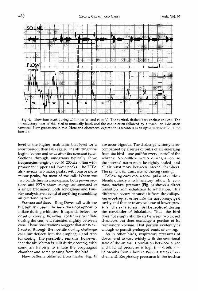
Fig. 4. Flow into mask during whinnies (w) and coos (c). The vertical, dashed bars enclose one coo. The introductory hoot of this bird is unusually loud, and the coo is often followed by a "wah" on inhalation (arrows). Flow gradations in ml/s. Here and elsewhere, expiration is recorded as an upward deflection. Time bar: 1 s.
level of the higher, maintains that level for a short period, then falls again. The shifting tone begins before and ends after the constant tone. Sections through sonograms typically show frequencies ranging over 50-200 Hz, often with prominent upper and lower peaks. The FFTA also reveals two major peaks, with one or more minor peaks, for most of the call. Where the two bands fuse in a sonogram, both power sec- tions and FFTA show energy concentrated at a single frequency. Both sohograms and Fou- rier analysis are devoid of anything resembling an overtone pattern.
Pressure and flow .--Ring Doves call with the bill tightly closed. The neck does not appear to inflate during whinnies. It expands before the onset of cooing, however, continues to inflate during the coo, and subsides slightly between coos. These observations suggest that air is ex- hausted through the nostrils during challenge calls but deflects into the esophagus and crop for cooing. The possibility remains, however, that the air column is split during cooing, with some air helping to inflate the esophageal chamber and some passing from the bird.
Flow patterns obtained from masks (Fig. 4)
are unambiguous. The challenge whinny is ac- companied by a series of puffs of air emerging from the bird--one puff for every "note" of the whinny. No outflow occurs during a coo, so the internal nares must be tightly sealed, and all air must move between internal chambers.
The system is, thus, closed during cooing. Following each coo, a short pulse of outflow
blends quickly into inhalatory inflow. In con- trast, tracheal pressure (Fig. 6) shows a direct transition from exhalation to inhalation. This
difference occurs because air from the collaps- ing esophagus rushes into the nasopharyngeal cavity and thence to any volume of lower pres- sure. The exhaled air must be replaced during the remainder of inhalation. Thus, the bird does not simply shuttle air between two closed chambers but does exchange a portion of its respiratory volume. That portion evidently is enough to permit prolonged bouts of cooing.
As in other birds, respiratory pressures of doves tend to vary widely with the emotional state of the animal. Correlation between airsac
and tracheal pressures is high (r = 0.965; n = 63 breaths from a bird in various states of ex-
citement). Respiratory pressures in the trachea
JULY 1982] Syringeal Mechanics 481
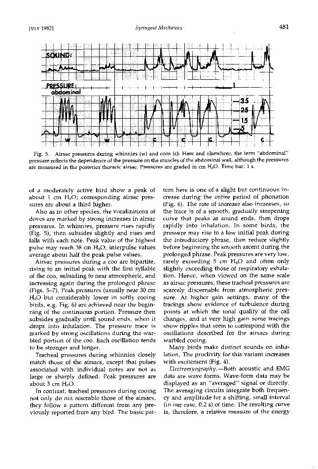
Fig. 5. Airsac pressures during whinnies (w) and coos (c). Here and elsewhere, the term "abdominal" pressure reflects the dependence of the pressure on the muscles of the abdominal wall, although the pressures are measured in the posterior thoracic airsac. Pressures are graded in cm H20. Time bar: I s.
of a moderately active bird show a peak of about 1 cm H20; corresponding airsac pres- sures are about a third higher.
Also as in other species, the vocalizations of doves are marked by strong increases in airsac pressures. In whinnies, pressure rises rapidly (Fig. 5), then subsides slightly and rises and falls with each note. Peak value of the highest pulse may reach 38 cm H20; interpulse values average about half the peak pulse values.
Airsac pressures during a coo are bipartite, rising to an initial peak with the first syllable of the coo, subsiding to near atmospheric, and increasing again during the prolonged phrase (Figs. 5-7). Peak pressures (usually near 30 cm H•O but considerably lower in softly cooing birds, e.g. Fig. 6) are achieved near the begin- ning of the continuous portion. Pressure then subsides gradually until sound ends, when it drops into inhalation. The pressure trace is marked by strong oscillations during the war- bled portion of the coo. Each oscillation tends to be stronger and longer.
Tracheal pressures during whinnies closely match those of the airsacs, except that pulses associated with individual notes are not as
large or sharply defined. Peak pressures are about 3 cm H•O.
In contrast, tracheal pressures during cooing not only do not resemble those of the airsacs, they follow a pattern different from any pre- viously reported from any bird. The basic pat-
tern here is one of a slight but continuous in- crease during the entire period of phonation (Fig. 6). The rate of increase also increases, so the trace is of a smooth, gradually steepening curve that peaks as sound ends, then drops rapidly into inhalation. In some birds, the pressure may rise to a low initial peak during the introductory phrase, then reduce slightly before beginning the smooth ascent during the prolonged phrase. Peak pressures are very low, rarely exceeding 5 cm H•O and often only slightly exceeding those of respiratory exhala- tion. Hence, when viewed on the same scale as airsac pressures, these tracheal pressures are scarcely discernable from atmospheric pres- sure. At higher gain settings, many of the tracings show evidence of turbulence during points at which the tonal quality of the call changes, and at very high gain some tracings show ripples that seem to correspond with the oscillations described for the airsacs during warbled cooing.
Many birds make distinct sounds on inha- lation. The proclivity for this variant increases with excitement (Fig. 4).
Electrornyography.--Both acoustic and EMG data are wave forms. Wave-form data may be displayed as an "averaged" signal or directly. The averaging circuits integrate both frequen- cy and amplitude for a shifting, small interval (in our case, 0.2 s) of time. The resulting curve is, therefore, a relative measure of the energy

482 GAVNT, GAVNT, AND CASEY [Auk, Vol. 99
Fig. 6. Tracheal and abdominal pressures in a softly cooing dove. Pressure graded in cm H20. Time bar: 1 s.
of the signal at a given moment. Patterns, but not quantities, of energy use and production can be compared. For much of our work, the averaging mode permits the separation of a weak signal from background noise. One sim- ply adjusts the instruments to "see" back- ground as zero, and any departure from that is revealed by deflection of the pen or light beam. When EMG signals are sufficiently strong and clean, however, we prefer a direct display, if for no other reason than that the temporal resolution is better.
EMG signals from a muscle contracting steadily against a constant load will show small but continuous fluctuations in both amplitude and frequency. Hence, the mere observation that sound and an EMG are both fluctuating is not in itself of great interest. Rather, we must establish that the fluctuations of the EMG sig- nal constitute a pattern that is repeatable from performance to performance within or among individuals and/or that changes in activity levels can be correlated with some change in the sound.
A. Abdominal muscles: The pattern of activ- ity in the muscles of the abdominal body wall
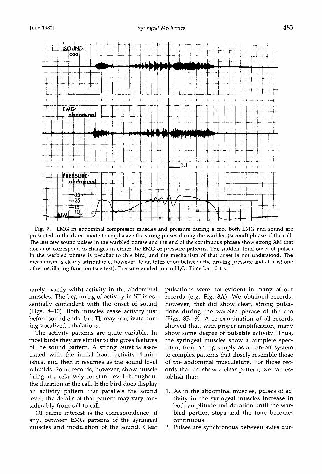
closely corresponds with the pattern of sound. Activity for cooing begins about 10-30 ms be- fore the onset of the initial hoot and ceases just before the pressure drops sharply. Sound may continue for some time after activity stops (Figs. 7-8). Shortly thereafter, EMG begins again at a barely detectable level. That low level is maintained as a continuous background during the warbled phrase but is rapidly aug- mented by a series of short, sharp pulses of much stronger activity. The first pulses may be little more than single spikes that are barely detectable from background, but each succeed- ing pulse waxes in both duration and ampli- tude. The final, largest and longest pulses are accompanied by a distinct warbling of the sound. Finally, activity becomes constant at a relatively high level and is so maintained until near the end of the call. The pattern is essen- tially constant for an individual, and variation among birds is confined to the development of the pulsatile portion. All birds show pulsatile activity during the warbled phrase, but the pattern is more distinct in some than others.
B. Syringeal muscles: In TL, activity for cooing begins within 10 ms before or after (but
Jt•L¾ 1982] SyringeaI Mechanics 483
Fig. 7. EMG in abdominal compressor muscles and pressure during a coo. Both EMG and sound are presented in the direct mode to emphasize the strong pulses during the warbled (second) phrase of the call. The last few sound pulses in the warbled phrase and the end of the continuous phrase show strong AM that does not correspond to changes in either the EMG or pressure patterns. The sudden, loud onset of pulses in the warbled phrase is peculiar to this bird, and the mechanism of that onset is not understood. The mechanism is clearly attributable, however, to an interaction between the driving pressure and at least one other oscillating function (see text). Pressure graded in cm H20. Time bar: 0.1 s.
rarely exactly with) activity in the abdominal musdes. The beginning of activity in ST is es- sentially coincident with the onset of sound (Figs. 8-10). Both muscles cease activity just before sound ends, but TL may reactivate dur- ing vocalized inhalations.
The activity patterns are quite variable. In most birds they are similar to the gross features of the sound pattern. A strong burst is asso- ciated with the initial hoot, activity dimin- ishes, and then it resumes as the sound level rebuilds. Some records, however, show muscle firing at a relatively constant level throughout the duration of the call. If the bird does display an activity pattern that parallels the sound level, the details of that pattern may vary con- siderably from call to call.
Of prime interest is the correspondence, if any, between EMG patterns of the syringeal muscles and modulation of the sound. Clear
pulsations were not evident in many of our records (e.g. Fig. 8A). We obtained records, however, that did show clear, strong pulsa- tions during the warbled phrase of the coo (Figs. 8B, 9). A re-examination of all records showed that, with proper amplification, many show some degree of pulsatile activity. Thus, the syringeal muscles show a complete spec- trum, from acting simply as an on-off system to complex patterns that closely resemble those of the abdominal musculature. For those rec-
ords that do show a clear pattern, we can es- tablish that:
1. As in the abdominal muscles, pulses of ac- tivity in the syringeal muscles increase in both amplitude and duration until the war- bled portion stops and the tone becomes continuous.
2. Pulses are synchronous between sides dur-
484 GAV•T, GAV•T, •v C•s•¾ [Auk, Vol. 99
LJ_-LI•[ I I I I
.LLLLLLJ_L•.L L .l_J;:;•E;I I:1 I I J_ ¾: :• 7-T'I•TTTH•7T•
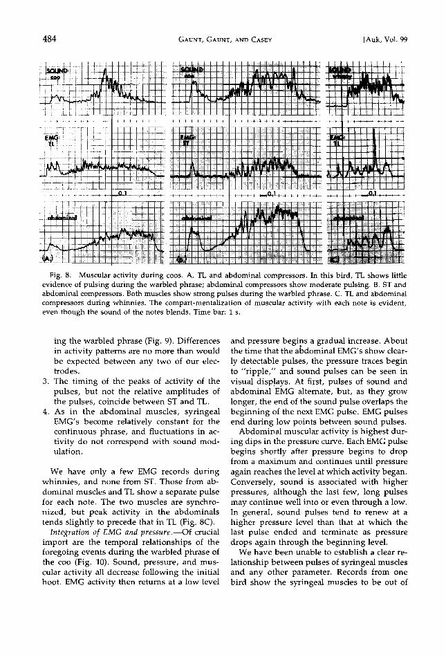
Fig. 8. Muscular activity during coos. A. TL and abdominal compressors. In this bird, TL shows little evidence of pulsing during the warbled phrase; abdominal compressors show moderate pulsing. B. ST and abdominal compressors. Both muscles show strong pulses during the warbled phrase. C. TL and abdominal compressors during whinnies. The compart-mentalization of muscular activity with each note is evident, even though the sound of the notes blends. Time bar: 1 s.
ing the warbled phrase (Fig. 9). Differences in activity patterns are no more than would be expected between any two of our elec- trodes.
3. The timing of the peaks of activity of the pulses, but not the relative amplitudes of the pulses, coincide between ST and TL.
4. As in the abdominal muscles, syringeal EMG's become relatively constant for the continuous phrase, and fluctuations in ac- tivity do not correspond with sound mod- ulation.
We have only a few EMG records during whinnies, and none from ST. Those from ab- dominal muscles and TL show a separate pulse for each note. The two muscles are synchro- nized, but peak activity in the abdominals tends slightly to precede that in TL (Fig. 8C).
Integration of EMG and pressure.--Of crucial import are the temporal relationships of the foregoing events during the warbled phrase of the coo (Fig. 10). Sound, pressure, and mus- cular activity all decrease following the initial hoot. EMG activity then returns at a low level
and pressure begi,ns a gradual increase. About the time that the aDdominal EMG's show clear-
ly detectable pulses, the pressure traces begin to "ripple," and sound pulses can be seen in visual disp!ays. At first, pulses of sound and abdominal EMG alternate, but, as they grow longer, the end of the sound pulse overlaps the beginning of the next EMG pulse. EMG pulses end during low points between sound pulses.
Abdominal muscular activity is highest dur- ing dips in the pressure curve. Each EMG pulse begins shortly after pressure begins to drop from a maximum and continues until pressure again reaches the level at which activity began. Conversely, sound is associated with higher pressures, although the last few, long pulses may continue well into or even through a low. In general, sound pulses tend to renew at a higher pressure level than that at which the last pulse ended and terminate as pressure drops again through the beginning level.
We have been unable to establish a clear re-
lationship between pulses of syringeal muscles and any other parameter. Records from one bird show the syringeal muscles to be out of

Jury 1982] Syringeal Mechanics 485
Fig. 9. Correspondence of activity between right and left syringeal muscles. All of these muscles show strong pulses during the warbled phrase. Minor differences in pattern between right and left sides can be attributed to differences in electrodes or electrode placement. Time bar: 1 s.
phase with abdominal pulses and in phase with amplitude maxima of the sound, but this relationship is not obvious in other records.
DISCUSSION
Pressure and fiow.--Compression of the air- sacs generates the internal pressure that drives vocalization. Peak internal pressures are some- what higher than those of a Starling (Sturnus vulgaris) during a distress call (Gaunt et al. 1973), but considerably below those of a crow- ing rooster (Gaunt et al. 1976). As in other birds, the dove's internal pressure closely re- sembles the curve for averaged sound (loud- ness) (Fig. 10). Once again, the control of "on/ off" and major ch.anges in loudness are depen- dent on the driving pressure, i.e. ultimately on the action of the abdominal compressor mus- cles. It is also apparent that more subtle AM may be traced to the same source. We will re- turn to this point.
Far more unusual are the pressures recorded from the trachea (Fig. 6), which resemble nei- ther the extremely low pressure pattern of os-
cines (Gaunt et al. 1973) nor the damped ab- dominal pattern of roosters (Gaunt et al. 1976). The differences may reflect, in part, the can- nulation technique. In previous experiments, the tracheal cannulae were directed into the
outflow, thus recording velocity head. The cannulae in doves were at right angles to the flow and measured pressure head. An expla- nation for the observed pattern has two com- ponents. First, the fact that tracheal pressures do not parallel those of the airsacs indicates that the dove's vocal system is not a low- impedance-high-flow system like that of a rooster. Rather, it acts more like a valve, but a less effective valve than the syrinx of a Star- ling. Second, unlike the case in Starlings, the air emerging from the valve does not exhaust into open space but is diverted into the esoph- agus. As long as the esophagus is only partially inflated and its walls can easily expand, pres- sure remains low. As the neck swells, the fi- brous portions of the esophageal walls and overlying skin become taut, expansion slows, and internal pressure climbs. What is mea- sured, then, is the relationship between the

486 GAu•T, GA•J•T, A•I• CAsœ¾ [Auk, Vol. 99
2 3 SOUND:
FLOW: MASK
PRI
AIR SAC
EMC•: AB
TL
Fig. 10. Summary of flow, pressure, and muscular events during a coo. Flow and pressure traces are not to the same scale. EMG traces are scaled to the percentage of peak value. Dotted lines indicate relatively common variants (see text for discussion).
influx of air to the esophageal chamber and the latter's increasing resistance to expansion.
The resonant properties of the esophageal chamber probably are not involved in deter- mining the frequency of the coo, because the chamber expands during cooing without a concommittant drop in mean frequency. One dove that could not inflate cooed very softly, but a primary role for the esophageal chamber as a' sound disseminator seems unlikely. In this example, the injury that prevented inflation may have had other effects. More generally, a
feathered surface would not be a good sound radiator, and, judging from the singing behav- ior of other birds, sound disseminates quite well from a widely opened mouth. Neck infla- tion in the Columbidae may be primarily a vi- sual display, with any acoustic effects being secondary.
Sound analysis.--An analysis of the sound- producing mechanism encounters a severe dif- ficulty in applying the standard model, for the dove's call is devoid of overtones. This situa-
tion is not in itself unusual; Greenewalt (1968)

JvL¾ 1982] Syringeal Mechanics 487
?
Fig. 11. Models of membrane contribution to sound generation. A. "Classic" model, with membrane vibrating normal to the flow of air. Vibrations engender alternate zones of compressed and rarified air (sound waves) that pass down the trachea. B. "Ripple" model, in which oscillation of the membrane is a wave travelling along the length of the membrane. C. "Whistle" model, in which the flexible membrane constricts the passage (not necessarily as shown) to form a slot. Friction along the walls slows peripheral flow and initiates the formation of vortices. Oscillation of the membrane, if any, is in response to the vortices. P equals pressure (sound) over some period of time (T) at a point downstream from the source. The T axis could also represent distance from source along the trachea at a given time.
assembled a "portfolio" of 60 examples and presents many more throughout his book. He observes (p. 144), "For all birds, whistled phrases are much more common than those with harmonic content; for the Passeriformes as a whole phrases with harmonic content are rare; for the more primitive families a rough approximation would equate phrases with har- monic spectra to those which are whistled." Both he and D/in'wang (1974) have discussed at some length the conditions of overtone for- mation. But this is not the key question.
Although it may be possible to define con- ditions in which a membrane, clamped at its edges and set into free oscillation, might vi- brate without overtones, these conditions are
extremely rigid and would only rarely be met by any biological system. Further, most of the overtones will be at partials rather than har- monic intervals (Casey 1981). Only rectangular membranes will produce true harmonics, and these will comprise but a few of many over- tones. Other shapes may produce some partials that are sufficiently close to harmonic values to be indistinguishable in a sonogram (but not the FFTA). These, too, will be only a few among many.
The Greenewalt-Stein model (see especially Greenewalt 1969, figure on page 132) implies, and Gaunt and Wells (1973) assumed, that the membrane vibration originates centrally and
is normal to the flow (Fig. 11A). In fact, the stimulus begins at one end and flows across the surface of the membrane. As Greenewalt
states (1968:155), except under high tension, the membranes will be deformed downstream
and, if quite lax, will develop ripples. It is but a small step to the appreciation of Klatt and Stefanski (1974) and D/irrwang (1974), who state that the principle oscillation will be a travelling wave (Fig. 11B). We have been un- able to find even a first order approximation to describe the behavior of a membrane oscillat-
ing in this manner. It is possible that the rip- ples might form standing waves at harmonic intervals, as in vibrating strings, but nothing we have seen in the analysis of simpler mem- brane vibrations suggests that overtones could be absent. The key question, then, is not how to produce overtones, but how to avoid them. One solution to that problem is provided in Greenewalt's term "whistled phrase," for whistles produce pure tones. The possibility that a whistle, either in pure form or interact- ing with a membrane, might be the actual source of the sound is implicit in the analyses of Abs (1980) and Gaunt and Wells (1973) and is directly invoked by Nottebohm (1976) in ref- erence to some psittacid calls. Yet even these authors emphasize the action of membranes.
Characteristics of whistles.--Whistles are pe- riodic disturbances, vortices or vortex rings

488 GAUNT, GAVNT, ANY CASEY [Auk, Vol. 99
(Fig. 11C), formed in or in association with a column of flowing fluid. All whistles involve some form of feedback loop that establishes and maintains their periodicity (Chanaud 1970). The tones produced can be extremely loud, even with the flow of small volumes of fluid. They characteristically lack harmonics. A whistle can interact with a resonator that
may determine the frequency and may or may not produce harmonics, but no whistle, either pure or resonance coupled, normally produces partials. Whistles can be produced by a variety of techniques that fall into two or three cate- gories, depending on whether the classifica- tion is based on an analogy with one or two rods oriented across the air stream (Morse and Ingard 1968) or on the nature of the feedback mechanism (Chanaud 1970). A form of hole tone, the presumed mechanism of human lip and tongue whistles (Wilson et al. 1971), would seem appropriate to many syringes. In hole tones, the disturbances are generated by shear- ing forces as a column of air is forced through a narrow aperture. A two-hole tone derives its feedback from an interaction of the second ap- erture with a perturbance started by the first aperture. In the constricted columbid syrinx (Figs. lB, 11C), the narrowed entrances of the bronchi into the trachea are appropriate first apertures. If a tone generated at either or both of those slits should interact with the gap be- tween the infolded LTM's, then the action of the columbid syrinx would be that of a two- hole tone of a type known, appropriately, as a Rayleigh bird whistle.
In many whistles, e.g. flutes and recorders, the disturbance is used to activate a resonating structure, the properties of which may then determine the frequency and/or harmonic structure of the sound. Such whistles are fre-
quency modulated by altering the properties of the resonator. The MTM's of the syrinx seem to be appropriate resonators. An interaction between turbulence in the air column and the
syringeal membranes has been suggested by Abs (1980) and Gaunt and Wells (1973). Such interactions may play a role in vocalizations of some species. If membrane oscillations are driven, then they are not restricted to the dis- crete values of free vibrations (Kinsler and Frey 1962). A membranous resonator will react most strongly when the driving frequency matches the membrane's natural frequency--hence, the coupling of AM and FM (Gaunt and Wells
1973). If membrane vibration becomes part of the feedback that controls the whistle's fre-
quency, then membrane tension determines frequency as previously supposed. Different syringes, or different species, might use dif- ferent couplings. In extreme cases, designation of "source" becomes an exercise in sophistry.
Evidence suggestive of such interactions may be found in sonograms of coos (Fig. 3), for the two torres suggest the action of different but closely coupled mechanisms. Unfortu- nately, it is possible to erect equally plausible models of mechanisms in which either the con-
stant or the shifting tone represents either the membrane or the whistle, depending on initial assumptions.
Modulation.--In Streptopelia the AM shows two important characteristics: it is of irregular period, and it is not linked to FM. Modulation by tracheal resonance is improbable for all the reasons cited by Greenewalt. Harmonic mod- ulation can be eliminated, both because we see no overtones and because the modulations are
irregular (Greenewalt 1968). Irregular modula- tions might be produced by true modulation at source, but again we encounter two objec- tions. First, sonograms of coos do not show the side-band pattern characteristic of such mod- ulations. Second, the mechanism postulated for such rapid modulations requires changes of syringeal configuration that would produce distinct FM. This leaves us with three, not mutually exclusive, techniques: changes in the input energy, beats, and mufflers.
A. Changed input energy. Gross aspects of loudness of bird calls are clearly linked to the driving pressure (Gaunt et al. 1973, 1976; Brackenbury 1977). In the coos of $treptopelia, more subtle aspects are also affected. Both the abdominal EMG's and the associated pressure pattern exactly fit our fluctuating flow model for the production of trills (Gaunt and Gaunt 1977). If the velocity of flow drops below threshold, a whistle will stop, and such a com- plete interruption of sound is seen in the war- bled portion of some calls (Figs. 2, 7). Changes in internal pressure and flow will have less in- fluence on the frequency of a hole-tone whistle than on the frequency of a membrane, because the tension of the syringeal membrane is in part determined by internal pressure (Gaunt and Wells 1973).
Although changes of driving pressure are clearly related to some of the modulation in the

1982] Syringeal Mechanics 489
warbled portion, they cannot account for mod- ulation elsewhere in the coo or even all the modulation in the warble. Here we find a sec-
ond kind of AM that is much faster than any that could be directly attributed to the rather slow changes in internal pressure. This is per- ceived by the human ear not as a warble but as timbre, just as if the call had harmonics. These modulations divide the wave train into
a series of ellipsoid boll. Such a pattern can be generated by either of the next two techniques.
B. Beats. Both Greenewalt and Stein dis- cussed the effects of beats in some detail. Beats
are not a true modulation of a carrier frequency but rather the interaction of two tones of sim-
ilar amplitude but slightly different frequency. The difference causes the two wave trains
gradually to shift phase relative to one another. When the tones are in phase, they augment each other to produce a louder resultant; when out of phase, they cancel. If the two compo- nents are of equal amplitude, negative inter- ference is complete, and resultant amplitude drops to zero when the phase difference is 180 ø . These points are called nodes. The phase of the resultant wave reverses across a node.
If one component is significantly louder than the other, interference is incomplete and phase does not shift at the minima. The beat fre-
quency is equal to the difference between the two component frequencies. If the two com- ponents are sufficiently different, one hears not a beat but the two tones. Sufficiency is deter- mined by the receiver, not by the wave form.
To demonstrate the presence of a beat, we should show: (1) the presence of two compo- nent frequencies at the correct interval to pro- duce the beat frequency, and/or (2) the pres- ence of 180 ø phase shifts (phase reversal) at the nodes.
The resolution of the acoustic energy of a coo into two peaks is clbarest near the beginning and end of the prolonged phrase. Power sec- tions of these zones in the coos of D16 show
peaks that would produce beats with periods of 12-20 ms. The FFTA confirms these results, e.g. a beat with a period of 15 ms would be produced by interaction of the 510 and 577.5 Hz peaks in Fig. 3. Pulses of this duration are found in the overlap zones of D16's call (Fig. 2, arrows).
The second test is less persuasive. Almost 40% of the nodes in the calls of three doves
show no phase shift across the nodes, and in
only 45% of those showing any shift is the change near the expected 180 ø . Much of the observed shifting appears to involve changes in the instantaneous frequencies of the waves immediately adjacent to nodes, a phenomenon that could obscure phase reversals.
In short, many of the very rapid "modula- tions" can be attributed to beats. As discussed
by Greenewalt, beats are most easily explained as a product of the two-voice phenomenon. The two sides of the dove syrinx will not be exactly identical and will not be subject to identical patterns of flow. The differences will be slight, presenting the ideal situation for the formation of beats.
Many of the presumed beats appear to be superimposed on modulations of longer and often irregular duration. This superposition is most evident in the warbled phrase of all coos and the concluding portion of the coos of D16 (Fig. 2). Some modulations of the warbled por- tion could be attributed to the previously dis- cussed pressure changes, but not those in the continuous phrase. This, then, suggests the presence of a third type of modulation.
C. Mufflers. An example of an oscillating muffler in the phonation of a toad (Bufo val- liceps) has been described by Martin and Gans (1972). Sound generated in a column of flowing air passes down a tubular pathway that is pe- riodically constricted. In B. valliceps, the ary- tenoid cartilages of the larynx are the constric- tors (Fig. 12). They are set into oscillation by the same airstream that stimulates the vocal
cords, but, as they are far more massive than the vocal cords, their oscillation is of a much lower frequency. When the tube is patent, sound passes freely; when constricted, the sound is muffled. Here the modulation fre-
quency is the frequency of the muffler, not the difference between the frequencies of two os- cillators. Because the source remains un-
changed, AM and FM can be separated. The pattern of modulation can be changed rapidly by changing the oscillatory pattern of the muf- fler.
The larynx is unlikely to be an oscillating muffler in doves. Changing the glottal bore would elicit strong tracheal pressure fluctua- tions out of phase with sound pulses; no such changes were observed. The infolded LTM's, however, are ideally placed to play the role. In B. vaIliceps the modulating pattern is regular because the arytenoid cartilages are of constant

490 GAozaT, GAUZaT, •zar• C•sœ¾ [Auk, Vol. 99
• Ampli t ude ½ '• modulated
• tone
Modulator: vibrating arytenoid
pI' carr'*r frequency
Sou• *o•rc.: I / vibrating / | vocal card [
(A.) (•.)
Sound source: aerodynamic whistle
tar:
vibrating LTM
Fig. 12. Amplitude modulation of the tone by action of mufflers. Comparison of models for (A) toad (modified from Gans 1974) and (B) dove.
mass. In doves, the elastic properties of the LTM's vary with their tension. Stein and Greenewalt have suggested similar roles for either the LTM's or external labia (fleshy pads continuous with the LTM in oscines), although Greenewalt's mechanism is somewhat differ-
ent from the one proposed by Stein and ex- panded here.
Tension in the LTM's in doves is controlled
by action of the syringeal muscles, particularly TL. We have shown that the activity of the sy- ringeal musculature may, in some doves, in- clude regular pulsations that presumably effect regular changes of syringeal configuration. Such pulsations are confined to the warbled phrase, however, where they interact with os- cillations of the driving pressure. In the un- warbled portion of the coo, the activity of the syringeal musculature is essentially continu- ous, with no more fluctuation than would be
expected from any contraction. Hence, the pat- tern of activity in the tracheal musculature may influence the early portions of the call, but, unless it initiates a free-running oscillation that continues elastically with little damping, it is unlikely to be a factor in modulations of the continuous phrase. An equally important point is that the activity of the syringeal mus-
cles does not elicit FM in doves, as it should according to traditional models.
The LTM muffler offers an additional, com- plicating possibility. The amplitude of a hole tone is closely related to the flow rate, which, in turn, is determined by the driving pressure and the resistance. The resistance of a tube var-
ies inversely with the fourth power of the ra- dius, so that very small changes in the position of the LTM could have a direct and strong effect on a whistle. The small changes in flow would probably pass undetected by our tracheal can- nulae, especially given the overriding effect of esophageal wall tension.
The sound patterns in Fig. 2 may be inter- preted as being the result of an interaction be- tween a beat frequency and varying pressure, or beat and muffler, or all three. Silence could result from complete closure of the muffler, re- duction of flow below threshold, or coinci-
dence of the minima of separate modulations. Thus, the number of potential interactions is sufficient that the complex patterns of AM seen in some calls are not surprising.
We have not commented on the determina-
tion of frequency, nor do we deem it profitable to consider either this problem or the forma-
JULY 1982] Syringeal Mechanics 491
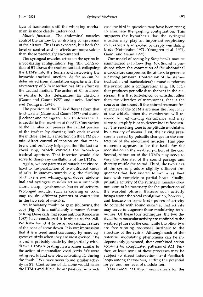
tion of harmonics until the whistling mecha- nism is more clearly understood.
Muscle function.--The abdominal muscles control the airflow by controlling compression of the airsacs. This is as expected, but both the level of control and its effects are more subtle
than those previously encountered. The syringeal muscles act to set the syrinx in
a vocalizing configuration (Fig. lB). Contrac- tion of ST draws the trachea caudad, collapsing the LTM's into the lumen and narrowing the bronchio tracheal junction. As far as can be determined from stimulation experiments, the asymmetry of ST's insertion has little effect on the caudad motion. The action of ST in doves is similar to that determined for chickens
(Gaunt and Gaunt 1977) and ducks (Lockner and Youngren 1976).
The position of the TL is different from that in chickens (Gaunt and Gaunt 1977) and ducks (Lockner and Youngren 1976). In doves the TL is caudal to the insertion of the ST. Contraction
of the TL also compresses the caudal portion of the trachea by drawing both ends toward the middle. The TL's insertion on the LTM per- mits direct control of tension on that mem-
brane and probably helps position the last tra- cheal ring, which controls the bronchio- tracheal aperture. That insertion would also serve to damp any oscillations of the LTM's.
Again, we see patterns of muscle activity re- lated to the production of two different kinds of calls. In staccato sounds, e.g. the clucking of chickens and whinnying of doves, abdom- inal and syringeal muscles act as a unit with short, sharp, synchronous bursts of activity. Prolonged sounds, such as crowing or coos, may require different patterns of contraction in the two sets of muscles.
An inhalatory "wah" or gasp (following the coo) (Fig. 4) is a sufficiently common feature of Ring Dove calls that some authors (Goodwin 1967) have considered it intrinsic to the call. We have found it to be an occasional feature
of the coos of some doves. It is our impression that it is uttered most commonly by more ag- gressive birds when they are most excited. The sound is probably made by the partially with- drawn LTM's vibrating in a manner similar to the action of mammalian vocal cords. We were
intrigued to find one bird activating TL during the "wah." We have never found similar activ-
ity in ST. Contraction of TL alone would tense the LTM's and dilate the air passage, in which
case the bird in question may have been trying to eliminate the gasping configuration. This supports the hypothesis that the syringeal muscles may play an important respiratory role, especially in excited or deeply ventilating birds (Nottebohm 1971, Youngren et al. 1974, Gaunt and Gaunt 1977).
Our model of cooing by Streptopelia may be summarized as follows (Fig. 10). Sound is pro- duced when the contraction of the abdominal
musculature compresses the airsacs to generate a driving pressure. Contraction of the sterno- trachealis and tracheolateralis muscles reforms
the syrinx into a configuration (Fig. lB, 11C) that produces periodic disturbances in the air- stream. It is that turbulence, or whistle, rather than the vibration of membranes, that is the source of the sound. If the natural resonant fre-
quencies of the MTM's are near the frequency of the whistle, then the membranes will re- spond to that driving disturbance and may serve to amplify it or to determine its frequen- cy. The resulting tone is amplitude modulated by a variety of means. First, the driving pres- sure is varied by pulsatile changes in the con- traction of the abdominal muscles. This phe- nomenon appears to be the basis for the modulation in the warbled portion of the coo. Second, vibration of the LTM's may serve to vary the diameter of the sound passage and thereby muffle the sound. Third, the two sides of the syrinx produce slightly different fre- quencies that then interact to form a resultant tone with complete or partial beats. Finally, pulsatile activity of the syringeal muscles does not seem to be necessary for the production of the warbled phrase. Because such activity brings about the vocal configuration, however, and because in some birds pulses of activity do coincide with sound maxima, that activity may serve to augment these modulating tech- niques. Of these four techniques, the two de- rived from muscular activity are confined to the warbled phrase of the coo, while the other two are free-running processes intrinsic to the structure of the syrinx. Although each of the potential modulating phenomena can be in- dependently generated, their combined action accounts for complicated patterns of AM. Fur- ther, at least some of these processes may be subject to direct interactions and feedback loops among themselves, adding the potential for yet another level of modulation.
This model has major implications for the

492 G^v•T, G^v•T, ^•v C^sE¾ [Auk, Vol. 99
study of avian vocal mechanics in general. First, whistles may play a very important role in bird calls. Whistles are advantageous in that they produce loud sounds with the consump- tion of only small amounts of air.
This interpretation has both happy and wor- risome aspects. On the one hand, it provides a ready explanation for the production of har- monic-free calls that are rapidly modulated in amplitude and/or frequency. On the other hand, whistles are intricate devices, and the workings of even commonly encountered ones, e.g. human lip and tongue whistles or the American police whistle, are not entirely understood. Even a dove's simple syrinx could produce whistles by several, not necessarily mutually exclusive, techniques. Further, most whistles used by humans are controlled by coupling them to resonating devices. The membranous portions of a syrinx might serve such a function, but their ability to do so would depend much on the nature of the tone generator.
A second implication is not entirely new. We again find that the syrinx is only one compo- nent in a vocal system. Here we have direct evidence that the activity of the abdominal muscles affects AM in far more subtle ways than had been previously apparent. Similar evidence has been presented for trilling calls of chicks, with pulse rates of up to 50/s (Phillips and Youngren 1981). Those authors did not measure internal pressures, but we have since determined that airsac pressures in chicks also oscillate in synchrony with the notes of the trill. Hence, an abdominal mechanism for pro- ducing warbles and trills may be widespread. This technique is predicted by the Pulsatile- Input Model (Gaunt and Gaunt 1977) of Cal- der's (1970) "minibreath" data. Indeed, given that (1) Calder's instruments could provide no direct measure of either flow or internal pres- sure, (2) these measurements, when obtained from a variety of species, have not shown either flow or priessure reversal, and (3) we have here demonstrated an alternative expla- nation, we are extremely skeptical of the entire minibreath hypothesis.
The present data also introduce a need for caution. If avian voice is modulated even in
part by nonsyringeal mechanisms, then sy- ringeal function cannot be deduced from acoustic analysis of the call alone, regardless of the sophistication of that analysis. Further,
acoustic evidence alone may fit contradictory models equally well.
In presenting this hypothesis, we do not wish to suggest that all syringeal sounds de- pend on, or even use, whistles. An abundance of evidence (Beebe 1925; Miller 1934; Miskimen 1951; Gross 1964, 1979) testifies to the impor- tance of vibrating, or at least flexible, mem- branes in syringeal function. Many harsh and broad-band calls are certainly produced di- rectly by membrane vibration. The demonstra- tion that a flexible structure is necessary for sound production, however, is not a demon- stration that the vibration of that structure gen- erates the sound. Flexible membranes would
be necessary to produce whistles, as we use flexible tongue and lips. This possibility is supported by Beebe's (1925) observation that the frequencies of sounds produced by a tin- amou's syrinx decreased as the syrinx relaxed and the membranes were withdrawn from the
lumen. That observation is directly opposed to the predictions of the Greenewalt-Stein model, because the tension of the membranes in-
creases as they are withdrawn, and the fre- quency of oscillation should thus also increase. Finally, vibration of membranes may be nec- essary to initiate, maintain, or amplify an aero- dynami.c oscillation. Stiffening or otherwise injuring the membranes may silence a bird (Abs pers. comm., Gross 1979), and indirect interference may produce aberrant sounds (Cohen and Cheng 1979).
If our hypothesis is correct, many questions remain. What is the exact nature of the whis-
tling mechanism? We have suggested a hole tone, but other mechanisms are possible. We do not suppose that one mechanism would be universal among species. What is the relation- ship between whistles and membranes? Do the latter amplify the tone? Are they part of a feed- back system? Can they determine the frequen- cy of the whistle? How are harmonic overtones generated?
Some of these questions may be technically difficult to answer. For instance, stroboscopic views of membranes in actual or modelled sy- ringes can distinguish between the competing hypotheses only if those hypotheses predict different kinds of vibration. Such evidence
might help determine which, if either, of the differently behaving frequency bands in our sonograms is related to membrane vibration. But stroboscopic evidence that a membrane

JULY 1982] Syringeal Mechanics 493
vibrates in accordance with the wave form of
a sound means nothing alone, because a driven oscillator will be forced toward conformity. Similar difficulties in deciphering cause and effect characterize many experiments with scale models. Mathematical analysis of even simple situations is difficult and tedious. Yet construction and evaluation of precisely de- fined and controlled mathematical and scale
models, perhaps in conjunction with computer simulation (Titze 1973, 1974), can play a critical role. At presentß this area of avian biology could benefit greatly from additional theoreti- cal treatments, if only to define the nature and direction of physiological experiments.
ACKNOWLEDGMENTS
Many persons have helped with various aspects of these studies. Among those deserving special thanks are: Dr. Donald R. Houser, Department of Mechan- ical Engineering, The Ohio State University, for ac- cess to the Fast Fourier Transform Analyzer; Mr. Ro- land Hawkins, Conservatory Aviary, Pittsburgh, for providing birds; Drs. Luis F. Baptista, William E. Evansß Lincoln Fairchildß and Lewis Greenwaldß for commentary, encouragementß and assistance; and especially Dr. Richard E. Phillips for a detailed and perceptive critique of an earlier manuscript. Three reviewers chosen by the Editor provided valuable suggestions. This work was supported by NSF grant DEB-7911774.
LITERATURE CITED
ABs, M. 1980. Zur Bioakustik des Stimmbruchs bei
V6geln. Zool. Jhb. Physiol. 84: 289-382. [3ASMA]IAN, J. V., & G. A. $TECKO. 1962. A new bi-
polar indwelling electrode for electromyogra- phy. J. Appl. Physiol. 17: 849.
BEEBE, W. 1925. The Variegated Tinamue, Crypturus variegatus variegatus (Gmelin). Zoologica 6: 195- 227.
BRACRENBUR¾, J. H. 1977. Physiological energetics of cock-crow. Nature 270: 433-435.
CARDER, W. A. 1970. Respiration during song in the Canary (Serinius canaria). Comp. Biochem. Physiol. 32: 251-258.
CASEY, R. M. 1981. Theoretical analysis of tympanic membranes in avian syrinx. Unpublished M.$. thesis. Columbus, Ohio, Ohio State Univ.
CHANAUD, R. C. 1970. Aerodynamic whistles. Sci. Amer. 222: 40-46.
COHEN, J., & M.-F. CHENG. 1979. Role of vocaliza- tion in the reproductive cycle of Ring Doves (Streptopelia risoria): effects of hypoglossal nerve section on the reproductive behavior and phys- iology of the female. Hormone Behav. 213: 113- 127.
DORRWANG, R. 1974ß Funktionelle Biologie, Anato- mie und Physiologie der V6gelstimme. Unpub- lished Ph.D. dissertation, Baselß Switzerland, Universit•it Basel.
GANS, C. 1974. Biomechanics: an approach to ver- tebrate biology. Ann Arborß Univ. Mich. Press.
GAUNT, A. $., & $. L. L. GAUNT. 1977. Mechanics of the syrinx in Gallus gallus: II. Electromy- ographic studies of ad libiturn vocalization. J. Morphol. 152: 1-20.
, & M. E. WELLS. 1973. Models of syringeal mechanisms. Amer. Zool. 13: 1227-1247.
ß S. L. L. GAUNT, & D. HECTOR. 1976ß Me-
chanics of the syrinx in Gallus gallus. I. A com- parison of pressure events in chickens to those in oscines. Condor 73: 208-233.
--, R. C. STEIN, & S. L. L. GAUNT. 1973ß Pres- sure and air flow during distress calls of the Star- lingß Sturnus vulgaris (Aves: Passeriformes). J. Exp. Zool. 183: 241-262.
GAUNT, S. L. L. 1980. Thermoregulation in doves (Columbidae): a novel esophageal heat exchang- er. Science 210: 445-447.
GOODWIN, D. 1967. Pigeons and doves of the world. Londonß England, Brit. Mus. Nat. Hist.
GREENEWALT, C. H. 1968. Bird song: acoustics and physiology. Washington, D.C., Smithsonian Inst. Press.
ß 1969. How birds sing. Sci. Amer. 221: 126- 139.
GRoss, W. [3. 1964. Devoicing the chicken. Poultry Sci. 43: 1143-1144.
ß 1979. An operation for reducing the vocal intensity of Peafowl. Avian Dis. 23: 1031-1036.
HERSH, G. L. 1966. Bird voices and resonant tuning in helium-air mixtures. Unpublished Ph.D. dis- sertation. Berkeley, California, Univ. California.
KINGß A. S. 1979. Systema respiratorium. In: Nom- ina Anatomica Avium (J. J. Baumel, Ed.). New York, Academic Press.
KINSLER, L. E., & A. R. FREY. 1962. Fundamentals
of acoustics, 2nd ed. New York, John Wiley & Sons.
KLATT, D. H., & R. A. STEFANSKI. 1974. HOW does a Mynah bird imitate human speech? J. Acoust. Soc. Amer. 55: 822•832.
LOCKNER, F. R., & O. M. YOUNGREN. 1976. Func-
tional syringeal anatomy of the Mallard. I. In situ electromyograms during ESB elicited calling. Auk 93: 324-342.
MARTIN, W. F., & C. GANS. 1972. Muscular control of the vocal tract during release signaling in the toad Bufo valliceps. J. Morphol. 137: 1-28.
MILLER, A. H. 1934. The vocal apparatus of some North American owls. Condor 36: 204-213.
MILLER, W. J., & L. S. MILLERß 1958. Synopsis of behaviour traits of the Ring-neck Dove. Anim. Behav. 6: 3-8.

494 GAVNT, GAVNT, AND CASEY [Auk, Vol. 99
MISKIMEN, M. 1951. Sound production in passefine birds. Auk 68: 493-504.
MORSE, P.M., & K. U. IN6ARD. 1968. Theoretical acoustics. New York, McGraw Hill.
MYERS, J. A. 1917. Studies on the syrinx of Gallus domesticus. J. Morphol. 29: 165-214.
NOTTEBOHM, F. 1971. Neural lateralization of vocal
control in a passefine bird. I. Song. J. Exp. Zool. 177: 229-262.
--. 1976. Phonation in the Orange-winged Am- azon Parrot, Amazona amazonica. J. Comp. Phys- iol. 108A: 157-170.
--, & M. E. NOTTEBOHM. 1971. Vocalizations
and breeding behaviour of surgically deafened Ring Doves (Streptopelia risoria). Anim. Behav. 19: 313-327ß
PEEK, F. W., O. M. YOUN6REN, & R. E. PHILLIPS. 1975. Repetitive vocalizations evoked by elec- trical stimulation of avian brains. IV. Evoked
and spontaneous activity in expiratory and in- spiratory nerves and muscles of the chicken (Gallus gallus). Brain, Behav. Evol. 12: 1-42.
PHILLIPS, R. E., & O. M. YOUNGREN. 1981. Effects
of denervation of the tracheo-syfingeal muscles on frequency control in chicks. Auk 98: 299-306.
SIBLE¾, C. G., & J. E. ALQUIST. 1972. A comparative study of the egg white proteins of non-passerine birds. Yale Univ. Bull. 39: 1-276.
STEIN, R. C. 1968. Modulation in bird sounds. Auk 85: 229-243.
SUTHERLAND, C. A., & D. S. McCHEsNEY. 1965. Sound production in two species of geese. Liv- ing Bird 4: 99-106.
TITZE, I. R. 1973. The human vocal cords. A math- ematical model. Part I. Phone fica 28: 129-170.
ß 1974. The human vocal cords. A mathemat-
ical model. Part 2. Phonefica 29: 1-29.
WILSON, T. A., G. S. BEAVERS, M. A. DECOSTER, D.
K. HOLGER, & M.D. REGENFUSS. 1971. Exper- iments on the fluid mechanics of whistling. J. Acoust. Soc. Amer. 50: 366-372.
WARNER, R. W. 1972. The syrinx in the family Co- lumbidae. J. Zool. 166: 385-390.
YOUNGREN, O. M., F. W. PEEK, & R. E. PHILLIPS. 1974. Repetitive vocalizations evoked by local electrical stimulation of avian brains. Ill. Evoked
activity in the tracheal muscles of the chicken (Gallus gallus). Brain, Behav. Evol. 9: 393-421.