Gaze failure, drifting eye movements, centripetal nystagmus in ... · seconds, andin...
8
British Journal of Ophthalmology, 1977, 61, 774-781 Gaze failure, drifting eye movements, and centripetal nystagmus in cerebellar disease J. LEECH,' M. GRESTY,1 K. HESS,2 AND PETER RUDGE' From the 'Medical Research Council's Hearing and Balance Unit, Institute of Neurology, National Hospital, Queen Square, London, WCJ, and 2Neurological Clinic of the University of Zurich, Switzerland SUMMARY Three abnormalities of eye movement in man are described which are indicative of cerebellar system disorder, namely, centripetally beating nystagmus, failure to maintain lateral gaze either in darkness or with eye closure, and slow drifting movements of the eyes in the absence of fixation. Similar eye movement signs follow cerebellectomy in the primate and the cat. These abnormalities of eye movement, together with other signs of cerebellar disease, such as rebound, alternating, and gaze paretic nystagmus, are explained by the hypothesis that the cerebellum helps to maintain lateral gaze and that brain stem mechanisms which monitor gaze position generate compensatory biases in the absence of normal cerebellar function. Cerebellar disease in man causes a variety of ab- normalities of eye movement, including dysmetria (Orzechowski, 1927), gaze paretic nystagmus (Holmes, 1917), and rebound nystagmus (Hood et al., 1973). Examination of the direct-coupled elec- tro-oculographic (EOG) records obtained in the Medical Research Council's Hearing and Balance Unit has revealed three additional abnormalities not previously reported in man which we consider are also indicative of cerebellar system disorder, namely, centripetal nystagmus, in which the fast phase of the nystagmus beats towards the position of primary gaze, failure of gaze maintenance after eye closure or in darkness, and drifting movements of the eyes in the absence of fixation. Methods PATIENTS All patients with centripetal nystagmus were sur- veyed. It was found that centripetal nystagmus occurred in patients with peripheral vestibular lesions, those intoxicated with drugs or suffering from congenital abnormalities, and in patients with disorders of the central nervous system. The characteristics of nystagmus in peripheral lesions are well documented and are discussed later. Drug- intoxicated and congenitally abnormal patients were excluded from the study because of the inability to specify the lesions in these cases. The remaining 17 patients, ranging in age from 23 to 73 years, had Address for reprints: Dr P. Rudge, Nationa! Hospital, Queen Square, London WCIN 3BG subacute or chronic disorders of the central nervous system. The only clinical feature common to all was evidence of a cerebellar system disorder, although the majority did have a wide variety of signs indica- tive of involvement of other systems-for example, pyramidal system, cranial nerve palsies. A detailed clinical description of the cases used to illustrate abnormalities of eye movement in this paper is given in the Appendix. Of the 17 cases 5 had a cerebellar tumour, 4 multiple sclerosis, 7 degenerative and vascular disease, and 1 Arnold-Chiari malformation. The diagnosis of cerebellar tumour was verified at operation in all five cases; one patient had a hae- mangioblastoma and the remainder had astrocy- tomas. All the patients with a diagnosis of multiple sclerosis were classified as definite by McAlpine's criteria (McAlpine et al., 1972). Three of these had a history of relapse and remission, while the fourth had the progressive form of the disease. A diagnosis of vascular disease was made in patients who had a history with an acute onset and who had hyper- tension or other signs of vascular disease. Degenera- tive disease of the central nervous system was diag- nosed in patients with a known family history, pro- gressive neurological disturbance in patients older than 50 years, or if there was radiological evidence of atrophy of the cerebellum. The diagnosis in the patient with Arnold-Chiari malformation was con- firmed at operation. Results CENTRIPETAL NYSTAGMUS Centripetally beating nystagmus was present, by 774 copyright. on 28 March 2019 by guest. Protected by http://bjo.bmj.com/ Br J Ophthalmol: first published as 10.1136/bjo.61.12.774 on 1 December 1977. Downloaded from
Transcript of Gaze failure, drifting eye movements, centripetal nystagmus in ... · seconds, andin...
British Journal of Ophthalmology, 1977, 61, 774-781
Gaze failure, drifting eye movements, andcentripetal nystagmus in cerebellar diseaseJ. LEECH,' M. GRESTY,1 K. HESS,2 AND PETER RUDGE'From the 'Medical Research Council's Hearing and Balance Unit, Institute of Neurology, National Hospital,Queen Square, London, WCJ, and 2Neurological Clinic of the University of Zurich, Switzerland
SUMMARY Three abnormalities of eye movement in man are described which are indicative ofcerebellar system disorder, namely, centripetally beating nystagmus, failure to maintain lateral gazeeither in darkness or with eye closure, and slow drifting movements of the eyes in the absence offixation. Similar eye movement signs follow cerebellectomy in the primate and the cat. Theseabnormalities of eye movement, together with other signs of cerebellar disease, such as rebound,alternating, and gaze paretic nystagmus, are explained by the hypothesis that the cerebellumhelps to maintain lateral gaze and that brain stem mechanisms which monitor gaze positiongenerate compensatory biases in the absence of normal cerebellar function.
Cerebellar disease in man causes a variety of ab-normalities of eye movement, including dysmetria(Orzechowski, 1927), gaze paretic nystagmus(Holmes, 1917), and rebound nystagmus (Hood etal., 1973). Examination of the direct-coupled elec-tro-oculographic (EOG) records obtained in theMedical Research Council's Hearing and BalanceUnit has revealed three additional abnormalities notpreviously reported in man which we consider arealso indicative of cerebellar system disorder,namely, centripetal nystagmus, in which the fastphase of the nystagmus beats towards the position ofprimary gaze, failure of gaze maintenance after eyeclosure or in darkness, and drifting movements ofthe eyes in the absence of fixation.
Methods
PATIENTSAll patients with centripetal nystagmus were sur-veyed. It was found that centripetal nystagmusoccurred in patients with peripheral vestibularlesions, those intoxicated with drugs or sufferingfrom congenital abnormalities, and in patients withdisorders of the central nervous system. Thecharacteristics of nystagmus in peripheral lesions arewell documented and are discussed later. Drug-intoxicated and congenitally abnormal patients wereexcluded from the study because of the inability tospecify the lesions in these cases. The remaining 17patients, ranging in age from 23 to 73 years, had
Address for reprints: Dr P. Rudge, Nationa! Hospital,Queen Square, London WCIN 3BG
subacute or chronic disorders of the central nervoussystem. The only clinical feature common to all wasevidence of a cerebellar system disorder, althoughthe majority did have a wide variety of signs indica-tive of involvement of other systems-for example,pyramidal system, cranial nerve palsies. A detailedclinical description of the cases used to illustrateabnormalities of eye movement in this paper is givenin the Appendix. Of the 17 cases 5 had a cerebellartumour, 4 multiple sclerosis, 7 degenerative andvascular disease, and 1 Arnold-Chiari malformation.The diagnosis of cerebellar tumour was verified at
operation in all five cases; one patient had a hae-mangioblastoma and the remainder had astrocy-tomas. All the patients with a diagnosis of multiplesclerosis were classified as definite by McAlpine'scriteria (McAlpine et al., 1972). Three of these had ahistory of relapse and remission, while the fourthhad the progressive form of the disease. A diagnosisof vascular disease was made in patients who had ahistory with an acute onset and who had hyper-tension or other signs of vascular disease. Degenera-tive disease of the central nervous system was diag-nosed in patients with a known family history, pro-gressive neurological disturbance in patients olderthan 50 years, or if there was radiological evidenceof atrophy of the cerebellum. The diagnosis in thepatient with Arnold-Chiari malformation was con-firmed at operation.
Results
CENTRIPETAL NYSTAGMUSCentripetally beating nystagmus was present, by
774
copyright. on 28 M
arch 2019 by guest. Protected by
http://bjo.bmj.com
/B
r J Ophthalm
ol: first published as 10.1136/bjo.61.12.774 on 1 Decem
ber 1977. Dow
nloaded from
Gaze failure, drifting eye movements, and centripetal nystagmus in cerebellar disease
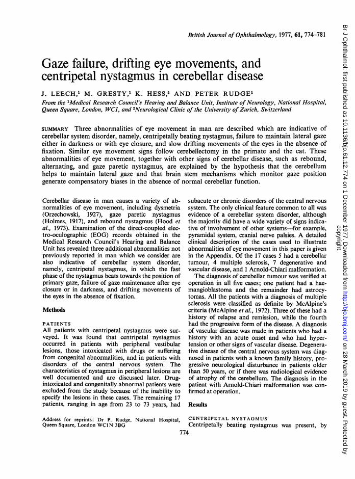
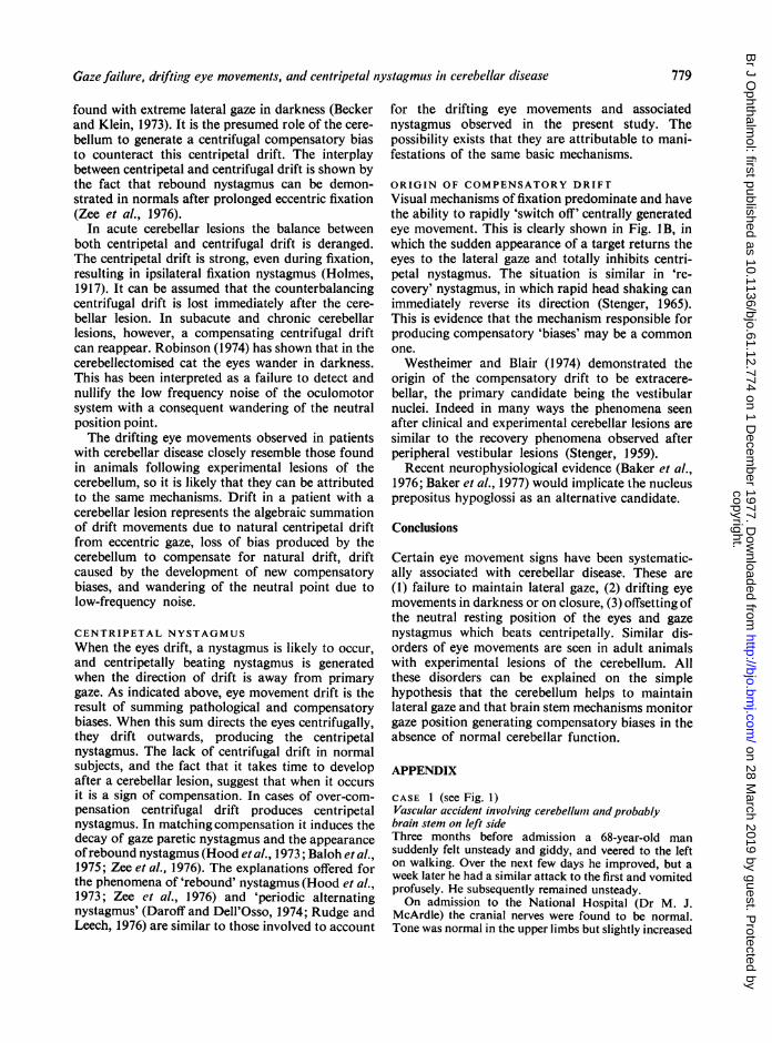
A Fig. 1 EOG recordings o,r g h t horizontal eye movements in a
1. patient with vascular diseaseA ~~~~~~~~~~~~~~~~~300 (case 1).
S o,fof A. The patient's eyes areinitially in primary gaze, fixated
3 00 on a central target light in anotherwise dark environment. Asecond target light appears 30°
left to the right and the central lighton v is extinguished. The patient
executes saccadic eyecont s ~movement(s), s, to fixate the
new target. Subsequently thetarget is turned offbr I minute,and in total darkness the eyes
right drift further to the right; smallB saccadic movements beating to
1 the left begin and attempts are/Qo30 made to correct the drift
(cont = continuation of the
s o f f 0 recording). Eventually a distinct30 nystagmus is evident. The targetjz finally reappears 300 to the right
e f t and corrective movements, s, aremade for refixation.
on B. The experimental regime iscont > ,g/ s off the same as in A, but this time
- i v the lateral target appears at 300left. When it is extinguished, theeyes drift to the extreme rightand then back to the left with theassistance of small saccades(earliest portion of the firstcontinuation trace). The eyescontinue to drift to the left, and
____________________ right-beating nystagmus develops.10 s The target light is turned on for
4 seconds to correct the positionof the eyes, s, and a fine left-beating nystagmus is immediately evident. The target is turned offfor a further 28seconds, and in this period of darkness the second continuation trace shows that the eyes drift further to the left and aright-beating nystagmus appears. A final rightward correction, s, is made when the target light is turned on again.Net EOG drift estimated with reference to eye position during target refixations-1°/minute.
definition, in all patients in the survey. Such nystag-mus occurred in the presence of optic fixation, indarkness, or on eye closure. An example of abilateral centripetal nystagmus occurring in darknessis presented in Fig. 1. When illumination is turnedoff, the patient's eyes drift about in the darkness anda centripetally beating nystagmus develops. Thecentripetal nystagmus is immediately abolished whenthe illumination is turned on again and is replaced bycentrifugal nystagmus (Fig. 1B). In some otherpatients a centripetal nystagmus was seen im-mediately the eyes attained an eccentric gazeposition.An example of predominantly unilateral centri-
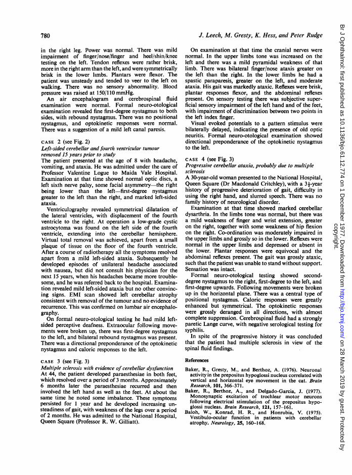
petal nystagmus following eye closure is presented in
Fig. 2B. After closure there is a failure to maintainlateral gaze, and the eyes assume a more neutralposition. During the movement towards the neutralposition a centripetal nystagmus appears. Fifteenpatients had a unilateral centripetal nystagmus,which could occur under conditions of darkness,closure, or fixation, or any combination of these. Ofthe 2 remaining patients one had bilateral centri-petal nystagmus in darkness; the other had a uni-lateral centripetal nystagmus in one direction indarkness and in the other direction on eye closure.
FAILURE OF GAZE MAINTENANCEA failure of gaze maintenance means that the eyesmove rapidly from an eccentric gaze position towards
775
copyright. on 28 M
arch 2019 by guest. Protected by
http://bjo.bmj.com
/B
r J Ophthalm
ol: first published as 10.1136/bjo.61.12.774 on 1 Decem
ber 1977. Dow
nloaded from
J. Leech, M. Gresty, K. Hess, and Peter Rudge
right
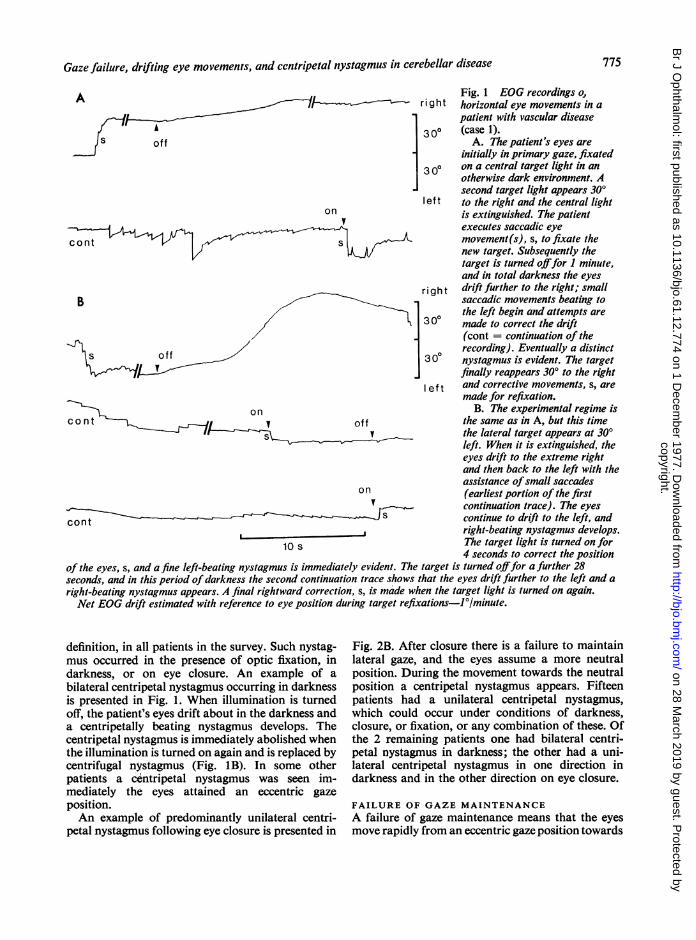
A 1300_0s eyes close '.a1 3ApenI, 0open ]
B
s
A
close
5 s
Fig. 2 EOG recordings of horizontal eye movements in a patient (case 2) after surgical removal of a tumour arisingfrom the lateral part of the floor of the fourth ventricle and affecting the left cerebellar hemisphere.
A. Initially the eyes fixate a target light in primary gaze. A second target light appears 30° to the right and thepatient fixates it, s. Immediately a right-beating nystagmus (J°) develops. After eye closure there is a rapid drifttowards primary gaze, on which is superimposed a left-beating nystagmus such that the net eye movement is towardsprimary gaze, but the slow-phase movement is to the right. Eventually the nystagmus ceases and the eyes drift to theright and show small irregular saccadic movements with occasional isolated, nystagmic beats. The eyes open, and aftera short series of inappropriate movements the 30° right target is refixated and the right-beating (J°) nystagmus reappears.
B. A target 300 left is fixated, s, with the appearance of a left-beating (J°) nystagmus. After eye closure the eyesdrift towards and through primary gaze. Superimposed on the drift is a right-beating nystagmus, of which the slowphase is to the left. Eventually the eyes are positioned in the right side of the orbit. When the eyes are opened,inappropriate movements are made until the eyes refixate the target and a left beating nystagmus is again seen.EOG drift negligible.
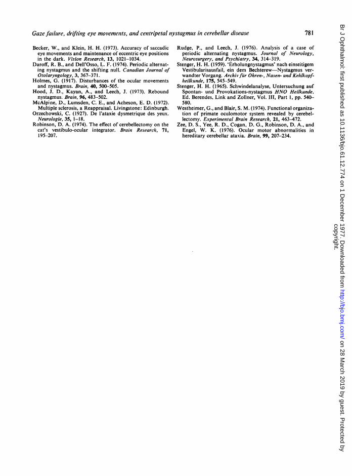
primary gaze. The movement is 'triggered' either byclosing the eyes or darkness and takes the approxi-mate shape of an exponential decay rather than asaccade. The whole movement lasts between 2 and 6seconds. Eleven of the patients were unable tomaintain eccentric gaze on eye closure (Figs. 2 and3), though all maintained eccentric gaze withfixation. Gaze failure was present in darkness in2 patients, both of whom had multiple sclerosis.With eye closure the patients failed to maintain gazeto both sides except in one case with a left-sidedcerebellar tumour and brain stem involvement, inwhich the failure was only to the left. When the eyesreturned towards centre the new neutral or nullposition of the eyes was not necessarily the primaryposition. This is illustrated in Fig. 3A, where theeyes tended to come to rest some 100 to the right ofthe primary position whether the initial gaze was tothe left or right. The movement of the eyes followsan approximately exponential time course with a
time constant of 0-5 to 1-5 seconds. Encouragementto maintain an eccentric position of gaze during eye
closure resulted in feeble and unsuccessful correc-tive movements.
DRIFT MOVEMENTThe term 'drift movements' means wandering move-
ments of the eyes, attaining velocities of only 1 or 2°per second and enduring for many tens of seconds.Removal of fixation by eye closure, darkness, or
both resulted in slow drifting movements of the eyes
in 9 of the patients. An example occurring in dark-ness is shown in Fig. IB, where the eyes move nearly400 to the right of the primary position, then driftslowly back past the midline. In other cases the driftappeared to be unidirectional, as in Fig. 2A and B,where there is a slow drift to the right of the midline.In the 5 cases in which there was evidence of a pre-
dominantly unilateral lesion the drift tended to beaway from that side. Furthermore, in those cases
where there was a failure to maintain eccentric gaze
and the neutral position was displaced to one sideof the position of primary gaze, the drift was alsopredominantly to the same side of the midline as this
A
A
open
300
3O0
lef t
776
copyright. on 28 M
arch 2019 by guest. Protected by
http://bjo.bmj.com
/B
r J Ophthalm
ol: first published as 10.1136/bjo.61.12.774 on 1 Decem
ber 1977. Dow
nloaded from
Gaze failure, drifting eye movements, and centripetal nystagmus in cerebellar disease
AR close
P A 300cp~~~~~~oeCP A
0
0v
LA I IC
5 s
B CR V
p ~~~~~~~0
Fig. 3 A. EOG recordings froma patient (case 3) with multiplesclerosis. When the eyes fixate atarget 30° to the right a 1°nystagmus appears. Eye closureproduces a rapid drift towardscentre and abolishes thenystagmus. With eye closure (C)on primary gaze (P) there is amovement to the right with theappearance ofa right-beatingnystagmus. Eye closure (C) onleft gaze (L) produces a rapidmovement to centre with theappearance ofa right-beatingnystagmus.
B. EOG recordings from apatient (case 4) with a progressivecerebellar syndrome, probablydue to multiple sclerosis. Aright-beating nystagmus isevident on primary (P) and right(R) gaze; on left (L) gaze thereis a left-beating nystagmus. Oneye closure (C) there is a rapidmovement of the eyes to thecentre. On left (L) gaze aright-beating nystagmus developswith eye closure (C).EOG drift negligible.
0 = eyes open.
C~~~~~~Lc
3 S
new neutral position. When nystagmus was presentduring drifting eye movements, either the fast or slowphase could be in the direction of the drift (compareFig. 2B with Fig. IB). Of the 9 patients with driftingmovements of the eyes all had gaze nystagmus and8 had additional failure of gaze maintenance. Fourpatients had bilateral drift, the remainder had uni-lateral drift.
OTHER EYE MOVEMENT DISORDERSOf the patients surveyed 14 had centrifugal nystag-mus on lateral gaze, 10 had deranged pursuit move-ments, 7 had exaggerated caloric responses, and 6had rebound nystagmus.
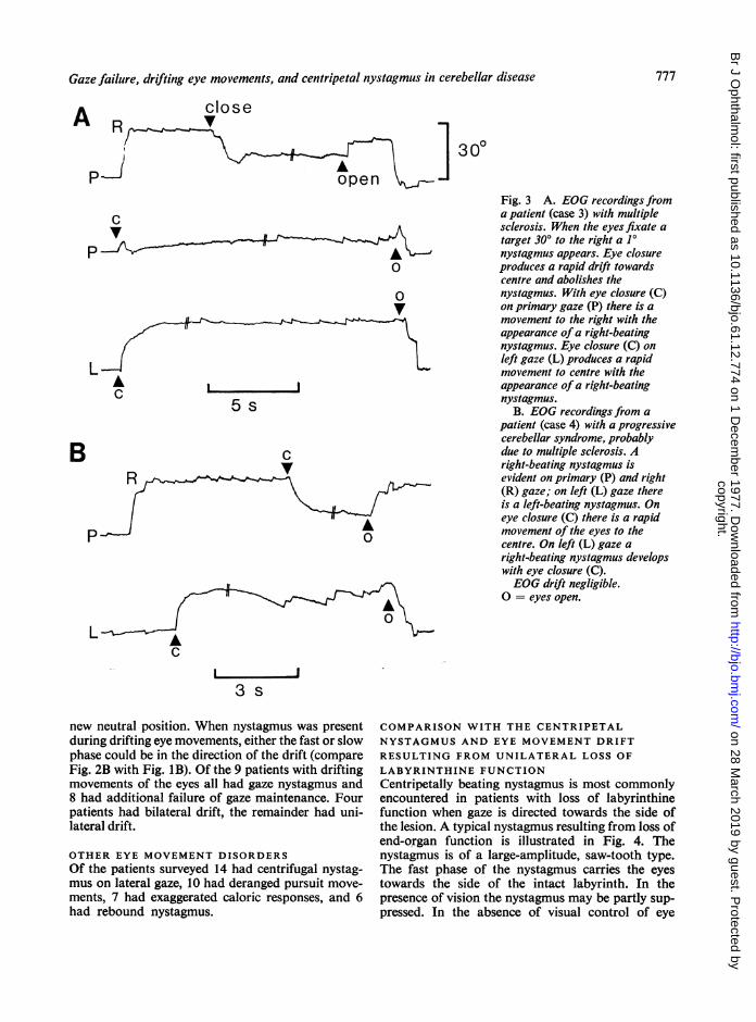
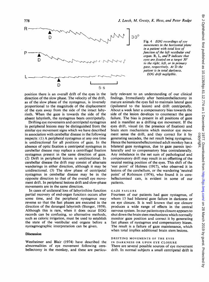
COMPARISON WITH THE CENTRIPETALNYSTAGMUS AND EYE MOVEMENT DRIFTRESULTING FROM UNILATERAL LOSS OFLABYRINTHINE FUNCTIONCentripetally beating nystagmus is most commonlyencountered in patients with loss of labyrinthinefunction when gaze is directed towards the side ofthe lesion. A typical nystagmus resulting from loss ofend-organ function is illustrated in Fig. 4. Thenystagmus is of a large-amplitude, saw-tooth type.The fast phase of the nystagmus carries the eyestowards the side of the intact labyrinth. In thepresence of vision the nystagmus may be partly sup-pressed. In the absence of visual control of eye
777
copyright. on 28 M
arch 2019 by guest. Protected by
http://bjo.bmj.com
/B
r J Ophthalm
ol: first published as 10.1136/bjo.61.12.774 on 1 Decem
ber 1977. Dow
nloaded from
J. Leech, M. Gresty, K. Hess, and Peter Ruidge
D
p ,_ ,
D I ID 5 s
position there is an overall drift of the eyes in thedirection of the slow phase. The velocity of the drift,as of the slow phase of the nystagmus, is inverselyproportional to the magnitude of the displacementof the eyes away from the side of the intact laby-rinth. When the gaze is towards the side of theabsent labyrinth, the nystagmus beats centripetally.
Drifting eye movements and centripetal nystagmusin peripheral lesions may be distinguished from thesimilar eye movement signs which we have describedin association with cerebellar disease in the followingrespects: (1) A peripheral nystagmus at any one timeis unidirectional for all positions of gaze. In theabsence of optic fixation a centripetal nystagmus incerebellar disease may replace a centrifugal fixationnystagmus present in the same direction of gaze.(2) Drift in peripheral lesions is unidirectional. Incerebellar disease the drift may consist of alternatewanderings in either direction, although it may beunidirectional. (3) The slow phase of centripetalnystagmus in cerebellar disease may be in theopposite direction to that of the overall eye move-ment drift. In peripheral lesions drift and slow-phasemovements are in the same direction.
In cases of unilateral loss of labyrinthine functionpartial recovery of end-organ function occurs aftersome time, and the peripheral nystagmus mayreverse so that the fast phases are executed in thedirection of the deranged labyrinth (Stenger, 1959).Although this is rare, when it does occur EOGrecords can be confusing, so alternative methods,such as caloric irrigation, must be used to establishthe state of the vestibular end organs before anystagmographic interpretation can be given.
Discussion
Westheimer and Blair (1974) have described theabnormalities of eye movement following cere-bellectomy in the monkey, and these are particu-
30OFig. 4 EOG recordings of eyemovements in the horizontal planein a patient with total loss offunction of the left vestibular endorgan. R, L, and P indicate thateyes are fixated on a target 300to the right, left, or in primarygaze, respectively. At D thepatient is in total darkness.EOG drift negligible.
larly relevant to an understanding of our clinicalfindings. Immediately after hemicerebellectomy inmature animals the eyes fail to maintain lateral gaze(ipsilateral to the lesion) and drift centripetally.About a week later a compensatory bias towards theside of the lesion develops to counteract the gazefailure. The bias is present in all positions of gazeand is manifest as a drifting eye movement. If theeyes drift, visual (in the presence of fixation) andbrain stem mechanisms which monitor eye move-ment sense the drift, and they correct for it bygenerating saccades, the net result being nystagmus.Hence the hemicerebellectomised adult monkey has abilateral gaze nystagmus, due to gaze paresis ipsi-laterally and to compensatory bias contralaterally.Any imbalance in the net sum of pathological andcompensatory drift may result in an offsetting of theneutral resting position of the eyes. This shift of the'rest point' of Holmes (1917), who observed it inlesions of the cerebellum, or the wandering 'neutralpoint' of Robinson (1974), who found it in cere-bellectomised cats, is evident in some of ourpatients.
GAZE FAILUREFourteen of our patients had gaze nystagmus, ofwhom 13 had bilateral gaze failure in darkness oron eye closure. It is well known that eye closureproduces a wide range of effects in the centralnervous system. In our patients eye closure appears toshut down the brain stem mechanisms which normallymonitor gaze position and correct it by generatingfast phases of nystagmus and compensatory biases.The result is a failure of gaze maintenance, whichwhen total implies additional brain stem lesions.
DRIFTING MOVEMENTS OF THE EYESIN DARKNESS OR UPON EYE CLOSUREThere are several possible sources of eye movementdrift. In normal subjects a small centripetal drift is
778
copyright. on 28 M
arch 2019 by guest. Protected by
http://bjo.bmj.com
/B
r J Ophthalm
ol: first published as 10.1136/bjo.61.12.774 on 1 Decem
ber 1977. Dow
nloaded from
Gaze failure, drifting eye movements, and centripetal nystagmus in cerebel/ar disease
found with extreme lateral gaze in darkness (Beckerand Klein, 1973). It is the presumed role of the cere-bellum to generate a centrifugal compensatory biasto counteract this centripetal drift. The interplaybetween centripetal and centrifugal drift is shown bythe fact that rebound nystagmus can be demon-strated in normals after prolonged eccentric fixation(Zee et al., 1976).
In acute cerebellar lesions the balance betweenboth centripetal and centrifugal drift is deranged.The centripetal drift is strong, even during fixation,resulting in ipsilateral fixation nystagmus (Holmes,1917). It can be assumed that the counterbalancingcentrifugal drift is lost immediately after the cere-bellar lesion. In subacute and chronic cerebellarlesions, however, a compensating centrifugal driftcan reappear. Robinson (1974) has shown that in thecerebellectomised cat the eyes wander in darkness.This has been interpreted as a failure to detect andnullify the low frequency noise of the oculomotorsystem with a consequent wandering of the neutralposition point.The drifting eye movements observed in patients
with cerebellar disease closely resemble those foundin animals following experimental lesions of thecerebellum, so it is likely that they can be attributedto the same mechanisms. Drift in a patient with acerebellar lesion represents the algebraic summationof drift movements due to natural centripetal driftfrom eccentric gaze, loss of bias produced by thecerebellum to compensate for natural drift, driftcaused by the development of new compensatorybiases, and wandering of the neutral point due tolow-frequency noise.
CENTRIPETAL NYSTAGMUSWhen the eyes drift, a nystagmus is likely to occur,and centripetally beating nystagmus is generatedwhen the direction of drift is away from primarygaze. As indicated above, eye movement drift is theresult of summing pathological and compensatorybiases. When this sum directs the eyes centrifugally,they drift outwards, producing the centripetalnystagmus. The lack of centrifugal drift in normalsubjects, and the fact that it takes time to developafter a cerebellar lesion, suggest that when it occursit is a sign of compensation. In cases of over-com-pensation centrifugal drift produces centripetalnystagmus. In matching compensation it induces thedecay of gaze paretic nystagmus and the appearanceofrebound nystagmus (Hood etal., 1973; Baloh etal.,1975; Zee et al., 1976). The explanations offered forthe phenomena of 'rebound' nystagmus (Hood et al.,1973; Zee et al., 1976) and 'periodic alternatingnystagmus' (Daroff and Dell'Osso, 1974; Rudge andLeech, 1976) are similar to those involved to account
for the drifting eye movements and associatednystagmus observed in the present study. Thepossibility exists that they are attributable to mani-festations of the same basic mechanisms.
ORIGIN OF COMPENSATORY DRIFTVisual mechanisms of fixation predominate and havethe ability to rapidly 'switch off' centrally generatedeye movement. This is clearly shown in Fig. I B, inwhich the sudden appearance of a target returns theeyes to the lateral gaze and totally inhibits centri-petal nystagmus. The situation is similar in 're-covery' nystagmus, in which rapid head shaking canimmediately reverse its direction (Stenger, 1965).This is evidence that the mechanism responsible forproducing compensatory 'biases' may be a commonone.Westheimer and Blair (1974) demonstrated the
origin of the compensatory drift to be extracere-bellar, the primary candidate being the vestibularnuclei. Indeed in many ways the phenomena seenafter clinical and experimental cerebellar lesions aresimilar to the recovery phenomena observed afterperipheral vestibular lesions (Stenger, 1959).
Recent neurophysiological evidence (Baker et al.,1976; Baker et al., 1977) would implicate the nucleusprepositus hypoglossi as an alternative candidate.
Conclusions
Certain eye movement signs have been systematic-ally associated with cerebellar disease. These are(1) failure to maintain lateral gaze, (2) drifting eyemovements in darkness or on closure, (3) offsetting ofthe neutral resting position of the eyes and gazenystagmus which beats centripetally. Similar dis-orders of eye movements are seen in adult animalswith experimental lesions of the cerebellum. Allthese disorders can be explained on the simplehypothesis that the cerebellum helps to maintainlateral gaze and that brain stem mechanisms monitorgaze position generating compensatory biases in theabsence of normal cerebellar function.
APPENDIX
CASE I (see Fig. 1)Vascular accident involving cerebelluln andprobablybrain stem on left sideThree months before admission a 68-year-old mansuddenly felt unsteady and giddy, and veered to the lefton walking. Over the next few days he improved, but aweek later he had a similar attack to the first and vomitedprofusely. He subsequently remained unsteady.On admission to the National Hospital (Dr M. J.
McArdle) the cranial nerves were found to be normal.Tone was normal in the upper limbs but slightly increased
779
copyright. on 28 M
arch 2019 by guest. Protected by
http://bjo.bmj.com
/B
r J Ophthalm
ol: first published as 10.1136/bjo.61.12.774 on 1 Decem
ber 1977. Dow
nloaded from
J. Leech, M. Gresty, K. Hess, and Peter Rudge
in the right leg. Power was normal. There was mildimpairment of finger/nose/finger and heel/shin/kneetesting on the left. Tendon reflexes were rather brisk,more in the right arm than the left, and were symmetricallybrisk in the lower limbs. Plantars were flexor. Thepatient was unsteady and tended to veer to the left onwalking. There was no sensory abnormality. Bloodpressure was raised at 150/110 mmHg.An air encephalogram and cerebrospinal fluid
examination were normal. Formal neuro-otologicalexamination revealed fine first-degree nystagmus to bothsides, with rebound nystagmus. There was no positionalnystagmus, and optokinetic responses were normal.There was a suggestion of a mild left canal paresis.
CASE 2 (see Fig. 2)Left-sided cerebellar and fourth ventricular tumourremoved 15 years prior to studyThe patient presented at the age of 8 with headache,vomiting, and ataxia. He was admitted under the care ofProfessor Valentine Logue to Maida Vale Hospital.Examination at that time showed normal optic discs, aleft sixth nerve palsy, some facial asymmetry-the rightbeing lower than the left-first-degree nystagmusgreater to the left than the right, and marked left-sidedataxia.
Ventriculography revealed symmetrical dilatation ofthe lateral ventricles, with displacement of the fourthventricle to the right. At operation a low-grade cysticastrocytoma was found on the left side of the fourthventricle, extending into the cerebellar hemisphere.Virtual total removal was achieved, apart from a smallplaque of tissue on the floor of the fourth ventricle.After a course of radiotherapy all the symptoms resolvedapart from a mild left-sided ataxia. Subsequently -hedeveloped episodes of unilateral headache associatedwith nausea, but did not consult his physician for thenext 15 years, when his headaches became more trouble-some, and he was referred back to the hospital. Examina-tion revealed mild left-sided ataxia but no other convinc-ing signs. EMI scan showed left cerebellar atrophyconsistent with removal of the tumour and no evidence ofrecurrence. This was confirmed on lumbar air encephalo-graphy.On formal neuro-otological testing he had mild left-
sided perceptive deafness. Extraocular following move-ments were broken up, there was first-degree nystagmusto the left, and bilateral rebound nystagmus was present.There was a directional preponderance of the optokineticnystagmus and caloric responses to the left.
CASE 3 (see Fig. 3)Multiple sclerosis with evidence of cerebellar dysfunctionAt 44, the patient developed paraesthesiae in both feet,which resolved over a period of 3 months. Approximately6 months later the paraesthesiae recurred and theninvolved the left hand as well as the feet. At about thesame time he noted some imbalance. These symptomspersisted for 1 year and he developed increasing un-steadiness of gait, with weakness of the legs over a periodof 2 months. He was admitted to the National Hospital,Queen Square (Professor R. W. Gilliatt).
On examination at that time the cranial nerves werenormal. In the upper limbs tone was increased on theleft and there was a mild pyramidal weakness of thatlimb. There was bilateral finger/nose ataxis greater onthe left than the right. In the lower limbs he had aspastic paraparesis, greater on the left, and moderateataxia. His gait was markedly ataxic. Reflexes were brisk,plantar responses flexor, and the abdominal reflexespresent. On sensory testing there was subjective super-ficial sensory impairment of the left hand and of the feet,with impairment of discrimination between two points inthe left index finger.
Visual evoked potentials to a pattern stimulus werebilaterally delayed, indicating the presence of old opticneuritis. Formal neuro-otological examination showeddirectional preponderance of the optokinetic nystagmusto the left.
CASE 4 (see Fig. 3)Progressive cerebellar ataxia, probably due to multiplesclerosisA 30-year-old woman presented to the National Hospital,Queen Square (Dr Macdonald Critchley), with a 3j-yearhistory of progressive deterioration of gait, difficulty inusing the right hand, and slurred speech. There was nofamily history of neurological disorder.
Examination at that time showed marked cerebellardysarthria. In the limbs tone was normal, but there wasa mild weakness of finger and wrist extension, greateron the right, together with some weakness of hip flexionon the right. Co-ordination was moderately impaired inthe upper limbs and grossly so in the lower. Reflexes werenormal in the upper limbs and depressed or absent inthe lower. Plantar responses were equivocal and theabdominal reflexes present. The gait was grossly ataxic,such that the patient was unable to stand without support.Sensation was intact.Formal neuro-otological testing showed second-
degree nystagmus to the right, first-degree to the left, andfirst-degree upwards. Following movements were brokenup in the horizontal plane. There was a central type ofpositional nystagmus. Caloric responses were greatlyenhanced but symmetrical. The optokinetic responseswere grossly deranged in all directions, with almostcomplete suppression. Cerebrospinal fluid had a stronglyparetic Lange curve, with negative serological testing forsyphilis.
In spite of the progressive history it was concludedthat the patient had multiple sclerosis in view of thespinal fluid findings.
References
Baker, R., Gresty, M., and Berthoz, A. (1976). Neuronalactivity in the prepositus hypoglossi nucleus correlated withvertical and horizontal eye movement in the cat. BrainResearch, 101, 366-371.
Baker, R., Berthoz, A., and Delgado-Garcia, J. (1977).Monosynaptic excitation of trochlear motor neuronsfollowing electrical stimulation of the prepositus hypo-glossi nucleas. Brain Research, 121, 157-161.
Baloh, W., Konrad, H. R., and Honrubia, V. (1975).Vestibulo-ocular function in patients with cerebellaratrophy. Neurology, 25, 160-168.
780
copyright. on 28 M
arch 2019 by guest. Protected by
http://bjo.bmj.com
/B
r J Ophthalm
ol: first published as 10.1136/bjo.61.12.774 on 1 Decem
ber 1977. Dow
nloaded from
Gaze failure, drifting eye movemenlts, and centripetal nystagmus in cerebellar disease
Becker, W., and Klein, H. H. (1973). Accuracy of saccadiceye movements and maintenance of eccentric eye positionsin the dark. Vision Research, 13, 1021-1034.
Daroff, R. B., and Dell'Osso, L. F. (1974). Periodic alternat-ing nystagmus and the shifting null. Canadian Journal ofOtolaryngology, 3, 367-371.
Holmes, G. (1917). Disturbances of the ocular movementsand nystagmus. Brain, 40, 500-505.
Hood, J. D., Kayan, A., and Leech, J. (1973). Reboundnystagmus. Brain, 96, 483-502.
McAlpine, D., Lumsden, C. E., and Acheson, E. D. (1972).Multiple sclerosis, a Reappraisal. Livingstone: Edinburgh.
Orzechowski, C. (1927). De l'ataxie dysmetrique des yeux.Neurologie, 35, 1-18.
Robinson, D. A. (1974). The effect of cerebellectomy on thecat's vestibulo-ocular integrator. Brain Research, 71,195-207.
Rudge, P., and Leech, J. (1976). Analysis of a case ofperiodic alternating nystagmus. Journal of Neurology,Neurosurgery, and Psychiatry, 34, 314-319.
Stenger, H. H. (1959). 'Erholungnystagmus' nach einseitigemVestibularisausfail, ein dem Bechterew-Nystagmus ver-wandter Vorgang. Archivfur Ohren-, Nasen- und Kehlkopf-heilkunde, 175, 545-549.
Stenger, H. H. (1965). Schwindelanalyse, Untersuchung aufSpontan- und Provokations-nystagmus HNO Heilkunde.Ed. Berendes, Link and Zollner, Vol. III, Part 1, pp. 540-580.
Westheimer, G., and Blair, S. M. (1974). Functional organiza-tion of primate oculomotor system revealed by cerebel-lectomy. Experimental Brain Research, 21, 463-472.
Zee, D. S., Yee, R. D., Cogan, D. G., Robinson, D. A., andEngel, W. K. (1976). Ocular motor abnormalities inhereditary cerebellar ataxia. Brain, 99, 207-234.
781
copyright. on 28 M
arch 2019 by guest. Protected by
http://bjo.bmj.com
/B
r J Ophthalm
ol: first published as 10.1136/bjo.61.12.774 on 1 Decem
ber 1977. Dow
nloaded from