Gastrulation in Amphibians Ppt
20
-
Upload
anonymous-mpd5isjva -
Category
Documents
-
view
81 -
download
2
Transcript of Gastrulation in Amphibians Ppt


GASTRULATION IN AMPHIBIANSIf we look at a cross
section of an embryo of the frog Xenopus, we can see that at this point it is a ball of cells with a fluid-filled cavity. The cavity is the blastocoels, and the embryo is in the blastula stage of development. A blastula contains large yolk-filled cells at the vegetal pole and smaller cells at the animal pole.

The three colors repredent the three tissue layers that become defined early in embryogenesis. Yellow indicates endoderm, red indicates mesoderm, and blue indicates ectoderm.

At the beginning of gastrulation, a few surface cells, called bottle cells, constrict at their apical ends and expand at their basal ends. These cells move into the interior of the embryo, followed by other surface cells. We can track the movement of cells into the embryo if we add dye to a few surface cells.

The movement of cells into the embryo creates a lip, called the dorsal lip, over which sheets of cells continue to move inside. At the same time, the ectoderm extends around the embryo’s surface in a process called epiboly. As gastrulation proceeds, a cavity, called the archenteron, forms while the blastocoel shrinks.

The archenteron is the primitive gut and is completely surrounded by endodermal tissue. The endoderm at the roof of the cavity originated from the outside, via the blastopore, which eventually becomes the anus of the animal.

As the ectoderm extend around the embryo, another set of bottle cells forms. These cells migrate into embryo, and other surface cells follow them, creating the ventral lip of the blastopore.
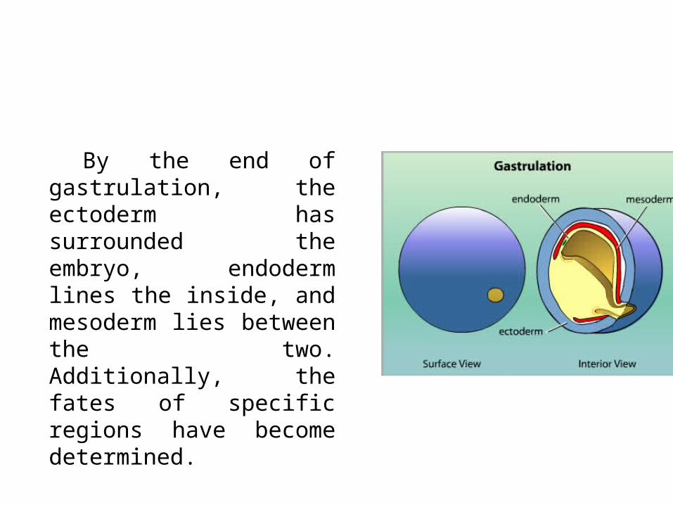
By the end of gastrulation, the ectoderm has surrounded the embryo, endoderm lines the inside, and mesoderm lies between the two. Additionally, the fates of specific regions have become determined.

The endoderm gives rise to the digestive and respiratory tracts and associated structures. The mesoderm gives rise to the skeleton, circulation system, muscles, excretory system, and most of the reproductive system. The ectoderm gives rise to the skin, sense organs, and nervous system.

Gastrulation in Aves
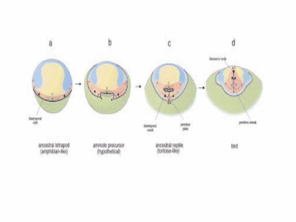
In the chick, the process of gastrulation is prolonged and highly modified than that of frog and Amphioxus. It is already started when the egg of chick is laid and completes well into the second day of incubation. The main characteristic of avian gastrulation is primitive streak.


The main characteristic of gastrulation Aves is the area element of the primitive (primitive streak). This area initially appears as a thickening in the middle of the posterior part pelucida area due to the migration of cells to the center of the area posteriolateral pelucida area. Part thickening narrows, moves anteriorly and constricted forming a trench called the primitive elements.

The arches are called primitive grooves and act as blastoporus. At the anterior end of the thickening called Hensen node (Hensen node). The central portion shaped as a node Hensen wells and through the edges will be traversed by the cells that enter the cavity of the blastula.

Gastrulation in Aves carried by cells that move independently and coordinated, from the outside into the embryo, rather than through movement along the cell in the form of a slab. Gastrulation in the form Aves archentron not true. After the endoderm is formed, which becomes archentron is topped subgerminal cavity bounded by the endoderm, is essentially the yolk. These cells move anteriorly, joining hipoblast hipoblas and eventually replace the anterior part of the embryo.

The next cell that goes through Hensen node also moves anteriorly, but did not move as far as going to the endoderm. These cells remain in the epiblast and endoderm to form the mesoderm and notochord head. Cells that enter this all moves anteriorly, pushing the middle to upper epiblast and eventually formed folds head. Meanwhile, more and more cells migrate in through the primitive elements that enter into the cavity of the blastula after they split off into two directions, one to go deeper and join hipoblast hipoblast and pushing to the edge.

These cells will form all the organs of endodermal and most of the extra-embryonic membranes. The second group spreads to form a sheet that lies between the epiblast and hipoblas. These slabs that form part of the embryonic mesoderm and extra-embryonic membranes. While the formation of the mesoderm takes place, the primitive elements began retracts so Hensen node in the middle of moving the location of the area pellucida be located in the posterior part.


In other words, Hensen node moves posteriorly and the posterior notochord is formed. Eventually shifted node reaches the most posterior position and shape anal area. At this stage it would be entirely composed of epiblast cells to the ectoderm that surrounds the yolk berepiboli. Gastrulation has been completed with the establishment eksoderm, replacement hipoblas with endoderm and mesoderm in between lay the two layers.