Gastrointestinal pathology Part III The stomach. The normal gastric mucosa Cardia – mainly...
37
Gastrointestinal pathology Part III The stomach
-
Upload
evelyn-lyons -
Category
Documents
-
view
225 -
download
1
Transcript of Gastrointestinal pathology Part III The stomach. The normal gastric mucosa Cardia – mainly...
Gastrointestinal pathology Part III
The stomach
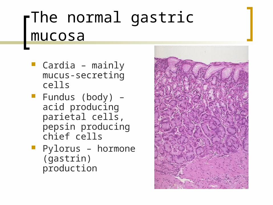
The normal gastric mucosa
Cardia – mainly mucus-secreting cells
Fundus (body) – acid producing parietal cells, pepsin producing chief cells
Pylorus – hormone (gastrin) production
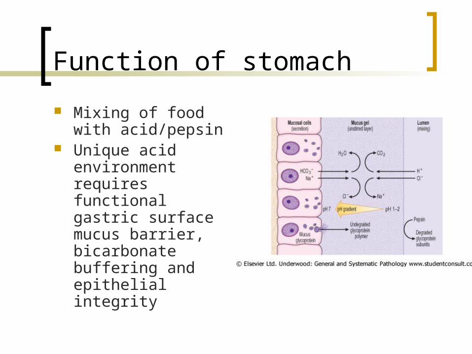
Function of stomach
Mixing of food with acid/pepsin
Unique acid environment requires functional gastric surface mucus barrier, bicarbonate buffering and epithelial integrity
Congenital disorders
Hiatus hernia Diaphragmatic hernia (through a non-
physiological defect) Congenital pyloric stenosis. Male
infants with hypertrophy of pyloric smooth muscle leading to projectile vomiting
Gastritis
Acute gastritis often due to chemical injury (alcohol, drugs,special foods)
Chronic gastritis: Helicobacter pylori infection Chemical damage (bile reflux, drugs) Autoimmune (associated with vitamin
B12 malabsorption (pernicious anaemia)
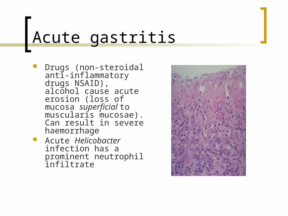
Acute gastritis
Drugs (non-steroidal anti-inflammatory drugs NSAID), alcohol cause acute erosion (loss of mucosa superficial to muscularis mucosae). Can result in severe haemorrhage
Acute Helicobacter infection has a prominent neutrophil infiltrate
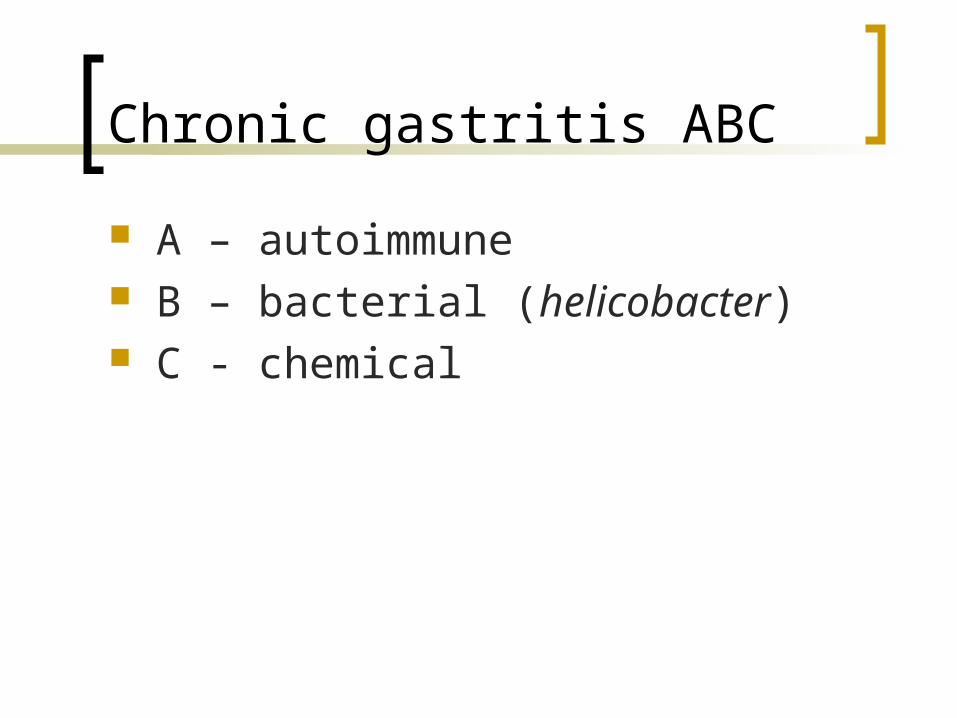
Chronic gastritis ABC
A – autoimmune B – bacterial (helicobacter) C - chemical
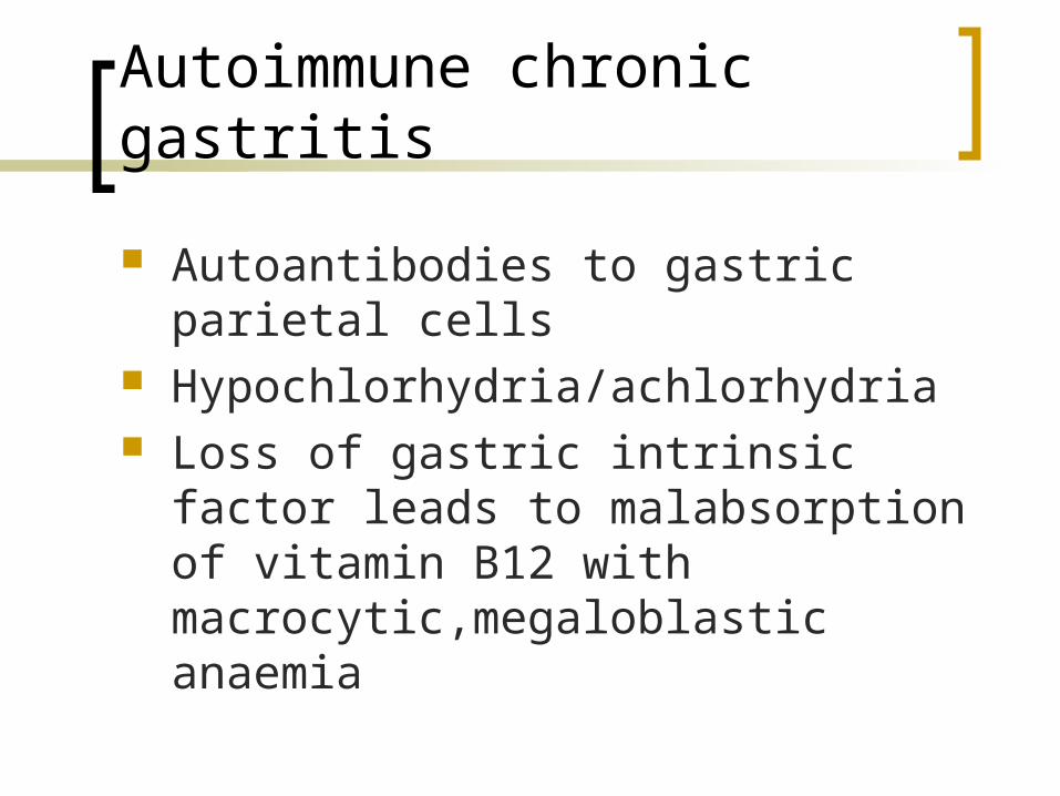
Autoimmune chronic gastritis
Autoantibodies to gastric parietal cells Hypochlorhydria/achlorhydria Loss of gastric intrinsic factor leads to
malabsorption of vitamin B12 with macrocytic,megaloblastic anaemia
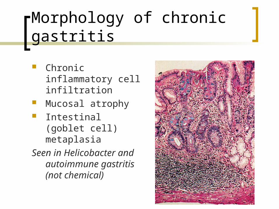
Morphology of chronic gastritis
Chronic inflammatory cell infiltration
Mucosal atrophy Intestinal (goblet cell)
metaplasia
Seen in Helicobacter and autoimmune gastritis (not chemical)
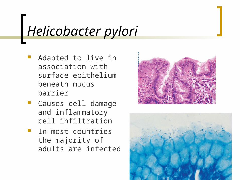
Helicobacter pylori
Adapted to live in association with surface epithelium beneath mucus barrier
Causes cell damage and inflammatory cell infiltration
In most countries the majority of adults are infected
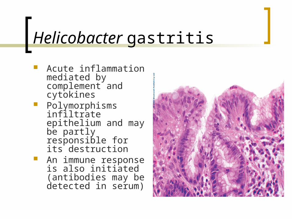
Helicobacter gastritis
Acute inflammation mediated by complement and cytokines
Polymorphisms infiltrate epithelium and may be partly responsible for its destruction
An immune response is also initiated (antibodies may be detected in serum)
Helicobacter gastritis
2 patterns of infection Diffuse involvement of body and antrum
(“pan gastritis” associated with diminishing acid output)
Infection confined to antrum (antral gastritis, associate with increased acid output)

Chemical gastritis
Commonly seen with bile reflux (toxic to cells)
Prominent hyperplastic response (inflammatory cells scanty)
With time – intestinal metaplasia
Consequences of gastritis
Peptic ulcer disease (Helicobacter) Adenocarcinoma (all types)
The “African enigma” – are complications of H.pylori infection less frequent in
Africans? Case not yet resolved
Peptic ulcer disease
A surface breach of mucosal lining of GI tract occurring as a result of acid and pepsin attack
Sites: Duodenum (DU) Stomach (GU) Oesophagus Gastro-enterostomy stoma Related to ectopic gastric mucosa (e.g. in
Meckel’s diverticulum)
Acute peptic ulcer
Acute erosion but breaching muscularis mucosae
Specific examples Curling’s ulcer (following severe burns) Cushing’s ulcer (following head injury) Preventive ante acid therapy
Chronic peptic ulcer
Complex epidemiology DU most common in Europe, GU in
Japan Incidence of DU declining, GU stable
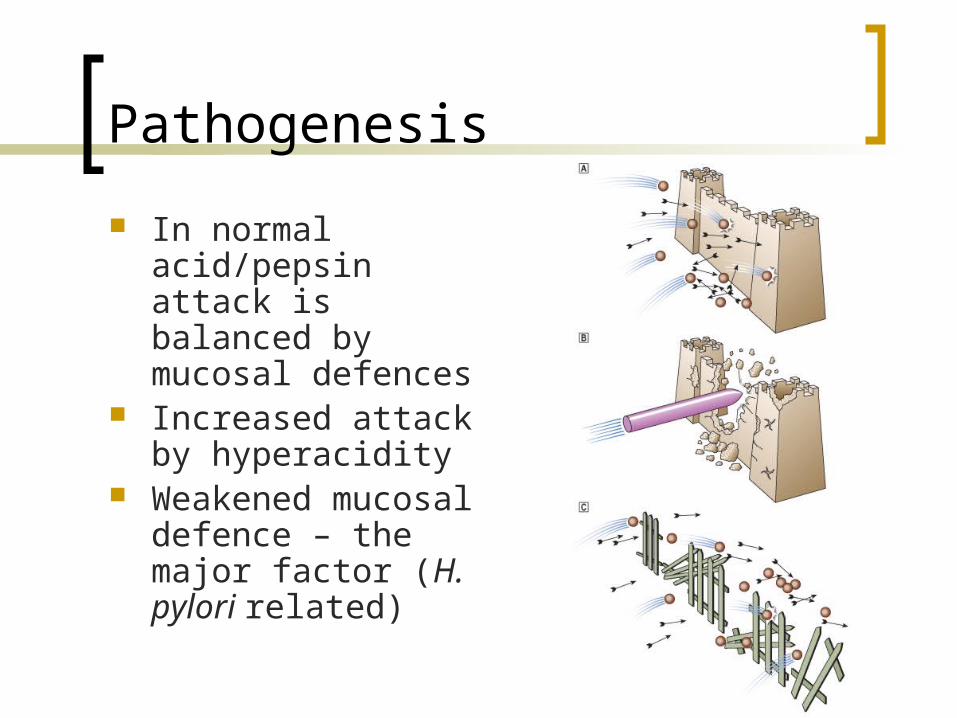
Pathogenesis
In normal acid/pepsin attack is balanced by mucosal defences
Increased attack by hyperacidity
Weakened mucosal defence – the major factor (H. pylori related)

Acid production
Tends to be high in DU patients. Antral gastritis causes increased gastrin production and acid secretion
Acid stimulates development of gastric metaplasia in the duodenum
Helicobacter organisms colonise the metaplastic epithelium and cause inflammatory damage leading to ulceration
Acid in GU
Pan gastritis diminishes acid secretion Ongoing gastritis and epithelial
damage is the main causal factor for ulceration

Helicobacter factors in pathogenesis
Some strains are more pathogenic than others. The Cag A (cytotoxic) antigen is one important virulence factor
Human variability also plays a part (e.g. individuals who produce high levels of IL-1 in inflammation get pan gastritis and GU, lower levels associated with antral gastritis and DU)
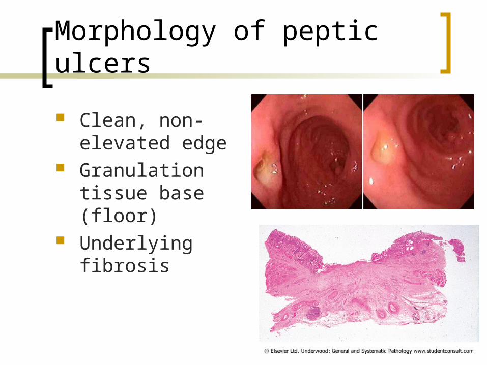
Morphology of peptic ulcers
Clean, non-elevated edge
Granulation tissue base (floor)
Underlying fibrosis
Complications of peptic ulcer
Perforation leading to peritonitis Haemorrhage by erosion of vessel in base Penetration of surrounding organ
(liver/pancreas) Obstruction (by scarring) – pyloric stenosis (Cancer – rare event in true peptic ulcer)
Gastric neoplasms
Polyps are common but usually not neoplastic (hyperplastic polyps. Hamartomas, ectopic pancreas)
Adenomas occur but are rare

Carcinoma of the stomach
The second most common fatal malignancy in the world
(after lung cancer) Commonest in Far East (Japan) Incidence declining High mortality unless disease detected
early
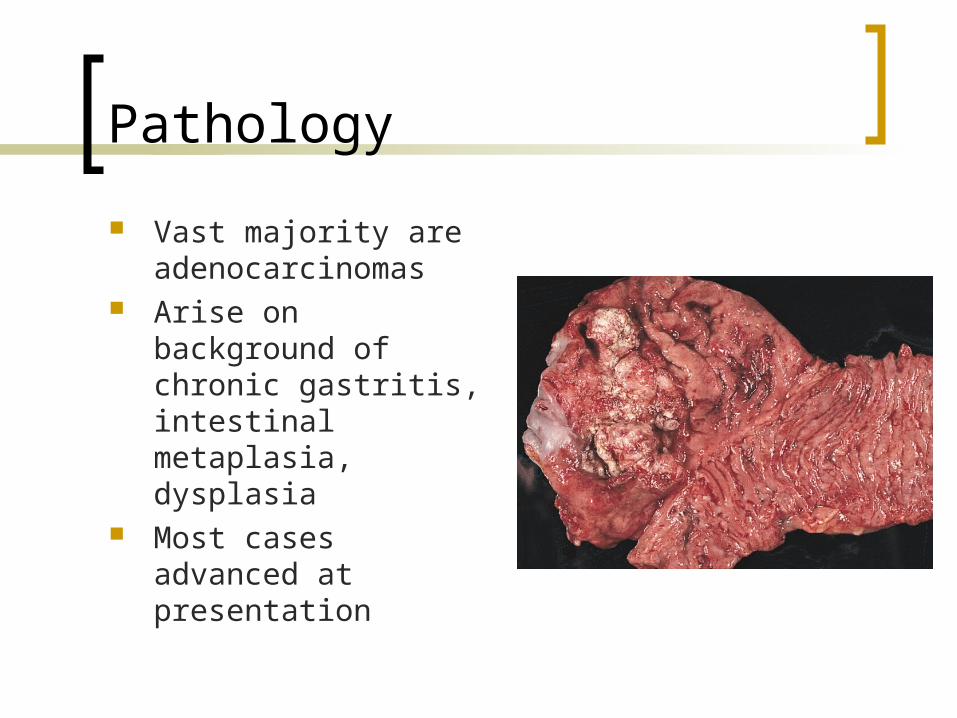
Pathology
Vast majority are adenocarcinomas
Arise on background of chronic gastritis, intestinal metaplasia, dysplasia
Most cases advanced at presentation
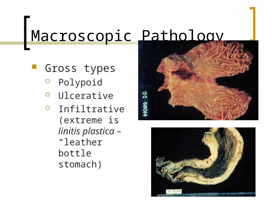
Macroscopic Pathology
Gross types Polypoid Ulcerative Infiltrative (extreme
is linitis plastica – “leather bottle stomach)
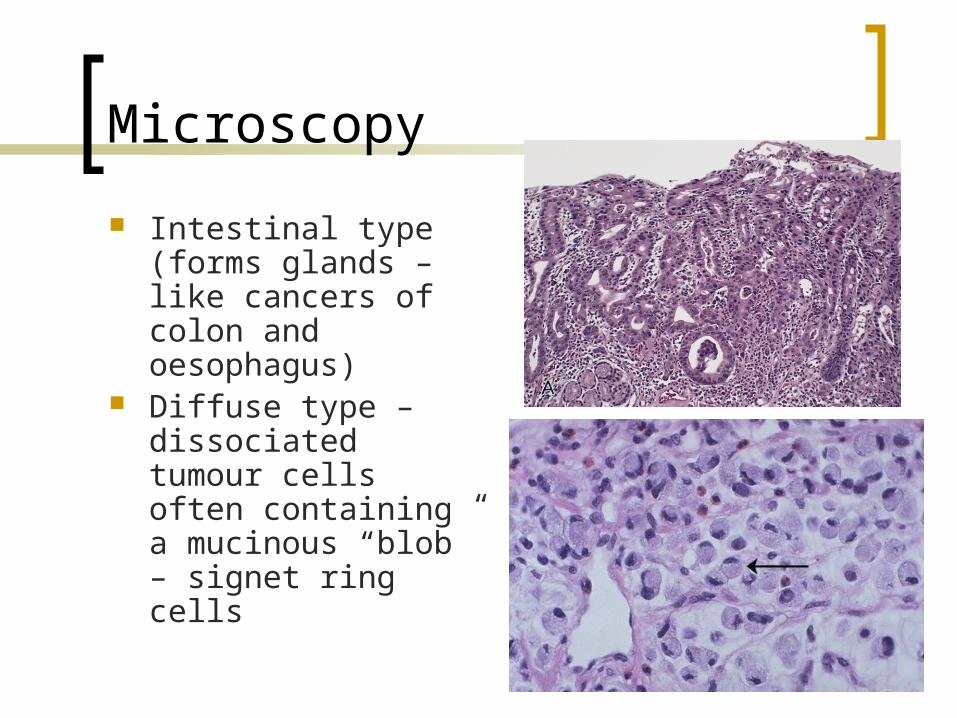
Microscopy
Intestinal type (forms glands – like cancers of colon and oesophagus)
Diffuse type – dissociated tumour cells often containing a mucinous “blob” – signet ring cells
Spread of gastric carcinoma
Local infiltration (through wall of stomach to peritoneum, pancreas etc)
Lymphatic – local and regional lymph nodes Blood – liver, lungs Transcoelomic (across peritoneal cavity).
Often involves ovaries (esp. signet ring cancer) – Krukenberg tumour.
Less common gastric neoplasms
Lymphoma Gastrointestinal stromal tumour (GIST) Neuroendocrine (carcinoid) tumours
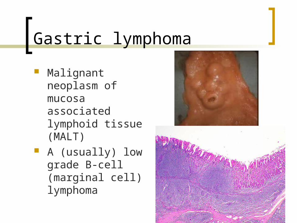
Gastric lymphoma
Malignant neoplasm of mucosa associated lymphoid tissue (MALT)
A (usually) low grade B-cell (marginal cell) lymphoma

Gastric lymphoma (maltoma)
Neoplastic cells infiltrate the epithelium (lymphoepithelial lesions)
Strongly associated with H. pylori and can be cured by eliminating infection.
Gastrointestinal stromal tumours (GIST)
Mesenchymal neoplasms Derived from interstitial cells of Cajal
(pacemaker cells controlling peristalsis)
Overexpress c-kit oncogene Used as diagnostic aid on tissue A target for therapy with tyrosine kinase
inhibitor (also used in CML)

GIST-spindle cell neoplasm of GI tract
GIST
Larger tumours with high mitotic rate tend to behave malignantly
Stomach is commonest site
Neuroendocrine tumours
Carcinoids are tumours of resident neuroendocrine cells in gastric glands
Usually seen in context of chronic atrophic gastritis (driven by gastrin)
Clinical behaviour variable

Any question?