Gastrointestinal lymphoma in childhoodjcp.bmj.com/content/jclinpath/23/6/459.full.pdf ·...
5
J. clin Path., 1970, 23, 459-463 Gastrointestinal lymphoma in childhood C. L. BERRY AND JEAN W. KEELING From the Department of Morbid Anatomy, Institute of Child Health and Hospital for Sick Children, Great Ormond Street, London SYNOPSIS Thirteen cases of lymphoma of the bowel in infancy and childhood are presented. There is a marked preponderance in males. The tumour most commonly presents with abdominal pain and intestinal obstruction; the prognosis is poor. Histological findings characteristic of the 'Burkitt' type of lymphosarcoma do not apparently influence survival. Primary neoplasms of the gastrointestinal tract are infrequent in childhood and comprise less than 1 % of tumours seen in this age group. The majority of the recorded cases are tumours of the reticuloendothelial system. Several centres have published small series describing intra- abdominal lymphosarcomata alone or as part of a review of malignant lymphomata in childhood (Bailey, Burgert, and Dahlin, 1961; Jones and Klingberg, 1963; Rosenberg, Diamond, Dargeon, and Craver, 1958; Sullivan, 1962; Cutler, Stark, and Scott, 1945; Charache, 1956; Maxwell, 1954; Mestel, 1959). We present here a review of 13 primary malignant gastrointestinal lymphosarcomata, with an assessment of their course, response to therapy, and histological appearance. Materials and Methods During the 10-year period 1960-69, 15 primary tumours of the gastrointestinal tract have been seen in this hospital (upper age limit is approxi- mately 13 years). Two were teratomata arising in the gastric wall and have been reported else- where (Atwell, Claireaux, and Nixon, 1967; Berry, Keeling, and Hilton, 1969). From the remaining 13 cases, sections of the surgical specimens were stained with haematoxylin and eosin, haematoxylin-van Gieson, Gordon and Received for publication 2 December 1969. Sweet's reticulin stain, and the Unna-Pappenheim techniques. Necropsy material was available from one case (case 2). Results The clinical details, age at presentation, sex, duration of symptoms, operative findings, sub- sequent treatment, and progress are shown in Table I. PATHOLOGICAL FINDINGS Typical macroscopic appearances are seen in Figs. 1 and 2 (case 5) and Figs. 3 and 4 (case 7). Microscopic findings are summarized in Table II. Ten tumours are lymphoblastic lympho- sarcomata and are composed of closely packed round cells having prominent nuclei with little cytoplasm (Fig. 5). Five of these tumours contain large phagocytic reticular cells giving a 'starry sky' appearance and in three instances other cytological characteristics of the 'Burkitt' type of lymphoma (ill-defined cytoplasmic border, small cytoplasmic vacuoles, prominent nucleoli, frequent mitoses, occasional pyroninophilia) are present (Fig. 6). Involvement of the muscle coats of the bowel wall leads to the appearance of altered muscle cells within the tumour in some instances. Three tumours are considered to be reticulum copyright. on 5 September 2018 by guest. Protected by http://jcp.bmj.com/ J Clin Pathol: first published as 10.1136/jcp.23.6.459 on 1 September 1970. Downloaded from
Transcript of Gastrointestinal lymphoma in childhoodjcp.bmj.com/content/jclinpath/23/6/459.full.pdf ·...

J. clin Path., 1970, 23, 459-463
Gastrointestinal lymphoma in childhood
C. L. BERRY AND JEAN W. KEELINGFrom the Department of Morbid Anatomy, Institute of Child Health and Hospital for Sick Children,Great Ormond Street, London
SYNOPSIS Thirteen cases of lymphoma of the bowel in infancy and childhood are presented.There is a marked preponderance in males. The tumour most commonly presents withabdominal pain and intestinal obstruction; the prognosis is poor. Histological findingscharacteristic of the 'Burkitt' type of lymphosarcoma do not apparently influence survival.
Primary neoplasms of the gastrointestinal tractare infrequent in childhood and comprise lessthan 1% of tumours seen in this age group. Themajority of the recorded cases are tumours ofthe reticuloendothelial system. Several centreshave published small series describing intra-abdominal lymphosarcomata alone or as part ofa review of malignant lymphomata in childhood(Bailey, Burgert, and Dahlin, 1961; Jones andKlingberg, 1963; Rosenberg, Diamond, Dargeon,and Craver, 1958; Sullivan, 1962; Cutler, Stark,and Scott, 1945; Charache, 1956; Maxwell,1954; Mestel, 1959).We present here a review of 13 primary
malignant gastrointestinal lymphosarcomata,with an assessment of their course, response totherapy, and histological appearance.
Materials and Methods
During the 10-year period 1960-69, 15 primarytumours of the gastrointestinal tract have beenseen in this hospital (upper age limit is approxi-mately 13 years). Two were teratomata arisingin the gastric wall and have been reported else-where (Atwell, Claireaux, and Nixon, 1967;Berry, Keeling, and Hilton, 1969). From theremaining 13 cases, sections of the surgicalspecimens were stained with haematoxylin andeosin, haematoxylin-van Gieson, Gordon andReceived for publication 2 December 1969.
Sweet's reticulin stain, and the Unna-Pappenheimtechniques. Necropsy material was availablefrom one case (case 2).
Results
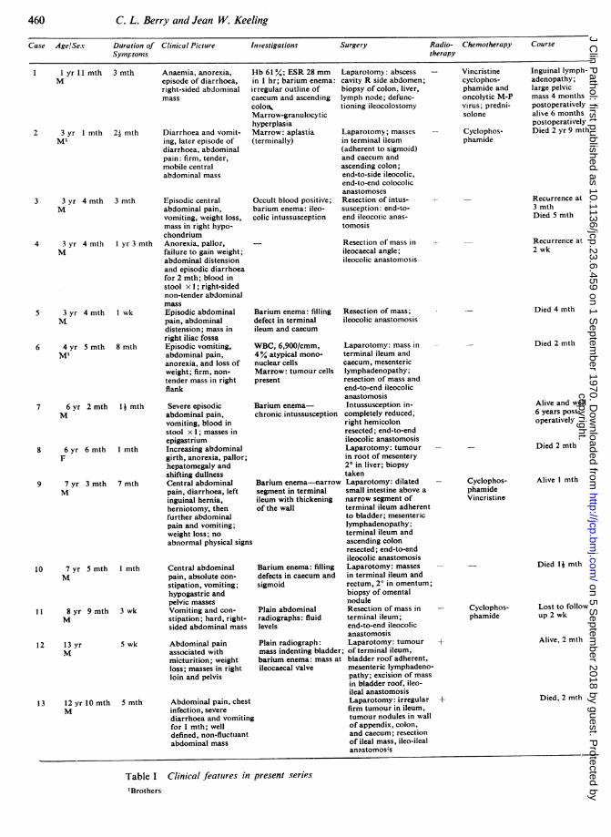
The clinical details, age at presentation, sex,duration of symptoms, operative findings, sub-sequent treatment, and progress are shown inTable I.
PATHOLOGICAL FINDINGSTypical macroscopic appearances are seen inFigs. 1 and 2 (case 5) and Figs. 3 and 4 (case 7).
Microscopic findings are summarized in TableII. Ten tumours are lymphoblastic lympho-sarcomata and are composed of closely packedround cells having prominent nuclei with littlecytoplasm (Fig. 5). Five of these tumours containlarge phagocytic reticular cells giving a 'starrysky' appearance and in three instances othercytological characteristics of the 'Burkitt' typeof lymphoma (ill-defined cytoplasmic border,small cytoplasmic vacuoles, prominent nucleoli,frequent mitoses, occasional pyroninophilia) arepresent (Fig. 6). Involvement of the muscle coatsof the bowel wall leads to the appearance ofaltered muscle cells within the tumour in someinstances.
Three tumours are considered to be reticulum
copyright. on 5 S
eptember 2018 by guest. P
rotected byhttp://jcp.bm
j.com/
J Clin P
athol: first published as 10.1136/jcp.23.6.459 on 1 Septem
ber 1970. Dow
nloaded from
C. L. Berry and Jean W. Keeling
Case Age/Sex Duration of Clinical Picture Investigations Surgery Radio- Chemotherapy Course
Symptoms therapy
Anaemia, anorexia,episode of diarrhoea,right-sided abdominalmass
Diarrhoea and vomit-ing, later episode ofdiarrhoea, abdominalpain: firm, tender,mobile centralabdominal mass
Episodic centralabdominal pain,vomiting, weight loss,mass in right hypo-chondriumAnorexia, pallor,failure to gain weight;abdominal distensionand episodic diarrhoeafor 2 mth; blood instool x 1; right-sidednon-tender abdominalmassEpisodic abdominalpain, abdominaldistension; mass inright iliac fossaEpisodic vomiting,abdominal pain,anorexia, and loss ofweight; firm, non-tender mass in rightflank
7 6 yr 2 mth 1 mth Severe episodicM abdominal pain,
vomiting, blood instool x 1; masses inepigastrium
8 6 yr 6 mth I mth Increasing abdominalF girth, anorexia, pallor;
hepatomegaly andshifting dullness
9 7 yr 3 mth 7 mth Central abdominalM pain, diarrhoea, left
inguinal hernia,herniotomy, thenfurther abdominalpain and vomiting;weight loss; noabnormal physical signs
10 7 yr 5 mth 1 mth Central abdominalM pain, absolute con-
stipation, vomiting;hypogastric andpelvic masses
1t 8 yr 9 mth 3 wk Vomiting and con-M stipation; hard, right-
sided abdominal mass
1 2 13 yrM
13 12 yr 10 mthM
5 wk Abdominal painassociated withmicturition; weightloss; masses in rightloin and pelvis
5 mth Abdominal pain, chestinfection, severediarrhoea and vomitingfor 1 mth; welldefined, non-fluctuantabdominal mass
Hb 61 %; ESR 28 mm Laparotomy: abscess - Vincristine Inguinal Iympn-in I hr; barium enema: cavity R side abdomen;irregular outline of biopsy of colon, liver,caecum and ascending lymph node; defunc-coloN tioning ileocolostomyMarrow-granulocytichyperplasiaMarrow: aplastia Laparotomy; masses(terminally) in terminal ileum
Occult blood positive;barium enema: ileo-colic intussusception
Barium enema: fillingdefect in terminalileum and caecum
(adherent to sigmoid)and caecum andascending colon;end-to-side ileocolic,end-to-end colocolicanastomosesResection of intus-susception: end-to-end ileocotic anas-tomosis
Resection of mass inileocaecal angle;ileocolic anastomosis
cyclophos-phamide andoncolytic M-Pvirus; predni-solone
Cyclophos-phamide
adenopathy;large pelvicmass 4 monthspostoperativelyalive 6 monthspostoperativelyDied 2 yr 9 mth
Recurrence at3 mthDied 5 mth
Recurrence at2 wk
Died 4 mthResection of mass;ileocolic anastomosis
WBC, 6,900/cmm, Laparotomy: mass in4% atypical mono- terminal ileum andnuclear cells caecum, mesentericMarrow: tumour cells lymphadenopathy;present resection of mass and
end-to-end ileocolicanastomosis
Barium enema- Intussusception in-chronic intussusception completely reduced,
right hemicolonresected; end-to-endileocolic anastomosisLaparotomy: tumourin root of mesentery2° in liver; biopsytaken
Barium enema-narrow Laparotomy: dilatedsegment in terminal small intestine above aileum with thickening narrow segment ofof the wall terminal ileum adherent
to bladder; mesentericlymphadenopathy;terminal ileum andascending colonresected; end-to-endileocolic anastomosis
Barium enema: filling Laparotomy: massesdefects in caecum and in terminal ileum andsigmoid rectum, 2° in omentum;
biopsy of omentalnodule
Plain abdominal Resection of mass inradiographs: fluid terminal ileum;levels end-to-end ileocolic
anastomosisPlain radiograph: Laparotomy: tumour +mass indenting bladder; of terminal ileum,barium enema: mass at bladder roof adherent,ileocaecal valve mesenteric lymphadeno-
pathy; excision of massin bladder roof, ileo-ileal anastomosisLaparotomy: irregular +firm tumour in ileum,tumour nodules in wallof appendix, colon,and caecum; resectionof ileal mass, ileo-ilealanastomosis
Died 2 mth
Alive and well6 years post-operatively
Died 2 mth
Cyclophos-phamideVincristine
Alive 1 mth
- Died Ij mth
Cyclophos- Lost to followphamide up 2 wk
Alive, 2 mth
Died, 2 mth
Clinical feattires in present series
460
1 I yr II mth 3 mthM
2 3 yr I mth 2. mthMl
3 3 yr 4 mth 3 mthM
4 3 yrM
4 mth I yr 3 mth
5 3 yr 4 mthM
6 4 yr S mthMl
I wk
8 mth
Table I'Brothers
Id
r
II
Ec
I
i4
copyright. on 5 S
eptember 2018 by guest. P
rotected byhttp://jcp.bm
j.com/
J Clin P
athol: first published as 10.1136/jcp.23.6.459 on 1 Septem
ber 1970. Dow
nloaded from

461 Gastrointestinal lymphoma in childhood
"1 2 3 4 b 6 7 91 1s0 1II 12
Fig. 1 Case 5: white nodules can be seen throughthe serosa distorting the normal intestinal profile.Enlarged lymph nodes are seen in the mesentery.
2h
Fig. 2 Case 5: Hemisection shows a homogeneouswhite tumour involving the bowel wall with ulcerationof the overlying mucosa and affecting local lymphnodes by direct extension.
Fig. 4 Case 7: Hemisection shows an area of whitetumour within the bowel wall at the apex of theintussusception.
t̂'" 'T l
i z:.. 2 4
Fig. 3 Case 7: the terminal ileum (right) andcaecum are diffusely thickened.
.I
r.
.lI,:146 .Alk
..*.I
...1:4.,
copyright. on 5 S
eptember 2018 by guest. P
rotected byhttp://jcp.bm
j.com/
J Clin P
athol: first published as 10.1136/jcp.23.6.459 on 1 Septem
ber 1970. Dow
nloaded from

462 C. L. Berry and Jean W. Keeling
Fig. 5 Typical appearance of lymphosarcoma(haematoxylin and eosin x 500).
Fig. 6 Lymphosarcoma with histiocytes containingnuclear debris scattered in the tumour mass
(haematoxylin and eosin x 540).
Case Site HistologicalAppearance
1 Caecum and ascending colon, Lymphosarcoma20 liver
2 Terminal ileum and caecum Reticulum cell sarcoma3 Terminal ileum and caecum Lymphosarcoma'4 Terminal ileum and caecum Lymphosarcoma5 Terminal ileum and caecum, 20 Lymphosarcomal
mesenteric nodes6 Terminal ileum, caecum, Reticulum cell sarcoma
ascending colon7 Terminal ileum and caecum Lymphosarcoma8 Root of mesentery, liNer 20 Lymphosarcoma9 Terminal ileum Lymphosarcoma10 Ileum, rectum, omental 2° Lymphosarcomal11 Terminal ileum and caecum Lymphosarcomal12 Ileum 20, bladder vault Reticulum cell sarcoma13 Ileum, nodules in caecum and Lymphosarcoma'
colon, appendix
Table II Histology ofpresent series'Burkitt-like morphology
cell sarcomas, and are composed of pleomorphicreticulin cells; binucleate cells are seen. Reticulinfibres are seen between cells and thicker con-densations of reticulin divide cell masses.Bone marrow examination was normal in
cases 3, 4, 9, 11, 12, and 13. The narrow aspiratein case 1 showed granulocytic hyperplasia butno tumour infiltration. Appearances were withinnormal limits in case 2 initially but terminalaplasia was found. In case 6 the tumour cells werefound terminally. Bone marrow examination wasnot performed in cases 5, 7, 8, and 10.Necropsy was performed on case 2; he died
two years and nine months after resection of anileocaecal tumour. Bone marrow aplasia andpulmonary fibrosis had developed and wasapparently associated with cytotoxic drugtherapy.There was cultural and histological evidence
of disseminated moniliasis with involvement ofthe central nervous system. Residual tumour wasnot found.Tumour had involved bone marrow terminally
in case 6, with the appearance of tumour cells inthe blood. Metastases occurred in cases 1, 3, 4,8, 10, and 12. Transcoelomic spread of tumourto other abdominal viscera was probable incase 13.
Discussion
The age range at presentation was from 1 year11 months to 12 years 10 months in our series.Twelve of the 13 patients were male; a
similar degree of male preponderance was notedin the series described by Mestel (1959), 11 maleand two female cases, but was less marked inother studies. Bailey et al (1961) found a male-to-female ratio of 3-4:1, and Jones and Klingberg(1963) had a male-to-female ratio of 2-6:1.The 13 tumours constitute 0-6% of all tumours
copyright. on 5 S
eptember 2018 by guest. P
rotected byhttp://jcp.bm
j.com/
J Clin P
athol: first published as 10.1136/jcp.23.6.459 on 1 Septem
ber 1970. Dow
nloaded from

Gastrointestinal lymphoma in childhood
seen at this hospital since 1957 (excludingleukaemia) and 37% of the lymphomata.The presence of Burkitt-like morphology was
not associated with a better prognosis and thephagocytic activity of histiocytes included in thetumour does not appear to be evidence of aneffective host response to the tumour.Two of the cases were brothers (cases 2 and 6):
the familial tendency to develop lymphomata hasbeen discussed elsewhere (Rigby, Rosenlof,Pratt, and Lemon, 1966) and will not be con-sidered further here.Abdominal pain and distension were the most
frequent presenting symptoms, as in most seriesof both adult and childhood age groups (seeMestel, 1959, for bibliography).
Investigation showed no distinctive features,and in general findings were those of intestinalobstruction and intussusception.
Primary treatment was surgical and was de-signed to achieve complete resection (cases 2, 3,4, 5, 6, 7, 9, 11, and 12) or palliation (cases 1 and13). In cases 8 and 10 biopsy only was performedand no further treatment was given. Cases 3, 4, 5,and 13 were treated by radiotherapy and cases 1,2, 9, 11, and 12 by cytotoxic drugs in addition.Four patients are alive but two have been diag-nosed only in the last few months. Of the remain-ing two, case 1 is alive with evidence of extensivedisease after six months. There is one long-termsurvivor (case 7) who is free of disease six yearsafter radical surgery alone. These dismal resultscompare with those found in other series (Char-ache, 1956; Mestel, 1959). Bailey et al (1961)have achieved some success with surgery andradiotherapy in a small series (three long-termsurvivors from nine cases).
These cases presented over a period of 10 years,and policy regarding chemotherapy and radio-therapy probably reflects common usage at thattime, except in cases 8 and 10 where extensivemetastatic spread was present at laparotomy andspecific antitumour therapy was withheld. Bonemarrow examination was not performed in allcases, and the use of antitumour therapy was notbased on the findings in the blood.
In general this is a tumour with an exceedinglygrave prognosis. Its relative infrequency and thevaried methods of treatment employed prevent arational assessment of the efficiency of varioustherapeutic regimes but it seems likely that radicalsurgery with radiotherapy aimed at preventinglocal recurrence is a rational mode of treatmentin view of the present state of knowledge of thisdisease.
C. L. Berry is the Gilson scholar of the Worship-ful Society of Apothecaries of London.We should like to thank the Photographic
Department of the Hospital for Sick Children forFigures 1-4.
References
Atwell, J. D., Claireaux, A. E., and Nixon, H. H. (1967). Teratomaof the stomach in the newborn. J. pediat. Surg., 2, 197-204.
Bailey, R. J., Jr., Burgert, E. O., Jr., and Dahlin, D. C. (1961).Malignant lymphoma in children. Pediatrics., 28, 985-992,
Berry, C. L., Keeling, J., and Hilton, C. (1969). Teratomata ininfancy and childhood: a review of 91 cases. J. Path., 98,241-252.
Charache, H. (1956). Lymphosarcoma in infancy and childhood:including a case of twenty-two years' survival. Amer. J.Roentgenol., 76, 594-598.
Cutler, G. D., Stark, R. B., and Scott, H. W. (1945). Lympho-sarcoma of the bowel in childhood. New Engl. J. Med., 232,665-670.
Jones, B., and Klingberg, W. G. (1963). Lymphosarcoma inchildren. A report of 43 cases and review of the recentliterature. J. Pediat., 63, 11-20.
Maxwell, G. M. (1954). Twelve cases of lymphoblastomata inchildren. Arch. Dis. Childh., 29, 155-159.
Mestel, A. L. (1959). Lymphosarcoma of the small intestine ininfancy and childhood. Ann. Surg., 149, 87-94.
Rigby, P. G., Rosenlof, R. C., Pratt, P. T., and Lemon, H. M.(1966). Leukemia and lymphoma. J. Amer. med. Ass.,197, 25-30.
Rosenberg, S. A., Diamond, H. D., Dargeon, H. W., and Craver,L. F. (1958). Lymphosarcoma in childhood. New Engi. J.Med., 259, 505-512.
Sullivan, M. P. (1962). Leukemic transformation in lympho-sarcoma of childhood. Pediatrics, 29, 589-599.
Addendum
Since the preparation of this paper, case 9 hasdied, eight months after diagnosis.
463copyright.
on 5 Septem
ber 2018 by guest. Protected by
http://jcp.bmj.com
/J C
lin Pathol: first published as 10.1136/jcp.23.6.459 on 1 S
eptember 1970. D
ownloaded from


![Reference list concerning Hodgkin's lymphoma · [Letter] The role of cytomegalovirus in inflammatory bowel disease and gastrointestinal lymphoma. Gastroenterology, 123(1) pp 390;](https://static.fdocuments.in/doc/165x107/6048c4c56c1e0c19fb3bc548/reference-list-concerning-hodgkins-letter-the-role-of-cytomegalovirus-in-inflammatory.jpg)