Gastric and duodenal ulcer disease. Anatomy Arterial blood supply Lymphatic drainage Nerve supply.
60
Gastric and duodenal Gastric and duodenal ulcer disease ulcer disease
-
Upload
sherman-matthews -
Category
Documents
-
view
224 -
download
3
Transcript of Gastric and duodenal ulcer disease. Anatomy Arterial blood supply Lymphatic drainage Nerve supply.

Gastric and duodenal ulcer Gastric and duodenal ulcer diseasedisease

AnatomyAnatomy Arterial blood supply Lymphatic drainage Nerve supply

PHYSIOLOGYPHYSIOLOGYFunction:
1. Digestion of food, reduce the size of food
2. Acts as reservoir
3. Absorption of Vit. 12, iron and calcium
Stimulant of Gastric secretion:1. Gastrin -----> (+) parietal cell
2. Acetylcholine (vagus) ---> (+) gastric cells
3. Histamine (mast cells) ---> parietal & chief cells

PHYSIOLOGYPHYSIOLOGYBAO: 2 – 5 meq of acid/hr. (vagal tone and basal
histamine secretion)
MAO: 1. Cephalic (vagus) ---> (+) parietal & G cell
10 meq acid/hr.
2. Gastric: ---> (+) vagus & G cell 15 – 25 meq of acid/hr pH = < 2.0
3. Intestinal: Chyme enters the duodenum (-) gastric release Secretin, gastric inhibitory peptide, peptide YY
– ACID condition sterilized the area, except for HELICOBACTER PYLORI

Protective factors Protective factors vs.vs. hostile factors hostile factors
Peptic Ulcer DiseasePeptic Ulcer Disease
PathogenesisPathogenesis : :

Peptic ulcerPeptic ulcerPathogenesis:1. For both Duodenal & Gastric Ulcers:
a. Infection w/ H. pylori: Decreases resistance of mucus layer from acid
permeation (hydrophobicity)
Increase acid secretion
Slow duodenal emptying
Reduced both duodenal and gastric bicarbonate secretion

Clinical ManifestationClinical Manifestation1. Abdominal pain:
– Due to irritation of afferent nerves w/in the ulcer by the acid or due to peristaltic waves passing through the ulcer
Duodenal: colicky or burning pain relieved w/ food intake
Gastric: gnawing or burning usually during or after eating.
2. N/V
3. Weight loss
4. Epigastric tenderness

Peptic ulcerPeptic ulcerPathogenesis:
b. Effects of NSAIDs Decreases ProstagladinProstaglandin – inhibits acid secretion, stimulates mucus
and HCO3 secretion and mucosal blood flow
c. Zollinger-Ellison Syndrome (1%): Massive secretion of HCL due to ectopic gastrin
production from non-beta islet cell tumor (gastrinoma) Associated w/ type I (MEN) PPP 20% multiple, 2/3 malignant, w/ slow growing Parietal cell mass is increased > gastrin 3-6 x the normal

Symptoms of gastric ulcer disease:
epigastric pain after meal or during meal
upper dyspeptic syndrome – loss of appetite, nauzea, vomiting, flatulence
vomiting brings relief
reduced nutrition
loss of weight

Comparing Duodenal Comparing Duodenal and Gastric Ulcersand Gastric Ulcers

Symptoms of duodenal ulcer disease:
epigastric pain 2 hours after meal or on a empty stomach or during night
pyrosis
good nutrition
obstipation
seasonal dependence (spring, autumn)
Diagnosis:Diagnosis:
1. UGIS (double contrast)
2. Endoscopy

Therapy:
Conservative • regular lifestyle• prohibition of the smoking and alcohol• diet (proteins, milk and milky products)• pharmacology (antagonists of H2 receptors,
antacids, anticholinergics
Surgical • BI, BII resection• proximal selective vagotomy• vagotomy with pyloroplastic• suture of perforated or haemorrhagic ulcer

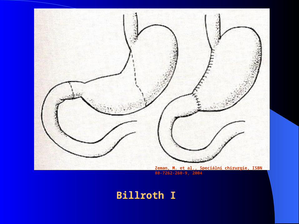
Stomach Stomach resections:resections:
BillrothBillroth I (BI) I (BI) – – gastro-duodenoanastomosis end-to-endgastro-duodenoanastomosis end-to-end
Billroth II (BII)Billroth II (BII) – gastro-jejunoanastomosis end-to-side – gastro-jejunoanastomosis end-to-side with blind closure of duodenumwith blind closure of duodenum
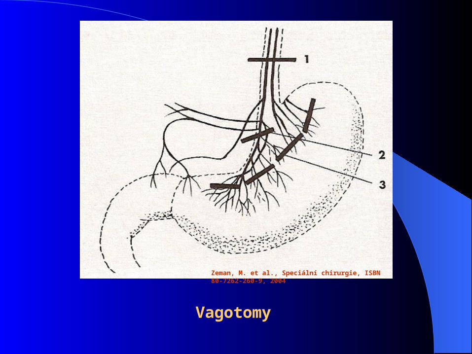
ProximalProximal selective vagotomyselective vagotomy – denervation – denervation of parietalof parietal gastric cellsgastric cells
Zeman, M. et al., Speciální chirurgie, ISBN 80-7262-260-9, 2004
Billroth I

Billroth II
Zeman, M. et al., Speciální chirurgie, ISBN 80-7262-260-9, 2004

Zeman, M. et al., Speciální chirurgie, ISBN 80-7262-260-9, 2004
Gastro-enteroanastomosis on Roux Y crankle
Zeman, M. et al., Speciální chirurgie, ISBN 80-7262-260-9, 2004
Vagotomy
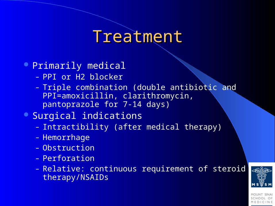
TreatmentTreatment
Primarily medical– PPI or H2 blocker– Triple combination (double antibiotic and PPI=amoxicillin,
clarithromycin, pantoprazole for 7-14 days) Surgical indications
– Intractibility (after medical therapy)– Hemorrhage– Obstruction– Perforation– Relative: continuous requirement of steroid therapy/NSAIDs
Treatment:Treatment:
Mechanism of Pharmacologic Therapy:4. For eradication of H. pylori:
a. Bismuth based triple therapy Bismuth + Tetracycline + Metronidazole
b. Proton pump inhibitor Omeprazole + Amoxicillin/Clarithromycin
+ metronidazole

Treatment:Treatment:
Surgical Treatment:Indication:1. Intractability:
– Highly selective vagotomy Low septic complication, (-) dumping and diarrhea
– For gastric ulcer: Total or subtotal gastrectomy w/ or w/o vagotomy

Zeman, M. et al., Speciální chirurgie, ISBN 80-7262-260-9, 2004
A – penetration B – perforation
C – bleeding D - stenosis

GI BleedingGI Bleeding

Ulcer with recent bleedUlcer with recent bleed
Treatment:Treatment:Surgical Treatment:
Indication:2. Hemorrhage: s/sx
– Critically ill– Endoscopy– Surgery: a. continue bleeding for more
than 6 units
b. recurrent bleeding after endoscopically controlled
- pyloroduodenostomy + HSV
- pyloroduodenostomy + vagotomy + pyloroplasty

Ulcer Perforation

Treatment:Treatment:Surgical Treatment:
Indication:3. Perforation: S/Sx
Graham omental patch only for shock, perforation > 48 hrs or other medical problem
Vagotomy + pyloroplasty; HSV Vagotomy + Gastrojejunostomy
4. Obstruction: S/Sx; Saline loading test Vagotomy + Antrectomy Vagotomy + Gastroenterostomy

Gastric outlet obstructionGastric outlet obstruction

Elective Surgical TherapyElective Surgical Therapy
Rare; most uncomplicated ulcers heal within 12 weeks
If don’t, change medication, observe addition 12 weeks
Check serum gastrin (antral G-cell hyperplasia or gastrinoma)
EGD: biopsy all 4 quadrants of ulcer (rule out malignant ulcer) if refractory
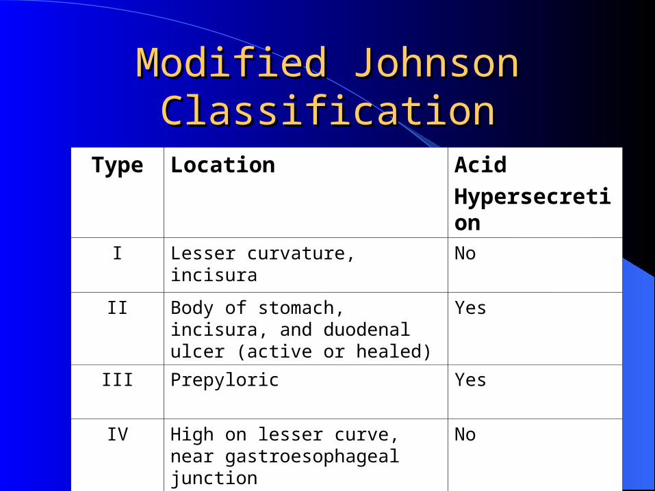
Modified Johnson Modified Johnson ClassificationClassification
Type Location Acid
HypersecretionI Lesser curvature, incisura No
II Body of stomach, incisura, and duodenal ulcer (active or healed)
Yes
III Prepyloric Yes
IV High on lesser curve, near gastroesophageal junction
No
V Anywhere (medication induced) No

Elective Surgical TherapyElective Surgical Therapy
Type I

Type IType I
Lesser curvature; incisura
MOST COMMONDecreased mucosal
protection (no vagotomy)
Distal gastrectomy (INCLUDING UCLER) with BI


Billroth IBillroth I

Elective Surgical TherapyElective Surgical Therapy
Type II/III

Type 2/3 UlcersType 2/3 Ulcers
Acid hypersecretion Antrectomy with ulcer and
bilateral truncal vagotomy Billroth II or Billroth I
depending on technical difficulty
Parietal cell vagotomy option but higher recurrence

Billroth IIBillroth II

R-Y limb (subtotal R-Y limb (subtotal gastrectomy)gastrectomy)

Elective Surgical TherapyElective Surgical Therapy
Type IV

Type 4 UlcersType 4 Ulcers Least common (5% of all
gastric ulcers) Ulcers 2-5cm from cardia
can be treated with distal gastrectomy, extending resection along the lesser curvature and BI (Pauchet/Shoemaker procedure)
Ulcers closer to GEJ, tongue-shaped resection high onto lesser curve (Csendes’ procedure with Roux-en-Y reconstruction)
Cardia
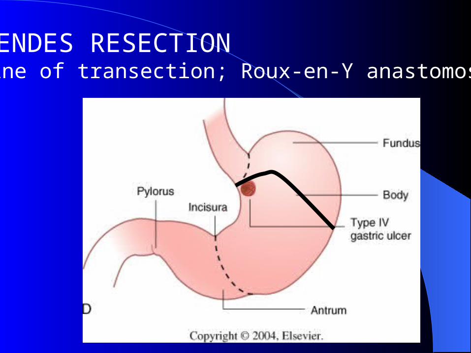
CSENDES RESECTION (Line of transection; Roux-en-Y anastomosis)

Elective Surgical TherapyElective Surgical Therapy
Giant Gastric Ulcer
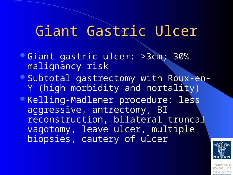
Giant Gastric UlcerGiant Gastric Ulcer
Giant gastric ulcer: >3cm; 30% malignancy risk
Subtotal gastrectomy with Roux-en-Y (high morbidity and mortality)
Kelling-Madlener procedure: less aggressive, antrectomy, BI reconstruction, bilateral truncal vagotomy, leave ulcer, multiple biopsies, cautery of ulcer

Complications after stomach resection:
Early – dehiscence, stenosis of anastomosis, bleeding, pancreatitis, obstructive icterus, affection of neighbour tissues
Late - days, weeks
- early dumping syndrome
- late dumping syndrome
- incoming crankle syndrome
- outcoming crankle syndrome
- ulcer in anastomosis or in outcoming crankle

Early Complications (1)Early Complications (1)1. Failure of the stomach or stomach remnant to empty occurs after
any procedure. It was formerly common after vagotomy and drainage. Causes are:
A. Prolonged paralysis of stomach (doubtful)
B. Edema at a stoma
C. Fluid and electrolyte disorder, especially hypokalaemia.
Management is conservative with NG suction, fluid, electrolyte and nutritional replacement.

Early Complications (2)Early Complications (2)
2. Intestinal obstruction.
Causes are:
A. Adhesive.
B. As a consequences:
(a) Twisting of the loop of a gastrojejunostomy after polya gastrectomy.
(b) Herniation of loops through a mesenteric defect.
(c) Retrograde intussusception of the efferent loop of a gastrojejunostomy (rare).
Prophylaxis: avoid causes – such as mesenteric cul de sacs or holes
Treatment: operative
Early Complications (3)Early Complications (3)
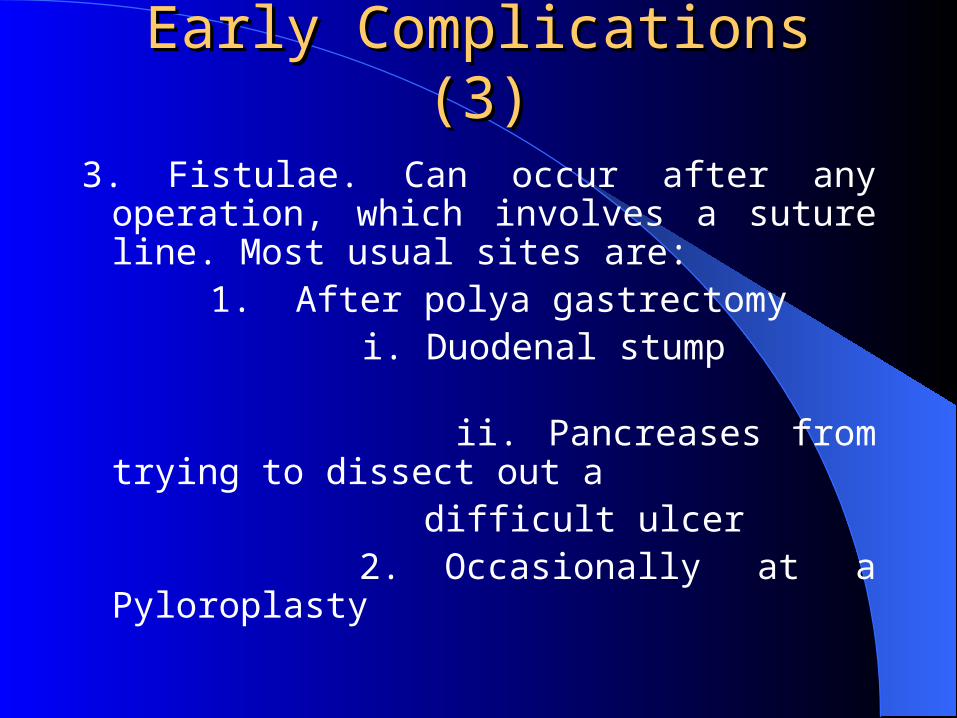
3. Fistulae. Can occur after any operation, which involves a suture line. Most usual sites are:
1. After polya gastrectomy i. Duodenal stump ii. Pancreases from trying to dissect
out a difficult ulcer 2. Occasionally at a Pyloroplasty

Early Complications (4)Early Complications (4)
4. Acute pancreatitis. May follow any procedure. Its etiology is unknown, but some cases are traumatic

Late Complications (1)Late Complications (1)1. Anastomotic and recurrent ulceration Causes: a. Inadequate resection of parietal cell mass.b. Isolated antrum left after polya gastrectomy.c. Zollinger – Ellison syndrome.d. Incomplete vagotmy.e. Persistent suture in the anastomosis. More usually this is merely a suture
exposed as a consequence of ulceration from another cause.
Prophylaxis: adequate primary treatment.
Management is related to cause and requires investigation to ascertain the level of acid secretion or the completeness of vagotomy. Recurrence after vagotomy is best managed by polya gastrectomy.

Late Complications (1)Late Complications (1)2. Gastrojejunocolic fistulae.
Occurs when a recurrent ulcer after gastrojejual anastomosis penetrates into the colon. It should arouse the suspicion of Zollinger-Ellison syndrome.
Clinical features: Severe diarrhea occurs due to enteritis caused by cronic contents passing directly into the small bowel and acidosis, dehydration, potassium loss, anaemia and cachexia will result in death if the fastula is not interrupted surgically.
Treatment:
1. Good risk patient. Excision of the gastric, jejunal and colonic components and the construction of a higher gastrectomy.
2. Poor risk patient. A staged procedure:
(a) Stage 1: Proximal colostomy which, diverts the faecal stream from the fistula and thus stops the enteritis.
(b) Stage 2: Excision of fistula and its visceral components and the construction of a higher gastrectomy and colonic anastomosis.
(c) Stage 3: Closure of colostomy.

Early dumping syndrome:
group of symptoms approved shortly after meal
appears after BII resection
vasomotoric sy. - face redness, fall of blood pressure, dizziness
GI sy. - vomiting, diarrhoea
Th.: diet, no sugar, low quantities of food, change BII to BI resection

LateLate dumping syndrome: dumping syndrome:
hhypoglycaemiaypoglycaemia (sugar is not enough digested)(sugar is not enough digested)
appears after BII resectionappears after BII resection
weakness, perspiration, dizzinessweakness, perspiration, dizziness, , tremor ccatremor cca 3h 3h afterafter mealmeal
Th.:Th.: no sugar, change BII to BI resection no sugar, change BII to BI resection
Anemia Anemia
Partial gastrectomy and polya reconstruction interferes with duodenal absorption of iron and a macrocytic anemia may result
More rarely, sufficient stomach has been removed to cause failure of release of intrinsic factor and thus a macrocytic anemia
Malnutrition may contribute to both.

Weight loss and its complications Weight loss and its complications
Particulary after partial gastrectomy when patients are unwilling to eat sufficiently, weight loss is common
Severe malnutrition is rare, but there is an increased risk of nutrition-associated diseases such as tuberculosis.

Bilious vomiting Bilious vomiting Any operation which, destroys or bypasses the
pylorus allows bile to reach the stomach.Not only does this produce atrophic gastritis but
also it may be associated with bilious vomiting.This is more likely after a polya gastrectomy
where characteristically a patient eats a meal and some to 10 to 20 minutes later vomits bile only.
In severe cases, either normal anatomy should be restored or the bile diverted more distally into the intestine.
DiarrheaDiarrhea
Apart from the dumping syndrome, all vagotomies except highly selective ones seem to cause diarrhea
Matters are made worse if cholecystectomy has been done or is subsequently done

Acute Gastritis (erosive)Acute Gastritis (erosive) Stress erosions are usually multiple, small punctuate
lesion in the proximal acid secreting portion of the stomach
Clinical Settings:1. Severe illness, trauma, burns (Cushing ulcer) or
sepsis– Due to (-) mucosal defense (ischemia)
2. Drug and Chemical ingestion– Aspirin / NSAIDs
3. CNS trauma:– Increase gastrin ---> elevated acid secretion– Curling ulcer

Acute GastritisAcute GastritisPathogenesis:
1. Aspirin, bile salts (backflow), alcohol2. Mucosal ischemia
Clinical manifestations:1. Gastrointestinal bleeding2. Abdominal pain
Diagnosis:– Endoscopy / radionuclide scanning / visceral
angiography

Acute GastritisAcute Gastritis
Treatment:– NPO– NGT / Saline lavage– Antacids / omeprazole / sucralfate– Intra-arterial infusion of vasopressin– Surgery --> if 6-8 units over 24 hrs
Mortality ---> 40%
1. Near total gastrectomy
2. Vagotomy + pyloroplasty + over sewing of bleeder
3. Partial gastrectomy + vagotomy
Zollinger-Ellison Syndrome Zollinger-Ellison Syndrome (Gastrinoma)(Gastrinoma) Symptoms tends to be more severe, unrelenting and less
responsive to therapy.
Clinical Manifestation:1. Pain2. Diarrhea3. Steatorrhea
Diagnosis:1. Acid secreting studies (50meq/hr)2. UGIS3. Radio-immuno assay for serum Gastrin level
Diff: a) Pernicious anemia b) Renal insufficiency c) Antral gastrin hyperplasia or hyperfunction
4. CT scan and angiography to localize gastrinoma5. Venous sampling