Gangrene - AST n Identify complications that can contribute to gangrene n Recognize treatment...
9
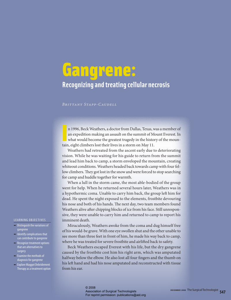
DECEMBER 2008 The Surgical Technologist 547 LEARNING OBJECTIVES n Distinguish the variations of gangrene n Identify complications that can contribute to gangrene n Recognize treatment options that are alternatives to surgery n Examine the methods of diagnosis for gangrene n Explore Maggot Debridement Therapy as a treatment option Gangrene: Recognizing and treating cellular necrosis Brittany Stapp-Caudell I n 1996, Beck Weathers, a doctor from Dallas, Texas, was a member of an expedition making an assault on the summit of Mount Everest. In what would become the greatest tragedy in the history of the moun- tain, eight climbers lost their lives in a storm on May 11. Weathers had retreated from the ascent early due to deteriorating vision. While he was waiting for his guide to return from the summit and lead him back to camp, a storm enveloped the mountain, creating whiteout conditions. Weathers headed back towards camp with four fel- low climbers. ey got lost in the snow and were forced to stop searching for camp and huddle together for warmth. When a lull in the storm came, the most able-bodied of the group went for help. When he returned several hours later, Weathers was in a hypothermic coma. Unable to carry him back, the group leſt him for dead. He spent the night exposed to the elements, frostbite devouring his nose and both of his hands. e next day, two team members found Weathers alive aſter chipping blocks of ice from his face. Still unrespon- sive, they were unable to carry him and returned to camp to report his imminent death. Miraculously, Weathers awoke from the coma and dug himself free of his would-be grave. With one eye swollen shut and the other unable to see more than three feet in front of him, he made his way back to camp, where he was treated for severe frostbite and airliſted back to safety. Beck Weathers escaped Everest with his life, but the dry gangrene caused by the frostbite cost him his right arm, which was amputated halfway below the elbow. He also lost all four fingers and the thumb on his leſt hand and had his nose amputated and reconstructed with tissue from his ear. © 2008 Association of Surgical Technologists For reprint permission: [email protected]
-
Upload
nguyennguyet -
Category
Documents
-
view
219 -
download
0
Transcript of Gangrene - AST n Identify complications that can contribute to gangrene n Recognize treatment...
deCember 2008 The Surgical Technologist 547
L E A R N I N G O B J E C T I V E Sn Distinguish the variations of
gangrenen Identify complications that
can contribute to gangrenen Recognize treatment options
that are alternatives to surgery
n Examine the methods of diagnosis for gangrene
n Explore Maggot Debridement Therapy as a treatment option
Gangrene: Recognizing and treating cellular necrosis
Brittany Stapp-Caudell
In 1996, Beck Weathers, a doctor from Dallas, Texas, was a member of an expedition making an assault on the summit of Mount Everest. In what would become the greatest tragedy in the history of the moun-
tain, eight climbers lost their lives in a storm on May 11.Weathers had retreated from the ascent early due to deteriorating
vision. While he was waiting for his guide to return from the summit and lead him back to camp, a storm enveloped the mountain, creating whiteout conditions. Weathers headed back towards camp with four fel-low climbers. They got lost in the snow and were forced to stop searching for camp and huddle together for warmth.
When a lull in the storm came, the most able-bodied of the group went for help. When he returned several hours later, Weathers was in a hypothermic coma. Unable to carry him back, the group left him for dead. He spent the night exposed to the elements, frostbite devouring his nose and both of his hands. The next day, two team members found Weathers alive after chipping blocks of ice from his face. Still unrespon-sive, they were unable to carry him and returned to camp to report his imminent death.
Miraculously, Weathers awoke from the coma and dug himself free of his would-be grave. With one eye swollen shut and the other unable to see more than three feet in front of him, he made his way back to camp, where he was treated for severe frostbite and airlifted back to safety.
Beck Weathers escaped Everest with his life, but the dry gangrene caused by the frostbite cost him his right arm, which was amputated halfway below the elbow. He also lost all four fingers and the thumb on his left hand and had his nose amputated and reconstructed with tissue from his ear.
© 2008 Association of Surgical Technologists For reprint permission: [email protected]
548 The Surgical Technologist deCember 2008
300 deCember 2008 2 Ce Credits
Gangrene is a general term that can be used to describe a number of conditions that involve the death and subsequent decay of tissue in one regional portion of the body.1 A complication of necrosis, gangrene can arise as a result of critical-ly insufficient blood supply,2,3 which is often asso-ciated with comorbid conditions such as diabetes and long-term smoking. It can develop when the blood supply is cut off to the affected area of the body as a result of various processes, including infection, vascular disease or trauma. If the gan-grene is widespread, shock can occur, and if left untreated, it can result in death.4 Due to its ten-dency to spread quickly and the possibility of the necrosis of entire appendages, urgent diagnosis and treatment of the condition is necessary for the well-being of the patient. Antibiotics, wound debridement and surgery are the primary treat-ments for gangrene.
A B O U T g A N g R E N EThere are several types of gangrene, but the three most common variations are wet, dry and gas gangrene. Less common variations include internal and Fournier’s gangrene. Gangrene can involve any part of the body, but the most common sites include the toes, fingers, feet and hands.3 Additionally, gangrene can affect the muscles and internal organs.2 The best treat-ment for gangrene is revascularization of the affected tissue, thus reversing some of the effects of necrosis and ultimately allowing healing of the damaged tissue. Other treatments for gan-grene include debridement and surgical ampu-tation. The chosen method of treatment is gen-erally determined depending on the location of the affected tissue and extent of tissue damage, death or loss. Although gangrene can be poten-tially fatal, the prognosis for recovery is good if gangrene is identified early and treated quickly.2
h I S T O R yBefore the introduction of antibiotics, fly maggots were commonly used to treat chronic wounds or ulcers. The maggots were utilized to debride the necrotic tissue without harming the healthy, liv-ing tissue. This practice largely died out after the
introduction of antibiotics and enzymes as accept-able treatments for surgical, chronic and traumatic or accidental wounds. Recently, however, maggot therapy has regained some credibility and is some-times employed with great efficacy in cases of chronic tissue necrosis and gangrene infections.
C A U S E SGangrene occurs when a body part loses its blood supply. The affected tissue may be the skin, mus-cles or internal organs. Blood provides oxygen and nutrients to feed the tissue cells and immune system components, such as antibodies, to ward off infections. Without a substantially function-ing blood supply, the cells struggle to survive and ultimately die.2 This necrosis, or cell death, can result when a portion of the body’s tissues become infected, injured or constricted, inter-rupting the blood supply. In addition, tissue in a particular region of the body may have a decrease in the amount of blood supply due to a number of diseases or conditions such as arteriosclerosis, diabetes, smoking or wound infections – includ-ing those related to surgery.1 Any of these afflic-tions can significantly increase a person’s likeli-hood of contracting gangrene. Another indicator for susceptibility is a suppressed immune sys-tem. Patients with HIV or who are undergoing chemotherapy are at a far greater risk of infec-tion due to the weakened state of their immune system. Severe burns or frostbite can also cause gangrene in body tissues due to the necrosis that results from such injuries or conditions.
S y M P T O M SThe symptoms of gangrene depend on both the location and cause of the condition.1 If the skin is involved, or the gangrene is close to the skin, the symptoms may include discoloration (blue or black if the skin is affected; red or bronze if the affected area is beneath the skin), foul-smelling discharge and/or loss of feeling in the area.1 If the affected area is inside the body, the symptoms may include, but are not limited to, confusion, fever, gas in tissues beneath the skin, a general ill feeling, low blood pressure and persistent or severe pain.1
© 2008 Association of Surgical Technologists For reprint permission: [email protected]
deCember 2008 The Surgical Technologist 549
A condition called septic shock can occur if a bacterial infection that originated in the gangre-nous tissue spreads throughout the body.2 Symp-toms of septic shock include low blood pressure, an increased heart rate, lightheadedness, short-ness of breath and confusion.2
T y P E S O F g A N g R E N ED r y G a n g re n eDry gangrene is caused by a reduction in the blood flow through the arteries of certain tissues. It typically appears gradually and progresses slowly. In most people, the affected area does not become infected. In this type of gangrene, the tissue becomes necrotic, cold and black, begins to dry, and eventually sloughs off as a result of the decreased blood supply to the said tis-sue . 2 Dr y gang rene is commonly seen in pat ients who suf fer from arteriosclerosis, a result of increased levels of cholesterol, diabetes, cigarette smoking and other genetic factors.
Dry gangrene typically begins at the distal part of the limb, due to ischemia, and often occurs in the toes and feet. This type of gangrene usually spreads slowly until it reaches the point where the blood supply is inadequate to keep tissue viable.1 Macroscopically, the affected tissue becomes dry, shrunken and blackened. The dark coloration is due to the liberation of hemoglobin from hemo-lyzed red blood cells, which are acted upon by hydrogen sulfide that is produced by the bacteria that causes gangrene, resulting in formation of black iron sulfide that remains in the tissues. The line of separation usually brings about complete severance between the healthy and necrotic tis-sue, ultimately resulting in the gangrenous tissue falling off if it is not surgically removed.
If the blood flow is interrupted for a reason other than severe bacterial infection, the result is a case of dry gangrene. People with impaired peripheral blood flow, such as diabetics, are at
greater risk of contracting dry gangrene.The early signs of dry gangrene are a dull ache
and sensation of coldness in the affected area along with pallor of the flesh. If caught early, the process can sometimes be reversed by vascular surgery. However, if necrosis sets in, the affected tissue must be removed just as with wet gangrene.
We t G a n g re n eWet or moist gangrene develops as a complica-tion of an untreated bacterial infection, such as in an open wound. Swelling, blistering and a wet appearance are common features of wet gan-grene. It can develop in victims of severe burns, frostbite or other injuries in which blood supply is
compromised.2 In addi-tion, wet gangrene often presents in patients with comorbid conditions such as obesity or dia-betes, where the patient u n k n o w i n g l y g e t s injured and then the wound becomes infect-ed. Wet gangrene needs to be treated immedi-ately because it spreads
quickly and can be fatal.2Swelling resulting from the bacterial infec-
tion causes a sudden stoppage of blood flow, which causes tissue necrosis. Cessation of blood flow facilitates invasion of the muscles by bacte-ria, which multiply because disease-fighting cells (white blood cells) cannot reach the affected part.
Wet gangrene occurs in naturally-moist tis-sue and organs such as the mouth, bowel, lungs, cervix and vulva. Bedsores occurring on body parts such as the sacrum, buttocks and heels are also categorized as wet gangrene infections. In wet gangrene, the tissue is infected by saprogenic microorganisms that cause tissue to swell and emit a fetid smell. Wet gangrene usually develops rapidly due to blockage of venous and/or arterial blood flow. The affected part is saturated with stagnant blood, which promotes the rapid growth of bacteria. The toxic products formed by bacte-ria are absorbed causing systemic manifestation
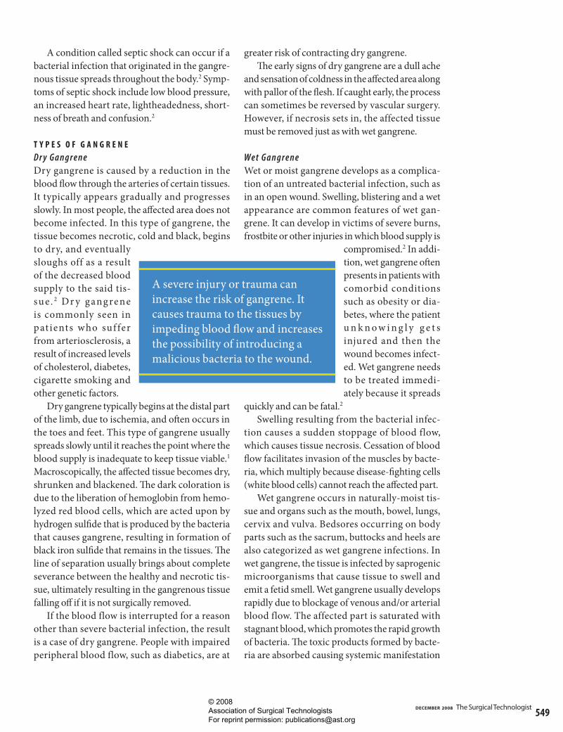
A severe injury or trauma can increase the risk of gangrene. It causes trauma to the tissues by impeding blood flow and increases the possibility of introducing a malicious bacteria to the wound.
© 2008 Association of Surgical Technologists For reprint permission: [email protected]
550 The Surgical Technologist deCember 2008
of septicemia and finally, death. Macroscopically, the affected part is edematous, soft, putrid, rotten and dark. The darkness in wet gangrene occurs due to the same mechanism as in dry gangrene.
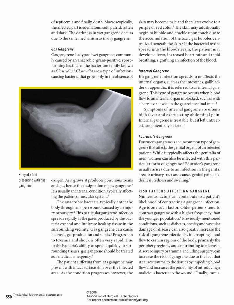
G a s G a n g re n eGas gangrene is a type of wet gangrene, common-ly caused by an anaerobic, gram-positive, spore-forming bacillus of the bacterium family known as Clostridia.5 Clostridia are a type of infection-causing bacteria that grow only in the absence of
oxygen. As it grows, it produces poisonous toxins and gas, hence the designation of gas gangrene.5 It is usually an internal condition, typically affect-ing the patient’s muscular system.2
The anaerobic bacteria typically enter the body through an open wound caused by an inju-ry or surgery.2 This particular gangrene infection spreads rapidly as the gases produced by the bac-teria expand and infiltrate healthy tissue in the surrounding vicinity. Gas gangrene can cause necrosis, gas production and sepsis.5 Progression to toxemia and shock is often very rapid. Due to the bacteria’s ability to spread quickly to sur-rounding tissues, gas gangrene should be treated as a medical emergency.5
The patient suffering from gas gangrene may present with intact surface skin over the infected area. As the condition progresses however, the
skin may become pale and then later evolve to a purple or red color.5 The skin may additionally begin to bubble and crackle upon touch due to the accumulation of the toxic gas bubbles cen-tralized beneath the skin.2 If the bacterial toxins spread into the bloodstream, the patient may develop a fever, increased heart rate and rapid breathing, signifying an infection of the blood.
I n te rn a l G a n g re n eIf a gangrene infection spreads to or affects the internal organs, such as the intestines, gallblad-der or appendix, it is referred to as internal gan-grene. This type of gangrene occurs when blood flow to an internal organ is blocked, such as with a hernia or a twist in the gastrointestinal tract.2
Symptoms of internal gangrene are often a high fever and excruciating abdominal pain. Internal gangrene is treatable, but if left untreat-ed, can potentially be fatal.2
Fo u rn i e r ’s G a n g re n eFournier’s gangrene is an uncommon type of gan-grene that affects the genital organs of an infected patient. While it typically affects the genitalia of men, women can also be infected with this par-ticular form of gangrene.6 Fournier’s gangrene usually arises due to an infection in the genital area or urinary tract and causes genital pain, ten-derness, redness and swelling.2
R I S k F A C T O R S A F F E C T I N g g A N g R E N E Numerous factors can contribute to a patient’s likelihood of contracting a gangrene infection. Age is one such factor. Older patients tend to contract gangrene with a higher frequency than the younger population.4 Previously-mentioned conditions, such as diabetes, obesity and vascular damage or disease can also greatly increase the risk of a gangrene infection by interrupting blood flow to certain regions of the body, primarily the periphery regions, and contributing to necrosis. A severe injury or trauma, including surgery, can increase the risk of gangrene due to the fact that it causes trauma to the tissues by impeding blood flow and increases the possibility of introducing a malicious bacteria to the wound.1 Finally, immu-
X-ray of a foot presenting with gas gangrene.
© 2008 Association of Surgical Technologists For reprint permission: [email protected]
deCember 2008 The Surgical Technologist 551
nosuppression can increase the likelihood of a gangrene infection due to the fact that the body cannot effectively fight off a pathogenic invader.2
D I A g N O S I SThe diagnosis of gangrene is based on the patient’s history, physical examination, blood tests and other exams.4 The practitioner must investigate the patient’s history of injury, history of any and all possible chronic diseases or conditions (especially those that affect the vasculature of certain regions, such as diabetes and arteriosclerosis), sur-gery, cigarette smoking and possible exposure to extreme cold is usu-ally investigated when attempting to diagnose a gangrene infection.1
A physical exami-nation of the affected area is performed in an attempt to look for pos-sible local signs of a wet gangrene infection. The patient’s blood test results will ultimately show an increase in the number of white blood cells if the patient is suffering from a wet gangrene infection as the body attempts to fight off the bacteria. If possible, a sample of drainage from the gangre-nous wound is examined to identify the bacteria causing the infection.1 If the analysis of the drain-age from the wound of a wet gangrene case does not initially yield the cause of the condition, a cul-ture will be taken and grown in an attempt to iden-tify the type of pathogen present in the wound, as well as aiding in possible treatment options.
In order to diagnose a potential case of gas gangrene, an X-ray can be used in an attempt to examine the affected tissue for the presence of gas bubbles, signifying a potential case of gas gan-grene. Imaging studies, including but not limited to a CT scan or an MRI, can additionally aid in the determination of the extent of tissue damage as well as the amount of gas present.1 In people with dry gangrene, an arteriogram may be performed in order to visualize any obstruction in the artery that supplies blood to the affected part.1
T R E A T M E N TIn general, treatment of gangrene infections should include the removal of necrotic tissue in an attempt to allow healing of the surrounding living tissue. It is also an important step towards the prevention of further infection. The treatment options of the various types of gangrene, however, differ due to the different natures of the condi-tions.1 Antibiotics are usually administered intra-venously to a patient suffering from gangrene in an effort to control the spread of an aggressive infec-
tion. Additionally, pain relievers are adminis-tered to control the pain of the infection, while anticoagulants are given to prevent blood clot-ting. Intravenous fluids, such as dextrose in solu-tion and isotonic saline are dispensed to replen-ish electrolytes and reestablish fluid balance
within the infected individual’s body.4 Because the cause of dry gangrene is a lack of blood flow to certain tissues, restoring the blood supply is a vital characteristic of effective treatment.1
For a wet gangrene infection, surgical debri-dement, or removal of the dead tissue from the infected wound, can be performed to evacuate any dead tissue. Additionally, intravenous antibi-otics are administered to potentially control the infection causing the wet gangrene.
Due to the threat of rapid spreading of the gas gangrene infection via the bloodstream of the affected individual, this condition needs to be treated aggressively and quickly. The wound resulting from gas gangrene requires immedi-ate debridement. Additionally, antibiotics are administered immediately to the affected patient in an effort to both control and kill the imped-ing infection. Depending on the area that has the gangrene, the person’s overall condition and the cause of the gangrene, treatment may include amputation of the infected body part. Emer-gency operations to locate and debride any and all dead tissue, surgical interventions to improve
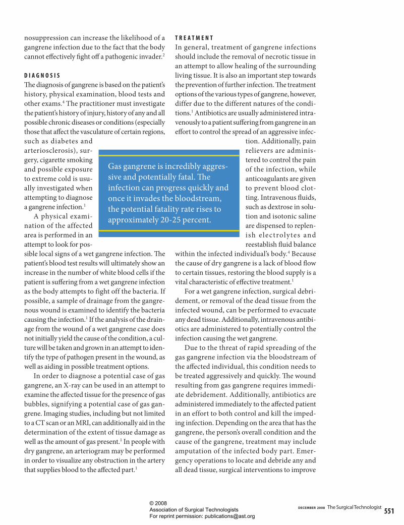
Gas gangrene is incredibly aggres-sive and potentially fatal. The infection can progress quickly and once it invades the bloodstream, the potential fatality rate rises to approximately 20-25 percent.
© 2008 Association of Surgical Technologists For reprint permission: [email protected]
552 The Surgical Technologist deCember 2008
blood supply to the given area, and repeated deb-ridement operations to remove all affected tissue in an attempt to reduce the risk of the spread of the infection to healthy surrounding tissues are standard operating procedures.
If the infection cannot be controlled with surgi-cal debridement and the consecutive administra-tion of antibiotics, amputation of the affected part becomes necessary to prevent further deteriora-tion of the surrounding, healthy tissues. Amputa-tion is usually the last effort to be exhausted in the treatment of gangrene, but due to the infection’s rapid spread and aggressive presentation, a large handful of patients routinely lose appendages or possibly limbs as a result of gangrene.
A L T E R N A T I V E T R E A T M E N T O P T I O N SOne alternative to standard practice is the use of a hyperbaric oxygen chamber as a means to reoxy-genate the damaged tissues. In this method, the patient is entirely enclosed in a pressure chamber breathing oxygen at a pressure greater than one atmosphere, a process known as hyperoxygen-ation.7 Breathing oxygen at three times the nor-mal atmospheric pressure can deliver up to 15 times the amount of physically dissolved oxygen as breathing regular air. This extra supply of oxy-gen dissolved in the blood plasma generates new capillaries in the wound area. Hyperbaric oxygen therapy has also been shown to inhibit the growth of many anaerobic and aerobic organisms. This effect, known as bacteriostasis, complements the improved ability of the host to combat disease and is useful in conditions where resistance factors are compromised, such as dysvascular conditions and immunosuppressive disorders.7 Patients receiving hyperbaric oxygen therapy must be monitored for symptoms of oxygen toxicity, such as profuse sweating, difficulty breathing and convulsions.2
P R O g N O S I SThe outlook for a person with gangrene depends on the portion of the body that is affected, the extent of the gangrene, the cause of the infection and the overall health of the patient.1 Addition-ally, the outlook for the patient recovering from a gangrene infection is generally favorable except in
people in whom the infection has spread through the blood stream.1
Gangrene is usually curable in the early stages with intravenous antibiotic treatment and debri-dement of the infected wound. In the absence of treatment however, gangrene may lead to a fatal infection once the pathogens invade the blood-stream and affect surrounding, healthy tissues and organ systems. If treatment is delayed, the gangrene is extensive, or the person has other significant medical problems, he or she may die.1
Gas gangrene, in particular, is incredibly aggressive and potentially fatal. The infection can progress quickly and once the infection invades the bloodstream, the potential fatality rate of the con-dition rises to approximately 20-25 percent. How-ever, if it is diagnosed and treated early, approxi-mately 80 percent of people with gas gangrene survive without the need for any amputation.1
Alternatively, patients suffering from dry gan-grene usually have many other comorbid condi-tions that ultimately complicate recovery and can prove fatal.
A B O U T T h E A U T h O RBrittany Stapp-Caudell is a second term surgical technology student at San Joaquin Valley College in Fresno, California. She is scheduled to begin her clinical experience in February 2009, and will graduate in September 2009.
References1. Carson-DeWitt R. Gangrene. 2007. Available at http://www.
bidmc.harvard.edu/YourHealth/ConditionsAZ.aspx?ChunkID=11839. Accessed September 20, 2008.
2. Diseases and conditions: Gangrene. 2007. Available at http://www.mayoclinic.com/health/gangrene/DS00993. Accessed September 18, 2008.
3. Ho H. Gas Gangrene. 2006. Available at http://www.emedicine. com/med/topic843.htm. Accessed September 18, 2008.
4. McGuigan B. What is gangrene? 2003. Available at http://www.wisegeek.com/what-is-gangrene.htm. Accessed Septem-ber 18, 2008.
5. Sitham SO. Gangrene. 2008. Available at http://www.nlm.nih.gov/medlineplus/ency/article/007218.htm#Causes,%20incidence,%20and%20risk%20factors. Accessed September 18, 2008.
6. Uppot RN. Case 54: Fournier gangrene. 2001. Available at http://radiology.rsnajnls.org/cgi/content/full/226/1/115. Accessed September 18, 2008.
7. Wound Care Institute Newsletter. 1996. “Commonly Asked Questions on Hyperbaric Oxygen Therapy.”Available at: http://www.woundcare.org/newsvol1n3/ar8a.htm. Accessed October 27, 2008.
© 2008 Association of Surgical Technologists For reprint permission: [email protected]
deCember 2008 The Surgical Technologist 553
A patient hobbles into the private exami-nation room at her doctor’s office and carefully seats herself on the examination table. It has been two days since her last visit and she is anxious to check on the status of the diabetic ulcer that is threaten-ing to claim her foot. She has exhausted all possible remedies for her ailment, including antibiotic regimens and surgical procedures to remove the necrotic tissue. Nothing, however, has been able to force the growing wound into remission.
Her doctor enters the room with a smile and asks how she’s feeling.
“I have a slight tingling sensation in my foot,” she says, “but overall, I feel fine.”
The doctor nods, pulls up a stool and sits in front of her. A medical assistant positions a trash can beneath the patient’s foot and the doctor begins to remove the covering from the wound site. As the gauze pad is slowly pulled away from the wound, a wriggling ball of maggots falls from the wound into the trash can below.
Unfazed, the doctor examines the wound. The necrotic tissue that had been prevalent two days earlier is completely gone. Live, pink tissue is all that remains. The doctor smiles at the patient and says, “Even better than I expected!”
F ly larvae, or maggots, are making a comeback in the modern medical community. Once a very common and popular means of clean-
ing infected wounds in the United States, mag-got debridement therapy (MDT) fell out of favor with the mainstream medical establishment with the development of advanced pharmacological antibiotic treatments after World War II.1
The practice was revisited in the 1970s and 80s, used only after all other means of wound care had been exhausted, and ultimately led to the first modern clinical studies of the practice in 1989.1 The results of those trials, and the studies and reports that followed, indicated that MDT is still an extremely viable treatment tool for cer-
Maggot Debridement Therapy: Biosurgical Treatment for Infected WoundsTom Borak
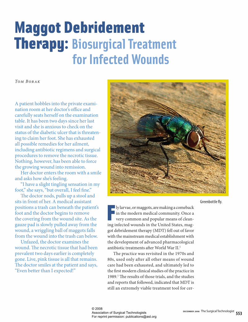
Greenbottle fly.
© 2008 Association of Surgical Technologists For reprint permission: [email protected]
554 The Surgical Technologist deCember 2008
tain types of wounds. In addition, the studies suggest that MDT does not need to be an option of last resort. In fact, while published accounts of “pre-amputation MDT” show a limb salvage rate of more than 40 percent, the success of MDT when used earlier in the course of treatment is even more dramatic.1
MDT serves three primary functions:n Clean the wound by dissolving dead and
infected tissue.n Disinfect the wound. Preliminary studies sug-
gest that maggots are even able to eradicate antibiotic-resistant bacteria, such as MRSA, from infect-ed wounds.2 This theory is currently under investigation and could have seri-ous implications for post-surgical infec-tion patients.
n Speed the rate of healing.1 It is also believed, though it has not yet been confirmed in a clinical trial, that the larvae actually stimulate the production of granula-tion tissue,2 the perfused, fibrous connective tissue that replaces a fibrin clot in healing wounds and aids vascularization. This effect has been previously reported in historical records and possible mechanisms for this occurrence are currently being sought.
Of course, the thought of introducing maggots to an open wound is difficult for some patients—and even some practitioners—to handle. Com-mon misconceptions include maggots generat-ing bacteria and increasing the risk of infection, burrowing deeper into the tissue and breeding more maggots. All of these fears, however, are unfounded.
While it is true that certain fly species, such as the screw worm fly, hatch larvae that burrow down into the living tissue, causing massive tis-sue damage and sometimes death,2, 3 many spe-
cies are much less aggressive. The species most commonly used in MDT is the blow fly (Lucilia sericata), commonly called the greenbottle for its metallic green color.2
When introduced to the wound, the blow fly larvae produce a mixture of proteolytic enzymes,2, 4 including collagenase, which breaks down the dead tissue into a semi-liquid, which is reabsorbed and digested.2 The larvae will not bur-row under the skin or attack healthy tissue and there is no danger that they will stay within the wound and breed. A mature larva must leave the
wound to pupate (the stage before it becomes an adult insect) or else it will die. In fact, once the larvae are fully grown they will come to the surface of the wound, where they are easily removed.2
The application pro-cess is very simple. A dressing is created by making a tracing of the wound on a sterile plas-
tic sheet, which is then cut out and transferred to a hydrocolloid dressing. The shape of the wound is cut from the hydrocolloid and discarded.2 The sheet with the wound-sized hole is then applied to the patient. This dressing serves two func-tions. It provides a sound base for the second component of the dressing system and protects the healthy tissue from the potent proteolytic enzymes released by the maggots.
The larvae, initially about 2 mm long, are introduced to the wound using a sterile piece of gauze to transfer them from their shipping con-tainer. The number of maggots used depends on several factors, including the size of the wound and the amount of necrotic tissue that is pres-ent. General guidelines indicate that the wound should contain no more than 10 maggots per square centimeter.2
After the larvae have been introduced to the wound, a sterile piece of fine nylon mesh, a lit-tle larger than the wound, but smaller than the
MDT is still an extremely viable treatment tool for certain types of wounds… While published accounts of “pre-amputation MDT” show a limb salvage rate of more than 40 percent, the success of MDT when used earlier in the course of treatment is even more dramatic.
© 2008 Association of Surgical Technologists For reprint permission: [email protected]
deCember 2008 The Surgical Technologist 555
hydrocolloid dressing, is applied to the back of the hydrocolloid with adhesive tape.2 An absor-bent pad is also applied to the outer surface of the net to catch any liquefied necrotic tissue. The outer absorbent dressing can be changed as often as required. Because the net is partially transpar-ent, the activity of the maggots can be determined without removing the primary dressing.2
The maggots are typically left in the wound for 24-48 hours. Their natural instinct tells them to leave the wound once the dead tissue is gone or they have consumed all that they can eat. When the dressing is removed, most of the maggots will crawl out of the wound on their own. Any that are left behind can be easily removed with gen-tle irrigation or forceps. If necrotic tissue is still present, additional applications of fresh maggots can be used as necessary. The contaminated mag-gots should be disposed of by the same means as other biological waste.
Before they can be shipped to medical facili-ties around the country, the maggots must be raised in a sterile envi-ronment. The external surface of the fly’s eggs are normally contami-nated with bacteria, which must be removed or killed before the eggs hatch i f the emerg-ing larva are to remain sterile.2
The eggs are collect-ed on raw liver in a con-trolled environment. They are then cleaned and sterilized under aseptic conditions, using equip-ment that is more commonly used for the produc-tion of sterile pharmaceuticals. 2
The sterilized eggs are then transferred asep-tically to sterile flasks, which contain an appro-priate substrate on which they will hatch. The substrate is formulated to maintain the viability of the larvae without allowing them to grow too rapidly. With sufficient oxygen, the larvae can be stored in a cool place for extended periods of time until they are ready for use.
In addition to the health benefits associated with MDT, patients can receive this therapy in the comfort of their own home or on an out-patient basis, which can reduce or eliminate the costs associated with hospitalization. It should always be remembered, however, that MDT is a potent therapeutic tool and should be used with caution by properly-trained staff.
References1. Sherman Ronald A. Maggot Debridement Therapy
(MDT). 2008. Available at: http://www.medicaledu.com/maggots.htm. Accessed October 30, 2008.
2. Jones M, Jones S, Shutler S, Thomas S. Maggots in Wound Debridement—an Introduction. 1999. Avail-able at: http://www.smtl.co.uk/WMPRC/Maggots/maggots.html. Accessed October 30, 2008.
3. Hall M, Smith K. Diptera Causing Myiasis in Man. In: Crosskey Roger W, Lane Richard P, eds. Medical Insects and Arachnids. 1995, 429-469.
4. Ksander G, Lee R, Vistnes L. Proteolytic Activity of Blowf ly Larvae Secretions in Experimental Burns. Surgery. 1981.
Preliminary studies suggest that maggots are even able to eradicate antibiotic-resistant bacteria, such as MRSA, from infected wounds. This theory is currently under investigation and could have seri-ous implications for post-surgical infection patients.
© 2008 Association of Surgical Technologists For reprint permission: [email protected]