
Gametogenesis - unimi.itusers.unimi.it/aluciano/didattica/download_files/Domestic_Animals... ·...
24
4 CHAPTER Gametogenesis At fertilization, the maternal and paternal genomes are united in the one-cell fertilized ovum, forming the zygote. In order to carry the two genomes to the site of unification in the oviduct, specialized cells known as the gametes have developed. The maternal gamete, the oocyte, is the largest cell of the body and has an inherent competence to initiate embryonic development once it has under- gone a process known as activation, described under fertilization in Chapter 5. This particular ability of the oocyte is used biotechnologically, especially in cloning by nuclear transfer. Cloning involves removing the oocyte’s own genome and then having its remaining cytoplasm initiate embryonic development based on the genome of a nuclear donor cell fused to the oocyte (see Chapter 21). The paternal gamete, the spermatozoon, on the other hand, has developed an ability for motion and penetration of the oocyte’s investments; it is these dynamic qualities of the spermatozoon that enable it to transport the paternal genome from the male to the female. In the following we will focus on how these spe- cialized gametes develop, how their genome is pre- pared for fertilization, and how the particular architecture of the cells is achieved. The first phase in this process involves the population of the devel- oping gonad by primordial germ cells from which the germ cells develop. The subsequent process, by which the maternal and paternal gametes are pro- duced from the primordial germ cells, is referred to as gametogenesis. Gametogenesis includes meiosis, to allow for recombination of genetic material and for reduction of the number of chro- mosomes from the diploid to the haploid comple- ment, and cytodifferentiation, to achieve the cellular structure characteristic of the female or male gamete. PRIMORDIAL GERM CELLS The primordial germ cells are the predecessors of the female and male gametes. When the embryo differentiates into the somatic germ layers (ecto- derm, mesoderm and endoderm) during the process of gastrulation (see Chapter 7), most cells lose their pluripotency (the ability to develop into all cell types of the mammalian body). However, one set of cells remains pluripotent. These are the primordial germ cells which, at least in the pig, first become recognizable in the posterior rim of the embryonic disc at gastrulation. From here they move into the newly formed mesoderm and endo- derm (Fig. 4-1). A few days later, the primordial germ cells are found in the visceral mesoderm sur- rounding the yolk sac and the allantois outside the embryo proper. Presumably, the primordial germ cells are brought to this location outside the forming embryo in order to ‘rescue’ them from the differentiation signals driving gastrulation within the embryo proper. By the time the first somites are formed, the primordial germ cells can be found in the mesoderm of both the yolk sac and the allan- tois, but also in the mesoderm of the incipient genital ridge where they are ready to populate the developing gonad (Figs 4-2, 4-3, 4-4). Thus, actively Poul Hyttel
Transcript of Gametogenesis - unimi.itusers.unimi.it/aluciano/didattica/download_files/Domestic_Animals... ·...

4CHAPTER
Gametogenesis
At fertilization, the maternal and paternal genomes are united in the one-cell fertilized ovum, forming the zygote. In order to carry the two genomes to the site of unification in the oviduct, specialized cells known as the gametes have developed. The maternal gamete, the oocyte, is the largest cell of the body and has an inherent competence to initiate embryonic development once it has under-gone a process known as activation, described under fertilization in Chapter 5. This particular ability of the oocyte is used biotechnologically, especially in cloning by nuclear transfer. Cloning involves removing the oocyte’s own genome and then having its remaining cytoplasm initiate embryonic development based on the genome of a nuclear donor cell fused to the oocyte (see Chapter 21).
The paternal gamete, the spermatozoon, on the other hand, has developed an ability for motion and penetration of the oocyte’s investments; it is these dynamic qualities of the spermatozoon that enable it to transport the paternal genome from the male to the female.
In the following we will focus on how these spe-cialized gametes develop, how their genome is pre-pared for fertilization, and how the particular architecture of the cells is achieved. The first phase in this process involves the population of the devel-oping gonad by primordial germ cells from which the germ cells develop. The subsequent process, by which the maternal and paternal gametes are pro-duced from the primordial germ cells, is referred to as gametogenesis. Gametogenesis includes meiosis, to allow for recombination of genetic
material and for reduction of the number of chro-mosomes from the diploid to the haploid comple-ment, and cytodifferentiation, to achieve the cellular structure characteristic of the female or male gamete.
PRIMORDIAL GERM CELLS
The primordial germ cells are the predecessors of the female and male gametes. When the embryo differentiates into the somatic germ layers (ecto-derm, mesoderm and endoderm) during the process of gastrulation (see Chapter 7), most cells lose their pluripotency (the ability to develop into all cell types of the mammalian body). However, one set of cells remains pluripotent. These are the primordial germ cells which, at least in the pig, first become recognizable in the posterior rim of the embryonic disc at gastrulation. From here they move into the newly formed mesoderm and endo-derm (Fig. 4-1). A few days later, the primordial germ cells are found in the visceral mesoderm sur-rounding the yolk sac and the allantois outside the embryo proper. Presumably, the primordial germ cells are brought to this location outside the forming embryo in order to ‘rescue’ them from the differentiation signals driving gastrulation within the embryo proper. By the time the first somites are formed, the primordial germ cells can be found in the mesoderm of both the yolk sac and the allan-tois, but also in the mesoderm of the incipient genital ridge where they are ready to populate the developing gonad (Figs 4-2, 4-3, 4-4). Thus, actively
Poul Hyttel

4Gametogenesis
33
A B
Fig. 4-1: Positioning of primordial germ cells in the endoderm. A: Embryonic disc of pig embryo at Day 14 of development. Note the primitive streak (1). The line ‘B’ indicates plane of section for Fig. 4-1B. B: Transverse section of the embryonic disc stained for Oct4. Note the individual Oct4 stained nuclei in the endoderm (1) identifying the primordial germ cells (arrows). 2: Mesoderm; 3: Ectoderm;.
56 4
3
2
8
7
1
Fig. 4-2: Migration of the primordial germ cells (red) from the yolk sac (1) along the yolk sac stalk (2) into the mesentery (4) of the primitive gut (3) and into the gonadal ridge (5) located medial to the mesonephros (6). 7: Allantois; 8: Urachus (allantois stalk). Courtesy Sinowatz and Rüsse (2007).

Essentials of Domestic Animal Embryology
34
Fig. 4-3: Porcine embryo at Day 21 of development stained for OCT4. Note the primordial germ cells (small dark dots) in the stalk of the yolk sac (arrow) where they are returning to the embryo proper.
or passively, the primordial germ cells are brought along the caudal aspect of the yolk sac and the allantois into the primitive mesentery and then into the still undifferentiated, but developing, gonad. During their relocation, the primordial germ cells can be recognized by their relatively large size and by the use of special staining techniques such as those for alkaline phosphatase activity or the transcription factor OCT4 which is involved in maintaining cellular pluripotency (Figs 4-3, 4-4).
During and after their migration, the primordial germ cells proliferate by mitoses. The female (XX) and male (XY) primordial germ cells become engaged in sex-specific differentiation of the gonads and become surrounded by somatic cells (see Chapter 15). They are now referred to as oogonia
and spermatogonia, respectively. These cells undergo further proliferation before they enter gametogenesis and, thus, meiosis.
THE CHROMOSOMES, MITOSIS AND MEIOSIS
The traits of a new individual are determined by specific genes identified as nucleotide sequences on the deoxyribonucleic acid (DNA). It is important to keep in mind that it is not the gene sequences them-selves, but the balanced and controlled expression of genes controlled by epigenetic regulation that is crucial for the behaviour of the cells and, thus, for the development of the conceptus. Together with

4Gametogenesis
35
A
B C
Fig. 4-4: Sagittal section of a porcine embryo at Day 16 of development (A). The section is stained for Oct4. The boxes ‘B’ and ‘C’ are enlarged. B: The nucleus of two primordial germ cells (arrows) located in the region of the developing gonadal ridge. C: The nuclei of three primordial germ cells (arrows) in the wall of the allantois (1). 2: Yolk sac; 3: Primitive gut; 4: Amniotic cavity; 5: Heart. Modified from Vejlsted et al. (2006). Reprinted with permission of John Wiley & Sons, Inc.

Essentials of Domestic Animal Embryology
36
Table 4.1: Chromosome numbers in various animal species (Rüsse and Sinowatz, 1998)
Species Chromosome number
Species Chromosome number
Dog (Canis familiaris) 78 Sheep (Ovis ammon aries) 54
Wolf (Canis lupus) 78 Gorilla (Gorilla gorilla) 48
Hen (Gallus gallus) 78 Man (Homo sapiens) 46
Camel (Camelus bactrianus)
74 Rabbit (Oryctolagus cuniculus) 44
Lama (Lama glama) 74 Rat (Rattus rattus) 42
Reindeer (Rangifer tarandus)
70 Rhesus monkey (Macca rhesus mulatta)
42
Wild horse (Equus przewalskii przewalskii)
66 Mouse (Mus musculus) 40
Domestic horse (Equus caballus)
64 Pig (Sus scrofa domesticus) 38
Donkey (Equus asinus) 62 Cat (Felis catus domesticus) 38
Domestic cattle (Bos primigenius taurus)
60 European wild boar (Sus scrofa) 36
Bison (Bison bison) 60 Pigeon (Columba livia) 16
Goat (Capra hircus) 60
numerous proteins, DNA makes up the chromo-somes. The chromosomes of an individual are inherited from the mother and the father. In humans, where the genes have been extensively mapped through the Human Genome Project (HUGO), it is estimated that there are about 25 000 genes on the 46 chromosomes. The number of func-tional genes in the domestic animals is probably similar.
In somatic cells, chromosomes appear as homol-ogous pairs to form the diploid chromosome com-plement. The diploid chromosome number is designated 2n as it includes a maternal and a pater-nal copy of each chromosome. The diploid chromo-some numbers in the domestic animals are listed in Table 4-1. One pair of chromosomes comprises the sex chromosomes, while the others are referred to as autosomes. If the sex chromosome pair is XX, the individual is genetically a female; if XY, genetically a male. One chromosome of each pair is inherited from the mother through the oocyte and the other
from the father through the spermatozoon. Thus, in order to result in a normal diploid chromosome complement at fertilization, the gametes must contain only one chromosome from each pair, referred to as the haploid chromosome comple-ment and designated 1n. Gametes, then, contain only half the number of chromosomes present in somatic cells.
Divisions of somatic cells occur through mitosis which transfers a copy of the full chromosome complement to each of the daughter cells. During gametogenesis, however, the special mechanisms of meiosis are responsible for producing the haploid chromosome complement in the germ cells.
Mitosis
Mitosis is the process whereby a cell divides its chro-mosome complement evenly between its daughter cells; karyokinesis describes the complete process, including mitosis, through which the nucleus of a

4Gametogenesis
37
cell gives rise to a nucleus in each of its daughter cells. Normally, karyokinesis is accompanied by cytokinesis, the division of the cytoplasm (includ-ing organelles and inclusions) between the daughter cells. Thus, karyokinesis and cytokinesis in combi-nation give rise to two daughter cells that in princi-ple are genetically identical to the parent cell, each receiving the complete diploid chromosomal com-plement (Fig. 4-5). Mitosis is one phase of the somatic cell cycle, the other phase being interphase. Interphase is subdivided into a gap phase 1 (G1), a DNA-synthetic phase (S) and a gap phase 2 (G2). During the S-phase, each chromosome replicates its DNA to produce two chromatids. As already men-tioned, the diploid chromosome complement is referred to as 2n. Before the S-phase, each chromo-some consists of a single DNA strand, and so each gene is present in two copies (2c), one of maternal and one of paternal origin. After the S-phase, because each chromosome now consists of two chromatids, each gene will be present in four copies (4c) although the chromosome number is maintained at 2n. During the G1-, S- and G2-phases the chromosomes are extremely long, spread through the nucleus in particular domains, and cannot be recognized with the light microscope.
Mitosis can be subdivided into pro-, meta-, ana- and telo- phases. When the cell moves from G2 of the interphase into the prophase of mitosis, the chromosomes coil, contract, condense and thicken (Fig. 4-5 B). They become visible with the light microscope and each can be seen to consist of two chromatids, joined at a narrow region known as the centromere. Also during the prophase, the centriole pair duplicates in the cytoplasm adjacent to the nucleus and the resultant two pairs become arranged at opposite poles of the nucleus. As the cell moves from pro- towards metaphase (a stage often referred to as pro-metaphase), microtubules starts to form from the two pairs of centrioles and the nuclear envelope begins to dismantle (Fig. 4-5 C). Some of the microtubules from each centriole pair then attach to structures on the chromatids, the kineto-chors, that are found in the centromere of each chromosome. This establishes the mitotic spindle with a centriole pair at each pole. Other microtu-
A B
C D
E
G
F
Fig. 4-5: Phases of mitosis: A: G2 of interphase; B: Prophase; C: Pro-metaphase; D: Metaphase; E: Anaphase; F: Telophase; G: Daughter cells in G1 of interphase. Courtesy Sinowatz and Rüsse (2007).
bules pass continuously from one centriole pair to the other without attaching to the chromosomes. As the spindle forms, the chromosomes line up in its equatorial plane, defining the metaphase of the cell

Essentials of Domestic Animal Embryology
38
tiated during fetal life, then becomes arrested at the diplotene stage of meiosis I until after puberty, and is only resumed shortly before ovulation. This pro-longed duration of the diplotene stage is also known as the dictyate stage. In the male, on the other hand, meiosis is not initiated until after puberty but it then becomes a continuous process.
Crossover and genetic recombinationAs in mitosis, maternal and paternal germ cells (oogonia and spermatogonia) undergo an S-phase before initiating meiosis. This forms two chromatids in each chromosome (2n, 4c) as in mitosis. This is the status of the germ cell when it enters the lepto-tene stage of the prophase of meiosis I (Fig. 4-6). In contrast to mitosis, however, the homologous chro-mosomes align in pairs during the prophase of meiosis I. Each chromosome pair therefore consists of four chromatids (4c) and is therefore referred to as a tetrad.
In the tetrad, each maternal chromatid becomes bound to its paternal counterpart over its full length by the synaptonemal complex. The chromatids match perfectly, point for point, in the female; so they do in the male, except for the XY-combination where the smaller Y-chromosome does not match the X-chromosome. This chromosome pairing allows for interchange of chromatid segments through a crossover process. The chromatids twist, or crossover, at certain points (the chiasmata) to form an X-like structure. Later during the prophase, the synaptonemal complex is gradually broken down, the DNA breaks in the chiasmata, and the free ends of the counterparts join up, thereby exchanging segments of DNA between maternal and paternal chromatids. This chromatin exchange, together with the random segregation of maternal and paternal chromosomes during meiosis II (see later), provide the molecular background for the genetic recombination that is characteristic of sexual reproduction.
During diakinesis, the homologous chromo-somes are gradually released from each other. With the transition from diakinesis to metaphase of meiosis I, the nuclear envelope disappears and
division (Fig. 4-5 D). Subsequently, the centromere of each chromosome divides, the two chromatids separate and, pulled by the microtubules, they start to migrate towards the spindle poles during ana-phase (Fig. 4-5 E). By this stage, each chromatid has transformed into a new chromosome. Arrangement of the chromosomes into two groups, one at each spindle pole, establishes the telophase of the divi-sion (Fig. 4-5 F). Further into telophase, the chro-mosomes uncoil and lengthen, and a nuclear envelope re-forms in each of the nascent daughter cells (Fig. 4-5 G). This event completes the division of the nucleus and its DNA (karyokinesis) into two daughter nuclei that contain the same DNA sequences and the same number of chromosomes as the initial parental nucleus (2n, 2c). Karyokinesis is followed by cytokinesis to divide the cytoplasm equally between the daughter cells.
Meiosis
Meiosis is a process involving two specialized cell divisions (meiosis I and II) that take place in the germ cells in order to generate haploid maternal and paternal gametes, the oocyte and the spermato-zoon. Each of the two meiotic divisions includes the same phases as are in mitosis: pro-, meta-, ana- and telophase except for the lack of prophase in meiosis II. Besides producing gametes with the haploid complement of chromosomes, a second, and equally important, function of meiosis is to allow for genetic recombination; the process that ensures that the genome of the next generation will be a unique combination of parental genomes. The most complex processes occur during meiosis I, which is characterized by a particularly long prophase that can be subdivided into leptotene, zygotene, pachytene and diplotene stages, and dia-kinesis. It is beyond the scope of this text to address each of these substages beyond saying that they take their names from the morphological appearance of the chromosomes. As soon as the germ cell has initiated meiosis I the maternal one is referred to as a primary oocyte while the paternal equivalent is a primary spermatocyte. In the female, meiosis is ini-

4Gametogenesis
39
1. F
irst m
eiot
ic di
visio
nSy
napt
onem
alco
mpl
ex
Com
plet
ion
ofS
- pha
se
Sepa
ratio
n of
hom
olog
ous
chro
mos
omes
Sepa
ratio
n of
chro
mat
ids
Sper
mat
ids
Sper
mat
ozoa
Lept
oten
eZy
gote
nePa
chyt
ene
(cro
ssin
g ov
er)
Dipl
oten
e
Prim
ary
sper
mat
ocyt
e
Met
apha
se I
Anap
hase
ICy
tokin
esis
Seco
ndar
ysp
erm
atoc
yte
2. S
econ
d m
eiot
ic di
visio
nM
etap
hase
IIAn
apha
se II
Met
apha
se I
Anap
hase
I
Met
apha
se II
Anap
hase
II
1. F
irst m
eiot
ic di
visio
nSy
napt
onem
alco
mpl
ex
Com
plet
ion
ofS
- pha
seLe
ptot
ene
Zygo
tene
Pach
yten
e(c
ross
ing
over
)Pr
imar
y oo
cyte
Prim
ordi
alfo
llicle
Ooc
yte
and
follic
ular
gro
wth
Zona
pel
lucid
a
Ooc
yte
Pola
r bod
y
Seco
ndar
y oo
cyte
Sepa
ratio
nof ho
mol
ogou
sch
rom
osom
es
2. S
econ
d m
eiot
ic di
visio
n
Sepa
ratio
nof ch
rom
atid
s
Sper
mat
ozoo
nin
zyg
ote
Pron
ucle
i
Pola
r bod
ies
Vork
erne
Ferti
lizat
ion
Dipl
oten
e (a
rrest
)
A: M
eios
is in
the
mal
eA:
Mei
osis
in fe
mal
e
Fig.
4-6
: P
hase
s of
mei
osis
in t
he m
ale
(A) a
nd fe
mal
e (B
). C
ourte
sy S
inow
atz
and
Rüs
se (2
007)
.

Essentials of Domestic Animal Embryology
40
go through a prophase. By telophase of meiosis II, the two chromatids of each chromosome have sepa-rated, and so each gamete receives only a single chromosome from each homologous pair (1n) con-taining a single DNA strand (1c). The secondary spermatocyte divides into two spermatids. The oocyte, on the other hand, again divides unequally giving rise to a large daughter cell, the precursor of the zygote and embryo, and a small second polar body. Moreover, in the oocyte, meiosis II becomes arrested in metaphase, the stage (M II) at which the oocyte is ovulated. Exceptions to this rule are found in the dog and the fox where the oocyte is ovulated in the prophase of meiosis I. In the large domestic species at least, the first and second polar bodies degenerate without dividing.
CYTODIFFERENTIATION OF THE GAMETES
While meiosis equips the gametes with the haploid chromosome number and allows for genetic recombination, there is a parallel need to construct the specialized cellular architecture that character-izes the two gametes. This cytodifferentiation forms oocytes from oogonia through oogenesis and spermatozoa from spermatogonia through spermatogenesis.
Oogenesis
Development of primordial folliclesAfter arriving and proliferating in the developing female gonad, the primordial germ cells become surrounded by follicular cells – flat somatic cells derived from the surface epithelium of the develop-ing ovary (Fig. 4-7). This turns the primordial germ cells into oogonia that continue to proliferate, but without completing cytokinesis, leaving them attached to each other by narrow cytoplasmic bridges. The resultant clusters of oogonia derived from individual primordial germ cells can be identi-fied in the embryo at stages of development that vary with species (Table 4-2).
meiotic spindles are formed. In the male germ cell, the poles of the meiotic spindle are constituted by centriole pairs as during mitosis. However, in at least the large domestic species and in contrast to those of mice, the oocyte lacks centrioles and the spindle poles are made up by unknown material ultrastructurally recognizable as small clusters of vesicles. The microtubules of the spindle attach to each homologous chromosome in a tetrad in meiosis I instead of to the chromatids as they do in mitosis. During the metaphase of meiosis I the chro-mosomes align in the equatorial plane but, during anaphase, it is the homologous chromosomes, not their chromatids, that are pulled apart to become grouped at the poles of the spindle during telophase. Thus, in contrast to mitosis, meiosis I separates homologous chromosomes rather than chromatids. Upon completion of meiosis I, the germ cell is haploid, containing only one chromo-some (1n) from each chromosome pair. It must be underlined, however, that the DNA content of the germ cell at this stage is still 2c because each chromosome consists of two chromatids.
Spermatocytes, spermatids, oocytes and polar bodiesAt the end of telophase of meiosis I, the primary spermatocyte divides into two secondary spermato-cytes by cytokinesis (Fig. 4-6). In the oocyte, however, the spindle is located at the periphery of the large spherical cell, and telophase is associated with a very uneven cytokinesis resulting in one of the two daughter cells being much larger than the other. The larger daughter cell is now referred to as a secondary oocyte while the small daughter cell, which is almost devoid of organelles, is the first polar body.
Immediately after completion of meiosis I, sec-ondary oocytes and secondary spermatocytes initi-ate meiosis II without an interphase. Consequently, there is no S-phase, no DNA replication, and the chromosomes remain with the same amount of DNA (2c). The chromosomes also remain con-tracted, ready to progress directly through meta-, ana- and telophase of meiosis II without needing to

4Gametogenesis
41
1 2
3
2
4
4
7
6
7
5
A
B C
D
E
Fig. 4-7: The development of the primordial follicle. A: Primordial germ cell (1) surrounded by pre-follicle cells (2). B: Cluster of oogonia (3) and oocytes (4) associated with pre-follicle cells. C: Multiplication of pre-follicle cells (5). D: Development of the pre-follicle cells into follicle cells (6) surrounded by a basal lamina (7) and associated with individual oocytes. E: The final isolated primordial follicle. Courtesy Sinowatz and Rüsse (2007).
Most oogonia continue their mitotic prolifera-tion but some differentiate into much larger primary oocytes. The latter immediately enter the S-phase of the cell cycle and then the prophase of meiosis I. Oogonia proliferate rapidly and in some species (notably the cow and human) are counted in mil-lions (Table 4-3). However, this proliferation is fol-lowed by apoptosis which leads to most oogonia and primary oocytes being lost with only a small population near the surface of the developing ovary surviving. All surviving primary oocytes have entered the prophase of meiosis I when they become arrested at the diplotene stage, still with an intact nucleus. Covered by flat follicular cells, these oocytes
form primordial follicles (Figs 4-8, 4-9, 4-10). The follicular cells rest on a basement membrane that separates them from the surrounding stromal cells. The primordial follicles constitute the pool of qui-escent follicles from which the female will recruit follicles for growth and ovulation for the rest of her reproductive life. The numbers of primordial germ cells, oogonia and oocytes in various animal species and man are listed in Table 4-3.
Follicular and oocyte growthFollicular growth occurs when follicles are recruited from the primordial pool and develop into

Essentials of Domestic Animal Embryology
42
Table 4-2: Chronology of events during the differentiation of the gonad in various species (Rüsse and Sinowatz, 1998)
Species Cattle Sheep Pig Horse Dog Cat
1PGCs in the gonadal ridge
9–10 mm 8 mm Days 30–32
9–10 mm Day 20
12 mm Day 21
Day 28 10 mm
Differentiation of male gonad
25 mm Day 40
20 mm Day 31
Day 26 – – –
Development of oogonia
55 mm Day 57
46 mm Day 43
Day 28 – 29 mm –
Onset of meiosis
125 mm Day 82
110 mm Day 55
Days 40–48 96 mm Day 73
At birth Days 40–50
First primordial follicles
160 mm Day 90
150 mm Day 66
97 mm Day 64
305 mm 3 weeks after birth
11 days after birth
Last mitoses of oogonia
Day 160 Day 82 Day 100 – 15–17 days after birth
8 days after birth
First primary follicles
325 mm Day 140
255 mm Day 95
– – – –
First secondary follicles
650 mm Day 210
320 mm Day 103
– – 2 months after birth
–
First tertiary follicles
740 mm Day 230
500 mm Day 150
– At birth 6 months after birth
–
Birth Day 280 Day 150 Day 115 Day 336 Day 62 Day 631Primordial germ cells.
primary, secondary and tertiary follicles. It should be emphasized that the vast majority of follicles that enter a growth phase fail to complete it; most degen-erate through a process referred to as atresia with only a minority completing their growth to the point of ovulation. At least in the large domestic species follicular growth is initiated during fetal life (Table 4-2). However, none of the oocytes enclosed in the follicles (except, perhaps, for some in atretic follicles) resume meiosis until puberty is reached.
Upon activation of the primordial follicle, the follicular cells start to proliferate and form a cuboi-dal monolayer around the oocyte to establish the primary follicle (Figs 4-8, 4-9, 4-10). The follicular cells are now referred to as granulosa cells. With this activation, a phase of oocyte growth is initiated in which the oocyte of the domestic species grows
from less than 30 µm to more than 120 µm in diameter. During this growth, the oocyte undergoes many morphological changes including the devel-opment of cortical granules (see below) in the cyto-plasm. Furthermore, it becomes competent to both resume meiosis and sustain embryonic develop-ment after fertilization.
Granulosa cells proliferate to form several layers around the oocyte in what becomes known as the secondary follicle (Figs 4-8, 4-9, 4-10). The oocyte and the surrounding granulosa cells synthesize certain glycoproteins that are deposited between itself and the surrounding granulosa cells as the zona pellucida. This structure is traversed by numer-ous projections from the innermost granulosa cells which thereby maintain contact with the oocyte through gap junctions. The stromal cells surround-ing the granulosa cells differentiate into an inner

4Gametogenesis
43
Table 4-3: Numbers of oogonia and oocytes at various ages in different species (Rüsse and Sinowatz, 2000)
Species Numbers of oogonia and/or oocytes
Cattle Day 50: 16 000Day 110: 2 700 000
Sheep At birth: 54 000–1 000 000Goat 6 months after birth: 24 000
3 years after birth: 12 000
Pig Day 110: 491 00010 days after birth: 60 000–509 0009 months after birth: 50 0002–10 years after birth: 16 000
Dog At birth: 700 0005 years after birth: 35 00010 years after birth: 500
Man Day 60: 600 000Day 150: 6 800 000At birth: 400 000–500 0007 years after birth: 13 000
12
3
4
5
6
7
8
9
10
11
Fig. 4-8: Follicular development in the bovine ovary from the primordial (1) through the primary (2) and secondary (3) to the tertiary follicle (4). In the oviduct, the mature oocyte at the metaphase II (5), the fertilized zygote (6) presenting two pronuclei, the 2- (7), 4- (8), and 8-cell stage (9) are seen, and in tip of the uterine horn, the morula (10) and the blastocyst (11) are shown.
theca interna, a layer of steroid-producing cells, and the outer theca externa, made up of concentric layers of cells that have supportive functions. The steroidogenic cells of the theca interna and the gran-ulosa cells are, together, responsible for the synthesis of estradiol in the follicle through a ‘two-cell’ system; the cells of the theca interna produce androgens that are transported to the granulosa cells where they are aromatized into oestrogens.
As development continues, fluid-filled spaces appear between the granulosa cells and coalesce into a single cavity, the antrum, characterizing the tertiary follicle (Figs 4-8, 4-9, 4-10). Such follicles are also referred to as antral follicles. In parallel with the expansion of the antrum, the oocyte becomes located in a protrusion of granulosa cells, the cumulus oophorus, extending into the antrum. The granulosa cells of the cumulus oophorus are referred to as cumulus cells. As the follicle develops, so does the oocyte until it achieves its characteristic
structure. The process of ovulation is triggered by the preovulatory LH surge, but some evidence suggests that even before this stimulus the oocyte undergoes changes in the developing antral follicle that build up its competence to be fertilized and to support initial embryonic development. This process may be referred to as oocyte capacitation.
4
2
13
3
1
2
7
6
5
4
1
2
3
5
2 3
1
1 2
2
34
2
5
6
1
5
A B
C
D
E
F
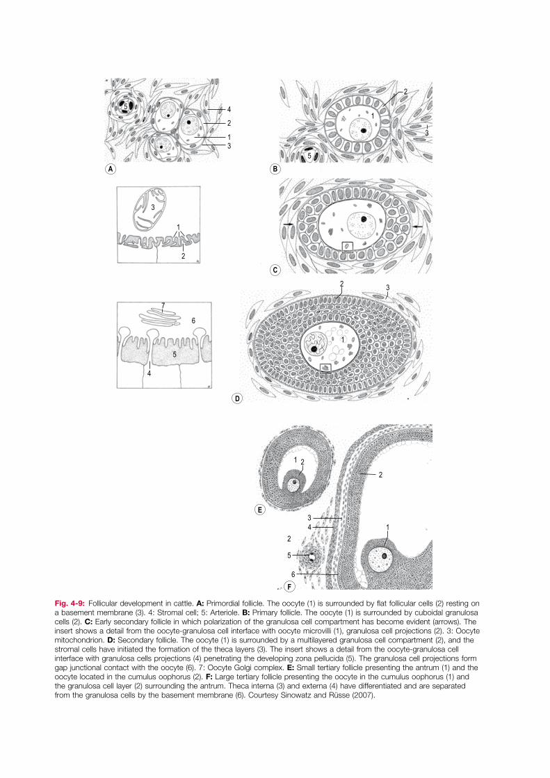
Fig. 4-9: Follicular development in cattle. A: Primordial follicle. The oocyte (1) is surrounded by flat follicular cells (2) resting on a basement membrane (3). 4: Stromal cell; 5: Arteriole. B: Primary follicle. The oocyte (1) is surrounded by cuboidal granulosa cells (2). C: Early secondary follicle in which polarization of the granulosa cell compartment has become evident (arrows). The insert shows a detail from the oocyte-granulosa cell interface with oocyte microvilli (1), granulosa cell projections (2). 3: Oocyte mitochondrion. D: Secondary follicle. The oocyte (1) is surrounded by a multilayered granulosa cell compartment (2), and the stromal cells have initiated the formation of the theca layers (3). The insert shows a detail from the oocyte-granulosa cell interface with granulosa cells projections (4) penetrating the developing zona pellucida (5). The granulosa cell projections form gap junctional contact with the oocyte (6). 7: Oocyte Golgi complex. E: Small tertiary follicle presenting the antrum (1) and the oocyte located in the cumulus oophorus (2). F: Large tertiary follicle presenting the oocyte in the cumulus oophorus (1) and the granulosa cell layer (2) surrounding the antrum. Theca interna (3) and externa (4) have differentiated and are separated from the granulosa cells by the basement membrane (6). Courtesy Sinowatz and Rüsse (2007).

4Gametogenesis
45
A
C D
B
Fig. 4-10: Follicular development in cattle. A: Primordial follicle presenting flat follicular cells (1), and the oocyte (2) with its nucleus (arrow). B: Primary follicle presenting cuboidal granulosa cells (3). 2: Oocyte; Arrow: Oocyte nucleus. C: Secondary follicle presenting a multilayered granulosa cell layer (3). 2: Oocyte; Arrow: Oocyte nucleus. D: Tertiary follicle presenting the granulosa cell layer (3) and cumulus cells (5) enclosing the oocyte (2). The theca cell layers (7) have started to form. 2: Oocyte; 4: Antrum; 6: Zona pellucida.
Follicular and oocyte maturationThe tertiary follicle continues its development and, if selected for ovulation, enters a final phase of fol-licular and oocyte maturation stimulated by the preovulatory LH surge. The period from the onset of the LH surge to ovulation is species-specific and varies from less than 12 h to more than 40 h. During
the preovulatory maturation of the follicle, steroid synthesis switches from oestradiol to progesterone production and the wall of the follicle prepares for rupturing to release the oocyte. The preovulatory maturation of the oocyte has nuclear as well as cytoplasmic components (Figs 4-11, 4-12). Nuclear

BeforeLH peak
UltrastructureGeneral structure
10 h afterLH peak
15 h afterLH peak
24 h afterLH peak
Fig. 4-11: Final oocyte maturation after the peak of the LH surge in cattle. Before the LH peak, the oocyte is in the diplotene stage and is characterized by a peripherally located nucleus (red) and a peripheral location of the organelles. At the ultrastructural level, the oocyte presents well developed smooth endoplasmic reticulum (SER; green), associated with lipid droplets (large black spheres) and mitochondria (blue), Golgi complexes (red), and clusters of cortical granules (small black spheres). The oocyte communicates through gap junctions with projections from the cumulus cells (arrows). At about 10 h after the LH peak, the oocyte has resumed meiosis and the nuclear envelope is dissolved into SER causing the nucleus, i.e. the germinal vesicle, to break down, and microtubules (black lines) appear adjacent to the condensing chromosomes (black in red nucleus). The perivitelline space between the oocyte and the zona pellucida develops, and in the oocyte the mitochondria tend to arrange around the lipid droplets and the Golgi complexes have decreased in size. The gap junctions between the oocyte and the cumulus cell projections are partially lost. At about 15 h after the LH peak, the oocyte has reached metaphase of the first meiotic division (metaphase I). The number and size of the lipid droplets have increased, mitochondria have assembled around the droplets, and these conglomerates have attained a more even distribution throughout the cytoplasm. Numerous ribosomes (black dots) have appeared, especially around the chromosomes, and the size of the Golgi complexes has decreased further. The gap junctions between the oocyte and the cumulus cell projections have been broken down. At about 24 h after the LH peak, the oocyte has reached the metaphase of the second meiotic division (metaphase II) and the first polar body has been abstricted. The bulk of the cortical granules are distributed at solitary positions along the plasma membrane. The lipid droplets and mitochondria have attained a more central location in the cytoplasm leaving a rather organelle-free peripheral zone in which the most prominent features are large clusters of SER. Golgi complexes are practically absent. Ovulation occurs around 24 h after the peak of the LH surge.

4Gametogenesis
47
A B
Fig. 4-12: Sections of bovine oocytes. A: Bovine oocyte at the diplotene stage. Arrow: Nucleus. 1: Zona pellucida; 2: Cumulus cells. B: Bovine oocyte at the metaphase of the second meiotic division (metaphase II). Arrow: Metaphase II plate with first polar body adjacent to it; 1: Zona pellucida; 2: Cumulus cells.
oocyte maturation refers to the process of meiosis which is resumed from the diplotene stage of meiosis I and continues to the metaphase of meiosis II when (except in the dog and fox) the oocyte is ovulated. The nucleus of the primary oocyte is often referred to as the germinal vesicle, and resumption of meiosis is morphologically evidenced by the breakdown of this structure (Fig. 4-11). Cytoplas-mic oocyte maturation involves the restructuring and modulation of many of the organelles of the oocyte. It is particularly conspicuous in cattle where the cortical granules (which, before the LH-surge, are found in large clusters) migrate to
solitary positions adjacent to the plasma membrane in preparation for exocytosis at fertilization (Fig. 4-11).
Spermatogenesis
Development of spermatogonia and seminiferous tubulesAfter arriving and proliferating in the male develop-ing gonad, the primordial germ cells become local-ized in solid cords of primitive sustentacular cells, the progenitors of the Sertoli cells, developed from

Essentials of Domestic Animal Embryology
48
the surface epithelium of the gonad. Shortly before puberty the cell cords acquire a lumen and develop into the seminiferous tubules of the testis. In paral-lel, the sustentacular cells gradually assume the characteristics of Sertoli cells and the primordial germ cells develop into spermatogonia (Figs 4-13, 4-14).
Spermatogenesis includes all the events by which the spermatogonia are transformed into spermato-zoa. This process can be subdivided into sperma-cytogenesis (the development of spermatocytes from spermatogonia), meiosis (the two meiotic divisions of the spermatocytes), and spermiogen-esis (the cellular re-structuring of the spermatids to spermatozoa without any cell divisions). Meiosis has already been described so the emphasis here will be on spermacytogenesis and spermiogenesis. There is a close cellular relationship between the spermatogenic cells and the Sertoli cells during sper-matogenesis; Sertoli cells are required for both phys-ical support and paracrine regulation of spermatogenesis, and also form the blood-testis barrier by sealing off the seminiferous tubules with tight junctions.
SpermacytogenesisSpermatogonia are located peripherally in the sem-iniferous tubules adjacent to the basal lamina and outside (i.e. on the blood side) of the blood–testis barrier. Three types can be identified: Type A, inter-mediate, and type B spermatogonia. Type A1 sper-matogonia are the stem cells for spermatogenesis. Thus, the first mitoses in a type A1 spermatogonium will result in a new type A1 stem cell and a second-generation type A2 spermatogonium with the ability to progress through spermatogenesis (Fig. 4-15). This ensures a perpetual population of stem cells for spermatogenesis. The type A2 spermatogonium will, at least in ruminants, give rise to another subse-quent generation of type A3 spermatogonia, which finally divide into intermediate spermatogonia sharing morphological characteristics with both type A and type B spermatogonia. The intermediate spermatogonia give rise to type B spermatogonia of
which there are two generations, at least in ruminants.
MeiosisThe last mitotic division of type B spermatogonia results in the formation of primary spermatocytes. These cells enter meiosis I, with its characteristically prolonged prophase. In contrast to the oocyte, the spermatocyte is not arrested at the diplotene stage in the prophase. The spermatocytes re-locate through the blood-testis barrier to the luminal compartment of the seminiferous tubules. This is brought about by a zipper-like mechanism involv-ing tight junctions; junctions are formed behind (basal to) the spermatocytes before the junctions ahead of them (on their luminal side) are dissolved to let the cells through. Completion of meiosis I results in the formation of two secondary sperma-tocytes that each divide into two spermatids through meiosis II. Throughout this series of divi-sions, from the second generation type A spermato-gonia to the spermatids, cytokinesis is incomplete, leaving all cells within a generation still connected through thin cytoplasmic bridges.
Spermiogenesis
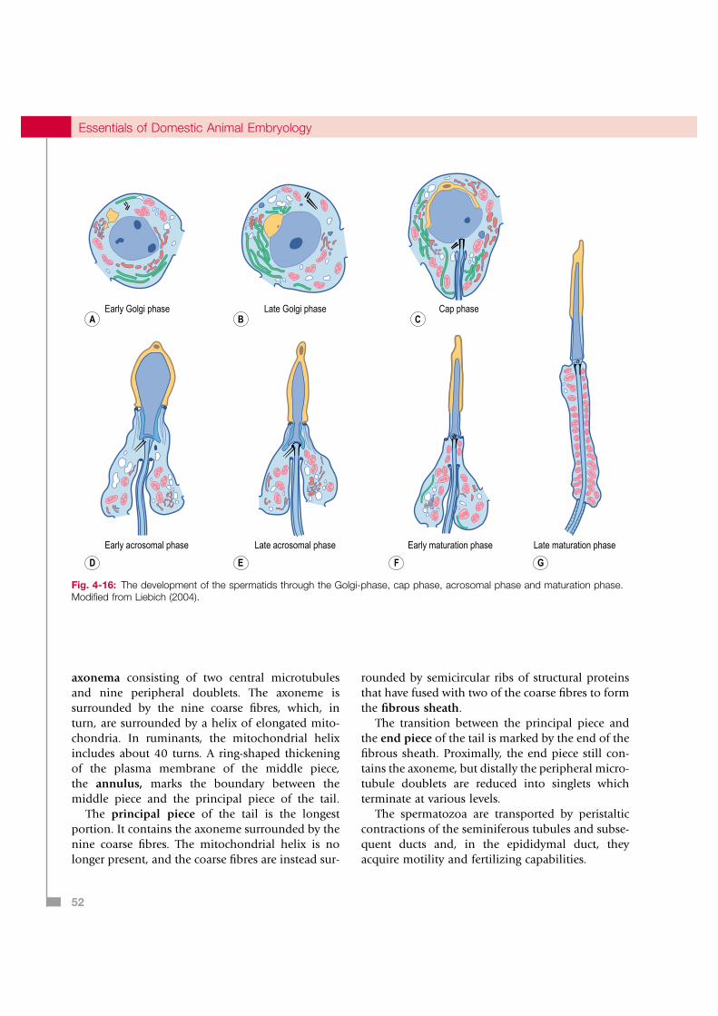
Spermatids are transformed into spermatozoa by spermiogenesis, which comprises four phases: the Golgi, cap, acrosome, and maturation phases. During the Golgi phase, the Golgi complex pro-duces acrosomal granules which fuse to form a single large acrosomal granule that becomes local-ized adjacent to the nucleus (Fig. 4-16). The centri-ole pair of the spermatid re-locate to the opposite pole of the nucleus where the proximal centriole becomes attached to the nucleus. At the same time an axoneme, consisting of two central microtubules surrounded by nine doublets, develops from the distal centriole.
During the cap phase, the acrosomal granule flat-tens and covers a larger portion of the spermatid nucleus. During the acrosomal phase, the chroma-tin in the nucleus condenses as histones are

4Gametogenesis
49
1
3
4
5
6
7
8
10
9
2
Fig. 4-13: Sertoli cell and spermatogenic cells in the seminiferous tubules of the testis. 1: Sertoli cell; 2: Spermatid – maturation phase; 3: Spermatid – cap phase; 4: Spermatid – acrosomal phase; 5: Spermatid – Golgi phase; 6: Primary spermatocytes connected by cytoplasmic bridges; 7: Blood-testis barrier; 8: Spermatogonium; 9: Basal lamina; 10: Sertoli cell nucleus. Modified from Liebich (2004).

Essentials of Domestic Animal Embryology
50
A B
Fig. 4-14: Sections of the testis of a boar (A) and a ram (B). A: Seminiferous tubules (1) of the boar testis with Leydig cells (2) between them. B: Seminiferous tubule of the ram with Sertoli cells (3), spermatogonia (4), spermatocytes (5) and spermatids (6).
exchanged with protamines. The acrosomal granule is restructured into the acrosome, which contains enzymes of importance for the spermatozoon’s pen-etration of the investments of the oocyte at fertiliza-tion. Eventually, the acrosome covers about two-thirds of the condensed nucleus of the sperma-tid, the cytoplasm is allocated to the development of the sperm tail, and the mitochondria are arranged around the growing axoneme. As these changes proceed, the spermatid rotates so that the acrosome faces the basal lamina of the seminiferous tubule and the developing tail faces the lumen.
During the maturation phase, the species-specific architecture of the sperm head and tail is developed. The major portion of the cytoplasm, including most of the organelles, is abstricted as the residual body (which is phagocytosed by the Sertoli cell) and the
spermatids are disconnected from each other. Finally, the spermatozoa are released from the Sertoli cells into the lumen of the seminiferous tubules.
Spermatozoa
The length of the spermatozoon at the time of release varies with species, but ranges from approximately 60 µm in the boar to 75 µm in the bull. At the light microscopical level, the spermatozoa appear to consist of two structures: the head and the tail. However, with the electron microscope, the tail can be subdivided into a neck, middle piece, principal piece, and end piece (Figs. 4-17).
The head of the spermatozoon contains the nucleus, which determines the shape of the head.

4Gametogenesis
51
Meiosis II
Meiosis I
A1A1 A2
Spermatids
Residual bodiesSpermatozoa
A2 spermatogonium
A3 spermatogonia
Intermediatespermatogonia
B1 spermatogonia
B2 spermatogonia
Primaryspermatocytes
Secondaryspermatocytes
Fig. 4-15: Spermatogenesis in the bull. The encircled A1 stem spermatogonium divides into an A2 spermatogonium, which enters differentiation into spermatozoa, and an A1 spermatogonium, which ensures the continuous supply of spermatogenic stem cells in the seminiferous tubules.
The anterior portion of the nucleus is covered by the acrosome delineated by an outer and inner acro-somal membrane. The acrosome contains hydro-lytic enzymes that are released during fertilization as a result of the acrosome reaction (see Chapter 5). The posterior region of the acrosome is narrow, and this region of the sperm head is referred to as the equatorial region, which continues posteriorly into the postacrosomal region. The nucleus of the spermatozoon has a cytoskeletal coat known as the
perinuclear theca which contains oocyte activating factors.
The neck is short and is connected to the head by a basal plate. It contains a proximal centriole and a distal centriole which continues into the axoneme of the tail. The centrioles are surrounded by nine peripheral coarse fibres, which are contin-ued in the coarse fibres of the tail.
The middle piece of the tail has the charac-teristic structure of a flagellum, containing a central
Essentials of Domestic Animal Embryology
52
Early Golgi phaseA
Late Golgi phaseB
Cap phaseC
Early acrosomal phase
D
Late acrosomal phase
E
Early maturation phase Late maturation phase
F G
Fig. 4-16: The development of the spermatids through the Golgi-phase, cap phase, acrosomal phase and maturation phase. Modified from Liebich (2004).
axonema consisting of two central microtubules and nine peripheral doublets. The axoneme is surrounded by the nine coarse fibres, which, in turn, are surrounded by a helix of elongated mito-chondria. In ruminants, the mitochondrial helix includes about 40 turns. A ring-shaped thickening of the plasma membrane of the middle piece, the annulus, marks the boundary between the middle piece and the principal piece of the tail.
The principal piece of the tail is the longest portion. It contains the axoneme surrounded by the nine coarse fibres. The mitochondrial helix is no longer present, and the coarse fibres are instead sur-
rounded by semicircular ribs of structural proteins that have fused with two of the coarse fibres to form the fibrous sheath.
The transition between the principal piece and the end piece of the tail is marked by the end of the fibrous sheath. Proximally, the end piece still con-tains the axoneme, but distally the peripheral micro-tubule doublets are reduced into singlets which terminate at various levels.
The spermatozoa are transported by peristaltic contractions of the seminiferous tubules and subse-quent ducts and, in the epididymal duct, they acquire motility and fertilizing capabilities.

4Gametogenesis
53
9µm
1µm
12µm
50µm
∅ 0.6µm
∅ 0.8µm
∅ 0.5µm
∅ 0.3µm
I
II
III
IV
4
10
5
8
2
1
310
67
9
11
I
II
III
IV
V
Fig. 4-17: The structure of the bull spermatozoon. The head (I) is connected to the middle piece (III) by the neck (II). The middle piece is continued in the principal piece (IV) and the end piece (V). The head presents a nucleus (1) with tightly packed chromatin, and the anterior portion of the nucleus is covered by the acrosome (2) with inner and outer acrosomal membranes located inside the plasma membrane (3). A basal plate (4) connects the head to the neck, which contains a proximal centriole (5) and a distal centriole extending into the axoneme located centrally in the middle piece and the principal piece of the tail consisting of two central (6) and 9 peripheral doublets (7) of microtubules. The middle piece presents 9 coarse fibres (8) surrounded by a mitochondrial helix (9). In the end piece, the axoneme is gradually lost. 10: Equatorial segment of the head; 11: Fibrous sheath. Courtesy Sinowatz and Rüsse (2007).

Essentials of Domestic Animal Embryology
54
Box 4-1 Molecular regulation of germ line development
As for the three principal germ layers, specification of the germ line occurs during the process of gastrulation. At least in mice, specification is initiated by local signals originating outside the embryo proper. These include the Bone Morphogenetic Proteins (BMP) 4 and 8b acting through the Smad pathway. Responding epiblast cells then initiate expression of fragilis/Ifitm3 and from this population of specified epiblast cells, germ line-restricted precursor cells are recruited. A key transcriptional regulator during this process is Blimp1/Prdm1. Functions related to Blimp1 include repression of the incipient somatic programs within the gastrulating epiblast cells, a hallmark being down-regulation of Hox gene expression. Other genes related to germ line specification include Stella and c-kit, the latter serving as a receptor for the stem cell factor/kit ligand being expressed in cells lining the path of germ cell transport to the developing gonad. Coupled to repression of somatic cell fate, germ line precursors maintain expression of pluripotency-associated genes including Oct4, Sox2 and Nanog and show substantial epigenetic modifications. The latter include global DNA demethylation as the germ cells populate the genital ridge followed by de novo methylation and acquisition of sex-specific imprints during subsequent gametogenesis (see Chapter 2, Fig. 2-3).
In the female embryo, germ cells enter meiosis early during ovarian development. Molecular markers expressed during this developmental period include Stra8 and SCP3, the latter a synaptonemal complex protein involved in pairing of homologue chromosomes. Apparently, entry into meiosis triggers loss of pluripotency in the germ cells. Oocytes guide early gonadal development. Folliculogenesis is initiated through expression of the transcription factor Figα, a factor specific for female germ cells, and essential for production of zona pellucida proteins for example. Further follicular development relies on a wealth of both endocrine and locally produced factors. In contrast to the situation in the female embryo, the presence of germ cells is apparently not necessary for testis development. Instead, differentiation of the gonad is controlled by the Sertoli cell lineage being induced by expression of the Y-linked Sry gene (Sex-determining region of the Y chromosome) in the somatic cells of the genital ridge. Sox9, Fgf9, and Dax1 are among the genes expressed after initial Sry expression. On arrival in the genital ridge, male germ cells are prevented from entering meiosis. Instead they enter a state of mitotic arrest when they have reached a species-specific appropriate number.
SUMMARY
Primordial germ cells migrate from the wall of the yolk sac to the developing indifferent gonad where they proliferate by mitoses. The primordial germ cells associate with somatic cells and develop into oogonia and spermatogonia. Subsequently, they initiate gametogenesis which includes meiosis and cytodifferentiation of the gametes. Meiosis provides the gametes with the haploid chromosome number (half of the diploid) and with recombination, while cytodifferentiation results in cellular re-
structuring into the characteristic forms of the two gametes, the oocyte and the spermatozoon. In the female, the oogonia form primary oocytes that enter meiosis I but become arrested at the diplotene stage of prophase. A primary oocyte surrounded by its somatic cells (follicular cells) makes up a primor-dial follicle, and this follicle type constitutes the resting pool of follicles from which follicles are recruited for growth through the primary, second-ary and tertiary stages. The primary oocyte does not resume meiosis and progress through its nuclear maturation to the metaphase of meiosis II until after puberty, shortly before ovulation. In parallel,

4Gametogenesis
55
of equine oocyte maturation in vivo. Mol. Reprod. Dev. 42:94–105.
Heuser, C.H. and Streeter, G.L. (1927): Early stages in the development of pig embryos, from the period of initial cleavage to the time of the appearance of limb buds. Contr. Embryol. Carneg. Inst. 20:1–19.
Hyttel, P., Farstad, W., Mondain-Monval, M., Bakke Lajord, K. and Smith, A.J. (1990): Structural aspects of oocyte maturation in the blue fox. Anat. Embryol. 181:325–331.
Hyttel, P., Fair, T., Callesen, H. and Greve, T. (1997): Oocyte growth, capacitation and final maturation in cattle. Theriogenology 47:23–32.
Liebich, H.-G. (2004): Funktionelle Histologie der Haussaugtiere. Schatter, Stuttgart, Germany.
Moor, R.M. and Warnes, G.M. (1979): Regulation of meiosis in mammalian oocytes. Br. Med. Bull. 35:99–103.
Rüsse, I. and Sinowatz, F. (1998): Lehrbuch der Embryologie der Haustiere. 2nd edn. Parey Buchverlag, Berlin.
Sutovsky, P., Manandhar, G., Wu, A. and Oko, R. (2003): Interactions of sperm perinuclear theca with the oocyte: implications for oocyte activation, anti-polysperm defense, and assisted reproduction. Microsc. Res. Tech. 61:362–378.
Vejlsted, M., Offenberg, H., Thorup, F. and Maddox-Hyttel P. (2006): Confinement and clearance of OCT4 in the porcine embryo at stereomicroscopically defined stages around gastrulation. Mol. Reprod. Dev. 73:709–718.
Wrobel, K.-H. and Süss, F. (1998): Identification and temporospatial distribution of bovine primordial germ cells prior to gonadal sexual differentiation. Anat. Embryol. 197:451–467.
the oocyte completes a cytoplasmic maturation. In the male, the spermatogonia are associated with somatic cells in solid cell cords that are the progeni-tors of the seminiferous tubules. Spermatogenesis begins after puberty and includes spermacytogene-sis, meiosis and spermiogenesis. Spermacytogene-sis includes several mitotic divisions of spermatogonia that divide into primary spermatocytes. By the two divisions of meiosis, primary spermatocytes produce haploid secondary spermatocytes and then sper-matids. Spermiogenesis is the process of re-struc-turing spermatids into spermatozoa.
FURTHER READING
Berndston, W.E. and Desjardins, C. (1974): The cycle of the seminiferous epithelium in the bovine testis. Am. J. Anat. 140:167–179.
Brennan, J. and Capel, B. (2004): One tissue, two fates: molecular genetic events that underlie testis versus ovary development. Nature Rev. Gen. 5:509–521.
Dieleman, S.J., Kruip, T.A.M., Fontijne, P., de Jong, W.H.R. and van dr Weyden, G.C. (1983): Changes in oestradiol, progesterone, and testosterone concentrations in follicular fluid and in the micromorphology of preovulatory bovine follicles relative to the peak of luteinizing hormone. J. Endocr. 97:31–42.
Grøndahl, C., Hyttel, P., Grøndahl, M.L., Eriksen, T., Godtfredsen, P. and Greve, T. (1995): Structural aspects


![Gametogenesis [Frog] - nepeducation](https://static.fdocuments.in/doc/165x107/61d5d4f7008d0e67e9698b62/gametogenesis-frog-nepeducation.jpg)


