GABAergic organization of the cat medial geniculate...
25
GABAergic Organization of the Cat Medial Geniculate Body CAMILLAN L. HUANG,* DAVID T. LARUE, AND JEFFERY A. WINER Division of Neurobiology, Department of Molecular and Cell Biology, University of California at Berkeley, Berkeley, California 94720-3200 ABSTRACT A study of neurons and processes (puncta) immunolabeled by antibodies to -aminobu- tyric acid (GABA) or glutamic acid decarboxylase was undertaken in the medial geniculate body of the adult cat. The proportion and types of GABAergic cells were determined with high resolution methods, including postembbedding immunocytochemistry on semithin plastic sections. A second goal was to draw parallels and differences between the auditory thalamus and other thalamic nuclei. Finally, the types of GABAergic puncta and their concentration in the three major subdivisions of the medial geniculate body were analyzed. The results were that (1) each division had many GABAergic neurons, averaging approximately 26% of the neuronal population; (2) the ventral division had the highest proportion of these cells (33%), the medial division the fewest (18%), and the dorsal division was intermediate (26%); (3) there was a gradient in the proportion of GABAergic neurons, i.e., the ventral and medial division values increased caudorostrally, whereas the value in the dorsal division declined; (4) the predominant GABAergic cell type in each division was a small neuron with a soma approximately 10–12 μm in diameter; (5) a small population of much larger GABAergic neurons was present mainly in the dorsal division; (6) in addition to the fine, granular puncta in each division, a type of giant GABAergic puncta was found only in the dorsal division nuclei. The results obtained with the two antibodies were essentially identical. These findings suggest a structural basis for qualitative differences in the distribution of GABAergic processing within the medial geniculate complex. The GABAergic arrangement in the ventral division was stereotyped, with only one type of putative GABAergic interneuron, and the puncta were correspondingly homogeneous. In contrast, the dorsal division had two types of GABAergic neurons, and the giant GABAergic puncta represent a new substrate for inhibitory interactions. The medial division also had more than one type of GABAergic neuron and a slightly lower concentration of puncta. These qualitative and quantitative distinctions suggest a morphologic basis for possible differences in inhibitory processing among medial geniculate body subdivisions. J. Comp. Neurol. 415:368–392, 1999. 1999 Wiley-Liss, Inc. Indexing terms: thalamus; inhibition; interneuron; local circuit; Golgi type II cell Interneurons play an essential role throughout the neuraxis in the processing and transmission of sensory and motor information (Horridge, 1968). Their contribu- tion in a few special instances to inhibitory and disinhibi- tory operations is comparatively well understood at both anatomic and physiological levels. Neurons such as Ren- shaw cells in the spinal cord (Windhorst, 1990), basket cells in the cerebral cortex (DeFelipe et al., 1986), Golgi type II cells in the lateral geniculate body (Friedlander et al., 1980; Cox et al., 1998), and Purkinje cells in the cerebellar cortex (Palay and Chan-Palay, 1974) and their associated circuits are among the best characterized inter- neuronal populations in the nervous system. However, in other parts of the thalamus, the physiological evidence available from intracellular recording and labeling studies is too limited to permit definitive conclusions about the specific role of intrinsic cells (Jahnsen and Llina ´ s, 1984a,b [guinea pig]). Moreover, there are comparatively few stud- ies available on the medial geniculate body, a structure critical for normal hearing (Winer, 1991) and about whose local circuit interneurons (Golgi type II cells) only a limited Grant sponsor: National Institutes of Health; Grant number: R01 DC02319-19. *Correspondence to: Dr. Camillan L. Huang, Division of Neurobiology, Room 285 Life Sciences Addition, Department of Molecular and Cell Biology, University of California at Berkeley, Berkeley, CA 94720-3200. E-mail: [email protected] Received 18 January 1999; Revised 18 August 1999; Accepted 23 August 1999 THE JOURNAL OF COMPARATIVE NEUROLOGY 415:368–392 (1999) 1999 WILEY-LISS, INC.
Transcript of GABAergic organization of the cat medial geniculate...
GABAergic Organization of the CatMedial Geniculate Body
CAMILLAN L. HUANG,* DAVID T. LARUE, AND JEFFERY A. WINERDivision of Neurobiology, Department of Molecular and Cell Biology,University of California at Berkeley, Berkeley, California 94720-3200
ABSTRACTA study of neurons and processes (puncta) immunolabeled by antibodies to !-aminobu-
tyric acid (GABA) or glutamic acid decarboxylase was undertaken in the medial geniculatebody of the adult cat. The proportion and types of GABAergic cells were determined with highresolution methods, including postembbedding immunocytochemistry on semithin plasticsections. A second goal was to draw parallels and differences between the auditory thalamusand other thalamic nuclei. Finally, the types of GABAergic puncta and their concentration inthe three major subdivisions of the medial geniculate body were analyzed. The results werethat (1) each division had many GABAergic neurons, averaging approximately 26% of theneuronal population; (2) the ventral division had the highest proportion of these cells (33%),the medial division the fewest (18%), and the dorsal division was intermediate (26%); (3) therewas a gradient in the proportion of GABAergic neurons, i.e., the ventral and medial divisionvalues increased caudorostrally, whereas the value in the dorsal division declined; (4) thepredominant GABAergic cell type in each division was a small neuron with a somaapproximately 10–12 µm in diameter; (5) a small population of much larger GABAergicneurons was present mainly in the dorsal division; (6) in addition to the fine, granular punctain each division, a type of giant GABAergic puncta was found only in the dorsal division nuclei.The results obtained with the two antibodies were essentially identical. These findingssuggest a structural basis for qualitative differences in the distribution of GABAergicprocessing within the medial geniculate complex. The GABAergic arrangement in the ventraldivision was stereotyped, with only one type of putative GABAergic interneuron, and thepuncta were correspondingly homogeneous. In contrast, the dorsal division had two types ofGABAergic neurons, and the giant GABAergic puncta represent a new substrate forinhibitory interactions. The medial division also had more than one type of GABAergic neuronand a slightly lower concentration of puncta. These qualitative and quantitative distinctionssuggest a morphologic basis for possible differences in inhibitory processing among medialgeniculate body subdivisions. J. Comp. Neurol. 415:368–392, 1999. ! 1999 Wiley-Liss, Inc.
Indexing terms: thalamus; inhibition; interneuron; local circuit; Golgi type II cell
Interneurons play an essential role throughout theneuraxis in the processing and transmission of sensoryand motor information (Horridge, 1968). Their contribu-tion in a few special instances to inhibitory and disinhibi-tory operations is comparatively well understood at bothanatomic and physiological levels. Neurons such as Ren-shaw cells in the spinal cord (Windhorst, 1990), basketcells in the cerebral cortex (DeFelipe et al., 1986), Golgitype II cells in the lateral geniculate body (Friedlanderet al., 1980; Cox et al., 1998), and Purkinje cells in thecerebellar cortex (Palay and Chan-Palay, 1974) and theirassociated circuits are among the best characterized inter-neuronal populations in the nervous system. However, inother parts of the thalamus, the physiological evidenceavailable from intracellular recording and labeling studies
is too limited to permit definitive conclusions about thespecific role of intrinsic cells (Jahnsen and Llinas, 1984a,b[guinea pig]). Moreover, there are comparatively few stud-ies available on the medial geniculate body, a structurecritical for normal hearing (Winer, 1991) and about whoselocal circuit interneurons (Golgi type II cells) only a limited
Grant sponsor: National Institutes of Health; Grant number: R01DC02319-19.
*Correspondence to: Dr. Camillan L. Huang, Division of Neurobiology,Room 285 Life Sciences Addition, Department of Molecular and CellBiology, University of California at Berkeley, Berkeley, CA 94720-3200.E-mail: [email protected]
Received 18 January 1999; Revised 18 August 1999; Accepted 23 August1999
THE JOURNAL OF COMPARATIVE NEUROLOGY 415:368–392 (1999)
! 1999 WILEY-LISS, INC.
amount of neurochemical information is available (Rinviket al., 1987; Rouiller et al., 1990). A more complete anal-ysis of medial geniculate body interneuronal organizationis the main object of the present study, with specialreference to parallels and differences among the threeprincipal divisions that comprise the auditory thalamus.
One proposed function for thalamic Golgi type II cells isto provide recurrent inhibition onto principal, type I,thalamocortical neurons. Such circuitry might play a rolein temporal coding (Morest, 1971) or as a source ofdisinhibition for nearby type II cells (Winer, 1992). In otherprimary sensory thalamic nuclei, such as the lateralgeniculate body and the ventrobasal complex, !-aminobu-tyric acid–accumulating (GABAergic) neurons mediatemany aspects of neuronal discharge, including receptivefield dynamics and global changes in neuronal excitability,to name just two (Lee et al., 1994; Kim et al., 1997; Ulrich
and Huguenard, 1997). A more refined picture of thedistribution of such cells in the main sensory thalamicnuclei in each modality is essential to understanding theirrole. Classic studies of Golgi impregnated material revealthat many neurons have local axons that contribute to thethalamic neuropil and could participate in these processes(Morest, 1964; Guillery, 1966; Scheibel and Scheibel, 1966).
The present study addresses the issues of the propor-tions and types of GABAergic neurons in the cat medialgeniculate body. These questions are pertinent to under-standing auditory thalamic function because each of thedifferent parts has a particular pattern of ascendingprojections from the midbrain (Calford and Aitkin, 1983), aspecific set of corticothalamic inputs (Diamond et al.,1969), and a unique physiological arrangement (Clareyet al., 1992). Prior work found that GABAergic neuronsand axon terminals and immunoreactive dendrites are ageneral feature of thalamic organization in many species(Mugnaini and Oertel, 1985; Winer et al., 1995; Arcelliet al., 1997) and that they are plentiful in the auditorythalamus (Rouiller et al., 1990). However, there has beenno systematic analysis of possible differences in the intrin-sic organization of medial geniculate body subdivisions,nor any quantitative treatment of the GABAergic pro-cesses (puncta) in the neuropil. Such differences couldhave a significant effect on how lemniscal and nonlemnis-cal streams of information are processed by these nuclei.The purpose of the present study is to contrast andcompare the GABAergic arrangements in the subdivisionsof the medial geniculate body. This will be accomplishedwith a qualitative and quantitative high-resolution, lightmicroscopic approach based on several antisera to GABAand glutamic acid decarboxylase (GAD).
A second rationale is to determine the different types ofGABAergic puncta within the medial geniculate body. Atleast two of the sources of GABA are extrinsic, whereasanother is intrinsic. One projection arises from the tha-lamic reticular nucleus (Crabtree, 1998), all of whoseneurons are GABAergic (Houser et al., 1980). Reticularnucleus axons have a wide range of terminal architectureswithin the thalamus (Cox et al., 1996); these could play arole in the diverse inhibitory effects seen in nuclei otherthan the medial geniculate body (Cox et al., 1997), and it islikely that analogous processes exist within the auditorythalamus. A second GABAergic projection originates in theinferior colliculus, where immunoreactive neurons with awide range of sizes and shapes exist (Oliver et al., 1994). Acorrespondingly diverse population of these—most notablythe largest cells in the inferior colliculus—project to eachsubdivision of the auditory thalamus (Winer et al., 1996),where they exert monosynaptic effects on their neuronaltargets (Peruzzi et al., 1997). It is unknown whetherspecific types of GABAergic puncta could represent theseintrinsic and extrinsic projection systems. A third originfor GABAergic influence is the several Golgi type II cellpopulations in the ventral (Morest, 1975), dorsal (Winerand Morest, 1983b), and medial (Winer and Morest, 1983a)divisions of the medial geniculate body. These vary in size,shape, and number and might contribute to the specificpatterns of neuropil organization found in each nucleus.The architectonic parcellation chosen follows that used inprevious studies (Morest, 1964, 1965; Winer and Morest,1984; Winer, 1992).
Although experimental identification of the origin ofeach presumptive type of puncta is beyond the scope of the
Abbreviations
BIC brachium of the inferior colliculusBSC brachium of the superior colliculusCG central grayCM centre median nucleusCN central nucleus of the inferior colliculusCP cerebral peduncleCSC commissure of the superior colliculusD dorsal nucleus of the medial geniculate bodyDC dorsal cortex of the inferior colliculusDCa caudal dorsal nucleus of the medial geniculate bodyDD deep dorsal nucleus of the medial geniculate bodyDS dorsal superficial nucleus of the medial geniculate bodyFF fields of ForelHa habenulaLD lateral dorsal nucleusLGB lateral geniculate bodyLL lateral lemniscusLMN lateral mesencephalic nucleusLN lateral nucleus of the inferior colliculusLP lateral posterior nucleusM medial division of the medial geniculate bodyMes V mesencephalic trigeminal nucleus and tractMd medial dorsal nucleusML medial lemniscusMRF mesencephalic reticular formationMZ marginal zone of the medial geniculate bodyOR optic radiationOT optic tractOv pars ovoidea of the ventral division of the medial geniculate
bodyPHy posterior hypothalamusPol posterior area of the thalamus, lateral regionPom posterior area of the thalamus, medial regionPt pretectal areaPul pulvinarPy pyramidal tractRe thalamic reticular nucleusRN red nucleusSGI intermediate gray layer of the superior colliculusSgl suprageniculate nucleus, lateral partSgm suprageniculate nucleus, medial partSGI intermediate gray layer of the superior colliculusSGP deep gray layer of the superior colliculusSGS superficial gray layer of the superior colliculusSNC substantia nigra, pars compactaSNR substantia nigra, pars reticulataSpf subparafascicular nucleusSpN suprapeduncular nucleusSt subthalamic nucleusV ventral division of the medial geniculate bodyVb ventrobasal complexVl ventrolateral nucleus of the medial geniculate bodyVTA ventral tegmental areaZI zona incertaIII oculomotor nucleus
GABA IN THE MEDIAL GENICULATE BODY 369
present study, an essential step to that end is first to definethe differences among subdivisions that could contributeto inhibitory circuits in the medial geniculate body. Be-cause these different sources of inhibitory control may notexert the same effect on postsynaptic neurons, a moreprecise analysis of their organization could predict pat-terns of inhibition within the thalamus and clarify modal-ity specific differences in these arrangements.
MATERIALS AND METHODSGeneral procedures
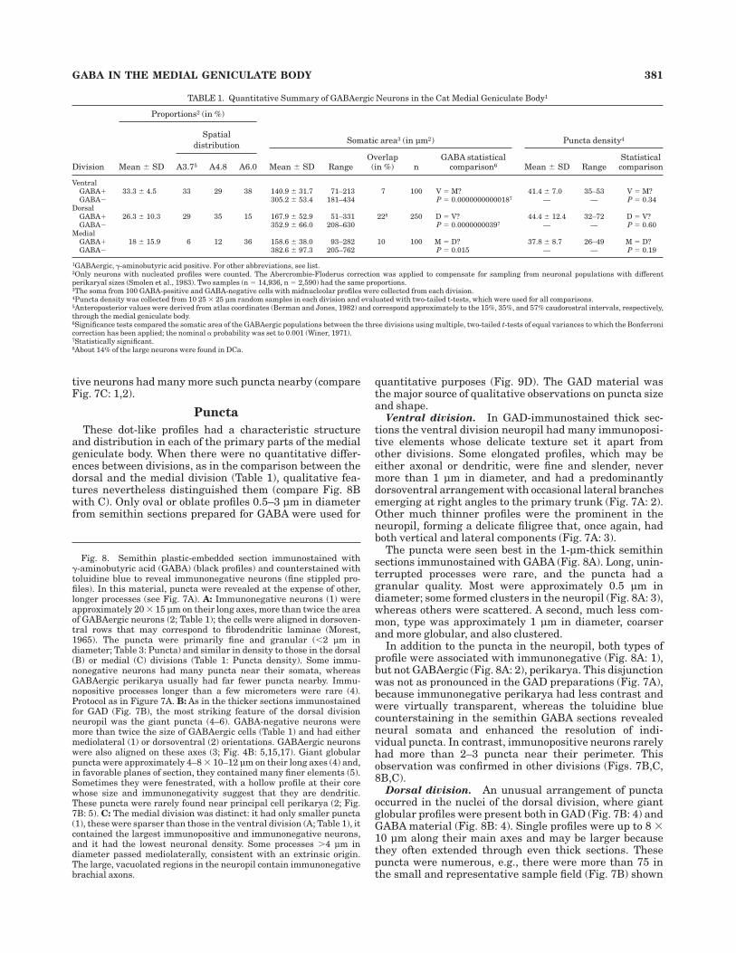
A complete series of medial geniculate bodies from 15healthy adult male cats, each weighing 2.5–4.0 kg, wereavailable. Experimental procedures followed the guide-lines of the institutional animal care and use committeeand standard veterinary protocols (Society for Neurosci-ence, 1991). Animals were anesthetized with sodium pen-tobarbital (Abbott Laboratories, North Chicago, IL; 40mg/kg, i.p.); the perfusion began when the animal wasareflexic to nociceptive stimuli. After perfusion and partialdissection, the brain was blocked in situ to approximatethe stereotaxic transverse or horizontal plane in a stan-dard atlas (Berman and Jones, 1982). To include the entiremedial geniculate body, the block extended approximately2 mm beyond the caudal and rostral poles of the auditorythalamus; this detail was important because the rostralextremity reaches the level of the ventrobasal complex(Fig. 1G: Vb) in the transverse plane (Winer, 1985).Several experiments were also available in which adultcats received cortical injections of horseradish peroxidase(Winer, 1984), after which the brain was prepared forpostembedding immunocytochemistry for the simulta-neous demonstration of retrograde labeling and immuno-staining (Larue and Winer, 1996). This material wasuseful as an independent estimate of the proportion oflocal circuit neurons and for comparative purposes incontrasting GABAergic and thalamocortical neurons (Fig.9B,C).
ImmunocytochemistryGABA immunocytochemistry for thick sections. Six
cats were perfused intracardially with a brief ("1 minute)washout (100–200 ml of phosphate buffered saline [PBS],pH 7.4, 25°C) followed by fixative (2,000 ml of 3–4%paraformaldehyde/0.25–3% glutaraldehyde/in phosphatebuffer [PB], pH 7.4, 4–10°C). After 1 hour, cryoprotectant(10% sucrose/0.1 M PB, 500 ml) was perfused and, afterdissection, the brain was immersed overnight in a secondcryoprotectant (30% sucrose at 4°C). Sections 50-µm-thickwere cut on a VibratomeTM (Oxford Laboratories, FosterCity, CA) and collected in buffer (cold 0.1 M PB). Sectionswere placed in blocking serum (5% normal goat serum/PBS; 60 minutes), then incubated in rabbit anti-GABA(DiaSorin, Clearwater, MN; 1:5,000 with 2% normal goatserum; overnight at 4°C). A 1:2 Nissl-stained series wasprepared for cytoarchitectonic analysis.
GABA immunocytochemistry for postembedded semi-thin sections. Three animals were perfused intracardi-ally with a brief washout ("1 minute; 100–200 ml of PBS,25°C) and then fixed (2,000 ml of 2% paraformaldehyde/3%glutaraldehyde; 4°C). Blocks 200 µm thick and 6 # 8 mmwide were cut on a Vibratome, and the slabs were osmi-cated, dehydrated in graded alcohols, flat-embedded in arecipe for soft Araldite epoxy suitable for semithin section-
ing, then polymerized (15 hours, 60°C). Semithin sections,1–1.5-µm-thick, were cut with 8-mm-wide glass knivesfrom blocks that contained an entire hemithalamus. Forpostembedding, sections were heat mounted on cleanslides, etched in ethanolic sodium hydroxide (approxi-mately 10% NaOH/100% EtOH), deosmicated, rehydrated,and immunostained on the slide (streptavidin-biotin kit[Histomark, Kirkegaard & Perry Laboratories, Inc., Gai-thersburg, MD], or with avidin/biotin at twice the recom-mended dilution ([Vector Laboratories, Inc., Burlingame,CA]; Larue and Winer, 1996). A 1:5 or a 1:3 series counter-stained with toluidine blue was used to determine cytoar-chitectonic borders.
GAD immunocytochemistry. Six cats were perfusedwith washout (200–300 ml of normal saline) and thenfixative (2,000 ml of 0.5% zinc salicylate/10% unbufferedformalin, 20°C for both solutions; Mugnaini and Dahl,1983). One hour later, cryoprotectant was perfused (10%sucrose/normal saline, 500 ml) and the brain was dis-sected, blocked, and stored overnight in a cryoprotectantsolution (30% sucrose/saline solution, 4°C). Frozen sec-tions 25 µm thick were collected (0.5 M Tris, pH 7.6) andplaced in blocking serum (10% normal rabbit serum/0.1 MDL-lysine in 0.5 M Tris, pH 7.6, 60 minutes; SigmaChemical Co., St. Louis, MO). They were incubated over-night with sheep anti–GAD-1440 (1:2,000 dilution with 2%normal rabbit serum, at 4°C) following standard protocols(Oertel et al., 1981; Mugnaini and Dahl, 1983; Larue andWiner, 1996). Antigen was localized with the immunoper-oxidase technique (ABC Vectastain; Vector Laboratories).A 1:2 Nissl series was also prepared (Winer and Larue,1988).
Data analysisArchitectonic boundaries. Subdivisions were deter-
mined independently from Nissl- or toluidine blue–stainedsections that were adjacent to the immunostained sec-tions. The architectonic scheme was derived from Golgistudies (Morest, 1965) and connectional experiments re-lated to auditory midbrain (Calford and Aitkin, 1983) andcortical (Niimi and Matsuoka, 1979) projections.As demon-strated below, the immunostaining patterns also distin-guished the medial geniculate body subdivisions from oneanother. The boundaries derived from the Nissl or thetoluidine blue–stained sections were superimposed on theimmunostained sections (Winer, 1992); precise alignmentwas achieved by a careful comparison of major vessels ineach set of preparations.
Neurons. GAD-positive neurons from each medial ge-niculate body division were drawn with a drawing tubewith an oil immersion objective (Figs. 3–6). Only darklyimmunostained cells with pale nuclei were consideredimmunopositive. These neurons were readily compared insize, shape, and dendritic origins to specimens recognizedin Golgi preparations (Morest, 1975; Winer and Morest,1983a,b).
Puncta. Many different types of immunopositive pro-files, including puncta, preterminal processes, and den-drites were drawn (Figs. 7, 8). For quantitative analysis(Table 1), however, only puncta were counted. Representa-tive examples of such processes appear in Figure 9D.
Quantitative analysis of neurons. The proportion ofGABAergic neurons in each medial geniculate body divi-sion was determined by using 150 sample areas of varioussizes; the total area sampled in one cat was approximately
370 C.L. HUANG ET AL.
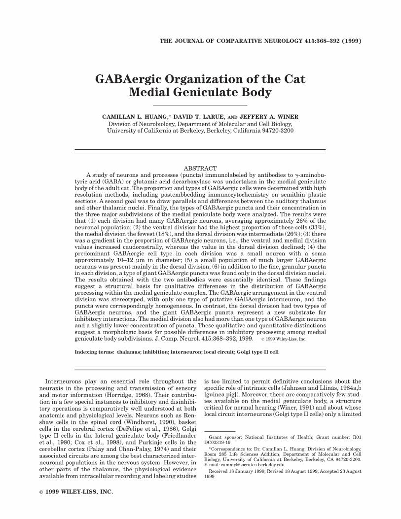
Fig. 1. Architectonic subdivisions of the medial geniculate body(gray stipple, left side) and plots of !-aminobutyric acid–positive(GABAergic) neurons (dots, right side) in a caudorostral (A–G) trans-verse sequence and in a representative horizontal section (H). Num-bers, percentage of the length of the medial geniculate body. Planach-romat, N.A. 0.30, #125. A: The caudal tip had a lower concentration ofGABAergic cells than more rostral sections due to the many brachialfibers. B: Nuclear differences in the proportion of GABAergic cells werepresent in the dorsal division. C: The dorsal superficial nucleus (DS)had far fewer GABAergic cells than the other parts of the medialgeniculate body. D: The brachium of the inferior colliculus (BIC) was
virtually defined by the absence of immunopositive neurons. E: Theconcentration of GABAergic neurons was similar in the medialgeniculate body and in the lateral geniculate body (LGB) and slightlylower in the adjoining pulvinar (Pul) and lateral posterior (LP) nuclei.F: Near the rostral pole of the medial geniculate complex (Pol), thethalamofugal and corticofugal axons marked an abrupt decline in thedensity of GABAergic neurons. G: Even the rostral pole had manyimmunopositive neurons. H: Caudal-to-rostral gradients in the propor-tion of GABAergic neurons in the dorsal division nuclei and in themedial division were present (see Fig. 2F, dorsal, medial). Asterisk,fiducial mark. For abbreviations, see list.
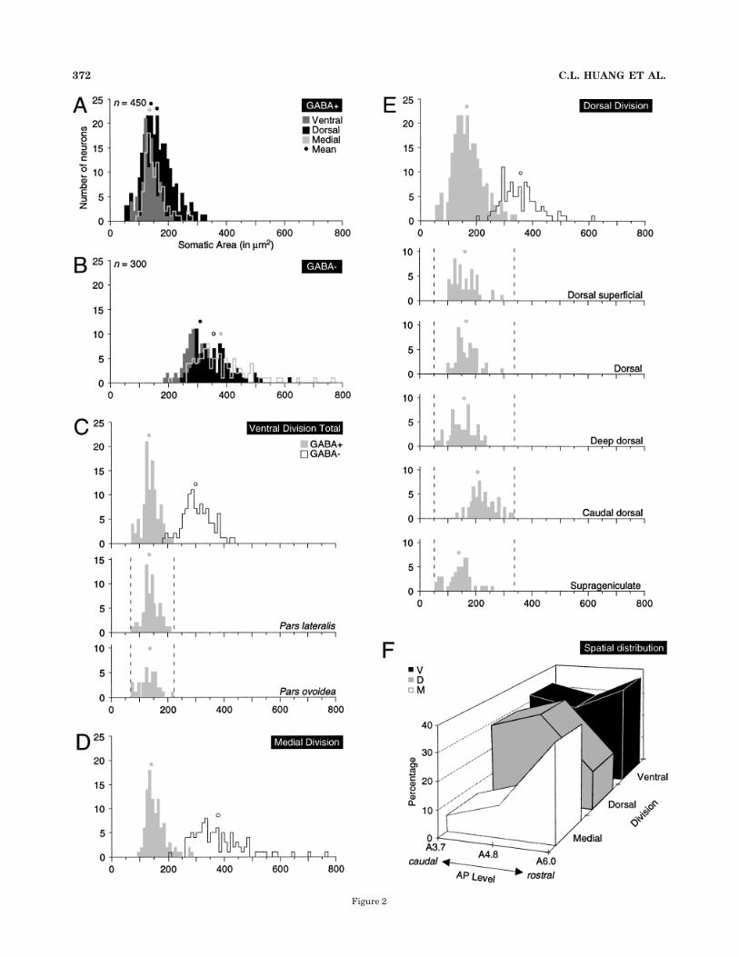
Figure 2
372 C.L. HUANG ET AL.
210 mm2. The quantitative results from this specimenwere compared with those from other animals; only if theywere indistinguishable statistically were the results in-cluded as representative. Sample fields avoided architec-tonic boundaries and excluded large blood vessels. GABAer-gic neurons were plotted from 1-µm-thick, plastic-embedded sections on an X-Y recorder coupled to amicroscope stage (Omnigraphics, Austin, TX; Figs. 1, 2F,Table 1). All the neurons from a corresponding toluidineblue–stained section were also plotted in a similar manner(Figs. 1, 2F, Table 1). Only perikarya with a nucleus wereselected. The Abercrombie-Floderus correction was ap-plied to compensate for counting errors caused by differ-ences in the mean caliper (nuclear) diameter between thesmaller, GABA-positive (Fig. 2A,F) and larger, GABA-negative (Fig. 2B,F) neurons (Smolen et al., 1983). Propor-tions were determined by averaging the ratios of GABAer-gic cells to all neurons/area at each anteroposterior level.
Quantitative analysis of perikarya. RepresentativeGABAergic and immunonegative perikarya were drawnfrom semithin, plastic-embedded GABA sections from eachdivision under oil immersion. A sample of 100 neurons/division was collected. The criteria for inclusion were thatneuronal somata were complete, midnucleolar, and remotefrom architectonic borders. These profiles were scannedand digitized with an image processing system (NIHImage, version 1.61; National Institutes of Health,Bethesda, MD). The perikaryal diameter was fitted withthe largest possible oval to measure its area. Two-tailedt-tests of equal variances with an $ level % 0.001 were usedto make multiple comparisons of the perikarya betweennuclear subdivisions. The Bonferroni correction was thenapplied to minimize the probability of a type I error byusing multiple, repeated measures (Winer, 1971).
Quantitative analysis of puncta. Ten representativesamples (each 25 # 25 µm) of immunopositive puncta weredrawn and counted from 1-µm-thick, deplasticized GABAsections. Sample zones were selected from a numberedgrid overlaid on the section; a random number sequencewas used to select a particular grid square from the sample
space. Each grid was distant from architectonic borders,and regions with blood vessels were excluded. Immunoposi-tive profiles that could not be classified with certainty wereomitted from analysis.
For Figure 9, conventional photomicrographs were takenwith high resolution, black and white film. Negatives werescanned into Photoshop 5.0 (Adobe Systems, San Jose, CA)on a Super Coolscan LS-1000 film scanner (Nikon Instru-ments Division, Garden City, NY). Images were importedand composed in Canvas 5.0 (Deneba Software, Miami,FL). For Figures 10 and 11, images were photographeddigitally on a Nikon Microphot-FXA equipped with aSpot" cooled CCD camera (Diagnosticx Instruments, Inc.,Sterling Heights, MI), and processed as above in Photo-shop and Canvas. No digital editing of the image contentwas performed.
RESULTSThe results in tissue immunostained for GAD (Fig. 7) or
GABA (Fig. 8) were indistinguishable: each antiserumrevealed the same types of neurons and comparable variet-ies of puncta. The term, GABAergic, therefore, refers tothe results obtained with both methods unless specificreference is made to one procedure. The superior immuno-penetration in the thick GAD sections often revealedimmunopositive axons and dendrites, whereas the semi-thin GABA material gave a high resolution view of punctaand somata that was essential for quantitative studies andfor determining their areal and regional distribution.
Regional distribution of GABAergic neuronsSurveys of the rostral midbrain and caudal diencepha-
lon showed that the transition between them was markedby an abrupt increase in the proportion of GABAergicneurons (Fig. 1H). From its caudal pole (Fig. 1A: DCa) toits rostral tip (Fig. 1G: Pol), the medial geniculate bodyhad many more GABAergic cells than the adjoining mesen-cephalic reticular formation (Fig. 1A: MRF), pretectum(Fig. 1D: Pt) or the fields of Forel (Fig. 1G: FF). Theauditory thalamic GABAergic cells were comparable nu-merically to those in the most superficial one-third of thesuperior colliculus (Fig. 1A: SGS), ventrobasal complex(Fig. 1G: Vb), and lateral geniculate body (Fig. 1E: LGB).The number of thalamic neurons was far larger than thatin the tegmental sector of the midbrain or in other parts ofthe diencephalon, such as, for example, in the posteriorhypothalamus (Fig. 1F: PHy).
Proportion of GABAergic neuronsAbout 26% of the medial geniculate body neurons were
GABAergic; their proportion ranged from 33% in theventral division to 18% in the medial division, with thedorsal division value intermediate (26%; Table 1). We nextconsidered whether the number of such cells was constantalong the caudorostral axis by sampling at levels approxi-mating transverse stereotaxic values of 3.7, 4.8, and 6 mm,respectively, rostral to interaural zero, or the central halfof the medial geniculate body. Two of the three divisions,the dorsal and the medial, showed significant changes butin opposite directions: the dorsal division value rose slightlyand then declined precipitously, whereas the medial divi-sion increased slightly at first and, later, more dramati-cally (Fig. 2F; Table 1). The three sampling intervalsrepresent a total of less than half the length of the medial
Fig. 2. Somatic size distribution of !-aminobutyric acid–positive(GABAergic) and non-GABAergic cells (A–E) and spatial gradients ofGABAergic neurons (F) in the three major parts of the medialgeniculate body. A: The distributions of GABAergic neurons over-lapped except for the smallest dorsal division cells and a few largercells in the dorsal and medial divisions. B: There was more dispersionin the size range among the non-GABAergic cells, mainly from themagnocellular neurons in the medial division and the large thalamo-cortical neurons in the suprageniculate nucleus. C: In the ventraldivision, there was little overlap between the GABAergic (stippled)and the non-GABAergic (outline) cell populations. The somatic size ofGABAergic neurons did not differ in the lateral (pars lateralis) andmedial (pars ovoidea) subnuclei of the ventral division (Table 2).D: Medial division GABAergic neurons were bimodally distributed, aswere those in the dorsal division (D). The largest non-GABAergicneurons, the magnocellular neurons and other subtypes, were promi-nent. E: In the dorsal division, the proportion of GABAergic cells wassignificantly lower than that in the ventral division (Table 1), with abimodal distribution. Three of the five dorsal division nuclei hadsignificantly smaller or larger GABAergic neurons (Table 2); the deepdorsal nucleus had the smallest such neurons, the caudal dorsalnucleus had the largest. F: Spatial gradients of GABAergic neurons incaudal-to-rostral traverses through the central 40% of the auditorythalamus showed a rapid increase in the medial division, slowergrowth in the ventral division, and a precipitous decline in the dorsaldivision. The latter decrease coincided with the emergence of theanterior dorsal nuclei (Winer and Morest, 1983b).
GABA IN THE MEDIAL GENICULATE BODY 373
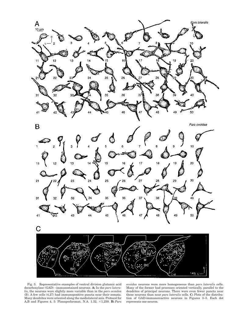
Fig. 3. Representative examples of ventral division glutamic aciddecarboxylase (GAD) –immunostained neurons. A: In the pars latera-lis, the neurons were slightly more variable than in the pars ovoidea(B). A few cells (4,27) had immunopositive puncta near their somata.Many dendrites were oriented along the mediolateral axis. Protocol forA,B and Figures 4, 5: Planapochromat, N.A. 1.32, #1,250. B: Pars
ovoidea neurons were more homogeneous than pars lateralis cells.Many of the former had processes oriented vertically, parallel to thedendrites of principal neurons. There were even fewer puncta nearthese neurons than near pars lateralis cells. C: Plots of the distribu-tion of GAD-immunoreactive neurons in Figures 3–5. Each dotrepresents one neuron.
geniculate body. Measurements were limited to this regionbecause the myeloarchitectonic volume and complexity ofthe neuropil at the caudal and rostral faces of the auditorythalamus, as seen in fiber stained preparations, made such
measurements in those regions difficult to interpret. Thepresent measures excluded the brachium and its associ-ated tracts, and represent a substantial part of the medialgeniculate body.
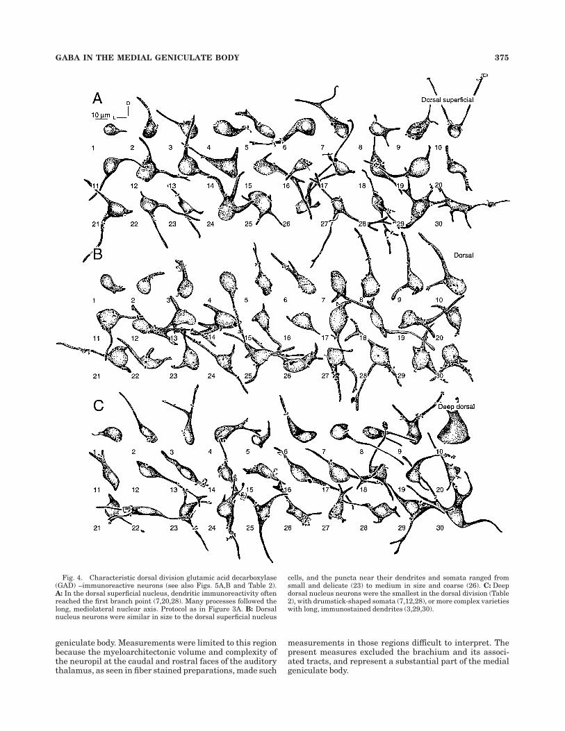
Fig. 4. Characteristic dorsal division glutamic acid decarboxylase(GAD) –immunoreactive neurons (see also Figs. 5A,B and Table 2).A: In the dorsal superficial nucleus, dendritic immunoreactivity oftenreached the first branch point (7,20,28). Many processes followed thelong, mediolateral nuclear axis. Protocol as in Figure 3A. B: Dorsalnucleus neurons were similar in size to the dorsal superficial nucleus
cells, and the puncta near their dendrites and somata ranged fromsmall and delicate (23) to medium in size and coarse (26). C: Deepdorsal nucleus neurons were the smallest in the dorsal division (Table2), with drumstick-shaped somata (7,12,28), or more complex varietieswith long, immunostained dendrites (3,29,30).
GABA IN THE MEDIAL GENICULATE BODY 375
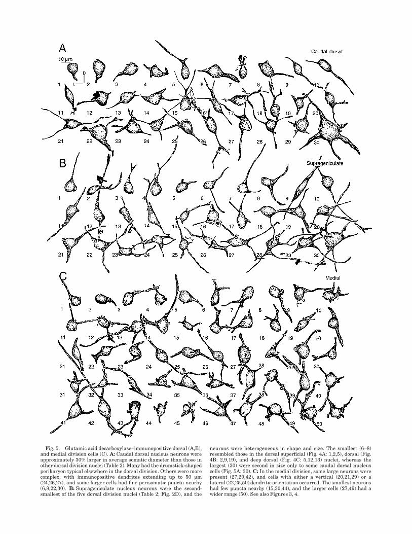
Fig. 5. Glutamic acid decarboxylase–immunopositive dorsal (A,B),and medial division cells (C). A: Caudal dorsal nucleus neurons wereapproximately 30% larger in average somatic diameter than those inother dorsal division nuclei (Table 2). Many had the drumstick-shapedperikaryon typical elsewhere in the dorsal division. Others were morecomplex, with immunopositive dendrites extending up to 50 µm(24,26,27), and some larger cells had fine perisomatic puncta nearby(6,8,22,30). B: Suprageniculate nucleus neurons were the second-smallest of the five dorsal division nuclei (Table 2; Fig. 2D), and the
neurons were heterogeneous in shape and size. The smallest (6–8)resembled those in the dorsal superficial (Fig. 4A: 1,2,5), dorsal (Fig.4B: 2,9,19), and deep dorsal (Fig. 4C: 5,12,13) nuclei, whereas thelargest (30) were second in size only to some caudal dorsal nucleuscells (Fig. 5A: 30). C: In the medial division, some large neurons werepresent (27,29,42), and cells with either a vertical (20,21,29) or alateral (22,25,50) dendritic orientation occurred. The smallest neuronshad few puncta nearby (15,30,44), and the larger cells (27,49) had awider range (50). See also Figures 3, 4.
Neuronal sizeSomatic measurements were made from semithin sec-
tions in plastic-embedded material. In each division, asample of midnucleolar GABAergic populations (ventraland medial divisions, n % 100; dorsal division, n % 250)and 100 GABA-negative neurons from each division wasdrawn and their dendrites excluded. In each division,there was a significant somatic size difference (P " 0.001)between the two neuronal populations (t-test; Table 1).The distributions of GABAergic and non-GABAergic popu-lations were largely non-overlapping (Fig. 2A,B), withGABAergic cells on average less than half the size of theGABA-negative neurons (see Figs. 10D: 2, 11B,C: 1 forexceptions).
Comparisons of the size of GABAergic neurons revealedstatistically significant differences in two of the threedivisions. Thus, dorsal division GABAergic neurons wereapproximately 20% larger than those in the ventral divi-sion, and medial division cells were approximately 15%larger than their ventral division counterparts (Table 1;Fig. 2C–E). However, dorsal division cells (Table 1; Fig.
2E) were not significantly larger than those in the medialdivision (Table 1; Fig. 2D). Among GABA-negative neu-rons, ventral division cells were the smallest and those inthe medial division were the largest (Table 1).
A subset of GABAergic neurons in the dorsal division(approximately 20%; see Fig. 2E) and in the medialdivision (approximately 5%; see Fig. 2D) were unusuallylarge and contributed to a bimodal, although not statisti-cally significant, distribution of somatic size. This findingsuggests that there might be more than one type ofGABAergic neuron (Fig. 2A).
Varieties of GABAergic neuronsFrom the GAD preparations, 30–50 neurons were drawn
from each division to show representative examples (Figs.3–5). There were three reasons for including these data: (1)they provide independent confirmation of the neuronaltypes recognized in GABA material; (2) they revealed moreof the dendritic configuration of the cells; (3) the superiorimmunopenetration showed the distribution of GAD-positive puncta in some detail.
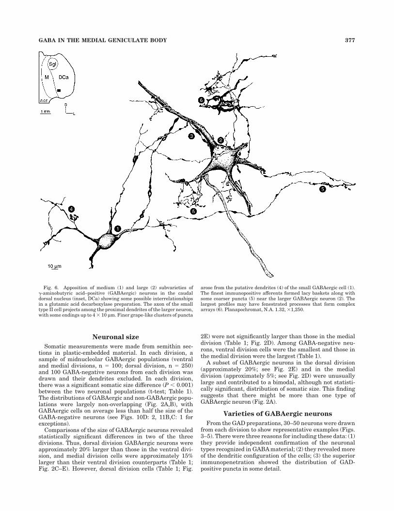
Fig. 6. Apposition of medium (1) and large (2) subvarieties of!-aminobutyric acid–positive (GABAergic) neurons in the caudaldorsal nucleus (inset, DCa) showing some possible interrelationshipsin a glutamic acid decarboxylase preparation. The axon of the smalltype II cell projects among the proximal dendrites of the larger neuron,with some endings up to 4 # 10 µm. Finer grape-like clusters of puncta
arose from the putative dendrites (4) of the small GABAergic cell (1).The finest immunopositive afferents formed lacy baskets along withsome coarser puncta (5) near the larger GABAergic neuron (2). Thelargest profiles may have fenestrated processes that form complexarrays (6). Planapochromat, N.A. 1.32, #1,250.
GABA IN THE MEDIAL GENICULATE BODY 377
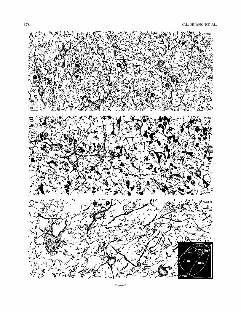
Figure 7
378 C.L. HUANG ET AL.
Ventral division. These neurons were the least vari-able in both the GABA (Table 1) and the GAD preparations(Fig. 3). The typical soma was oval and bipolar (Fig. 3A: 22;Fig. 3B: 18; Fig. 10A,B: 1), approximately 11 # 13 µm onits main axes (Fig. 7A: 1), with either a vertical (Fig. 3A: 7;Fig. 3B: 6) or a horizontal (Fig. 3A: 50; Fig. 3B: 19)orientation. Because of the thickness of the section and thecriterion that the perikaryon was confined to it, theseviews probably included most of the principal (and proxi-mal) dendritic trunks of these neurons, unless the immuno-reactivity among these processes was variable. Most cellshad two dendritic trunks that emerged from any part ofthe perikaryon (Fig. 3A: 16,26; Fig. 3B: 31,34) and werevariable in thickness, ranging from approximately 2 µm(Fig. 3A: 15; Fig. 3B: 30) to 4 µm (Fig. 3A: 18; Fig. 3B: 50). Asummary of the chief types of GABAergic neurons appearsin Figure 12D.
Although the puncta (see below for details) were numer-ous in the ventral division (Figs. 7A, 8A), they were foundless often near the perikarya of GAD-positive neurons(Fig. 9C: 1). Some of the largest neurons had no puncta intheir immediate vicinity (Fig. 3A: 34,46; Fig. 3B: 17,48),others had a few (Fig. 3A: 29,30; Fig. 3B: 3,43), and a rareneuron had up to five (Fig. 3A: 24,42; Fig. 3B: 14,37). Smallclusters of puncta near dendrites were sometimes noted(Fig. 3A: 10,38,47; Fig. 3B: 12,33,50).
Dorsal division. The mean somatic diameter of theseGABAergic neurons was the largest in the medial genicu-late body (Table 1). They were the most variable in size,and had a wider range in shape and a more heterogeneousdendritic architecture. Because there were regional differ-
ences among dorsal division nuclei, the results from fivesubdivisions were compared (Figs. 4, 5A,B).
GABAergic neurons in the dorsal superficial nucleuswere often oriented mediolaterally, with their dendritesparallel to those of the tufted principal cells that are one ofthe primary cell types in Golgi material (Winer andMorest, 1984). The smallest immunopositive somata wereapproximately 100 µm2 in area and had either a horizontalorientation (Fig. 4A: 1) or, more often, a multipolar, radiateconfiguration with 2–3 primary dendrites (Fig. 4A: 24)that may divide (Fig. 4A: 27). Some neurons had only oneprocess apparent (Fig. 4A: 5,6) and others had up to five(Fig. 4A: 30). Many dendrites were slender ("2 µm indiameter), sparsely branched, bifurcated simply ratherthan forming tufts, and radiated without an obviouspreference. The largest immunopositive perikarya werestellate shaped (Fig. 4A: 24,25,29), and their processescould cross the long axes of immunonegative tufted neurondendrites. The cell size range in the dorsal superficialnucleus was smaller than that elsewhere in the dorsaldivision (Figs. 4B,C, 5A,B). These neurons had less den-dritic immunoreactivity than ventral division neurons(compare Fig. 4A: 10,13,21–24 and Fig. 3A: 26,39,40).
A few dorsal superficial nucleus neurons had morepuncta near their somata (Fig. 4A: 7,9,12) than otherselsewhere in the dorsal division. These included some ofthe smallest cells (Fig. 4A: 16); the largest neurons had fewsuch puncta near them (Fig. 4A: 24,25). Clusters of punctanear their dendrites as seen in the ventral division wereuncommon.
The other dorsal division nuclei (dorsal: Fig 4B; deepdorsal: Fig. 4C; caudal dorsal: Fig. 5A; and supragenicu-late: Fig. 5B) shared most of these features, includingmainly small (approximately 150 µm2) immunopositivesomata (Fig. 4B: 5,16; Fig. 4C: 12,28; Fig. 5A: 13,19; Fig.5B: 3,17), and about 2–3 slender primary dendrites (Fig.4B: 20,28; Fig. 4C: 23,25; Fig. 5A: 21,27; Fig. 5B: 8,23); afew neurons had puncta near their somata (Fig. 4B: 6,10;Fig. 4C: 26,27; Fig. 5A: 6,14; Fig. 5B: 16,26). The chiefdifferences among the dorsal division nuclei were the fewunusually large neurons in the deep dorsal (Fig. 4C: 10),caudal dorsal (Fig. 5A: 30), and suprageniculate (Fig. 5B:30; see also Fig. 5C) nuclei; the large endings in somesubdivisions (Fig. 5A: 12); and the occasional aggregationof puncta near dendrites (Fig. 5B: 12,28).
Medial division. These neurons were slightly, but notsignificantly, smaller than dorsal division GAD-positivecells (Table 1: Medial % 158.6 µm2, Dorsal % 167.9 µm2). Acardinal feature was the heterogeneity in their dendriticstructure and orientation. There were small neurons withradiating dendritic origins (Fig. 5C: 45), medium-sizedcells oriented dorsoventrally (Fig 5C: 20) or horizontally(Fig. 5C: 25), or with both vertical and lateral processes(Fig. 5C: 17), neurons with intermediate orientations (Fig.5C: 41), and larger cells with a corresponding range ofdiversity. The horizontal subtype was the rarest, whereasthe vertical and multipolar neurons were the most com-mon. Even the longest immunostained processes had fewbranches (Fig. 5C: 39,42).
The density of puncta/625 µm2 was not statisticallydifferent from the value in the ventral division (Table 1),although it was the lowest of the three divisions. Manymedial division neurons had at least some puncta neartheir somata, including small (Fig. 5C: 12), medium-sized(Fig. 5C: 7), and large (Fig. 5C: 27,50) cells. Immunonega-
Fig. 7. Glutamic acid decarboxylase (GAD) –positive puncta in thethree principal divisions of the medial geniculate body (locus in C,inset). A: Ventral division immunopositive neurons (1) were small(approximately 10 # 12 µm in diameter), with a preferred verticalorientation (Fig. 3A: 16; 3B: 31). There were many GAD-positivepuncta near their primary dendrites (2). Some puncta were approxi-mately 1 µm in diameter and formed clusters in the neuropil. Thepuncta rarely were associated with GAD-positive somata. A second,larger and more globular profile with clusters, possibly of puncta, wasalso present (4). The neuropil was strikingly regular (compare withB,C), with a vertical orientation of the immunopositive neurons andthe processes ascending toward them. Protocol for A–C and for Figure8: Planapochromat, N.A. 1.32, #1,250. B: Despite the broad somaticsize range among dorsal division neurons (Table 2), most had drum-stick-shaped somata approximately 10–12 µm in diameter (1); the fullrange contained slightly larger cells (2) and other neurons (3) with astellate appearance. The puncta also distinguished the dorsal from theventral (A) and medial (C) divisions because giant processes fromapproximately 4–10 µm on their long axes were numerous in theneuropil. These puncta had a complex substructure, with vacuolatedregions that, in semithin material, might contain immunonegativedendrites (Fig. 12C: arrow). The profiles were elongated, formed shortrows or clusters, and appeared to be interconnected serially (4). Giantpuncta near somata were unusual (5), and a peridendritic locus wasmore common (6). Very fine preterminal processes, some with delicateboutons (7), made a lacy plexus in the dorsal division. C: Medialdivision GAD-immunoreactive puncta were larger and sparser thanthose of the ventral division (A), and smaller than those in the dorsaldivision (B). As in the other divisions, the small immunopositive somawas the most common neuronal profile (1). The medial division hadsome processes up to 4 µm thick that traversed the neuropil at obliqueor acute angles (3; but see Figure 11A: arrow). The largest medialdivision puncta (4) were much smaller than the giant dorsal divisionprofiles, and perhaps rarer than similar endings in the ventraldivision (A). An immunonegative principal cell soma had many punctanearby (2).
GABA IN THE MEDIAL GENICULATE BODY 379
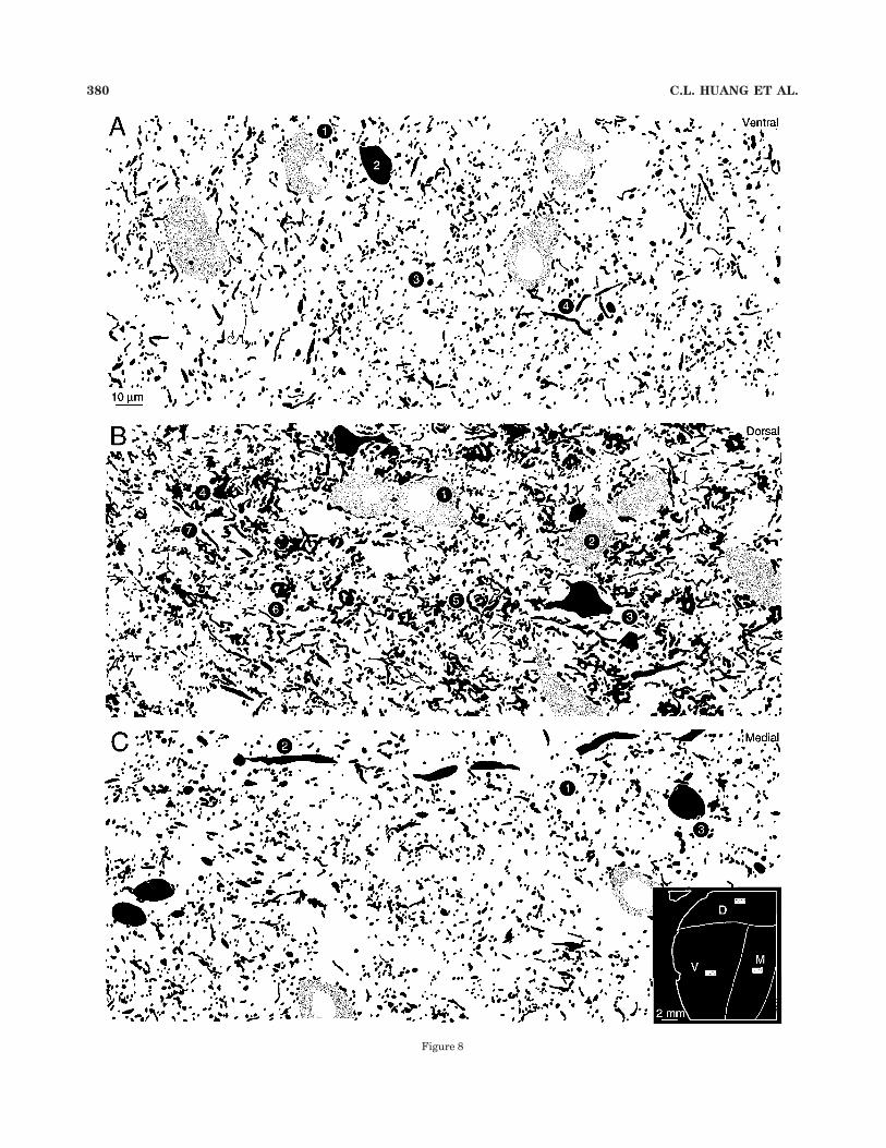
Figure 8
380 C.L. HUANG ET AL.
tive neurons had many more such puncta nearby (compareFig. 7C: 1,2).
PunctaThese dot-like profiles had a characteristic structure
and distribution in each of the primary parts of the medialgeniculate body. When there were no quantitative differ-ences between divisions, as in the comparison between thedorsal and the medial division (Table 1), qualitative fea-tures nevertheless distinguished them (compare Fig. 8Bwith C). Only oval or oblate profiles 0.5–3 µm in diameterfrom semithin sections prepared for GABA were used for
quantitative purposes (Fig. 9D). The GAD material wasthe major source of qualitative observations on puncta sizeand shape.
Ventral division. In GAD-immunostained thick sec-tions the ventral division neuropil had many immunoposi-tive elements whose delicate texture set it apart fromother divisions. Some elongated profiles, which may beeither axonal or dendritic, were fine and slender, nevermore than 1 µm in diameter, and had a predominantlydorsoventral arrangement with occasional lateral branchesemerging at right angles to the primary trunk (Fig. 7A: 2).Other much thinner profiles were the prominent in theneuropil, forming a delicate filigree that, once again, hadboth vertical and lateral components (Fig. 7A: 3).
The puncta were seen best in the 1-µm-thick semithinsections immunostained with GABA (Fig. 8A). Long, unin-terrupted processes were rare, and the puncta had agranular quality. Most were approximately 0.5 µm indiameter; some formed clusters in the neuropil (Fig. 8A: 3),whereas others were scattered. A second, much less com-mon, type was approximately 1 µm in diameter, coarserand more globular, and also clustered.
In addition to the puncta in the neuropil, both types ofprofile were associated with immunonegative (Fig. 8A: 1),but not GABAergic (Fig. 8A: 2), perikarya. This disjunctionwas not as pronounced in the GAD preparations (Fig. 7A),because immunonegative perikarya had less contrast andwere virtually transparent, whereas the toluidine bluecounterstaining in the semithin GABA sections revealedneural somata and enhanced the resolution of indi-vidual puncta. In contrast, immunopositive neurons rarelyhad more than 2–3 puncta near their perimeter. Thisobservation was confirmed in other divisions (Figs. 7B,C,8B,C).
Dorsal division. An unusual arrangement of punctaoccurred in the nuclei of the dorsal division, where giantglobular profiles were present both in GAD (Fig. 7B: 4) andGABA material (Fig. 8B: 4). Single profiles were up to 8 #10 µm along their main axes and may be larger becausethey often extended through even thick sections. Thesepuncta were numerous, e.g., there were more than 75 inthe small and representative sample field (Fig. 7B) shown
Fig. 8. Semithin plastic-embedded section immunostained with!-aminobutyric acid (GABA) (black profiles) and counterstained withtoluidine blue to reveal immunonegative neurons (fine stippled pro-files). In this material, puncta were revealed at the expense of other,longer processes (see Fig. 7A). A: Immunonegative neurons (1) wereapproximately 20 # 15 µm on their long axes, more than twice the areaof GABAergic neurons (2; Table 1); the cells were aligned in dorsoven-tral rows that may correspond to fibrodendritic laminae (Morest,1965). The puncta were primarily fine and granular ("2 µm indiameter; Table 3: Puncta) and similar in density to those in the dorsal(B) or medial (C) divisions (Table 1: Puncta density). Some immu-nonegative neurons had many puncta near their somata, whereasGABAergic perikarya usually had far fewer puncta nearby. Immu-nopositive processes longer than a few micrometers were rare (4).Protocol as in Figure 7A. B: As in the thicker sections immunostainedfor GAD (Fig. 7B), the most striking feature of the dorsal divisionneuropil was the giant puncta (4–6). GABA-negative neurons weremore than twice the size of GABAergic cells (Table 1) and had eithermediolateral (1) or dorsoventral (2) orientations. GABAergic neuronswere also aligned on these axes (3; Fig. 4B: 5,15,17). Giant globularpuncta were approximately 4–8 # 10–12 µm on their long axes (4) and,in favorable planes of section, they contained many finer elements (5).Sometimes they were fenestrated, with a hollow profile at their corewhose size and immunonegativity suggest that they are dendritic.These puncta were rarely found near principal cell perikarya (2; Fig.7B: 5). C: The medial division was distinct: it had only smaller puncta(1), these were sparser than those in the ventral division (A; Table 1), itcontained the largest immunopositive and immunonegative neurons,and it had the lowest neuronal density. Some processes &4 µm indiameter passed mediolaterally, consistent with an extrinsic origin.The large, vacuolated regions in the neuropil contain immunonegativebrachial axons.
TABLE 1. Quantitative Summary of GABAergic Neurons in the Cat Medial Geniculate Body1
Division
Proportions2 (in %)
Somatic area3 (in µm2) Puncta density4
Mean ' SD
Spatialdistribution
A3.75 A4.8 A6.0 Mean ' SD RangeOverlap(in %) n
GABA statisticalcomparison6 Mean ' SD Range
Statisticalcomparison
VentralGABA( 33.3 ' 4.5 33 29 38 140.9 ' 31.7 71–213 7 100 V % M? 41.4 ' 7.0 35–53 V % M?GABA) 305.2 ' 53.4 181–434 P % 0.00000000000187 — — P % 0.34
DorsalGABA( 26.3 ' 10.3 29 35 15 167.9 ' 52.9 51–331 228 250 D % V? 44.4 ' 12.4 32–72 D % V?GABA) 352.9 ' 66.0 208–630 P % 0.00000000397 — — P % 0.60
MedialGABA( 18 ' 15.9 6 12 36 158.6 ' 38.0 93–282 10 100 M % D? 37.8 ' 8.7 26–49 M % D?GABA) 382.6 ' 97.3 205–762 P % 0.015 — — P % 0.19
1GABAergic, !-aminobutyric acid positive. For other abbreviations, see list.2Only neurons with nucleated profiles were counted. The Abercrombie-Floderus correction was applied to compensate for sampling from neuronal populations with differentperikaryal sizes (Smolen et al., 1983). Two samples (n % 14,936, n % 2,590) had the same proportions.3The soma from 100 GABA-positive and GABA-negative cells with midnucleolar profiles were collected from each division.4Puncta density was collected from 10 25 # 25 µm random samples in each division and evaluated with two-tailed t-tests, which were used for all comparisons.5Anteroposterior values were derived from atlas coordinates (Berman and Jones, 1982) and correspond approximately to the 15%, 35%, and 57% caudorostral intervals, respectively,through the medial geniculate body.6Significance tests compared the somatic area of the GABAergic populations between the three divisions using multiple, two-tailed t-tests of equal variances to which the Bonferronicorrection has been applied; the nominal $ probability was set to 0.001 (Winer, 1971).7Statistically significant.8About 14% of the large neurons were found in DCa.
GABA IN THE MEDIAL GENICULATE BODY 381
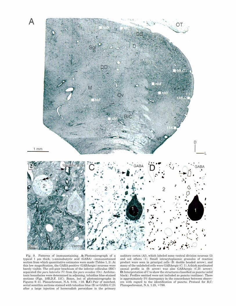
Fig. 9. Patterns of immunostaining. A: Photomicrograph of atypical 1 µm thick, !-aminobutyric acid (GABA) –immunostainedsection from which quantitative estimates were made (Tables 1, 2). Atthis low magnification, the GABA positive (GABAergic) neurons werebarely visible. The cell-poor brachium of the inferior colliculus (BIC)separated the pars lateralis (V) from the pars ovoidea (Ov). Architec-tonic boundaries were determined in adjoining, toluidine blue-stainedsections (Figs. 10B,D,F, 11C). Boxes, loci of photomicrographs inFigures 9–11. Planachromat, N.A. 0.04, #16. B,C: Pair of matched,serial semithin sections stained with toluidine blue (B) or GABA (C,D)after a large injection of horseradish peroxidase in the primary
auditory cortex (AI), which labeled some ventral division neurons (2)and not others (1). Small intracytoplasmic granules of reactionproduct were seen in principal cells (B: double headed arrow), andmany of the unlabeled cells were GABAergic (C: 1). A thick myelinatedaxonal profile in (B: arrow) was also GABAergic (C,D: arrow).D: Interpretation of C to show the structures classified as puncta (solidblack). Profiles omitted were not included as puncta (outlines). Thereis approximately 5% discrepancy in the concordance between observ-ers with regard to the identification of puncta. Protocol for B,C:Planapochromat, N.A. 1.25, #788.
here, and the dorsal division in its entirety, therefore, mustcontain thousands of them (Winer et al., 1999).
The processes that give rise to the giant profiles wereonly approximately 1–2 µm in diameter in both GAD (Fig.7B: 7) and GABA (Fig. 8B: 3) material. The giant punctathemselves were elaborate and had a substructure filledwith thorns, globules, and swellings that could rep-resent clusters of smaller puncta. The central part ofthe profile often had a hollow space. In the plastic-embedded semithin sections counterstained with toluidineblue, this region contained a pale, unmyelinated processthat was immunonegative and with a caliber suggestingthat it may be dendritic (Fig. 10C, arrow). Such punctawere less common near the perikarya of either immu-nonegative (Fig. 8B: 2) or immunopositive (Fig. 7B: 5)neurons.
The giant puncta seemed to be less numerous insemithin sections (Fig. 8B) than in the GAD prepara-tions (Fig. 7B), perhaps because of differences in sectionthickness. A second reason for their apparent enhance-ment in the GAD material was that far more of theprocesses associated with them were revealed, whereasin the GABA, the fine, puncta-sparse segments wererarely immunostained. Perhaps the abundant and appar-ently diffuse clusters of profiles in GABA material(Fig. 8B: 5) would be in continuity if the interveningprocesses were visible, as they are in the thicker GADpreparations.
In addition to the giant puncta, smaller profiles werealso present in the neuropil and near the somata ofimmunonegative neurons (Figs. 7B, 8B). These punctawere less obvious in the GAD preparations, suggestingthat some of the many fine puncta in the GABA materialaggregate in the thicker sections by virtue of immunostain-ing of their intervening processes.
Medial division. The GABAergic organization in themedial division (Figs. 7C, 8C) was unique and featured apopulation of puncta larger than those in the ventraldivision (Figs. 7A, 8A) and smaller than those in the dorsaldivision (Figs. 7B, 8B). It alone of the three divisions had asignificant number of thick GABAergic processes (Figs.7C: 3, 8C: 2), some of which were up to 5 µm in diameter(Saint Marie et al., 1997), and these coursed mediolater-ally, a feature consistent with an extrinsic origin (Wineret al., 1996). Other, finer processes, usually devoid ofpuncta, traversed the medial division neuropil (Fig. 7C).
The dominant medial division puncta were medium insize and globular, and they clustered in the neuropil (Figs.
7C, 8C: 1). These puncta were much smaller than even thefragments of the giant dorsal division puncta, and rarelyas granular as those in the ventral division. As with mostpuncta in the medial geniculate body, they were associ-ated, in descending order of density, with the neuropil, ornear the somata of immunonegative neurons (Fig. 7C: 2)and, in rare instances, near the somata of immunopositivecells (Fig. 8C: 3). Their absolute number was indistinguish-able among the three division (Table 1).
DISCUSSIONWe address six interrelated themes. The first issue is the
concordance of the present results with those of previousmorphological and immunocytochemical studies of the catmedial geniculate body. This is followed by a considerationof possible physiological implications. Next, we assess thesignificance of species specific patterns of thalamic GABA-ergic organization. We then contrast the different patternsof organization seen in specific thalamic sensory and motornuclei with one another. The features common to thalamicGolgi type II cells and the evidence for more than onevariety of Golgi type II cell are assessed. We conclude byevaluating the consequences of these results for parcella-tions of the auditory thalamus.
Relation to prior morphologic andimmunocytochemical results
Golgi studies. A key issue is whether the types ofneurons identified in Golgi preparations can be related tothe present, immunocytochemically identified profiles. Inthe ventral division, Golgi type II cells have a drumstick-shaped soma approximately 10–12 µm in diameter fromwhich approximately three to four thin primary dendritesarise without preferential orientation (Morest, 1964). Thisclass represents approximately 35% of ventral divisionneurons and, with the bushy tufted Golgi type I cells,constitutes the entire neuronal population in the ventraldivision (Morest, 1975). In semithin, plastic-embedded,GABA-immunostained material, the neuronal profile cor-responding to this description was immunopositive andhad a small (10–12 µm in diameter) soma with a highlyinvaginated nuclear envelope, scant cytoplasm, and fineprimary dendrites. In contrast, virtually all of the immu-nonegative neurons were significantly (approximately 25–30%) larger, had smooth somatic contours, and thickerprincipal dendrites with fewer processes, and these often
TABLE 2. Quantitative Summary of Ventral and Dorsal Division GABAergic Neurons1
Division Subdivision Mean ' SD Range
Somatic area (in µm2)
n Statistical comparison2
Ventral Pars lateralis 140.6 ' 26.4 78–204 64 V % Ov? P % 0.18Pars ovoidea 132.1 ' 32.7 71–213 36
Dorsal Dorsal superficial 165.3 ' 44.7 104–299 50 DS % D? P % 0.91DS % DD? P % 0.092DS % DCa? P % 0.0000000000123
DS % Sgl? P % 0.023Dorsal 167.0 ' 39.2 93–294 50 D % DD? P % 0.059
D % DCa? P % 0.00000000000243
D % Sgl? P % 0.012Deep dorsal 111.0 ' 38.7 51–230 50 DD % DCa? P % 0.00000000000000443
Caudal dorsal 216.8 ' 45.0 136–331 50 DCa % Sgl? P % 0.000000000000000373
Suprageniculate 133.3 ' 58.4 52–255 50 Sgl % DD? P % 0.53
1GABAergic, !-aminobutyric acid positive. For other abbreviations, see list.2See footnote 6 on Table 1.3Statistically significant.
GABA IN THE MEDIAL GENICULATE BODY 383
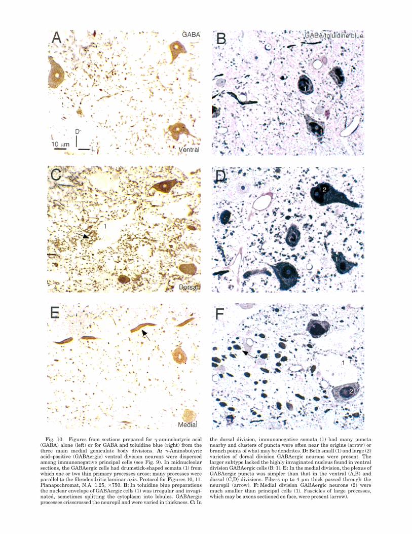
Fig. 10. Figures from sections prepared for !-aminobutyric acid(GABA) alone (left) or for GABA and toluidine blue (right) from thethree main medial geniculate body divisions. A: !-Aminobutyricacid–positive (GABAergic) ventral division neurons were dispersedamong immunonegative principal cells (see Fig. 9). In midnucleolarsections, the GABAergic cells had drumstick-shaped somata (1) fromwhich one or two thin primary processes arose; many processes wereparallel to the fibrodendritic laminar axis. Protocol for Figures 10, 11:Planapochromat, N.A. 1.25, #750. B: In toluidine blue preparationsthe nuclear envelope of GABAergic cells (1) was irregular and invagi-nated, sometimes splitting the cytoplasm into lobules. GABAergicprocesses crisscrossed the neuropil and were varied in thickness. C: In
the dorsal division, immunonegative somata (1) had many punctanearby and clusters of puncta were often near the origins (arrow) orbranch points of what may be dendrites. D: Both small (1) and large (2)varieties of dorsal division GABAergic neurons were present. Thelarger subtype lacked the highly invaginated nucleus found in ventraldivision GABAergic cells (B: 1). E: In the medial division, the plexus ofGABAergic puncta was simpler than that in the ventral (A,B) anddorsal (C,D) divisions. Fibers up to 4 µm thick passed through theneuropil (arrow). F: Medial division GABAergic neurons (2) weremuch smaller than principal cells (1). Fascicles of large processes,which may be axons sectioned en face, were present (arrow).
arose at the somatic poles. Given the many parallelsbetween the profile of the Golgi type II cell and that of theimmunostained ventral division neurons, we conclude thatthe correspondence between them is substantial.
In the cat dorsal division, two varieties of Golgi type IIcell were recognized in rapid Golgi preparations (Winerand Morest, 1983b,1984). One was much smaller (againapproximately 10–12 µm in diameter) and far more numer-ous; it had a thin (approximately 1 µm in diameter),unmyelinated and profusely branched local axon andslender and radiating dendrites with sparse, complexappendages. A second, much rarer Golgi type II cell had alarger soma (approximately 14–18 µm in diameter), thickerdendrites, longer and more variable spines, and a muchthicker axon (approximately 2–3 µm in diameter) withfewer branches than the smaller variety (Fig. 12B). As inthe ventral division, the correspondence between theGolgi-impregnated neuronal profiles and their immuno-stained counterparts was substantial. Both the large andsmall subvarieties were present in the GABA materialand, as expected, the larger neurons were rarer (Fig. 4C:10; see also Fig. 6: 2).
In the medial division, only a small Golgi type II neuronhas been characterized definitively in rapid Golgi material(Morest, 1964; Winer and Morest, 1983a), and it resembledthose described above in the ventral and dorsal divisions(in Winer, 1992, Table 6.1). The present, immunocytochemi-cal results have identified this neuron readily, and, inaddition, a second larger and rarer variety (Fig. 7C: 4) forwhich the corresponding, Golgi-impregnated neuron re-mains uncertain. There are several types of medial divi-sion neuron that might correspond to the large Golgi typeII neuron (for example, tufted or elongated cells). However,too little is known about the structure and local distribu-tion of their axon in the rapid Golgi preparations, and of
their dendritic configuration in immunostained material,to permit more secure conclusions about their identity.
The correlations suggested above should be regarded asprovisional rather than definitive for two reasons. First, nointracellular recordings in the medial geniculate bodyfrom Golgi type II/GABAergic neurons are available thatcould confirm independently their identity. Second, theresults from studies of the lateral geniculate body, in whichcells have been characterized physiologically and morpho-logically, suggest that certain neurons that might appearto be Golgi type II/class III/GABAergic on structuralgrounds are actually excitatory principal cells with anatypical morphology and that project to the cerebral cortex(Friedlander et al., 1980).
Immunocytochemical investigations. It is more diffi-cult to relate the present, neurochemical results directly toprevious studies (Table 3) for several reasons. First, fewprior investigations of the medial geniculate body made aneffort to relate the profiles of immunopositive neurons withthose of Golgi-impregnated cells. Second, our quantitativeresults are based on semithin postembedded material anduse the Abercrombie-Floderus correction, which was notapplied in other investigations; when numerical estimatesof the proportion of GABAergic neurons are not adjustedfor somatic size differences, quantitative differences amongstudies cannot be compared directly. Third, in studies thatused thick sections (&3 µm) for quantitative purposes,technical problems in assessing immunopenetration inthese frozen- or Vibratome-sectioned preparations cancomplicate quantitative estimates of immunonegative andimmunopositive neurons. Fourth, earlier work did notalways specify the nuclear locus of the observations withinthe medial geniculate body. We have treated nucleardifferences in neuronal subtypes and in puncta size, shape,and density as a primary datum. Fifth, we have validated
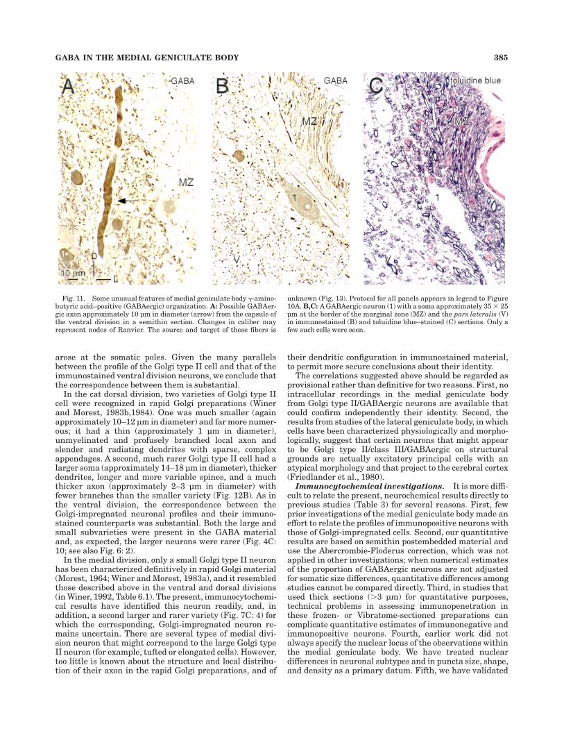
Fig. 11. Some unusual features of medial geniculate body !-amino-butyric acid–positive (GABAergic) organization. A: Possible GABAer-gic axon approximately 10 µm in diameter (arrow) from the capsule ofthe ventral division in a semithin section. Changes in caliber mayrepresent nodes of Ranvier. The source and target of these fibers is
unknown (Fig. 13). Protocol for all panels appears in legend to Figure10A. B,C: A GABAergic neuron (1) with a soma approximately 35 # 25µm at the border of the marginal zone (MZ) and the pars lateralis (V)in immunostained (B) and toluidine blue–stained (C) sections. Only afew such cells were seen.
GABA IN THE MEDIAL GENICULATE BODY 385
our results with different antibodies, whereas prior stud-ies have often used one only.
The present results are largely in accord with those inan earlier investigation of medial geniculate body GABAer-gic organization (Rinvik et al., 1987). The salient points inboth studies were that &20% of the neurons were immu-nopositive in each subdivision of the auditory thalamus,that there were qualitative and quantitative differencesbetween divisions, and that there was a strong gradient ofincreasing immunoreactivity along the caudorostral axisof the medial division. An unusually large variety of type IIcell also was recognized which may correspond to the classwe have identified. Furthermore, in the dorsal division,‘‘ring-like profiles’’ were described that seem closely tomatch the giant peridendritic GABAergic puncta in thepresent account in shape, although not in size.
There are fewer points of concordance between thepresent results and those of another immunocytochemicalstudy (Rouiller et al., 1990). Their maximum proportion ofGABAergic neurons was much lower than the value in thepresent investigation (27 vs. 38%, respectively; see Table3). They found only a modest range of change in thisproportion, i.e., from approximately 18 to approximately28%, along the caudorostral axis in the medial division atstereotaxic levels corresponding to those in the presentstudy, whereas we found a far larger difference (Fig. 2F:Medial). Moreover, the giant, and possibly peridendritic,puncta in the dorsal division were not recognized. Finally,they reported a caudorostral gradient of GABAergic immu-noreactivity for the ventral division that is the opposite ofthe present finding; the reason for the latter discrepancy isunknown. Some numerical differences may be attributableto methodological variables. We counted neurons fromsemithin sections only, and all estimates of proportionswere adjusted with the Abercrombie-Floderus correction.In contrast, their numerical estimates were based on acomparison of adjacent, immunoreacted or Nissl-stainedsections &5 µm thick, with no post hoc compensation forsampling bias due to mean caliper diameter. Differences intissue penetration between semithin immunostained sec-tions (present results) and the thicker, free-floating immu-nostained preparations and Nissl material in their studycould account for all or part of the disparity.
Physiological implicationsThe proportion of medial geniculate body GABAergic
neurons averaged 33% in this study, a value substantiallylarger than the 20% reported in the central nucleus of theinferior colliculus (Oliver et al., 1994), and the 25% seen inthe auditory cortex (Prieto et al., 1994). With the exceptionof the dorsal nucleus of the lateral lemniscus, nearly all ofwhose neurons are GABAergic (Adams and Mugnaini,1984), the medial geniculate body, at least in carnivoresand primates (Winer and Larue, 1996a), may have thelargest proportion of GABAergic cells in the auditorysystem (Aitkin, 1989) and among the highest number inthe brain (Emson, 1983). Thus, the degree of local process-ing and the proportion of interneurons may differ acrossauditory synaptic stations, assuming that some approxi-mate parity in the ratio of interneurons to their respectivesynaptic terminations is preserved from nucleus to nucleus.This finding suggests that GABAergic neurons must havea prominent role in medial geniculate body function.Intracellular recordings and iontophoretic studies thatwould clarify the role of interneurons are not yet available.
Without these data, we can only endorse ideas alreadypropounded as to how processing might be influenced byinterneurons. These include the temporal modulation ofspike activity of ventral division principal cells by means ofaxodendritic synapses, whereas the dendrodendritic end-ings might allow for extended regimens of recurringintraglomerular processing (Morest, 1971, 1975). In anyevent, the presence of only one variety of Golgi type IIGABAergic neuron suggests that the local circuit organiza-tion within pars lateralis and pars ovoidea is likely to besimilar, and that this pattern may differ from that in thedorsal and medial division, in which significantly differentproportions and types of Golgi type II/GABAergic neuronsoccur (Table 1).
Physiological investigations of the dorsal division haverevealed that its neurons respond sluggishly to tonalstimuli and more strongly to complex signals, that theirtuning curves are broad and have lower Q10 dB values thanthose of ventral division neurons, and that slow changes inglobal excitability are a cardinal feature (Aitkin andDunlop, 1969; Altman et al., 1970; Aitkin and Prain, 1974;Aitkin et al., 1981). Some of these attributes may beconferred by the input from the nucleus sagulum (Calfordand Aitkin, 1983; Beneyto et al., 1998), and there is adirect GABAergic projection from the inferior colliculus(Peruzzi et al., 1997) that could play an inhibitory roleonce delegated exclusively to Golgi type II cells (Wineret al., 1996). Indeed, the totality of extrinsic GABAergicinput from the inferior colliculus and the thalamic reticu-lar nucleus (Montero, 1983) could approximate that fromthe GABAergic Golgi type II cells, suggesting that thecollective impact of inhibition on thalamic processing mustbe enormous. In view of the coarse temporal coding andwide frequency tuning of dorsal division neurons, it canonly be surmised that such processes either cannot dependon Golgi type II cells, or that the behavior of dorsal division
Fig. 12. Summary of results and comparison with Golgi studies.A: Typical Golgi type II cell from the dorsal division of the cat medialgeniculate body; it had a small, approximately 10–12 µm in diameterdrumstick-shaped soma, 3–4 main dendrites projecting in a stellateconfiguration, and a local AXON whose distal processes extendedbeyond the section. The somatodendritic profile of this cell resembledthat of many immunostained dorsal division specimens (D: Average).1, the dendritic appendages were sparse and longer than those ofprincipal cells. Protocol for panels A–C: rapid Golgi impregnation. ForA–D: Planapochromat, N.A. 1.32, #1,250. Redrawn from Winer andMorest (1983b). B: A large Golgi type II (IIb; cf. Fig. 13) from the dorsaldivision. This cell was more than twice the somatic size of the smallGolgi type II (IIa; cf. Fig. 13) dorsal division neuron, had a much largerand more complex AXON, and longer and more elaborate dendriticappendages than those of classic type IIa cells (A). Dorsal divisionstellate principal cells (not shown) were still larger, with many shortdendritic appendages, and an unbranched axon. 1, long pedunculateddendritic spine. 2, complex multilobulated appendage. These irregularand contorted dendrites may be too small to be the source of the giantpuncta (Figs. 7B: 4; 8B: 6). Redrawn from Winer and Morest (1984).C: A small stellate Golgi type II cell from the ventral division, with acharacteristic drumstick-shaped soma, 3–4 main trunks parallel tothe long axis of the isofrequency laminae, and with various dendriticappendages (1). The AXON formed only local branches and wasthinner than that of the type IIb neuron (B). 1, the intermediate anddistal dendrites have long, stringy processes. Redrawn from Winer(1992). D: Representative GABAergic neurons from the primary me-dial geniculate body divisions. The largest such cells were in the dorsaldivision, and the medial division had the widest absolute size range.Glutamic acid decarboxylase preparation, frozen sections. Scale bar inA applies to A–D.
386 C.L. HUANG ET AL.
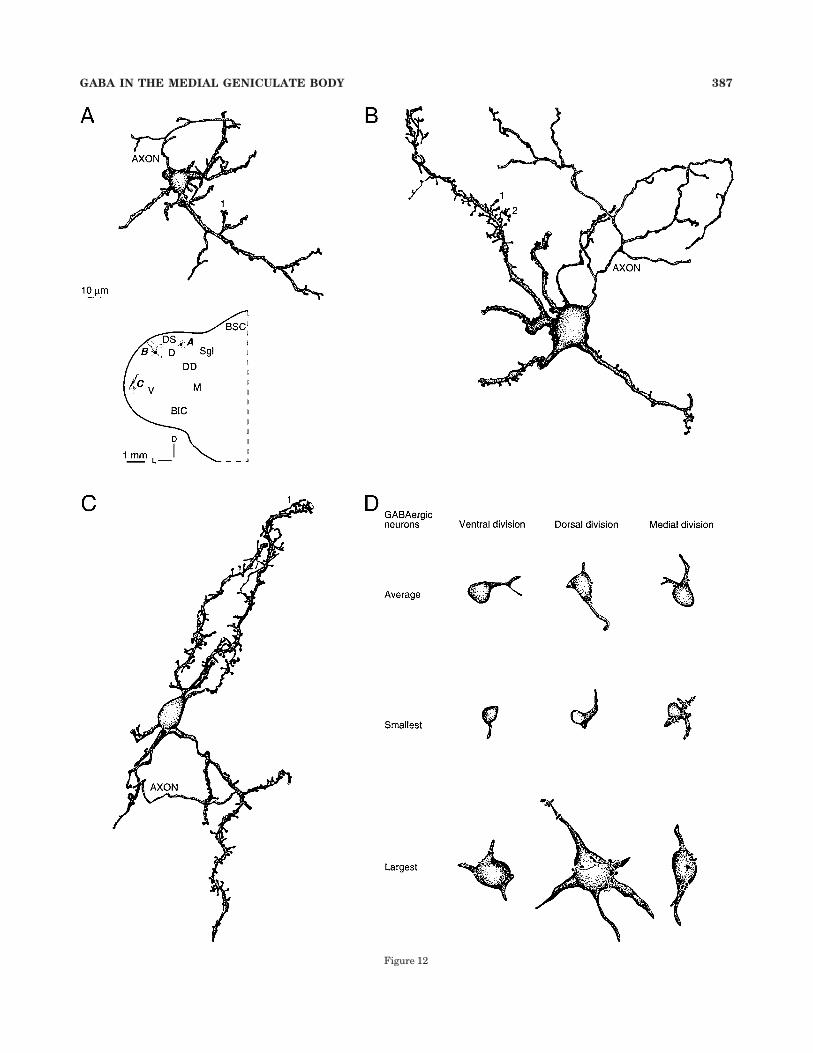
Figure 12
GABA IN THE MEDIAL GENICULATE BODY 387
interneurons differs fundamentally from that of correspond-ing cells in the ventral division. The presence of the largeGolgi type II cells, albeit in small numbers, could repre-sent yet other forms of local circuit processing presumablyunique to the dorsal division. The giant GABAergic puncta,many of which are 5 µm in diameter, are a significantdeparture from the far finer puncta predominating else-where in the medial geniculate body. Each of the six dorsaldivision nuclei has many of these profiles, and they are notpresent elsewhere in the medial geniculate body (Wineret al., 1998). They are often seen near large caliber,immunonegative dendrites and, thereby, may influencethe behavior of principal cells, although ultrastructuralconfirmation is not yet available.
Medial division neurons have even broader tuning thancells in the dorsal or ventral divisions, they prefer complexor polymodal stimuli, and they show a degree of physiologi-cal plasticity not evident in other auditory thalamic nuclei(Aitkin, 1973; Calford, 1983; Gerren and Weinberger, 1983).These attributes certainly seem incompatible with the cycle-by-cycle fine temporal control of signal processing by GABAergicGolgi type II cells that has been hypothesized for the ventraldivision (Morest, 1974), and they leave open what role thelarger GABAergic Golgi type II neuron might serve.
Comparative GABAergic arrangements in themedial geniculate body
Some anatomic features are common to all thalamicnuclei. These include projections to the cerebral cortex(Jones, 1985) which have varying degrees of topography(Jones, 1984), a substantial thalamocortical-corticotha-lamic reciprocity (Winer and Larue, 1987), and the preva-lence of bushy tufted Golgi type I neurons as the main
thalamocortical (Winer, 1984) and thalamosubcortical (Shi-nonaga et al., 1994) projection neuron. Other commondenominators are that many of these neurons are glutama-tergic (LeDoux and Farb, 1991 [rat]) and that their termi-nation is specific to certain cortical layers (Jones, 1981),and, in the case of the medial geniculate body, they have aprojection to the amygdala that targets specific nuclearsubdivisions (LeDoux et al., 1985 [rat]). A further feature,i.e., the presence of a population of GABAergic Golgi typeII cells, has a more variable and nucleus-specific expres-sion. Because these numerical differences have been ad-dressed by several studies (Ohara and Lieberman, 1993[rat]; Arcelli et al., 1997 [rat, guinea pig, cat, monkey]),only the salient points are summarized below.
The proportion of GABAergic neurons in the medialgeniculate body is species specific (Table 3), ranging fromnone (Ottersen and Storm-Mathisen, 1984 [mice]) to "1%(Winer and Larue, 1988 [rat], Winer et al., 1992 [mus-tached bat, Pteronotus p. parnellii]) to approximately 5%(J.A. Winer and D.T. Larue, unpublished observations[pallid bat, Antrozous pallidus]), to approximately 30%(present results), and to a substantial number in primates(Smith et al., 1987 [squirrel monkey]; Winer and Larue,1996a [macaque monkey]). In avians, the few speciesstudied suggest that the range is narrower, from none inchicken (Muller, 1988) to "1% in the barn owl (Winer andLarue, 1996b) nucleus ovoidalis, the thalamic homologuefor the medial geniculate body. In the few reptiles so farinvestigated, GABAergic thalamic cells are rare or nonex-istent in the nucleus reuniens, which is believed to behomologous to the medial geniculate body (Pritz andStritzel, 1994; Pritz, 1995). In some instances, the paucityof GABAergic neurons is limited to the auditory thalamus:
TABLE 3. Comparison With Other Studies of Thalamic GABAergic Organization1
Species Study Nucleus
Antisera2
Amount(in %)
Percent/Division
GABAergicneurons5
(diameteror area) Puncta6GAD3
GABA4
Thick Semithin V D M
Cat Rinvik et al. (1987) MGB # # 24–27 23 32 30 12–14, few 35 µm Thicker and with more irregularoutlines in (D)
Rouiller et al. (1990) # # 7–28 — 0 9–19 µm —Arcelli et al. (1997) # # # — — — — —Present study3 # # # 6–38 33 26 18 !12, rare 18 (D)* µm 1–3Fitzpatrick et al. (1984) LGB # "25 — — — 9–14 µm —Rinvik et al. (1987) # # 12–27 — — — 12–15, few 35 µm Light to dark, some large ring-like
profilesArcelli et al. (1997) # # # 24–27 — — — — —Penny et al. (1983) Vb # # 27–32 — — — 10–12 µm —Spreafico et al. (1983) # 19–21 — — — "10–14 µm Darkly stainedRinvik et al. (1987) # # 20–30 — — — 12–15, few 22–26 µm —Arcelli et al. (1997) # # # 24–27 — — — — —
Rat Muganini and Oertel (1985) MGB # "5 — — — — Very few (D)–few (V, M)Winer and Larue (1988) # "1 — — — V: 69 µm2
D: 64 µm2
M: 81 µm2
1–2
Arcelli et al. (1997) # # # "1 — — — — —Guinea pig Asanuma (1991) # # &10 — — — Vb: 10–15 µm Small
Arcelli et al. (1997) # # # "1 — — — — —Rabbit Arcelli et al. (1997) # # # "1 — — — — —Bat Vater et al. (1992) # # — — — — M: 7–11 µm Variable
Winer et al. (1992) # # "1 — — — V: 38 µm2
D: 33 µm21–2
Monkey Smith et al. (1987) # — — — — —Arcelli et al. (1997) # # # 27–33 — — — — —
1GABAergic, !-aminobutyric acid positive; MGB, medial geniculate body; approximate symbol; extrapolated/estimated data; —, not described; asterisk, from semithin sections. Forother abbreviations, see list.2GAD preparations were sectioned between 15 and 50 µm; GABA preparations were sectioned between 30 and 60 µm for thick sections or prepared from postembedded tissuesectioned at 1–1.5 µm (semithin sections).3GAD-1440 (Oertel et al., 1981).4GABA (DiaSorin; Stillwater, MN).5In µm or µm2.6Puncta types: 1, "0.5 µm in diameter; 2, 0.5–2.0 µm; 3, &2 µm.
388 C.L. HUANG ET AL.
in the case of the mustached bat and rat, for example, theinferior colliculus has many such cells, as does the cerebralcortex (Winer and Larue, 1996a). Perhaps numerical vari-ability across species with regard to GABAergic auditorythalamic neurons is either a departure from some un-known ancestral pattern, or the comparative differencesreflect regressive developmental events (Cowan et al.,1984) in which immature Golgi type II cells do not survivein some species. Although the present results cannotevaluate which of these scenarios is most plausible, theydo predict that, in the absence or severe reduction in thenumber of Golgi type II cells, there should be a similarreduction in the concomitant ultrastructural features thataccompany such cells, including dendrodendritic interac-tions (Morest, 1971), synaptic glomeruli (Ralston, 1971), ornests (Morest, 1975), and their associated intrinsic tha-lamic circuitry (Jones, 1985). This is in fact the case(Ohara and Lieberman, 1993; Arcelli et al., 1997). Howthese might affect thalamic processing is unknown.
We have speculated elsewhere that synaptic glomerulicould act as dynamic frequency-specific or aurally specifictemporal filters adapted to the analysis of the fine struc-ture of the stimulus, such as onset or offset behavior, orfrequency or amplitude modulation (Winer and Morest,1983b, 1984; Winer, 1985). Whatever their role proves ulti-mately to be, that they are largely absent in the auditorythalamus of many rodents, microchiropterans, and avians—and comparatively abundant in carnivores and primates—suggests a difference in processing regimens in the largebrains of advanced mammals, although the nature of thedifference is unclear. It would be interesting to know whichpattern prevails in the auditory thalamus of Cetacea (Kruger,1959)andwhether it entails specificneuraland/orpsychophysi-cal correlates. Because cetacean sensory neocortex appears tohave some features considered as regressive such as a poorlydifferentiated layer IV, a substantial layer II, and unex-pected overall thinness, perhaps the number of thalamicGABAergic Golgi type II cells would resemble the propor-tion in the rat more closely than the value in the cat ormonkey (Morgane et al., 1985; Krubitzer, 1995).
GABAergic organization in otherthalamic nuclei
There is now a sufficient body of data available on theGABAergic organization of the mammalian thalamus topropose some basic principles in carnivores and primates(Table 3). First, the main sensory nuclei each contain atleast approximately 20% GABAergic neurons (Spreafico etal., 1983; Winer, 1992). This finding suggests that theremay be a core of GABAergic operations common to eachmodality, although their nature remains a matter ofconjecture. Second, the proportion of GABAergic neuronsin the lateral geniculate body seems somewhat moreconserved across species (Hendrickson et al., 1983; Oharaet al., 1983; Fitzpatrick et al., 1984) than does the numberfor either the ventrobasal complex (Arcelli et al., 1997) orthe medial geniculate body (Winer and Larue, 1996a). Thisconservation suggests differences among thalamic nucleithat might reflect evolutionary, functional, and modalityspecific constraints. Third, the vast majority of GABAergicneurons match closely the description of the classic type IIcell in the lateral geniculate body (Guillery, 1966; Monteroand Zempel, 1985), medial geniculate body (Morest, 1975;present results: Fig. 13A), and ventrobasal complex (Schei-bel and Scheibel, 1966; Spreafico et al., 1983). This finding
is consistent with the view that Golgi type II and GABAer-gic neurons represent the same neuronal population.Fourth, the sole exception to the latter generalization isthe class of large Golgi type II cells in the dorsal division(Winer and Morest, 1983b), which is presumptively repre-sented by the large GABAergic neurons in immunostainedmaterial (Rinvik et al., 1987; present results). This findingsuggests that some nuclei may have unique GABAergicprocesses that the large type II cells subserve. Fifth, thethalamic reticular nucleus is a common source of GABAer-gic input to the main sensory nuclei in each modality(Jones, 1985). This is in accord with the prediction thatprincipal nuclei receive some common types of corticoreticu-lothalamic modulation (Jones, 1975). Sixth, the variousthalamic nuclei each have different patterns of extrinsicGABAergic input. Thus, there are substantial extrinsicGABAergic inputs to the medial geniculate body from theinferior colliculus (Winer et al., 1996), and from the pretec-tum to the lateral geniculate body (Cucchiaro et al., 1993).In contrast, the ventrobasal complex appears to lack suchinput entirely (De Biasi and Rustioni, 1990). This findingis congruent with the principle established in the auditorybrainstem, where GABAergic and glycinergic neurons areas likely to be extrinsic projection cells as are glutamater-gic or aspartatergic neurons (Winer et al., 1995). Seventh,the few descriptions of the thalamic puncta availablesuggest that they are subdivision specific in shape andperhaps in density (Winer et al., 1992). Perhaps synapticdensity, and the proportion of GABAergic neurons, differsamong nuclei. Eighth, none of the thalamic GABAergicneuronal populations appears to project outside the thala-
Fig. 13. Schematic diagram of documented and putative circuitryin the subdivisions of the cat medial geniculate body based onGolgi-impregnated material (Winer, 1992 [cat, bat, opossum, human]),electron microscopic investigations of synaptology (Morest, 1975), andthe present, immunocytochemical, results. The ventral division (leftside) contains primarily (1; Morest, 1975) small stellate Golgi type II(IIa) cells with axons presynaptic to principal neuron dendrites, andpresynaptic processes that make dendrodendritic contacts (2; Morest,1971). Predicted but unconfirmed synaptic relationships are indicatedby a small gap. Similar arrangements may be present in the dorsaldivision: small type IIa cell make axodendritic and dendrodendriticcontacts onto principal cells (3; Winer, unpublished observations),whereas the second, much larger Golgi type IIb neuron could formperidendritic rings near principal cell dendritic trunks (4; presentresults). Alternate sources (5) for the latter circuit include !-aminobu-tyric acid–positive inferior colliculus neurons (Oliver et al., 1994) thatproject to the medial geniculate body (Winer et al., 1996) monosynapti-cally (Peruzzi et al., 1997), or axons from the thalamic reticularnucleus (Crabtree, 1998).
GABA IN THE MEDIAL GENICULATE BODY 389
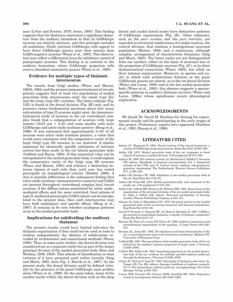
mus (LeVay and Ferster, 1979; Jones, 1985). This findingsuggests that the thalamus represents a significant depar-ture from the auditory brainstem in that its GABAergicneurons are entirely intrinsic, and this principle includesall modalities. Ninth, intrinsic GABAergic cells appear tohave fewer GABAergic puncta near their somata thanGABA-negative neurons (Winer et al., 1993). This observa-tion may reflect a differential locus for inhibitory control ofpostsynaptic neurons. This finding is in contrast to theauditory brainstem, where GABAergic projection cellsreceive abundant axosomatic puncta (Winer et al., 1995).
Evidence for multiple types of thalamicinterneuron
The results from Golgi studies (Winer and Morest,1983b, 1984) and the present immunocytochemical investi-gations suggests that at least two populations of medialgeniculate body interneurons exist: the small (type IIa)and the large (type IIb) varieties. The latter subtype (Fig.12D) is found in the dorsal division (Fig. 2E) only, and itspresence raises fundamental questions about how manysubvarieties of type II neurons might exist. An immunocy-tochemical study of neurons in the cat ventrobasal com-plex found that a subpopulation of neurons with largesomata (18.67 µm ' 2.45) and some smaller cells wereGABAergic and nitric oxide synthase positive (Meng et al.,1996). It was estimated that approximately 3–4% of allneurons were nitric oxide synthase positive, a value thatwould seem consistent with the comparative rarity of thelarge Golgi type IIb neurons in our material. A similarargument for chemically specific subclasses of intrinsicneuron has been made in the rat lateral geniculate body(Gabbott and Bacon, 1994). If this numerical value can beextrapolated to the medial geniculate body, it could explainthe comparative rarity of the Golgi type IIb neurons(Winer and Morest, 1984) and enhance a distinction be-tween classes of such cells that, until now, has restedprincipally on morphological criteria (Tombol, 1969). Aclue to possible differences is the subsequent finding thatnitric oxide synthase–immunoreactive material and GABAare present throughout ventrobasal complex local circuitneurons; if the diffuse nature postulated for nitric oxide–mediated effects and the more restricted, focal actions oftype II–mediated presynaptic GABArelease can be extrapo-lated to the present data, then such interneurons mayhave both modulatory and specific effects (Meng et al.,1997). It remains to be seen whether analogous patternsoccur in the medial geniculate body.
Implications for subdividing the auditorythalamus
The present results would have limited relevance forthalamic organization if they could not be used as tools toconfirm independently the identity of subdivisions re-vealed by architectonic and connectional methods (Winer,1992). Thus, in some early studies, the dorsal division wasconsidered not as a separate entity but as part of the largerprincipal division of the medial geniculate body (Rose andWoolsey, 1949, 1958). This scheme of classification or closevariants of it have persisted until rather recently (Imigand Morel, 1985, their Fig. 1; Rinvik et al., 1987). In thepresent study, the dorsal division could be defined virtu-ally by the presence of the giant GABAergic axon profilesalone (Winer et al., 1999). By the same token, many of thesmaller nuclei within the dorsal division such as the deep
dorsal and caudal dorsal nuclei have distinctive patternsof GABAergic organization (Fig. 2E). Other subnuclei,such as the pars ovoidea and the pars lateralis, areregarded as territorial subdivisions of a single nucleus, theventral division, that contains a homogeneous neuronalpopulation (Morest, 1964) and a continuous, althoughcomplex, arrangement of characteristic frequency (Imigand Morel, 1985). The latter nuclei are not distinguishedfrom one another, either on the basis of neuronal size orthe proportion of GABAergic neurons (Fig. 2C), or by theirthalamocortical connections (Winer, 1992), but solely ontheir laminar organization. Moreover, in species and nu-clei in which such architectonic features as the giantGABAergic puncta are absent, as in the rat dorsal division(Winer and Larue, 1988) and in the bat medial geniculatebody (Winer et al., 1992), this absence suggests a species-specific patterns in auditory thalamic circuitry (Winer andLarue, 1996a) whose significance awaits physiologicalexploration.
ACKNOWLEDGMENTSWe thank Dr. David M. Huchton for sharing his experi-
mental results and for participating in the early stages ofthis project. Preliminary reports have appeared (Huchtonet al., 1991; Huang et al., 1996).
LITERATURE CITEDAdams JC, Mugnaini E. 1984. Dorsal nucleus of the lateral lemniscus: a
nucleus of GABAergic projection neurons. Brain Res Bull 14:585–590.Aitkin LM. 1973. Medial geniculate body of the cat: responses to tonal
stimuli of neurons in medial division. J Neurophysiol 36:275–283.Aitkin LM. 1989. The auditory system. In: Bjorklund A, Hokfelt T, Swanson
LW, editors. Handbook of chemical neuroanatomy, Vol. 7. Integratedsystems of the CNS, part II: Central visual, auditory, somatosensory,gustatory. Amsterdam, The Netherlands: Elsevier Science PublishersB.V., p 165–218.
Aitkin LM, Dunlop CW. 1969. Inhibition in the medial geniculate body ofthe cat. Exp Brain Res 7:68–83.
Aitkin LM, Prain SM. 1974. Medial geniculate body: unit responses in theawake cat. J Neurophysiol 37:512–521.
Aitkin LM, Calford MB, Kenyon CE, Webster WR. 1981. Some facets of theorganization of the principal division of the cat medial geniculate body.In: Syka J, Aitkin, LM, editors. Neuronal mechanisms of hearing.London: Plenum Publishing Company. p 163–181.
Altman JA, Syka J, Shmigidina GN. 1970. Neuronal activity in the medialgeniculate body of the cat during monaural and binaural stimulation.Exp Brain Res 10:81–93.
Arcelli P, Frassoni C, Regondi MC, De Biasi S, Spreafico R. 1997. GABAer-gic neurons in mammalian thalamus: a marker of thalamic complexity?Brain Res Bull 42:27–37.
Beneyto M, Winer JA, Larue DT, Prieto JJ. 1998. Auditory connections andneurochemical organization of the sagulum. J Comp Neurol 401:329–351.
Berman AL, Jones EG. 1982. The thalamus and basal telencephalon of thecat: a cytoarchitectonic atlas with stereotaxic coordinates. Madison WI:University of Wisconsin Press.
Calford MB. 1983. The parcellation of the medial geniculate body of the catdefined by the auditory response properties of single units. J Neurosci3:2350–2364.
Calford MB, Aitkin LM. 1983. Ascending projections to the medial genicu-late body of the cat: evidence for multiple, parallel auditory pathwaysthrough the thalamus. J Neurosci 3:2365–2380.
Clarey JC, Barone P, Imig TJ. 1992. Physiology of thalamus and cortex. In:Popper AN, Fay RR, editors. Springer handbook of auditory research,Vol. 2: The mammalian auditory pathway: neurophysiology. New York:Springer-Verlag. p 232–334.
Cowan WM, Fawcett JW, O’Leary DDM, Stanfield BB. 1984. Regressiveevents in neurogenesis. Science 225:1258–1265.
390 C.L. HUANG ET AL.
Cox CL, Huguenard JR, Prince DA. 1996. Heterogeneous axonal arboriza-tions of rat thalamic reticular neurons in the ventrobasal nucleus. JComp Neurol 366:416–430.
Cox CL, Huguenard JR, Prince DA. 1997. Nucleus reticularis neuronsmediate diverse inhibitory effects in thalamus. Proc Natl Acad Sci USA94:8854–8859.
Cox CL, Zhou Q, Sherman SM. 1998. Glutamate locally activates dendriticoutputs of thalamic interneurons. Nature 394:478–482.
Crabtree JW. 1998. Organization in the auditory sector of the cat’s thalamicreticular nucleus. J Comp Neurol 390:167–182.
Cucchiaro JB, Uhlrich DJ, Sherman SM. 1993. Ultrastructure of synapsesfrom the pretectum in the A-laminae of the cat’s lateral geniculatenucleus. J Comp Neurol 334:618–630.
De Biasi S, Rustioni A. 1990. Ultrastructural immunocytochemical localiza-tion of excitatory amino acids in the somatosensory system. J Histo-chem Cytochem 38:1745–1754.
DeFelipe J, Hendry SHC, Jones EG. 1986. A correlative electron micro-scopic study of basket cells and large GABAergic neurons in the monkeysensory-motor cortex. Neuroscience 17:991–1009.
Diamond IT, Jones EG, Powell TPS. 1969. The projection of the auditorycortex upon the diencephalon and brain stem of the cat. Brain Res15:305–340.
Emson PC, editor. 1983. Chemical neuroanatomy. New York, NY: RavenPress.
Fitzpatrick D, Penny GR, Schmechel DE. 1984. Glutamic acid decarboxylase-immunoreactive neurons and terminals in the lateral geniculate nucleusof the cat. J Neurosci 4:1809–1829.
Friedlander MJ, Lin C-S, Stanford LR, Sherman SM. 1980. Morphology offunctionally identified neurons in lateral geniculate nucleus of the cat. JNeurophysiol 46:80–129.
Gabbott PLA, Bacon SJ. 1994. Two types of interneuron in the dorsallateral geniculate nucleus of the rat: a combined NADPH diaphorasehistochemical and GABA immunocytochemical study. J Comp Neurol350:281–301.
Gerren RA, Weinberger NM. 1983. Long term potentiation in the magnocel-lular medial geniculate nucleus of the anesthetized cat. Brain Res265:138–142.
Guillery RW. 1966. A study of Golgi preparations from the dorsal lateralgeniculate nucleus of the adult cat. J Comp Neurol 128:21–50.
Hendrickson AE, Ogren MP, Vaughn JE, Barber RP, Wu J-Y. 1983. Lightand electron microscopic immunocytochemical localization of glutamicacid decarboxylase in monkey geniculate complex: evidence for GABAer-gic neurons and synapses. J Neurosci 3:1245–1262.
Horridge GA. 1968. Interneurons. Their origin, action, specificity, growth,and plasticity. London: W.H. Freeman and Co.
Houser CR, Vaughn JE, Barber RP, Roberts E. 1980. GABA neurons are themajor cell type of the nucleus reticularis thalami. Brain Res 200:341–354.
Huang CL, Huchton DM, Larue DT, Winer JA. 1996. The GABAergicorganization of the cat medial geniculate body. Proc Soc Neurosci22:1069.
Huchton DM, Larue DT, Sun JY-M, Winer JA. 1991. The organization ofGABAergic neurons in the cat medial geniculate body: a quantitativeimmunocytochemical study of post-embedded material. Proc Soc Neuro-sci 17:300.
Imig TJ, Morel A. 1985. Tonotopic organization in lateral part of posteriorgroup of thalamic nuclei in the cat. J Neurophysiol 53:836–851.
Jahnsen H, Llinas R. 1984a. Electrophysiological studies of guinea-pigthalamic neurones: an in vitro study. J Physiol (Lond) 349:205–226.
Jahnsen H, Llinas R. 1984b. Ionic basis for the electroresponsiveness andoscillatory properties of guinea-pig thalamic neurones in vitro. JPhysiol (Lond) 349:227–248.
Jones EG. 1975. Some aspects of the organization of the thalamic reticularcomplex. J Comp Neurol 162:285–308.
Jones EG. 1981. Functional subdivision and synaptic organization of themammalian thalamus. Int Rev Physiol 25:173–245.
Jones EG. 1984. Organization of the thalamocortical complex and itsrelation to sensory processes. In: DarianSmith I, editor. Handbook ofphysiology, Sect. 1: The nervous system, Vol. III: Sensory processes,Part 1. Washington, DC: American Physiological Society. p 149–212.
Jones EG. 1985. The thalamus. New York, NY: Plenum Press.Kim U, Sanchez-Vives MV, McCormick DA. 1997. Functional dynamics of
GABAergic inhibition in the thalamus. Science 278:130–134.Krubitzer L. 1995. The organization of neocortex in mammals: are species
differences really so different? Trends Neurosci 18:408–417.
Kruger L. 1959. The thalamus of the dolphin (Tursiops truncatus) andcomparison with other mammals. J Comp Neurol 111:133–194.
Larue DT, Winer JA. 1996. Postembedding immunocytochemistry of largesections of brain tissue: an improved flatembedding technique. JNeurosci Methods 68:125–132.
LeDoux JE, Farb CR. 1991. Neurons of the acoustic thalamus that projectto the amygdala contain glutamate. Neurosci Lett 134:145–149.
LeDoux JE, Ruggiero DA, Reis DJ. 1985. Projections to the subcorticalforebrain from anatomically defined regions of the medial geniculatebody in the rat. J Comp Neurol 242:182–213.
Lee SM, Friedberg MH, Ebner FF. 1994. The role of GABA-mediatedinhibition in the rat ventral posterior medial thalamus. I. Assessment ofreceptive field changes following thalamic reticular nucleus lesions. JNeurophysiol 71:1702–1715.
LeVay S, Ferster D. 1979. Proportion of interneurons in the cat’s lateralgeniculate nucleus. Brain Res 164:304–308.
Meng X-W, Ohara PT, Ralston III HJ. 1996. Nitric oxide synthase immuno-reactivity distinguishes a sub-population of GABA-immunoreactiveneurons in the ventrobasal complex of the cat. Brain Res 728:111–115.
Meng X-W, Ohara PT, Ralston HJ III. 1997. Ultrastructural localization ofnitric oxide synthase immunoreactivity in the cat ventrobasal complex.J Neurocytol 27:833–842.
Montero VM. 1983. Ultrastructural identification of axon terminals fromthe thalamic reticular nucleus in the medial geniculate body in the rat:An EM autoradiographic study. Exp Brain Res 51:338–342.
Montero VM, Zempel J. 1985. Evidence for two types of GABA-containinginterneurons in the A-laminae of the cat lateral geniculate nucleus: adouble-label HRP and GABA-immunocytochemical study. Exp BrainRes 60:603–609.
Morest DK. 1964. The neuronal architecture of the medial geniculate bodyof the cat. J Anat (Lond.) 98:611–630.
Morest DK. 1965. The laminar structure of the medial geniculate body ofthe cat. J Anat (Lond.) 99:143–160.
Morest DK. 1971. Dendrodendritic synapses of cells that have axons: thefine structure of the Golgi type II cell in the medial geniculate body ofthe cat. Z Anat Entwicklungsgesch 133:216–246.
Morest DK. 1974. LCN’s in the medial geniculate body of the cat. NeurosciRes Prog Bull 13:367–377.
Morest DK. 1975. Synaptic relationships of Golgi type II cells in the medialgeniculate body of the cat. J Comp Neurol 162:157–193.
Morgane PJ, Jacobs MS, Galaburda A. 1985. Conservative features ofneocortical evolution in dolphin brain. Brain Behav Evol 26:176–184.
Mugnaini E, Dahl A-L. 1983. Zinc-aldehyde fixation for light-microscopicimmunocytochemistry of nervous tissues. J Histochem Cytochem 31:1435–1438.
Mugnaini E, Oertel WH. 1985. An atlas of the distribution of GABAergicneurons and terminals in the rat CNS as revealed by GAD immunocyto-chemistry. In: Bjorklund A, Hokfelt T, editors. Handbook of chemicalneuroanatomy, Vol. 4: GABA and neuropeptides in the CNS, Part 1.Amsterdam, The Netherlands: Elsevier Scientific Publishers BV. p436–608.
Muller CM. 1988. Distribution of GABAergic perikarya and terminals inthe centers of the higher auditory pathway of the chicken. Cell TissueRes 252:99–106.
Niimi K, Matsuoka H. 1979. Thalamocortical organization of the auditorysystem in the cat studied by retrograde axonal transport of horseradishperoxidase. Adv Anat Embryol Cell Biol 57:1–56.
Oertel WH, Schmechel DE, Tappaz ML, Kopin IJ. 1981. Production of aspecific antiserum to rat brain glutamic acid decarboxylase by injectionof an antigen-antibody complex. Neuroscience 6:2689–2700.
Ohara PT, Lieberman AR. 1993. Some aspects of the synaptic circuitryunderlying inhibition in the ventrobasal thalamus. J Neurocytol 2:815–825.
Ohara PT, Lieberman AR, Hunt SP, Wu J-Y. 1983. Neural elementscontaining glutamic acid decarboxylase (GAD) in the dorsal lateralgeniculate nucleus of the rat: immunohistochemical studies by lightand electron microscopy. Neuroscience 8:189–212.
Oliver DL, Winer JA, Beckius GE, Saint Marie RL. 1994. Morphology ofGABAergic cells and axon terminals in the cat inferior colliculus. JComp Neurol 340:27–42.
Ottersen OP, Storm-Mathisen J. 1984. GABA-containing neurons in thethalamus and pretectum of the rodent. An immunocytochemical study.Anat Embryol (Berl) 170:197–207.
Palay SL, Chan-Palay V. 1974. Cerebellar cortex: cytology and organiza-tion. Berlin: Springer-Verlag.
GABA IN THE MEDIAL GENICULATE BODY 391
Peruzzi D, Bartlett E, Smith PH, Oliver DL. 1997. A monosynapticGABAergic input from the inferior colliculus to the medial geniculatebody in rat. J Neurosci 17:3766–3777.
Prieto JJ, Peterson BA, Winer JA. 1994. Morphology and spatial distribu-tion of GABAergic neurons in cat primary auditory cortex (AI). J CompNeurol 344:349–382.
Pritz MB. 1995. The thalamus of reptiles and mammals: similarities anddifferences. Brain Behav Evol 46:197–208.
Pritz MB, Stritzel ME. 1994. Glutamic acid decarboxylase immunoreactiv-ity in some dorsal thalamic nuclei in Crocodilia. Neurosci Lett 165:109–112.
Ralston HJ III. 1971. Evidence for presynaptic dendrites and a proposal fortheir mechanism of action. Nature 230:585–587.
Rinvik E, Ottersen OP, Storm-Mathisen J. 1987. !-Aminobutyrate-likeimmunoreactivity in the thalamus of the cat. Neuroscience 21:787–805.
Rose JE, Woolsey CN. 1949. The relations of thalamic connections, cellularstructure, and evocable electrical activity in the auditory region of thecat. J Comp Neurol 91:441–466.
Rose JE, Woolsey CN. 1958. Cortical connections and functional organiza-tion of thalamic auditory system of cat. In: Harlow HF, Woolsey CN,editors. Biological and biochemical bases of behavior. Madison, WI:University of Wisconsin Press. p 127–150.
Rouiller EM, Capt M, Hornung JP, Streit P. 1990. Correlation betweenregional changes in the distributions of GABA-containing neurons andunit response properties in the medial geniculate body of the cat. HearRes 49:249–258.
Saint Marie RL, Stanforth DA, Jubelier EM. 1997. Substrate for rapidfeedforward inhibition of the auditory forebrain. Brain Res 765:173–176.
Scheibel ME, Scheibel AB. 1966. Patterns of organization in specific andnonspecific thalamic fields. In: Purpura DP, Yahr MD, editors. Thethalamus. New York, NY: Columbia University Press. p 13–46.
Shinonaga Y, Takada M, Mizuno N. 1994. Direct projections from thenon-laminated divisions of the medial geniculate nucleus to the tempo-ral polar cortex and amygdala in the cat. J Comp Neurol 340:405–426.
Smith Y, Seguela P, Parent A. 1987. Distribution of GABA-immunoreactiveneurons in the thalamus of the squirrel monkey (Saimiri sciureus).Neuroscience 22:579–591.
Smolen AJ, Wright LL, Cunningham TJ. 1983. Neuron numbers in thesuperior cervical sympathetic ganglion of the rat: a critical comparisonof methods for cell counting. J Neurocytol 12:739–750.
Society for Neuroscience. 1991. Handbook for the use of animals inneuroscience research. Washington, DC: Society for Neuroscience.
Spreafico R, Schmechel DE, Ellis LC Jr., Rustioni A. 1983. Cortical relayneurons and interneurons on the n. ventralis posterolateralis of cats: ahorseradish peroxidase, electron-microscopic, Golgi and immunocyto-chemical study. Neuroscience 9:491–509.
Tombol T. 1969. Two types of short axon (Golgi 2nd) interneurons in thespecific thalamic nuclei. Acta Morphol Acad Sci Hung 17:285–297.
Ulrich D, Huguenard JR. 1997. GABAA-receptor-mediated rebound burstfiring and burst shunting in thalamus. J Neurophysiol 78:1748–1751.
Windhorst U. 1990. Activation of Renshaw cells. Prog Neurobiol 35:135–179.
Winer BJ. 1971. Statistical principles in experimental design. New York,NY: McGraw-Hill.
Winer JA. 1984. Identification and structure of neurons in the medialgeniculate body projecting to primary auditory cortex (AI) in the cat.Neuroscience 13:395–413.
Winer JA. 1985. The medial geniculate body of the cat. Adv Anat EmbryolCell Biol 86:1–98.
Winer JA. 1991. Anatomy of the medial geniculate body. In: Altschuler RA,Bobbin RP, Clopton BM, Hoffman DW, editors. Neurobiology of hearing,Vol. II: The central auditory system. New York, NY: Raven Press, Ltd. p293–333.
Winer JA. 1992. The functional architecture of the medial geniculate bodyand the primary auditory cortex. In: Webster DB, Popper AN, Fay RR,editors. Springer handbook of auditory research, Vol. 1: The mamma-lian auditory pathway: neuroanatomy. New York, NY: Springer-Verlag.p 222–409.
Winer JA, Larue DT. 1987. Patterns of reciprocity in auditory thalamocorti-cal and corticothalamic connections: study with horseradish peroxidaseand autoradiographic methods in the rat medial geniculate body. JComp Neurol 257:282–315.
Winer JA, Larue DT. 1988. Anatomy of glutamic acid decarboxylase (GAD)immunoreactive neurons and axons in the rat medial geniculate body. JComp Neurol 278:47–68.
Winer JA, Larue DT. 1996a. Evolution of GABAergic circuitry in themammalian medial geniculate body. Proc Natl Acad Sci USA 93:3083–3087.
Winer JA, Larue DT. 1996b. GABA and glycine in the auditory thalamusand midbrain of the barn owl (Tyto alba). Proc Soc Neurosci 22:1069.
Winer JA, Morest DK. 1983a. The medial division of the medial geniculatebody of the cat: implications for thalamic organization. J Neurosci3:2629–2651.
Winer JA, Morest DK. 1983b. The neuronal architecture of the dorsaldivision of the medial geniculate body of the cat. A study with the rapidGolgi method. J Comp Neurol 221:1–30.
Winer JA, Morest DK. 1984. Axons of the dorsal division of the medialgeniculate body of the cat: a study with the rapid Golgi method. J CompNeurol 224:344–370.
Winer JA, Wenstrup JJ, Larue DT. 1992. Patterns of GABAergic immunore-activity define subdivisions of the mustached bat’s medial geniculatebody. J Comp Neurol 319:172–190.
Winer JA, Khurana SK, Prieto JJ, Larue DT. 1993. GABAergic axosomaticendings preferentially target non-GABAergic neurons in the cat medialgeniculate body. Proc Soc Neurosci 19:1426.
Winer JA, Larue DT, Pollak GD. 1995. GABA and glycine in the centralauditory system of the mustache bat: structural substrates for inhibi-tory neuronal organization. J Comp Neurol 355:317–353.
Winer JA, Saint Marie RL, Larue DT, Oliver DL. 1996. GABAergicfeedforward projections from the inferior colliculus to the medialgeniculate body. Proc Natl Acad Sci USA 93:8005–8010.
Winer JA, Larue DT, Huang CL. 1998. Novel convergence of giant axonalendings in the cat medial geniculate body. Proc Soc Neurosci 24:1881.
Winer JA, Larue DT, Huang CL. 1999. Two systems of giant axon terminalsin the cat medial geniculate body: convergence of cortical and GABAer-gic inputs. J Comp Neurol 413:181–197.
392 C.L. HUANG ET AL.


![Medial septum lesions disrupt exploratory trip ... · septohippocampal involvement in dead reckoning ... cholinergic and GABAergic projections to the hippocampus [16,17]. Second,](https://static.fdocuments.in/doc/165x107/5fa6e449750b7f31bc09c35f/medial-septum-lesions-disrupt-exploratory-trip-septohippocampal-involvement.jpg)