G Gynecology & Obstetrics Damiani et al, Gynecol bstet ... · Damiani et al, Gynecol bstet...
3
Damiani et al., Gynecol Obstet (Sunnyvale) 2014, 4:4 DOI: 10.4172/2161-0932.1000218 Case Report Open Access Volume 4 • Issue 4 • 1000218 Gynecol Obstet (Sunnyvale) ISSN: 2161-0932 Gynecology, an open access journal Severe Hepatic Rupture after Cesarean Section for Help Syndrome followed by Multiple Laparotomies: A Teaching Case Gianluca Raffaello Damiani*, Gianmaria Confalonieri, Luca Fumagalli, Paolo Faccioli, Alfredo Galluzzi and Antonio Pellegrino Department of Obstetrics and Gynecology, Alessandro Manzoni Hospital, Lecco, Italy Abstract A successful management of hepatic capsular rupture, with massive hemoperitoneum occurred 15 hours after an emergency cesarean section at 36 week’ gestation is meticulously descripted. The grade of hepatic involvement varies from minor capsular laceration to extensive parenchymal rupture. Our management involved a combination of surgical interventions and aggressive supportive care. The patient was discharged after 54 days and 4 laparotomies and a super-selective artery embolization. Ultrasound after 45 days from the last surgery showed uniform hepatic parenchyma free of focal lesions. Due to the rarity and the unpredictability nature of this devastating event we retain necessary to report our experience, reinforcing the importance of the post-surgery management. *Corresponding author: Damiani Gianluca Raffaello, Department of Obstetrics and Gynecology ,Alessandro Manzoni Hopsital, Dell’Eremo street 11, Lecco, Italy, Tel: 39- 080-5612443; E-mail: [email protected] Received March 04, 2014; Accepted April 16, 2014; Published April 26, 2014 Citation: Damiani GR, Confalonieri G, Fumagalli L, Faccioli P, Galluzzi A, et al. (2014) Severe Hepatic Rupture after Cesarean Section for Help Syndrome followed by Multiple Laparotomies: A Teaching Case. Gynecol Obstet (Sunnyvale) 4: 218. doi:10.4172/2161-0932.1000218 Copyright: © 2014 Damiani GR, et al. This is an open-access article distributed under the terms of the Creative Commons Attribution License, which permits unrestricted use, distribution, and reproduction in any medium, provided the original author and source are credited. Keywords: Hemoperitoneum; Super-selective artery embolization; Glisson’s capsule’s rupture; Bogota bag; Absorbable mesh Introduction Spontaneous Hepatic Rupture (HR) is an infrequent and life–threating condition of pregnancy that is virtually exclusively associated with severe Pre-eclampsia (PE) or HELLP Syndrome (HS) with an incidence of 1 per 2000 patients [1]. In the present case intraparenchymal and sub-capsular hemorrhage developed, resulting in a HR. Fatality rates are high despite the successes in hepatic surgery and critical care unit assistance. e crucial pivotal points are the atypical clinical presentation of PE or HS, taking into account that HR can occur without clinical signs or warning. Case A 31-years old nulliparous woman, aſter In vitro Fertilization (IVF), was referred our department at 35+5 weeks’ gestation presenting, elevated Blood Pressure (BP). e patient’s BP was occasionally borderline since the 27 th weeks of gestation. On admission BP was 178/90 mmhg, proteinuria was 0.59/24 g/h, ast 15 and alt 13 u/l, uric acid 6.8 mg/dl, platelets 268 10 9 /L, d-dimero 407 mg/L. Uterine and middle cerebral Doppler were within normal ranges. e patient was without any complaints. Her personal history was unremarkable. Labetalol was administered (15 ml/h). Subsequent BP values were within normal limits. Betamethasone was administered for prevention of RDS. Four days aſter admittance, the patient complained of mild epigastric pain, accompanied by vomiting. Labor was induced using Foley trans-cervical catheter. Labetalol was increased to 25 ml/h. Aſter 12 hours from labor induction, lab tests presented a slight increase in liver enzymes, while platelets were stable (Table 1). Proteinuria was 3.55 g/24 h. 8 hours later, there was a significant increase of aspartate-alanine aminotransferase (ast 204 u/l, alt 165 u/l) and lactic dehydrogenase (ldh 500 u/l). BP was 160/110, headache and moderate epigastric pain were complained by the patient. Cesarean section (CS) was performed owing to worsening clinical and laboratory parameters and a female infant was born with Apgar scores of 9 and 10 aſter 1 and 5 minutes, respectively. e CS was uncomplicated and there were no findings of hemoperitoneum. 4 hours later, the uterus was contracted and the onset of diuresis was optimal (200 ml/h). Proteinuria was 7.78 g/24 h. 18 hours aſter CS, the patient complained of general malaise and developed marked abdominal distension. Ultrasound showed free fluid in abdomen suggestive of hemoperitoneum with inferior vena cava diameter in M-mode of 13 mm. Blood sample revealed a severe anemic situation (hb 6.6 mg/dl, ht 20.6%, diuresis 15 ml/h). e patient was taken to the operating room for an explorative laparotomy. Intraoperative findings included massive hemoperitoneum, about 800 ml of fresh blood were aspirated, uterine incision was repaired, resulting hemostatic. Superior abdomen was explored and no active bleeding was evidenced. Post-surgery the patient was transferred to the Intensive Care Unit (ICU). Four units of fresh blood (1000 ml), 6 units of fresh frozen plasma (3600 ml) and 1 unit of platelets were transfused. Magnesium sulphate and labetalol were infused at 25 ml/h (1g/h). 14 hours later, abdominal ultrasound revealed a voluminous hematoma interesting the upper quadrant, right hypochondrium. A CT scan revealed a 14 cm intra-parenchymal hepatic hematoma with active bleeding from the right and leſt ramus of hepatic artery. A concomitant 11 cm–sub-capsular hematoma with an extra capsular extension into the paracolic gutter. Free fluid was present in the Douglas space (Figure 1). Super-selective embolization of hepatic right artery was performed, according to Seldinger technique (5 Fr Simmons2 catheters), with microcatheter Progreat (Terumo-0.035) and introduction of spongostan; also leſt artery was treated. A concomitant contraction of diuresis was present (35 ml/h) (Figures 2 and 3). CT scan showed outbreaks of bleeding in both the hepatic lobes. An explorative laparotomy with xifo ombelic pubic incision was performed to evacuate the voluminous clot in order to decompress the portal system: e colon and the ileal loops were compressed and stretched by the hematoma along the inferior mesocolic space. e falciform and round ligaments were sectioned. At the manual examination a wide laceration of Glisson’ capsule was noticed. Direct hemostasis was not achievable, because the liver was edematous. Evacuation of the hematoma compressing the leſt hepatic lobe was performed. Hematoma on the right lobe was not removed, where the original lesion was located, in order to avoid severe hemorrhage. Portal venous flussimetry resulted normal, and a perihepatic packing was applied (14 gauzes). A bogotà bag was necessary in effort to reduce the intra-abdominal pressure to restore system perfusion. Estimated blood losses were 1000 cc. In post-surgery course there was an improvement of the conditions of the patient, with a decrease of transaminases. Two packed red blood cells were G y n e c o l o g y & O b s t e t r i c s ISSN: 2161-0932 Gynecology & Obstetrics
Transcript of G Gynecology & Obstetrics Damiani et al, Gynecol bstet ... · Damiani et al, Gynecol bstet...
Damiani et al., Gynecol Obstet (Sunnyvale) 2014, 4:4 DOI: 10.4172/2161-0932.1000218
Case Report Open Access
Volume 4 • Issue 4 • 1000218Gynecol Obstet (Sunnyvale)ISSN: 2161-0932 Gynecology, an open access journal
Severe Hepatic Rupture after Cesarean Section for Help Syndrome followed by Multiple Laparotomies: A Teaching CaseGianluca Raffaello Damiani*, Gianmaria Confalonieri, Luca Fumagalli, Paolo Faccioli, Alfredo Galluzzi and Antonio PellegrinoDepartment of Obstetrics and Gynecology, Alessandro Manzoni Hospital, Lecco, Italy
AbstractA successful management of hepatic capsular rupture, with massive hemoperitoneum occurred 15 hours after
an emergency cesarean section at 36 week’ gestation is meticulously descripted. The grade of hepatic involvement varies from minor capsular laceration to extensive parenchymal rupture. Our management involved a combination of surgical interventions and aggressive supportive care. The patient was discharged after 54 days and 4 laparotomies and a super-selective artery embolization. Ultrasound after 45 days from the last surgery showed uniform hepatic parenchyma free of focal lesions. Due to the rarity and the unpredictability nature of this devastating event we retain necessary to report our experience, reinforcing the importance of the post-surgery management.
*Corresponding author: Damiani Gianluca Raffaello, Department of Obstetrics andGynecology ,Alessandro Manzoni Hopsital, Dell’Eremo street 11, Lecco, Italy, Tel: 39-080-5612443; E-mail: [email protected]
Received March 04, 2014; Accepted April 16, 2014; Published April 26, 2014
Citation: Damiani GR, Confalonieri G, Fumagalli L, Faccioli P, Galluzzi A, et al. (2014) Severe Hepatic Rupture after Cesarean Section for Help Syndrome followed by Multiple Laparotomies: A Teaching Case. Gynecol Obstet (Sunnyvale) 4: 218. doi:10.4172/2161-0932.1000218
Copyright: © 2014 Damiani GR, et al. This is an open-access article distributed under the terms of the Creative Commons Attribution License, which permits unrestricted use, distribution, and reproduction in any medium, provided the original author and source are credited.
Keywords: Hemoperitoneum; Super-selective artery embolization;Glisson’s capsule’s rupture; Bogota bag; Absorbable mesh
IntroductionSpontaneous Hepatic Rupture (HR) is an infrequent and
life–threating condition of pregnancy that is virtually exclusively associated with severe Pre-eclampsia (PE) or HELLP Syndrome (HS) with an incidence of 1 per 2000 patients [1]. In the present case intraparenchymal and sub-capsular hemorrhage developed, resulting in a HR. Fatality rates are high despite the successes in hepatic surgery and critical care unit assistance. The crucial pivotal points are the atypical clinical presentation of PE or HS, taking into account that HR can occur without clinical signs or warning.
CaseA 31-years old nulliparous woman, after In vitro Fertilization (IVF),
was referred our department at 35+5 weeks’ gestation presenting, elevated Blood Pressure (BP). The patient’s BP was occasionally borderline since the 27th weeks of gestation. On admission BP was 178/90 mmhg, proteinuria was 0.59/24 g/h, ast 15 and alt 13 u/l, uric acid 6.8 mg/dl, platelets 268 109/L, d-dimero 407 mg/L. Uterine and middle cerebral Doppler were within normal ranges. The patient was without any complaints. Her personal history was unremarkable. Labetalol was administered (15 ml/h). Subsequent BP values were within normal limits. Betamethasone was administered for prevention of RDS. Four days after admittance, the patient complained of mild epigastric pain, accompanied by vomiting. Labor was induced using Foley trans-cervical catheter. Labetalol was increased to 25 ml/h. After 12 hours from labor induction, lab tests presented a slight increase in liver enzymes, while platelets were stable (Table 1). Proteinuria was 3.55 g/24 h. 8 hours later, there was a significant increase of aspartate-alanine aminotransferase (ast 204 u/l, alt 165 u/l) and lactic dehydrogenase (ldh 500 u/l). BP was 160/110, headache and moderate epigastric pain were complained by the patient. Cesarean section (CS) was performed owing to worsening clinical and laboratory parameters and a female infant was born with Apgar scores of 9 and 10 after 1 and 5 minutes, respectively. The CS was uncomplicated and there were no findings of hemoperitoneum. 4 hours later, the uterus was contracted and the onset of diuresis was optimal (200 ml/h). Proteinuria was 7.78 g/24 h. 18 hours after CS, the patient complained of general malaise and developed marked abdominal distension. Ultrasound showed free fluid in abdomen suggestive of hemoperitoneum with inferior vena cava diameter in M-mode of 13 mm. Blood sample revealed a severe anemic situation (hb 6.6 mg/dl,ht 20.6%, diuresis 15 ml/h). The patient was taken to the operatingroom for an explorative laparotomy. Intraoperative findings included
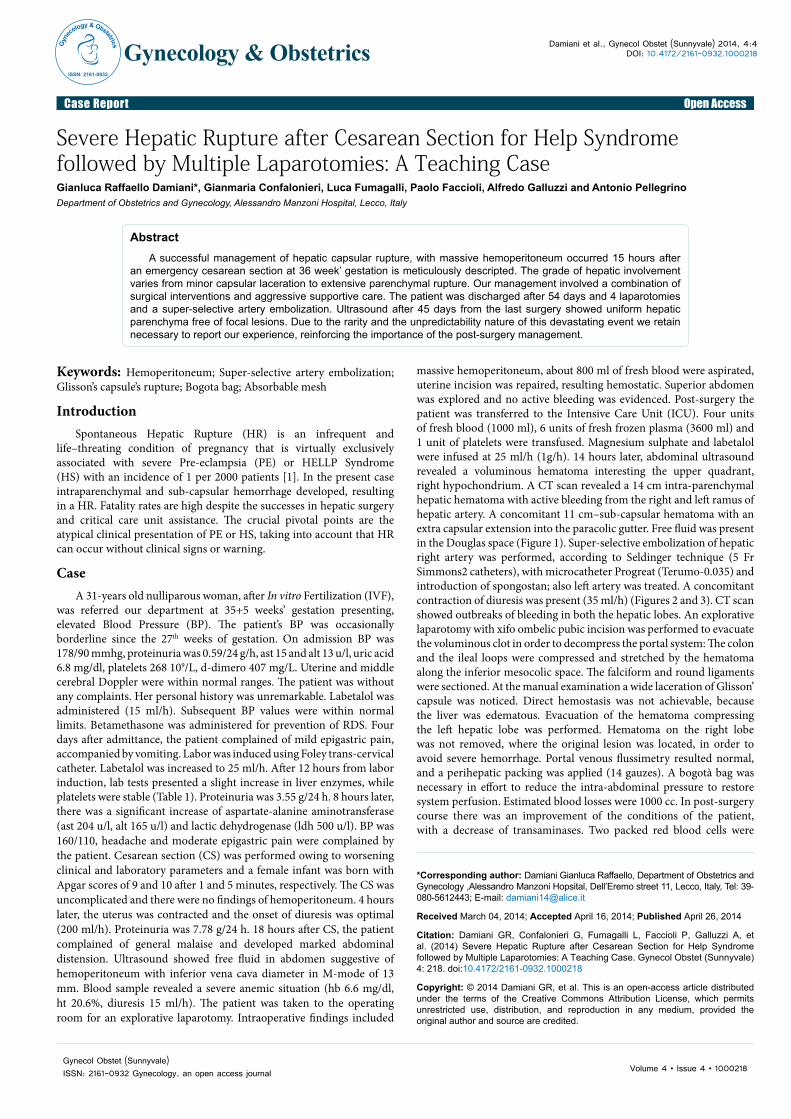
massive hemoperitoneum, about 800 ml of fresh blood were aspirated, uterine incision was repaired, resulting hemostatic. Superior abdomen was explored and no active bleeding was evidenced. Post-surgery the patient was transferred to the Intensive Care Unit (ICU). Four units of fresh blood (1000 ml), 6 units of fresh frozen plasma (3600 ml) and 1 unit of platelets were transfused. Magnesium sulphate and labetalol were infused at 25 ml/h (1g/h). 14 hours later, abdominal ultrasound revealed a voluminous hematoma interesting the upper quadrant, right hypochondrium. A CT scan revealed a 14 cm intra-parenchymal hepatic hematoma with active bleeding from the right and left ramus of hepatic artery. A concomitant 11 cm–sub-capsular hematoma with an extra capsular extension into the paracolic gutter. Free fluid was present in the Douglas space (Figure 1). Super-selective embolization of hepatic right artery was performed, according to Seldinger technique (5 Fr Simmons2 catheters), with microcatheter Progreat (Terumo-0.035) and introduction of spongostan; also left artery was treated. A concomitant contraction of diuresis was present (35 ml/h) (Figures 2 and 3). CT scan showed outbreaks of bleeding in both the hepatic lobes. An explorative laparotomy with xifo ombelic pubic incision was performed to evacuate the voluminous clot in order to decompress the portal system: The colon and the ileal loops were compressed and stretched by the hematoma along the inferior mesocolic space. The falciform and round ligaments were sectioned. At the manual examination a wide laceration of Glisson’ capsule was noticed. Direct hemostasis was not achievable, because the liver was edematous. Evacuation of the hematoma compressing the left hepatic lobe was performed. Hematoma on the right lobe was not removed, where the original lesion was located, in order to avoid severe hemorrhage. Portal venous flussimetry resulted normal, and a perihepatic packing was applied (14 gauzes). A bogotà bag was necessary in effort to reduce the intra-abdominal pressure to restore system perfusion. Estimated blood losses were 1000 cc. In post-surgery course there was an improvement of the conditions of the patient, with a decrease of transaminases. Two packed red blood cells were
Gyne
cology & Obstetrics
ISSN: 2161-0932
Gynecology & Obstetrics
Citation: Damiani GR, Confalonieri G, Fumagalli L, Faccioli P, Galluzzi A, et al. (2014) Severe Hepatic Rupture after Cesarean Section for Help Syndrome followed by Multiple Laparotomies: A Teaching Case. Gynecol Obstet (Sunnyvale) 4: 218. doi:10.4172/2161-0932.1000218
Page 2 of 3
Volume 4 • Issue 4 • 1000218Gynecol Obstet (Sunnyvale)ISSN: 2161-0932 Gynecology, an open access journal
transfused. Resumption of diuresis was noted, drainages were siero-hematic. Bilirubin and ammonemy were in normal ranges, while there was a further increase of transaminases. Parenteral and enteral nutrition was started. The surgical revaluation was programmed the following day. Remotion of further clots was performed, no active bleeding was evidenced. A new packing (8 gauzes) and a steri-drape in contact with both hepatic surface and was applied; 2 drainages were fixed in sub diaphragmatic region. 3 days later (8 days after CS) laparotomy was performed to remove further clotting and gauzes. For the increased intra-abdominal pressure was necessary to insert a dual mesh and perform a partial closure of the abdominal fascia. 2 packed red blood cells were transfused and pleuric-effusion resulted at the chest X-ray
with parenchymal disventilation. 48 hours later there was a little increase of the right effusion, partially loculated along the costal margin, while an omolateral atelectasis of the basal segment of inferior lobe. Over the course of the next 6 days hyperpyrexia was present and a CT was programmed: the parenchimografia was improved and the hematoma was organized under the rectum muscles, with hypoechoic aspect (18×6×25 cm). Bilateral pleural effusion was revealed, conditioning the atelectasy of the right inferior lobe. Thoracentesis was performed with evacuation of serous amber liquid (500 cc). Coltural exam was negative. On the 14th day after CS (Figures 4 and 5). A percutaneous drainage was inserted in the right hypochondrium towards the accumulation of blood. 1400 cc of serous hematic liquid was aspirated (bilirubin 4 mg/dl) Ultrasound showed a decrease of hematoma with thickness of 3 cm. Bilateral pleuric effusion was in resolution. There were no compressions of ileal bowel and parechimography was in resumption. Hepaticdrainage was removed and a retraction of 5 cm of pleuric drainage wasmade, and removed 48 hours later. On the 29th day after CS abdomenultrasound showed 2 residual areas of hematoma (thickness of 10-mm and 26-mm) in sub diaphragmatic region. Due to the presence ofiperpirexy, teicoplanin and imipemen were administered for 10 days
Figure 1: Abdominal ultrasound: revealed a voluminous hematoma interesting the upper quadrant, right hypochondrium and epigastrium (IV-VIII segment).
Figure 2: Abdominal ultrasound: revealed a voluminous hematoma interesting the upper quadrant, right hypochondrium and epigastrium (IV-VIII segment).
Figure 3: CT scan: intra-parenchymal hepatic hematoma with active bleeding from the right and left ramus of hepatic artery. A concomitant sub-capsular hematoma with an extra capsular caudal extension in the paracolic gutter.
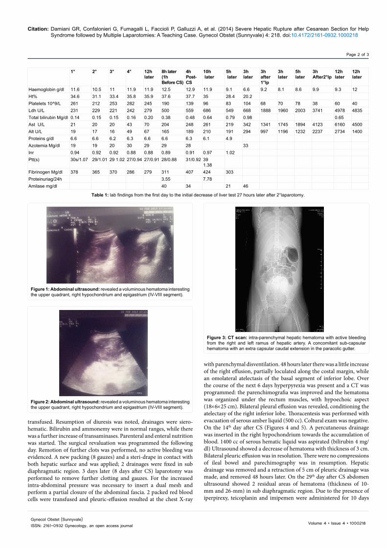
1° 2° 3° 4° 12h later
8h later(1hBefore CS)
4h Post-CS
10h later
5h later
3h later
3h after 1°lp
3hlater
5hlater
3hAfter2°lp
12hlater
12hlater
Haemoglobin g/dl 11.6 10.5 11 11.9 11.9 12.5 12.9 11.9 9.1 6.6 9.2 8.1 8.6 9.9 9.3 12Ht% 34.6 31.1 33.4 35.8 35.9 37.6 37.7 35 28.4 20.2Platelets 10^9/L 261 212 253 282 245 190 139 96 83 104 68 70 78 38 60 40Ldh U/L 231 229 221 242 279 500 559 686 549 668 1888 1960 2003 3741 4978 4835Total bilirubin Mg/dl 0.14 0.15 0.15 0.16 0.20 0.38 0.48 0.64 0.79 0.98 0.65Ast U/L 21 20 20 43 70 204 248 261 219 342 1341 1745 1894 4123 6160 4500Alt U/L 19 17 16 49 67 165 189 210 191 294 997 1196 1232 2237 2734 1400Proteins g/dl 6.6 6.6 6.2 6.3 6.6 6.6 6.3 6.1 4.9Azotemia Mg/dl 19 19 20 30 29 29 28 33Inr 0.94 0.92 0.92 0.88 0.88 0.89 0.91 0.97 1.02Ptt(s) 30s/1.07 29/1.01 29 1.02 27/0.94 27/0.91 28/0.88 31/0.92 39
1.38Fibrinogen Mg/dl 378 365 370 286 279 311 407 424 303Proteinuriag/24h 3.55 7.78Amilase mg/dl 40 34 21 46
Table 1: lab findings from the first day to the initial decrease of liver test 27 hours later after 2°laparotomy.
Citation: Damiani GR, Confalonieri G, Fumagalli L, Faccioli P, Galluzzi A, et al. (2014) Severe Hepatic Rupture after Cesarean Section for Help Syndrome followed by Multiple Laparotomies: A Teaching Case. Gynecol Obstet (Sunnyvale) 4: 218. doi:10.4172/2161-0932.1000218
Page 3 of 3
Volume 4 • Issue 4 • 1000218Gynecol Obstet (Sunnyvale)ISSN: 2161-0932 Gynecology, an open access journal
with discharge of the patient. An ultrasound performed 15 days later (45 days from the last surgery) shows uniform hepatic parenchyma free of focal lesions.
DiscussionSpontaneous liver hemorrhage with formation of sub-capsular
hematomas and rupture of Glissan’s capsule is a rare but often lethal
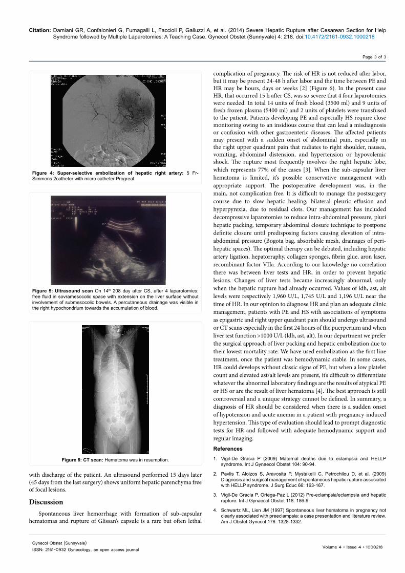
complication of pregnancy. The risk of HR is not reduced after labor, but it may be present 24-48 h after labor and the time between PE and HR may be hours, days or weeks [2] (Figure 6). In the present case HR, that occurred 15 h after CS, was so severe that 4 four laparotomies were needed. In total 14 units of fresh blood (3500 ml) and 9 units of fresh frozen plasma (5400 ml) and 2 units of platelets were transfused to the patient. Patients developing PE and especially HS require close monitoring owing to an insidious course that can lead a misdiagnosis or confusion with other gastroenteric diseases. The affected patients may present with a sudden onset of abdominal pain, especially in the right upper quadrant pain that radiates to right shoulder, nausea, vomiting, abdominal distension, and hypertension or hypovolemic shock. The rupture most frequently involves the right hepatic lobe, which represents 77% of the cases [3]. When the sub-capsular liver hematoma is limited, it’s possible conservative management with appropriate support. The postoperative development was, in the main, not complication free. It is difficult to manage the postsurgery course due to slow hepatic healing, bilateral pleuric effusion and hyperpyrexia, due to residual clots. Our management has included decompressive laparotomies to reduce intra-abdominal pressure, pluri hepatic packing, temporary abdominal closure technique to postpone definite closure until predisposing factors causing elevation of intra-abdominal pressure (Bogota bag, absorbable mesh, drainages of peri-hepatic spaces). The optimal therapy can be debated, including hepatic artery ligation, hepatorraphy, collagen sponges, fibrin glue, aron laser, recombinant factor VIIa. According to our knowledge no correlation there was between liver tests and HR, in order to prevent hepatic lesions. Changes of liver tests became increasingly abnormal, only when the hepatic rupture had already occurred. Values of ldh, ast, alt levels were respectively 1,960 U/L, 1,745 U/L and 1,196 U/L near the time of HR. In our opinion to diagnose HR and plan an adequate clinic management, patients with PE and HS with associations of symptoms as epigastric and right upper quadrant pain should undergo ultrasound or CT scans especially in the first 24 hours of the puerperium and when liver test function >1000 U/L (ldh, ast, alt). In our department we prefer the surgical approach of liver packing and hepatic embolization due to their lowest mortality rate. We have used embolization as the first line treatment, once the patient was hemodynamic stable. In some cases, HR could develops without classic signs of PE, but when a low platelet count and elevated ast/alt levels are present, it’s difficult to differentiate whatever the abnormal laboratory findings are the results of atypical PE or HS or are the result of liver hematoma [4]. The best approach is still controversial and a unique strategy cannot be defined. In summary, a diagnosis of HR should be considered when there is a sudden onset of hypotension and acute anemia in a patient with pregnancy-induced hypertension. This type of evaluation should lead to prompt diagnostic tests for HR and followed with adequate hemodynamic support and regular imaging.
References
1. Vigil-De Gracia P (2009) Maternal deaths due to eclampsia and HELLPsyndrome. Int J Gynaecol Obstet 104: 90-94.
2. Pavlis T, Aloizos S, Aravosita P, Mystakelli C, Petrochilou D, et al. (2009)Diagnosis and surgical management of spontaneous hepatic rupture associated with HELLP syndrome. J Surg Educ 66: 163-167.
3. Vigil-De Gracia P, Ortega-Paz L (2012) Pre-eclampsia/eclampsia and hepaticrupture. Int J Gynaecol Obstet 118: 186-9.
4. Schwartz ML, Lien JM (1997) Spontaneous liver hematoma in pregnancy notclearly associated with preeclampsia: a case presentation and literature review. Am J Obstet Gynecol 176: 1328-1332.
Figure 4: Super-selective embolization of hepatic right artery: 5 Fr- Simmons 2catheter with micro catheter Progreat.
Figure 5: Ultrasound scan On 14th 208 day after CS, after 4 laparotomies: free fluid in sovramesocolic space with extension on the liver surface without involvement of submesocolic bowels. A percutaneous drainage was visible in the right hypochondrium towards the accumulation of blood.
Figure 6: CT scan: Hematoma was in resumption.