G ARTICLE IN PRESS Electrochimica Actafrancescoriccilab.com/wp-content/uploads/2013/01/50.pdf ·...
10
Please cite this article in press as: F. Ricci, et al., A review of experimental aspects of electrochemical immunosensors, Electrochim. Acta (2012), http://dx.doi.org/10.1016/j.electacta.2012.06.033 ARTICLE IN PRESS G Model EA-18854; No. of Pages 10 Electrochimica Acta xxx (2012) xxx–xxx Contents lists available at SciVerse ScienceDirect Electrochimica Acta jou rn al hom epa ge: www.elsevier.com/locate/electacta Review article A review of experimental aspects of electrochemical immunosensors Francesco Ricci a,b , Gianluca Adornetto a , Giuseppe Palleschi a,b,∗ a Dipartimento di Scienze e Tecnologie Chimiche, University of Rome, Tor Vergata, Via della Ricerca Scientifica, 00133 Rome, Italy b Consorzio Interuniversitario Biostrutture e Biosistemi “INBB”, Viale Medaglie d’Oro 305, 00136 Rome, Italy a r t i c l e i n f o Article history: Received 28 November 2011 Received in revised form 8 June 2012 Accepted 8 June 2012 Available online xxx Keywords: Electrochemical Immunosensors Screen printed Portable Guide a b s t r a c t In this brief review we want to give a guidance to all the researchers who want to challenge themselves in the task of developing electrochemical immunosensors for the first time. We will focus here only on practical aspects trying to give the readers useful insights that should be considered such as the choice of the electrode to be used, the best electrochemical and immunological procedures, the immobilization procedures, the instruments to purchase and other basic aspects of electrochemical immunosensors. In doing so we offer a wide view of the research on electrochemical immunosensor applications that have appeared in the literature in the last 5 years. © 2012 Elsevier Ltd. All rights reserved. Contents 1. Introduction . . . . . . . . . . . . . . . . . . . . . . . . . . . . . . . . . . . . . . . . . . . . . . . . . . . . . . . . . . . . . . . . . . . . . . . . . . . . . . . . . . . . . . . . . . . . . . . . . . . . . . . . . . . . . . . . . . . . . . . . . . . . . . . . . . . . . . . . . . 00 2. Electrodes . . . . . . . . . . . . . . . . . . . . . . . . . . . . . . . . . . . . . . . . . . . . . . . . . . . . . . . . . . . . . . . . . . . . . . . . . . . . . . . . . . . . . . . . . . . . . . . . . . . . . . . . . . . . . . . . . . . . . . . . . . . . . . . . . . . . . . . . . . . . 00 3. Immobilization procedures . . . . . . . . . . . . . . . . . . . . . . . . . . . . . . . . . . . . . . . . . . . . . . . . . . . . . . . . . . . . . . . . . . . . . . . . . . . . . . . . . . . . . . . . . . . . . . . . . . . . . . . . . . . . . . . . . . . . . . . . . . 00 4. Use of nanostructured materials for electrochemical immunosensors . . . . . . . . . . . . . . . . . . . . . . . . . . . . . . . . . . . . . . . . . . . . . . . . . . . . . . . . . . . . . . . . . . . . . . . . . . . . . 00 5. Use of magnetic beads for electrochemical immunosensors . . . . . . . . . . . . . . . . . . . . . . . . . . . . . . . . . . . . . . . . . . . . . . . . . . . . . . . . . . . . . . . . . . . . . . . . . . . . . . . . . . . . . . . . 00 6. Enzymatic labels and electrochemical methods . . . . . . . . . . . . . . . . . . . . . . . . . . . . . . . . . . . . . . . . . . . . . . . . . . . . . . . . . . . . . . . . . . . . . . . . . . . . . . . . . . . . . . . . . . . . . . . . . . . . . 00 7. Instruments . . . . . . . . . . . . . . . . . . . . . . . . . . . . . . . . . . . . . . . . . . . . . . . . . . . . . . . . . . . . . . . . . . . . . . . . . . . . . . . . . . . . . . . . . . . . . . . . . . . . . . . . . . . . . . . . . . . . . . . . . . . . . . . . . . . . . . . . . . 00 8. Conclusions . . . . . . . . . . . . . . . . . . . . . . . . . . . . . . . . . . . . . . . . . . . . . . . . . . . . . . . . . . . . . . . . . . . . . . . . . . . . . . . . . . . . . . . . . . . . . . . . . . . . . . . . . . . . . . . . . . . . . . . . . . . . . . . . . . . . . . . . . . 00 References . . . . . . . . . . . . . . . . . . . . . . . . . . . . . . . . . . . . . . . . . . . . . . . . . . . . . . . . . . . . . . . . . . . . . . . . . . . . . . . . . . . . . . . . . . . . . . . . . . . . . . . . . . . . . . . . . . . . . . . . . . . . . . . . . . . . . . . . . . . 00 1. Introduction The detection of biological and chemical pathogens, contam- inants and other important analytes plays a crucial role in the prevention of disease spread, infections and pathologies [1]. In order to ensure rapid appropriate counteractions in case of con- tamination and to improve food control, simple, inexpensive, and quantitative tools for the detection of these contaminants are urgently needed [2,3]. Since its introduction, more than 30 years ago, ELISA (enzyme linked immunosorbent assay) has become probably the most used technique for such task [4]. The avail- ability of antibodies targeting potentially any molecule, the high sensitivity due to the easily assayed enzymatic amplification step ∗ Corresponding author at: Dipartimento di Scienze e Tecnologie Chimiche, Uni- versity of Rome, Tor Vergata, Via della Ricerca Scientifica, 00133 Rome, Italy. E-mail address: [email protected] (G. Palleschi). and the easiness of use are among the most important reasons for this success. Moreover, the 96-well plate typically employed in ELISA tests is suitable for high-throughput and multianalyte tests. Because of the flexibility in format assay (i.e. competitive, sandwich or direct), ELISA is also equally useful for the detection of both antigens (i.e. usually small molecules) and antibodies [4]. For all these reasons ELISA is now one of the preferred tools for determining serum antibody concentrations (such as with the HIV test [5] or West Nile Virus [6]) and has found applications in the food industry in detecting potential food allergens [7]. Because of these advantages, during recent years, several efforts have been directed to the improvement of the ELISA method. For exam- ple, chromogenic reporters and substrates have been replaced by fluorogenic [8], electrochemiluminescent [9], and real-time PCR [10] reporters which may offer higher sensitivities and are more suitable for multiplexing. Many efforts have also been spent to find alternative measurement approaches. In this context simple and rapid lateral flow immunochromatographic assays with quick 0013-4686/$ – see front matter © 2012 Elsevier Ltd. All rights reserved. http://dx.doi.org/10.1016/j.electacta.2012.06.033
Transcript of G ARTICLE IN PRESS Electrochimica Actafrancescoriccilab.com/wp-content/uploads/2013/01/50.pdf ·...
G
E
R
A
Fa
b
a
ARRAA
KEISPG
C
1
ipotquapas
v
0h
ARTICLE IN PRESS Model
A-18854; No. of Pages 10
Electrochimica Acta xxx (2012) xxx– xxx
Contents lists available at SciVerse ScienceDirect
Electrochimica Acta
jou rn al hom epa ge: www.elsev ier .com/ locate /e lec tac ta
eview article
review of experimental aspects of electrochemical immunosensors
rancesco Riccia,b, Gianluca Adornettoa, Giuseppe Palleschia,b,∗
Dipartimento di Scienze e Tecnologie Chimiche, University of Rome, Tor Vergata, Via della Ricerca Scientifica, 00133 Rome, ItalyConsorzio Interuniversitario Biostrutture e Biosistemi “INBB”, Viale Medaglie d’Oro 305, 00136 Rome, Italy
r t i c l e i n f o
rticle history:eceived 28 November 2011eceived in revised form 8 June 2012ccepted 8 June 2012vailable online xxx
a b s t r a c t
In this brief review we want to give a guidance to all the researchers who want to challenge themselvesin the task of developing electrochemical immunosensors for the first time. We will focus here only onpractical aspects trying to give the readers useful insights that should be considered such as the choiceof the electrode to be used, the best electrochemical and immunological procedures, the immobilizationprocedures, the instruments to purchase and other basic aspects of electrochemical immunosensors. In
eywords:lectrochemicalmmunosensorscreen printedortable
doing so we offer a wide view of the research on electrochemical immunosensor applications that haveappeared in the literature in the last 5 years.
© 2012 Elsevier Ltd. All rights reserved.
uide
ontents
1. Introduction . . . . . . . . . . . . . . . . . . . . . . . . . . . . . . . . . . . . . . . . . . . . . . . . . . . . . . . . . . . . . . . . . . . . . . . . . . . . . . . . . . . . . . . . . . . . . . . . . . . . . . . . . . . . . . . . . . . . . . . . . . . . . . . . . . . . . . . . . . 002. Electrodes . . . . . . . . . . . . . . . . . . . . . . . . . . . . . . . . . . . . . . . . . . . . . . . . . . . . . . . . . . . . . . . . . . . . . . . . . . . . . . . . . . . . . . . . . . . . . . . . . . . . . . . . . . . . . . . . . . . . . . . . . . . . . . . . . . . . . . . . . . . . 003. Immobilization procedures . . . . . . . . . . . . . . . . . . . . . . . . . . . . . . . . . . . . . . . . . . . . . . . . . . . . . . . . . . . . . . . . . . . . . . . . . . . . . . . . . . . . . . . . . . . . . . . . . . . . . . . . . . . . . . . . . . . . . . . . . . 004. Use of nanostructured materials for electrochemical immunosensors . . . . . . . . . . . . . . . . . . . . . . . . . . . . . . . . . . . . . . . . . . . . . . . . . . . . . . . . . . . . . . . . . . . . . . . . . . . . . 005. Use of magnetic beads for electrochemical immunosensors . . . . . . . . . . . . . . . . . . . . . . . . . . . . . . . . . . . . . . . . . . . . . . . . . . . . . . . . . . . . . . . . . . . . . . . . . . . . . . . . . . . . . . . . 006. Enzymatic labels and electrochemical methods. . . . . . . . . . . . . . . . . . . . . . . . . . . . . . . . . . . . . . . . . . . . . . . . . . . . . . . . . . . . . . . . . . . . . . . . . . . . . . . . . . . . . . . . . . . . . . . . . . . . . 007. Instruments . . . . . . . . . . . . . . . . . . . . . . . . . . . . . . . . . . . . . . . . . . . . . . . . . . . . . . . . . . . . . . . . . . . . . . . . . . . . . . . . . . . . . . . . . . . . . . . . . . . . . . . . . . . . . . . . . . . . . . . . . . . . . . . . . . . . . . . . . . 008. Conclusions . . . . . . . . . . . . . . . . . . . . . . . . . . . . . . . . . . . . . . . . . . . . . . . . . . . . . . . . . . . . . . . . . . . . . . . . . . . . . . . . . . . . . . . . . . . . . . . . . . . . . . . . . . . . . . . . . . . . . . . . . . . . . . . . . . . . . . . . . . 00
References . . . . . . . . . . . . . . . . . . . . . . . . . . . . . . . . . . . . . . . . . . . . . . . . . . . . . . . . . . . . . . . . . . . . . . . . . . . . . . . . . . . . . . . . . . . . . . . . . . . . . . . . . . . . . . . . . . . . . . . . . . . . . . . . . . . . . . . . . . . 00
. Introduction
The detection of biological and chemical pathogens, contam-nants and other important analytes plays a crucial role in therevention of disease spread, infections and pathologies [1]. Inrder to ensure rapid appropriate counteractions in case of con-amination and to improve food control, simple, inexpensive, anduantitative tools for the detection of these contaminants arergently needed [2,3]. Since its introduction, more than 30 yearsgo, ELISA (enzyme linked immunosorbent assay) has become
and the easiness of use are among the most important reasonsfor this success. Moreover, the 96-well plate typically employedin ELISA tests is suitable for high-throughput and multianalytetests. Because of the flexibility in format assay (i.e. competitive,sandwich or direct), ELISA is also equally useful for the detectionof both antigens (i.e. usually small molecules) and antibodies [4].For all these reasons ELISA is now one of the preferred tools fordetermining serum antibody concentrations (such as with the HIVtest [5] or West Nile Virus [6]) and has found applications in thefood industry in detecting potential food allergens [7]. Because of
Please cite this article in press as: F. Ricci, et al., A review of experimental ahttp://dx.doi.org/10.1016/j.electacta.2012.06.033
robably the most used technique for such task [4]. The avail-bility of antibodies targeting potentially any molecule, the highensitivity due to the easily assayed enzymatic amplification step
∗ Corresponding author at: Dipartimento di Scienze e Tecnologie Chimiche, Uni-ersity of Rome, Tor Vergata, Via della Ricerca Scientifica, 00133 Rome, Italy.
E-mail address: [email protected] (G. Palleschi).
013-4686/$ – see front matter © 2012 Elsevier Ltd. All rights reserved.ttp://dx.doi.org/10.1016/j.electacta.2012.06.033
these advantages, during recent years, several efforts have beendirected to the improvement of the ELISA method. For exam-ple, chromogenic reporters and substrates have been replaced byfluorogenic [8], electrochemiluminescent [9], and real-time PCR
spects of electrochemical immunosensors, Electrochim. Acta (2012),
[10] reporters which may offer higher sensitivities and are moresuitable for multiplexing. Many efforts have also been spent tofind alternative measurement approaches. In this context simpleand rapid lateral flow immunochromatographic assays with quick

ING Model
E
2 mica A
osatbmsfu
pmedlaasfcbshtTiosccigaeafeatgwndscoclpoaaobtrwswoeot
tb
ARTICLEA-18854; No. of Pages 10
F. Ricci et al. / Electrochi
bservation of results directly by the naked-eyes probably repre-ent the best example [11]. ELISA is in fact an optical approachnd, as such, can suffer of several drawabacks associated with thisype of measurement. These include a requirement for generallyulky and power-intensive light sources, detectors, and monochro-ators, and potential false signals arising from complex colored
amples. Moreover, because the sensitivity of optical methodsollows the well-known Lambert–Beer law, a minimum sample vol-me and path length is required to achieve certain performances.
In this context, electrochemical methods appear as the mostromising alternative to optical approaches. The possibility toiniaturize modern microelectronics allows to build micro-
lectrodes which are useful for multiplexing and well suited foretection of very small volumes of samples (microliters to nano-
iters), in fact the sensitivity of electrochemical methods is notffected by the volume used during measurements. The low costnd a large-scale production of electronic devices is another rea-on which makes the electrochemical approach more appealingor high-throughput analysis. Finally, the paucity of electrochemi-al interferents in real samples makes the electrochemical methodsetter suited for use with complex and colored samples. It is not aurprise, then, that during the last decade many research groupsave devoted part of their efforts to the development, optimiza-ion and characterization of electrochemical immunosensors [12].he adaptation of an ELISA assay to an electrochemical approachs not straightforward. In fact, despite the assay format and theverall analysis steps that remain usually the same, several factorshould be taken in serious consideration to achieve performancesomparable or even better than those of the original ELISA opti-al approach. The electrode surface to be used as support for themmobilization of the recognition element (i.e. antibody or anti-en), the choice of the enzymatic substrate (which must lead ton electroactive product), the selection of the most appropriatelectrochemical methods (instead of a simple optical absorbance)nd of the adequate instrument are among the most importantactors to be considered when adapting an optical ELISA to anlectrochemical platform. In this short review we intend to give
practical guidance to all the researchers who want to challengehemselves in this delicate task. A detailed discussion on the back-round of ELISA is not within the scope of this paper and for thise send to more general reviews [4,13,14]. It is also important toote that there are different possible formats that can be used toevelop an electrochemical immunosensor. Both competitive andandwich formats can in fact be used. Two approaches could beonsidered when dealing with competitive immunosensors. A firstne in which immobilized antibodies react with free antigens inompetition with labeled antigens. A second one, using immobi-ized antigens and labeled antibodies, is generally preferred andrevents all the problems related to antibody immobilization (lossf affinity, orientation of the immobilized protein). In a sandwichssay, after interaction between immobilized antibodies and freentigens, labelled antibodies (directed toward a second binding sitef the antigen) are added; at this point, antigen is “sandwiched”etween two antibodies. Further details on the possible formatso be used for immunosensors can be found in three completeeviews [12,15,16]. We will focus here only on practical aspectshich should be considered when an electrochemical immunosen-
or must be developed. We will give the readers useful insightshich also take in consideration prices, commercial availability and
ther basic aspects of reagents and consumables needed to developlectrochemical immunosensors. In doing so we offer a wide viewn the research on electrochemical immunosensor applications
Please cite this article in press as: F. Ricci, et al., A review of experimental ahttp://dx.doi.org/10.1016/j.electacta.2012.06.033
hat have appeared in the literature in the last 5 years.Electrochemical immunosensors are usually obtained through
he immobilization of a recognition element (i.e. antigen or anti-ody) on the electrode surface. The assay steps often end with the
PRESScta xxx (2012) xxx– xxx
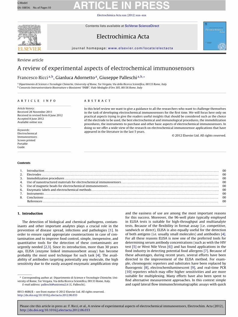
injection of a secondary enzyme-labelled antibody which followsthe addition of a proper enzymatic substrate (Fig. 1). In an electro-chemical format the enzymatic reaction leads to the production of amolecule which is electroactive (that is, it gives an electrochemicalsignal in presence of certain inputs). The signal is recorded with theuse of bench or portable instruments which are usually capable ofapplying different electrochemical techniques. We will begin oursurvey and this guidance with the first steps of an electrochemi-cal immunosensor, the selection of the electrode surface, and thechoice of the immobilization procedure of the recognition element.
2. Electrodes
The choice of the electrode to be used for an electrochemicalimmunosensor is of course crucial for several aspects includingthe sensitivity of the method, the cost of the assay and the pos-sibility to adopt different immobilization procedures. Electrodesused for this task are commonly made of inert metals such asplatinum [17], gold [18–27] and several forms of carbon includ-ing carbon fiber [28], epoxy graphite [29–33], graphene [34] orglassy carbon [35]. Many companies offer “conventional”-Tefloncoated rods as electrodes where one end is the sensing surface andthe other is for the connection to the instrument. Although thesesensors are widely used in electrochemical laboratories they arenot well suited for immunosensor applications. This is due to thefact that an electrochemical immunoassay usually requires severalsteps and many calibration points which would result in the needof a high number of such electrodes or in the reuse of the sameimmunosensor for several experiments. Moreover, although theybehave very well from an electrochemical point of view, these sen-sors are usually quite expensive. We have recently reviewed themost important Companies that offer conventional rod electrodesand found that gold electrode prices vary from a minimum of 60 Dto a maximum of ca. 300 D . Interestingly, the prices of rod glassycarbon electrodes span approximately the same range despite thecheaper nature of the electrode material. Moreover, these conven-tional electrodes are of course not intended for single use and oftenbefore each use they must be washed and cleaned very carefully, aprocedure that it is usually time consuming and reagent intensive(we note, however, that some examples have been proposed wherethe classic gold electrode surface can be regenerated and used sev-eral times [25,26]). As an additional drawback, these sensors are notvery amenable to high-throughput modification as they need to beimmersed in a quite large volume of solution. Finally, the measure-ment step is also not too practical because of the need of an externalreference electrode and, often, of a counter electrode. This usuallyrequires a working solution of not less than 1 mL (unless specialtricks are used) which is not suitable for large scale analysis. Forthese reasons, we believe that the use of conventional rod elec-trodes should be only limited to proof-of-principle applications ofnovel immunological approaches and should not regard the practi-cal applications of optimized immunosensors. In this perspective,the use of disposable screen-printed electrodes (SPEs) which arecharacterized by low-cost fabrication and the possibility of mass-production seem much better suited. Screen-printing (thick-film)technology involves the printing of various inks on planar ceramicor plastic supports [36–38]. Of note, the planar nature of the SPEallows the easy modification of the surface and, through the helpof an automatic dispenser, this can be done in a mass producibleway. Due to the miniaturized dimensions of SPEs, all immunologi-cal steps can be performed in a drop, using only a few microliters
spects of electrochemical immunosensors, Electrochim. Acta (2012),
of solution, thus reducing the reagent consumption. Gold [39–43],graphite [44–58] and silver screen printed electrodes are commer-cially available. The cost of each sensor usually spans 2–4 D /eachfor graphite and gold electrodes. Of note, the production of these
ARTICLE IN PRESSG Model
EA-18854; No. of Pages 10
F. Ricci et al. / Electrochimica Acta xxx (2012) xxx– xxx 3
Fig. 1. General overview of the main components of an electrochemical immunosensor. In this scheme, for the sake of clarity, we have depicted a generic immunosensorb elopmh e publ
sashtometes5ofttatafmthaf(im
ased on a sandwich format. The components that should be considered in the devighlight the reasons to choose among the multiple options that are available to th
creen printed electrodes is straightforward and, since the cost of screen printer machine (i.e. DEK, Unitech) is not too high (pricestart at ca. 20 k D ), this instrument may be of utility if the use of aigh number of electrodes is forecasted. Moreover, the possibilityo customize the production of screen printed electrodes may bef utility in case of special requirements or to test novel electrodeaterials (Table 1). The consumables needed to print a batch of
lectrodes are quite cheap especially in the case that graphite elec-rodes are produced. Silver ink, used to print the pseudo-referencelectrode of screen printed electrodes, is often the most expen-ive one (about 500–600 D /kg), while graphite ink costs less than0 D /kg and the insulating ink (used to delimitate the surface areaf the working electrode) is the most cost effective and can beound at less than 20 D /kg. Of note, 1 kg of ink is usually enougho print a huge number of electrodes (approximately 100,000 elec-rodes). Recently, arrays of eight screen printed electrodes [59–64]nd of a 96-well plate [65–67] have been developed and this makeshe use of these electrodes even more advantageous [68]. As anlternative to screen printed electrodes, the use of photolitographyor the production of electrodes well suited for integration with
icrofluidic systems and for the construction of miniaturized mul-iplex arrays has been reported [69–73]. As an additional note, itas to be stressed here that electrodes are not necessarily used asn immobilization support of the recognition element. Often, in
Please cite this article in press as: F. Ricci, et al., A review of experimental ahttp://dx.doi.org/10.1016/j.electacta.2012.06.033
act, the recognition element is immobilized on a separate supportmagnetic- and nano- beads) and only at the end of the immunolog-cal chain it is placed onto the surface of an unmodified electrode for
easurement of the enzymatic product. In these cases the electrode
ent of an electrochemical immunosensor are briefly overviewed. In this review weic in terms of electrodes, instruments, enzymatic labels and reagents.
only acts as the “measuring device” and its properties to “hold”biomolecules are not too crucial.
3. Immobilization procedures
The procedure and method used to immobilize the recognitionelement either directly on the electrode surface or on other solidsupports is a crucial step in the development of an electrochemicalimmunosensor. In this perspective a clear distinction with opticalELISA methods must be made. Generally, optical ELISA involves thenon-specific adsorption of the recognition element on the bottom ofpolystyrene wells. The immunological chain (the series of reactionand washing steps that leads to the final signal) ends with the injec-tion of a substrate of the label enzyme conjugated to the secondaryantibody [4,13,14]. Of note, the enzymatic product produced bythe enzyme label is detected in a homogeneous solution throughthe measurement of the absorbance at a selected wavelength. Forthis reason, in optical ELISA, the solution/well interface, and so themethods used to immobilize the recognition element on the solidsupport, is crucial for the optimal antibody/antigen interaction butdoes not directly affect the measurement step. This is not oftenthe case of electrochemical immunosensors. In electrochemicalmethods the measurement step occurs in a heterogeneous phase,more specifically at the solution/electrode interface. To achieve a
spects of electrochemical immunosensors, Electrochim. Acta (2012),
greater sensitivity it is thus preferable that the enzymatic productwould be generated directly on the surface of the working elec-trode in order to favor the diffusion of the analyte to the electrodesurface and to have a greater concentration of the analyte in the

ARTICLE IN PRESSG Model
EA-18854; No. of Pages 10
4 F. Ricci et al. / Electrochimica Acta xxx (2012) xxx– xxx
Table 1Conventional and screen printed electrodes used for immunosensors applications.
Screen printed electrodes (SPE)
Material Source/company, notes References
Graphite/Carbon DropSens [44,47,58]Alderon Biosciences [53]Home produced with DEK screen printer [46,48–52,54,56,57]Home produced [55]Home produced with DEK screen printer, Multi-8 electrodes [59–63]Ecobioservices and researches, Multi-8 electrodes [64]Alderon biosciences, 96 plate well [65–67]DropSens, 2 Working electrodes [40]Home produced with DEK screen printer, 4 Working electrodes [99]Home produced, 8 Working electrodes [74]
Gold DropSens [39–42]BVT technologies [43]
Conventional electrodes
Material Source/company, notes References
Platinum Wire electrode [17]Gold Commercial [18]
BAS, Rod electrode (3 mm ø) [19]Commercial, Rod electrode (3 mm ø) [20,23]BAS, Rod electrode (2 mm ø) [21,22]CHI, Rod electrode (2 mm ø) [24]Commercial, Rod electrode (1.6 mm ø) [25–27]
Graphite epoxy electrodes Home produced [33]Glassy carbon Commercial, Rod electrode (3 mm ø) [34,35]Carbon Commercial, Microelectrode [28]Indium tin oxide (ITO) Commercial, Modified with Amine-terminated G4 poly(amidoamine) dendrimer, ferrocenecarboxaldehyde [98]
Other electrodes
Material Source/company, notes References
Gold Home produced, Photolithographic chips of 16 working electrodes [1 mm × 1 mm] [69–73]Gold particles – Carbon nanotubes–Teflon powder Home produced, Composite electrode [92]Graphite epoxy electrodes Home produced, Magnetized electrodes [29–32]
dbdmatcmiTidhahvRetmomdptocTw
iffusion layer. Moreover, when the recognition element is immo-ilized directly on the electrode surface, the immobilization stepoes not only affect the antibody/antigen interaction but alsoodifies the sensing surface itself. For this reason, in order to have
n optimal electrochemical behavior it is essential that the elec-rode surface is not passivated by the recognition element. Theseonsiderations explain the wide range of different procedures andethods used for the immobilization of the recognition element
n the development of electrochemical immunosensors (Table 2).raditionally, electrochemical immunosensors were obtained bymmobilizing the recognition element (either antibody or antigen)irectly on the surface of the working electrode. For the reasons weave explained in the previous section, screen printed electrodesre preferred over other types of electrodes and several groupsave adopted a simple and straightforward adsorption procedureery similar to that adopted for optical ELISA [33,46,65,74,40].ecently, to achieve a better presentation of the recognitionlement to the target analyte [75], the use of ordered layers onhe surface of the electrode has been introduced. In this case the
ost used electrode material is gold (either as screen printedr traditional rod) which allows the formation of self assembledonolayers (SAMs) through thiol-gold chemistry in a very repro-
ucible and ordered way. In this perspective the immobilizationrocedure usually involves two separate steps. The first one ishe creation of the SAM with a linkage agent containing a thiol at
Please cite this article in press as: F. Ricci, et al., A review of experimental ahttp://dx.doi.org/10.1016/j.electacta.2012.06.033
ne end and a functional group (carboxylic acid, amine etc.) for aonvenient reaction with the recognition element at the other end.he second step involves the reaction of the recognition elementith the functional group of the linkage agent usually through
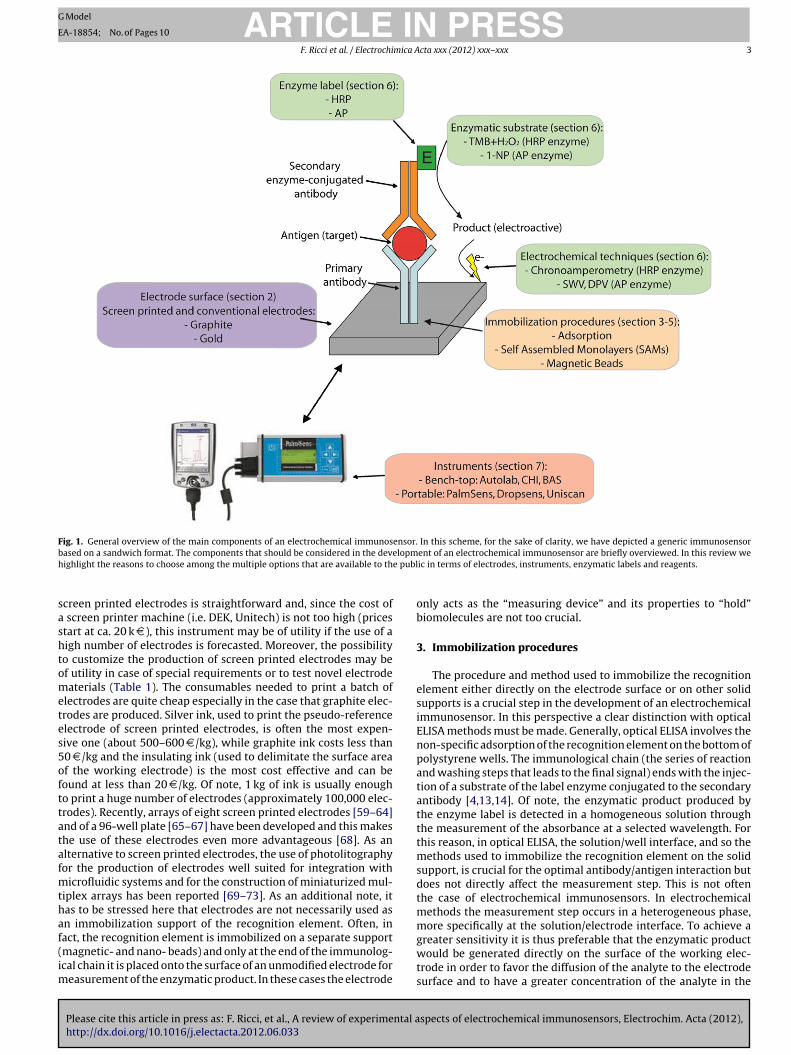
covalent links (such as amide bonds and Schiff’s base formation)directly with the functionalized thiol monolayer or by the aidof bridging molecules such as glutardealdehyde [76,77]. The useof 1-ethyl-3-(3-dimethylaminopropyl)-carbodiimide (EDC) andN-hydroxysuccinimide (NHS) [20,24,25,27,78] or of succinimidegroup by itself [41,72,73] has also found several applications. Theresultant monolayer is well oriented on the surface of the electrodeand this allows a better antibody/antigen interaction. Moreover,because SAMs are usually achieved through the use of a mixedsolution containing both the recognition element and a coadsor-bant (usually a short chain alkane-thiol), this leads to an optimalaverage spacing between the receptors and to a well-behavingelectrochemical surface [79]. Of note, we have found this kind ofimmobilization especially with novel approaches which proposeinnovative immunosensing platforms. For further details on selfassembled monolayers (SAM) for biosensor applications we sendthe readers to two very complete reviews on the argument [80,81].The use of thiolated antibodies (Fig. 2) [23,26], or systems usingthiolated scaffolds (Fig. 3) (e.g. DNA [21,22,82,83], protein G, andprotein A or antibody binding peptides [28,84–90]) have also beenreported with promising results. One of the limitations of SAMsis the preferential need of gold surfaces which can affect the costsof the immunosensor production. Alternative methods for the for-mation of SAMs are based on the use of diazonium salts which canbe easily applied to graphite and carbon electrodes [49]. Another
spects of electrochemical immunosensors, Electrochim. Acta (2012),
approach that has been recently proposed for the immobilizationof recognition elements on the electrode surface involves the useof different polymeric matrices such as polysulfone (PSU) polymer[91] and chitosan (CS). These matrices form porous networks with
ING Model
E
mica A
heai
4i
aahon[bmHaocitucntateetatba
5i
eomeitsmromkofupMnsaevgf
ARTICLEA-18854; No. of Pages 10
F. Ricci et al. / Electrochi
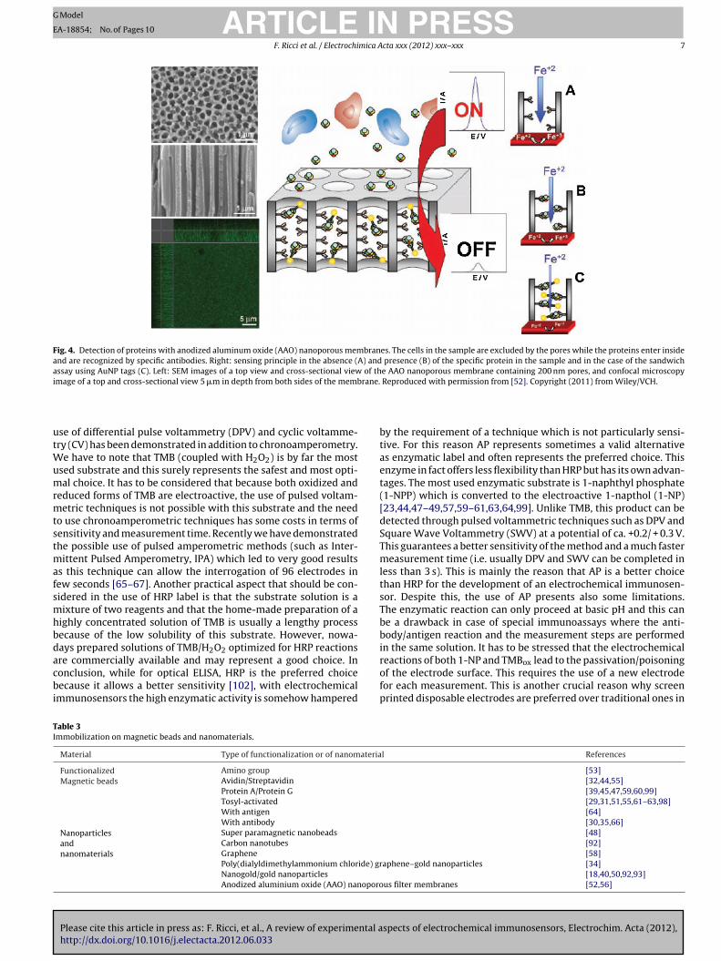
igh mechanical strength that allow the reagents to reach thelectrode (Fig. 4) [52]. Moreover, the presence of reactive aminond hydroxyl functional groups is well suited for biomoleculemmobilization [18,58].
. Use of nanostructured materials for electrochemicalmmunosensors
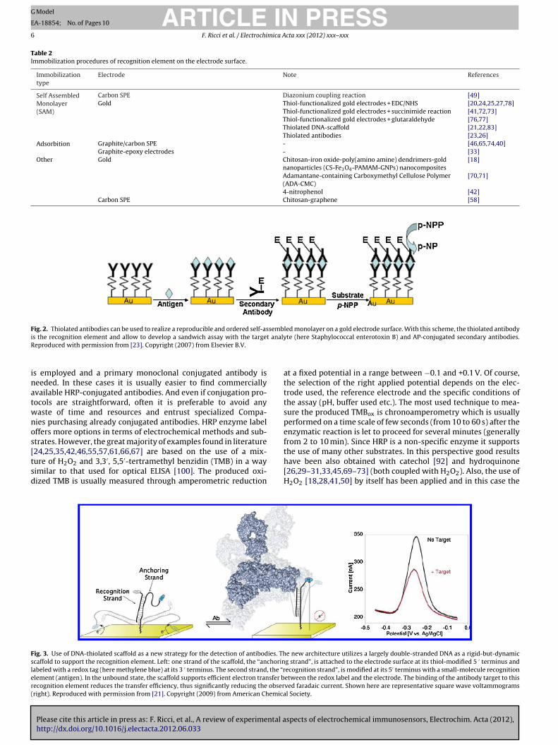
We have discussed earlier how the electrode surface is not only support for the immobilization of the recognition element butlso acts as the sensing surface. For this reason, several attemptsave been made in order to improve the electrochemical behaviorf traditional or screen-printed electrodes with the use of differentanomaterials. In this perspective the use of both carbon nanotubes92], gold nanoparticles [19,40,50,92–94], and graphene [34,58] haseen proposed and previous papers have reviewed the use of nano-aterials with improved electrochemical performances [95–97].owever, the use of such nanomaterials does not seem yet suit-ble for mass-production of the sensors, so their use in prototypesr commercialized instruments appears not likely. Surface modifi-ation protocols are in fact often complicated long processes or,n the best cases, are difficult to be implemented as an indus-rial process. Moreover, a cost assessment should be done whensing these materials since they are usually quite expensive espe-ially if purchased from specialized companies. Despite this weote that a leading company in screen printed electrodes produc-ion (DropSens) offers SPE which are already modified with singlend multi-walled carbon nanotubes. To the best of our knowledgehese sensors have not been yet applied to the development oflectrochemical immunosensors and for this reason we have nolements to suggest them to the readers. With regards to nanos-ructured materials it should be also noted that several interestingpproaches have been reported to date for the immobilization ofhe recognition element through the use of nanoporous filter mem-ranes in anodized aluminium oxide (AAO) to increase the surfacerea and the sensitivity of the sensor (Fig. 4) [52,56].
. Use of magnetic beads for electrochemicalmmunosensors
As we have seen above, the immobilization of the recognitionlement directly onto the electrode surface may prove difficultr may require several steps that are not always suitable forass-production. Another drawback in this perspective is that the
lectrode is used in the whole immunological chain thus lead-ng to possible passivation or poisoning of the electrode surfacehrough the non-specific adsorption of other species present in theample. Moreover, since the immunological chain usually requiresany washing steps, these can cause defects on the layer of the
ecognition elements that could compromise the reproducibilityf the results. Finally, the confinement of the recognition ele-ent onto the surface of an electrode can be an obstacle to the
inetic of antibody/antigen reaction and can limit the numberf biomolecules that can be immobilized on the electrode sur-ace. All these drawbacks have been recently overcome by these of magnetic beads (MBs), or magnetic nanobeads [48], as sup-ort for the immobilization of the recognition element (Table 3).agnetic beads are particles constituted from a dispersion of mag-
etic material (Fe2O3 and Fe3O4) and then covered with a thinhell of polymer that also serves to define a surface area for thedsorption or coupling of a large variety of molecules. MBs can be
Please cite this article in press as: F. Ricci, et al., A review of experimental ahttp://dx.doi.org/10.1016/j.electacta.2012.06.033
asily functionalized with different linkage groups such as strepta-idin (Fig. 5) [32,44,55], tosyl groups [29,31,51,55,61–63,98], aminoroups [53], antibodies or proteins [30,35,39,45,47,59,60,64,66,99]or fast and specific immobilization of the recognition element.
PRESScta xxx (2012) xxx– xxx 5
Moreover, because of their small size and spherical geometry, alarge number of biomolecules can be immobilized on the surfaceof each MB. This improves the sensitivity of the assay and allowsto reduce the reaction times and to use smaller volumes of solu-tion on the working electrode surface. The whole immunologicalchain is performed in micro-tubes using a rotation sample mixer.After each incubation or washing step, the MBs are concentrated onthe side wall of the micro-tubes by placing the tubes in a speciallydesigned magnetic particle separator allowing the supernatant tobe discarded. This allows fast immunoreactions between antigenand antibody and easy separation after washing and reaction steps.An additional advantage of the use of MBs is that the immobiliza-tion of the recognition element (coating step) can be performed inlarge quantities in a single step and the coated MBs can be storedfor several weeks without loss of activity. This is crucial when ahigh number of samples is expected and could lead to an importantreduction of the analysis time. Of note, in this case the electrode isused only as a sensing surface and so no passivation nor electro-chemical interferents are expected. The only problem representedby the use of MBs is that the recognition element, and thus thesecondary antibody at the end of the immunological chain, is notin direct contact with the electrode surface. This can pose somelimitations in the sensitivity of the method. To avoid this it hasbeen proposed the use of magnetized electrode surfaces eitherthrough the inclusion in the electrode material [29–32] of mag-netic particles or through the placement of small magnets belowthe surface of the working electrode [59–64]. This allows to concen-trate the MBs on the electrode surface for the final measurement atthe end of the immunological chain. MBs are sold (functionalizedor non-functionalized) by many companies (i.e. invitrogen, Pierce,Millipore, Promega etc.). The cost of the MBs depends on their sizeand their functionalitation. As an example the cost of tosylactivatedMBs is around 250 D for 2 mL of solution. From our experience anaverage of 10 �L of MBs is enough to generate a complete calibra-tion curve (8 replicated points) and 1 sample. It should be notedhere that several accessories are also needed to implement thistype of immunosensor. A magnetic separator (ca. 260 D ), a rotationsample mixer (this can be easily constructed in-house or purchasedfor ca. 300 D ) and strong magnets (neodinium magnets are prefer-able) are needed.
6. Enzymatic labels and electrochemical methods
A key step in the development of a new electrochemicalimmunosensor is the choice of the secondary antibody and ofthe procedure used to test its activity. Because of the easinesswith which they can be found from commercial sources and theirextended application with optical ELISA, the use of horse-radishperoxidase (HRP) and alkaline phosphatase (AP) conjugated anti-bodies has been the preferred route since the first examples ofelectrochemical immunosensors (Table 4). Of course, while withoptical ELISA the enzyme label should catalyze the production ofa colored species; in the case of electrochemical immunosensorsit is essential that the enzymatic product is electroactive so that itcan be easily measured through a voltammetric or amperometrictechnique. To this end several efforts have been devoted to findoptimal enzymatic substrates which could achieve the requiredsensitivity [100,101]. We will discuss pros and cons of both theseenzymatic labels and we will try to give valid reasons that should beconsidered when a choice is needed between these two options. Thefirst practical consideration that must be made when developing
spects of electrochemical immunosensors, Electrochim. Acta (2012),
a new electrochemical immunosensor is of course the commer-cial availability of the enzyme-conjugated antibody. In fact, notalways the required antibody is available conjugated with boththe enzymes. This is especially true when a sandwich format
ARTICLE IN PRESSG Model
EA-18854; No. of Pages 10
6 F. Ricci et al. / Electrochimica Acta xxx (2012) xxx– xxx
Table 2Immobilization procedures of recognition element on the electrode surface.
Immobilizationtype
Electrode Note References
Self AssembledMonolayer(SAM)
Carbon SPE Diazonium coupling reaction [49]Gold Thiol-functionalized gold electrodes + EDC/NHS [20,24,25,27,78]
Thiol-functionalized gold electrodes + succinimide reaction [41,72,73]Thiol-functionalized gold electrodes + glutaraldehyde [76,77]Thiolated DNA-scaffold [21,22,83]Thiolated antibodies [23,26]
Adsorbition Graphite/carbon SPE - [46,65,74,40]Graphite-epoxy electrodes - [33]
Other Gold Chitosan-iron oxide-poly(amino amine) dendrimers-goldnanoparticles (CS-Fe3O4-PAMAM-GNPs) nanocomposites
[18]
Adamantane-containing Carboxymethyl Cellulose Polymer(ADA-CMC)
[70,71]
4-nitrophenol [42]Carbon SPE Chitosan-graphene [58]
Fig. 2. Thiolated antibodies can be used to realize a reproducible and ordered self-assembled monolayer on a gold electrode surface. With this scheme, the thiolated antibodyi t analyR
inatwnos[tsd
Fsler(
s the recognition element and allow to develop a sandwich assay with the targeeproduced with permission from [23]. Copyright (2007) from Elsevier B.V.
s employed and a primary monoclonal conjugated antibody iseeded. In these cases it is usually easier to find commerciallyvailable HRP-conjugated antibodies. And even if conjugation pro-ocols are straightforward, often it is preferable to avoid anyaste of time and resources and entrust specialized Compa-ies purchasing already conjugated antibodies. HRP enzyme labelffers more options in terms of electrochemical methods and sub-trates. However, the great majority of examples found in literature
Please cite this article in press as: F. Ricci, et al., A review of experimental ahttp://dx.doi.org/10.1016/j.electacta.2012.06.033
24,25,35,42,46,55,57,61,66,67] are based on the use of a mix-ure of H2O2 and 3,3′, 5,5′-tertramethyl benzidin (TMB) in a wayimilar to that used for optical ELISA [100]. The produced oxi-ized TMB is usually measured through amperometric reduction
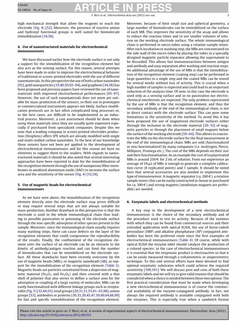
ig. 3. Use of DNA-thiolated scaffold as a new strategy for the detection of antibodies. Thcaffold to support the recognition element. Left: one strand of the scaffold, the “anchorinabeled with a redox tag (here methylene blue) at its 3 ′ terminus. The second strand, the “rlement (antigen). In the unbound state, the scaffold supports efficient electron transfer becognition element reduces the transfer efficiency, thus significantly reducing the obserright). Reproduced with permission from [21]. Copyright (2009) from American Chemica
te (here Staphylococcal enterotoxin B) and AP-conjugated secondary antibodies.
at a fixed potential in a range between −0.1 and +0.1 V. Of course,the selection of the right applied potential depends on the elec-trode used, the reference electrode and the specific conditions ofthe assay (pH, buffer used etc.). The most used technique to mea-sure the produced TMBox is chronoamperometry which is usuallyperformed on a time scale of few seconds (from 10 to 60 s) after theenzymatic reaction is let to proceed for several minutes (generallyfrom 2 to 10 min). Since HRP is a non-specific enzyme it supports
spects of electrochemical immunosensors, Electrochim. Acta (2012),
the use of many other substrates. In this perspective good resultshave been also obtained with catechol [92] and hydroquinone[26,29–31,33,45,69–73] (both coupled with H2O2). Also, the use ofH2O2 [18,28,41,50] by itself has been applied and in this case the
e new architecture utilizes a largely double-stranded DNA as a rigid-but-dynamicg strand”, is attached to the electrode surface at its thiol-modified 5 ′ terminus andecognition strand”, is modified at its 5′ terminus with a small-molecule recognitionetween the redox label and the electrode. The binding of the antibody target to thisved faradaic current. Shown here are representative square wave voltammogramsl Society.
ARTICLE IN PRESSG Model
EA-18854; No. of Pages 10
F. Ricci et al. / Electrochimica Acta xxx (2012) xxx– xxx 7
Fig. 4. Detection of proteins with anodized aluminum oxide (AAO) nanoporous membranes. The cells in the sample are excluded by the pores while the proteins enter insideand are recognized by specific antibodies. Right: sensing principle in the absence (A) and presence (B) of the specific protein in the sample and in the case of the sandwichassay using AuNP tags (C). Left: SEM images of a top view and cross-sectional view of the AAO nanoporous membrane containing 200 nm pores, and confocal microscopyi rane.
utWumrmtstmafsmhbdacbi
TI
mage of a top and cross-sectional view 5 �m in depth from both sides of the memb
se of differential pulse voltammetry (DPV) and cyclic voltamme-ry (CV) has been demonstrated in addition to chronoamperometry.
e have to note that TMB (coupled with H2O2) is by far the mostsed substrate and this surely represents the safest and most opti-al choice. It has to be considered that because both oxidized and
educed forms of TMB are electroactive, the use of pulsed voltam-etric techniques is not possible with this substrate and the need
o use chronoamperometric techniques has some costs in terms ofensitivity and measurement time. Recently we have demonstratedhe possible use of pulsed amperometric methods (such as Inter-
ittent Pulsed Amperometry, IPA) which led to very good resultss this technique can allow the interrogation of 96 electrodes inew seconds [65–67]. Another practical aspect that should be con-idered in the use of HRP label is that the substrate solution is aixture of two reagents and that the home-made preparation of a
ighly concentrated solution of TMB is usually a lengthy processecause of the low solubility of this substrate. However, nowa-ays prepared solutions of TMB/H2O2 optimized for HRP reactions
Please cite this article in press as: F. Ricci, et al., A review of experimental ahttp://dx.doi.org/10.1016/j.electacta.2012.06.033
re commercially available and may represent a good choice. Inonclusion, while for optical ELISA, HRP is the preferred choiceecause it allows a better sensitivity [102], with electrochemical
mmunosensors the high enzymatic activity is somehow hampered
able 3mmobilization on magnetic beads and nanomaterials.
Material Type of functionalization or of nanomateria
FunctionalizedMagnetic beads
Amino group
Avidin/Streptavidin
Protein A/Protein G
Tosyl-activated
With antigenWith antibody
Nanoparticlesandnanomaterials
Super paramagnetic nanobeads
Carbon nanotubes
Graphene
Poly(dialyldimethylammonium chloride) grNanogold/gold nanoparticles
Anodized aluminium oxide (AAO) nanoporo
Reproduced with permission from [52]. Copyright (2011) from Wiley/VCH.
by the requirement of a technique which is not particularly sensi-tive. For this reason AP represents sometimes a valid alternativeas enzymatic label and often represents the preferred choice. Thisenzyme in fact offers less flexibility than HRP but has its own advan-tages. The most used enzymatic substrate is 1-naphthyl phosphate(1-NPP) which is converted to the electroactive 1-napthol (1-NP)[23,44,47–49,57,59–61,63,64,99]. Unlike TMB, this product can bedetected through pulsed voltammetric techniques such as DPV andSquare Wave Voltammetry (SWV) at a potential of ca. +0.2/ + 0.3 V.This guarantees a better sensitivity of the method and a much fastermeasurement time (i.e. usually DPV and SWV can be completed inless than 3 s). This is mainly the reason that AP is a better choicethan HRP for the development of an electrochemical immunosen-sor. Despite this, the use of AP presents also some limitations.The enzymatic reaction can only proceed at basic pH and this canbe a drawback in case of special immunoassays where the anti-body/antigen reaction and the measurement steps are performedin the same solution. It has to be stressed that the electrochemical
spects of electrochemical immunosensors, Electrochim. Acta (2012),
reactions of both 1-NP and TMBox lead to the passivation/poisoningof the electrode surface. This requires the use of a new electrodefor each measurement. This is another crucial reason why screenprinted disposable electrodes are preferred over traditional ones in
l References
[53][32,44,55][39,45,47,59,60,99][29,31,51,55,61–63,98][64][30,35,66][48][92][58]
aphene–gold nanoparticles [34][18,40,50,92,93]
us filter membranes [52,56]

Please cite this article in press as: F. Ricci, et al., A review of experimental aspects of electrochemical immunosensors, Electrochim. Acta (2012),http://dx.doi.org/10.1016/j.electacta.2012.06.033
ARTICLE IN PRESSG Model
EA-18854; No. of Pages 10
8 F. Ricci et al. / Electrochimica Acta xxx (2012) xxx– xxx
Table 4Enzyme and other catalytic labels.
Alkaline phosphatase
Substrate Electrochemical method Measurement potentials (V) References
1-NPP DPV From −0.15 to +0.60 [44,47–49,57,59–61,63–65,99]1-NPP SWV From 0.0 to +0.45 [23]p-APP CA +0.3 [17]p-APP DPV From −0.2 to +0.6 [27]3-indoxyl phosphate–silver nitrate LSV From 0.0 to +0.4 [40]
Horseradish peroxidaseTMB – H2O2 CA 0.15 [24]TMB – H2O2 CA −0.1 [42,55,62]TMB – H2O2 CA +0.1 [46]TMB – H2O2 CA −0.2 [57]TMB – H2O2 CV From −0.5 to 0.8 [35]TMB – H2O2 IPA −0.1 [65–67]Hydroquinone – H2O2 CA ca. −0.15 [26,29–31,33,45,70–73]Catechol – H2O2 CA −0.05 [92]Tetrathiafulvalene – H2O2 CA −0.15 [39]H2O2–Chitosan–Toluidin Blue O CA −0.5 [74]5-methyl phenazinium methyl sulfate – H2O2 CA −0.2 [54]Thionine – H2O2 DPV From −0.05 to −0.5 [58]o-Phenylenediamine – H2O2 DPV From −0.3 to −0.8 [34]H2O2 DPV From −0.4 to 0.0 [50]H2O2 CV From −0.2 to 0.6 [18]H2O2 CA 0.0 [28,41]
Gold nanoparticlesHCl CA −1.0 [51]HCl DPV From +1.25 to 0.0 [32][Fe(CN)6]4 − DPV From −0.4 to 0.3 [52]4-nitrophenol + NaBH4 DPV – CV – [98]
1-NPP: 1-Naphthyl phosphate; p-APP: p-Aminophenyl phosphate; TMB: 3,3′ , 5,5′-Tetramethyl benzidin; [Fe(CN)6]4 −: Ferrocyanide. DPV: Differential pulse voltammetry;SWV: Square wave voltammetry; CA: Chronoamperometry; LSV: Linear sweep voltammetry; CV: Cyclic voltammetry; IPA: Intermittent pulse amperometry.
Fig. 5. Streptavidin-functionalized magnetic beads coupled with magnetized carbon SPE for immunosensor application. Biotinylated capture antibodies are immobilizedonto streptavidin-modified magnetic particles. A sandwich-type immunoassay with the target analyte (prolactin, PRL) and anti-prolactin antibodies labelled with alkalinephosphatase (AP) was used. The resulting bioconjugate formed is then trapped on the surface of the carbon SPE with a small magnet. The quantification of PRL was accomplishedby differential pulse voltammetric (DPV) determination of 1-naphtol formed upon 1-naphtyl phosphate additions. Reproduced with permission from [44]. Copyright (2011)from Elsevier B.V.

ING Model
E
mica A
elfiqewetaooritsm[jbrtrieiopb
7
iucacita(wftvtsaibUfa
8
ritetneT
ARTICLEA-18854; No. of Pages 10
F. Ricci et al. / Electrochi
lectrochemical immunosensing. In few recent examples, enzymeabels have been also recently replaced by inorganic “signal ampli-cation tags” such as catalytic gold nanoparticles [32,51,52,98] oruantum dots [19,53] that have been demonstrated to give inter-sting results in terms of sensitivity, analysis time and correlationith standard tests (Table 4). The use of amplification labels (either
nzymatic or “inorganic”) represents an attractive way to improvehe sensitivity of the immunological method. However, this is also
source of higher costs and higher analysis times. In fact, the needf a conjugated antibody, the addition of substrates and the usef multiple reactions and washing steps is a waste of time andesources that should be avoided. In this perspective electrochem-cal methods offer probably more flexibility than the optical oneso develop methods that do not need enzymatic labels. Recently,everal examples have appeared in the literature for the develop-ent of label-free and reagentless electrochemical immunosensors
20,82]. One of these is based on the use of scaffold DNA probes con-ugated with an antigen and a redox tag [21,22,83]. Upon antibodyinding, the DNA scaffold flexibility is reduced and this leads to aeduced electron transfer rate of the redox tag. This platform canhen detect in real-time and without any washing step or addedeagent the antibody/antigen binding event (Fig. 3). The sensitiv-ty achieved with these sensors is of course not comparable withnzyme-conjugated methods, however, the fact that electrochem-cal immunosensors are less prone to interferences than the opticalnes makes possible the use of such platforms in undiluted sam-les thus leading to detection limits that are often low enough toe useful for practical applications.
. Instruments
In the development of electrochemical immunosensors anmportant choice is represented by the instrumentation to besed. We have detailed above the methods used and among these,hronoamperometry, DPV and SWV are the techniques mainlypplied and, most of the commercially available electrochemi-al instruments, can perform these techniques. Electrochemicalnstruments can be divided in bench-top and portable. Bench-op instruments can usually perform a wider range of techniquesnd are more flexible. However, their cost is usually quite high>20,000 D ) and, from our experience, the performances achievedith these instruments are comparable to those portable, at least
or the specific application of electrochemical immunosensors. Inhis context, three companies (DropSens, PalmSens, Uniscan) offerery precise, low-cost and friendly-user platforms that perform allhe techniques usually required for electrochemical immunosen-ors. These instruments can be coupled with multi-array platformsnd can be also customized quite easily. The prices of these portablenstruments usually range between 2000 and 4000 D and they cane often customized for specific needs. They are all equipped withSB plugs for easy connection to PC, laptops or palm devices and,
rom our direct experience, they have friendly-user interfaces andre very reliable.
. Conclusions
In this short survey we have given practical information foresearchers that want to develop for the first time electrochem-cal immunosensors. We highlight the reasons to choose amonghe multiple options that are available to the public in terms oflectrodes, instruments, enzymatic labels and reagents. We believe
Please cite this article in press as: F. Ricci, et al., A review of experimental ahttp://dx.doi.org/10.1016/j.electacta.2012.06.033
hat electrochemical immunosensors may represent a valid alter-ative to optical ELISA. However, the most important advantages oflectrochemical detection have not yet been completely exploited.he mere adaptation of the optical immunoassay to an
PRESScta xxx (2012) xxx– xxx 9
electrochemical platform is not likely to give important advan-tages in terms of sensitivity, detection limit, analysis time and costs.Researchers should try to focus on the drawbacks of optical ELISAwhich can be overcome by electrochemical approaches and build,on these premises, immunoassays with better performances. Forexample, the possibility to use very small volumes and miniatur-ized array of electrochemical sensors has not been yet exhaustivelyemployed for immunosensors development. Also, too few exam-ples have been reported to date for the detection of antibodies in areagentless and label-free format. In our opinion electrochemistryis the best technique to achieve such important results and moreefforts should be dedicated to this goal. The low level of electro-chemical interferences in complex clinical or food samples is also animportant advantage over the optical approaches which makes thistechnique more suitable for point-of-care detection. An electro-chemical sensor for antibody detection that does not need reactionor washing steps or the addition of any external reagents coupledwith the low cost and easiness of use of electrochemical instrumen-tation would make such platform a perfect tool for the point-of-carediagnosis, prevention and treatment of many illnesses, includinginfectious and autoimmune diseases.
References
[1] C.A.K. Borrebaeck, Immunology Today 21 (2000) 379.[2] D. Huckle, Expert Review of Medical Devices 3 (2006) 421.[3] S.A. Soper, K. Brown, A. Ellington, B. Frazier, G. Garcia-Manero, V. Gau, S.I.
Gutman, D.F. Hayes, B. Korte, J.L. Landers, D. Larson, F. Ligler, A. Majum-dar, M. Mascini, D. Nolte, Z. Rosenzweig, J. Wang, D. Wilson, Biosensors andBioelectronics 21 (2006) 1932.
[4] R.M. Lequin, Clinical Chemistry 51 (2005) 2415.[5] E. Mylonakis, M. Paliou, M. Lally, T.P. Flanigan, J.D. Rich, American Journal of
Medicine 109 (2000) 568.[6] P. Shi, S.J. Wong, Expert Review of Molecular Diagnostics 6 (2003) 733.[7] A.J. Van Hengel, Analytical and Bioanalytical Chemistry 389 (2007) 111.[8] M.N. Levine, R.T. Raines, Analytical Biochemistry 418 (2011) 247.[9] R.W. Phillips, D. Abbott, Food Additives and Contaminants: Part A 25 (2008)
1084.[10] H. Tahk, M.H. Lee, K.B. Lee, D. Cheon, C. Choi, Journal of Virological Methods
175 (2011) 137.[11] B. Ngom, Y. Guo, X. Wang, D. Bi, Analytical and Bioanalytical Chemistry 397
(2010) 1113.[12] F. Ricci, G. Volpe, L. Micheli, G. Palleschi, Analytica Chimica Acta 605 (2007)
111.[13] A.H.W.M. Schuurs, B.K. van Weemen, Journal of Immunoassay 1 (1980) 229.[14] A. Voller, A. Bartlett, D.E. Bidwell, Journal of Clinical Pathology 31 (1978) 507.[15] C.A. Marquette, L.J. Blum, Biosensors and Bioelectronics 21 (2006) 1424.[16] E. Mallat, D. Barcelo, C. Barzan, G. Gaugliz, R. Abuknesha, TRAC-Trends in
Analytical Chemistry 20 (2001) 124.[17] A. Deng, H. Yang, Sensors and Actuators B 124 (2007) 202.[18] S. Liu, Q. Lin, X. Zhang, X. He, X. Xing, W. Lian, J. Huang, Sensors and Actuators
B 156 (2011) 71.[19] H. Yu, J. Lee, S. Kim, G.H. Nguyen, I.S. Kim, Analytical and Bioanalytical Chem-
istry 394 (2009) 2173.[20] Y. Huang, X. Nie, S. Gan, J. Jiang, G. Shen, R. Yu, Analytical Biochemistry 382
(2008) 16.[21] K.J. Cash, F. Ricci, K.W. Plaxco, Journal of the American Chemical Society 131
(2009) 6955.[22] K.J. Cash, F. Ricci, K.W. Plaxco, Chemical Communications 41 (2009) 6222.[23] M.P. Chatrathi, J. Wang, G.E. Collins, Biosensors and Bioelectronics 22 (2007)
2932.[24] S. Dulay, P. Lozano-Sánchez, E. Iwuoha, I. Katakis, C.K. O’Sullivan, Biosensors
and Bioelectronics 26 (2011) 3852.[25] R. Genc, D. Murphy, A. Fragoso, M. Ortiz, C.K. O’Sullivan, Analytical Chemistry
83 (2011) 563.[26] H.M. Nassef, L. Civit, A. Fragoso, C.K. O’Sullivan, Analytical Chemistry 81 (2009)
5299.[27] H.M. Nassef, M.C. Bermudo Redondo, P.J. Ciclitira, H.J. Ellis, A. Fragoso, C.K.
O’Sullivan, Analytical Chemistry 80 (2008) 9265.[28] D. Tang, R. Yuan, Y. Chai, Analytical Chemistry 80 (2008) 1582.[29] E. Zacco, J. Adrian, R. Galve, M.P. Marco, S. Alegret, M.I. Pividori, Biosensors
and Bioelectronics 22 (2007) 2184.[30] S. Liebana, A. Lermo, S. Campoy, M.P. Cortes, S. Alegret, M.I. Pividori, Biosen-
spects of electrochemical immunosensors, Electrochim. Acta (2012),
sors and Bioelectronics 25 (2009) 510.[31] A. Lermo, S. Fabiano, S. Hernández, R. Galve, M.P. Marco, S. Alegret, M.I. Pivi-
dori, Biosensors and Bioelectronics 24 (2009) 2057.[32] A. Ambrosi, M.T. Castaneda, A.J. Killard, M.R. Smyth, S. Alegret, A. Merkoci,
Analytical Chemistry 79 (2007) 5232.

ING Model
E
1 mica A
(2007) 9.[100] G. Volpe, D. Compagnone, R. Draisci, G. Palleschi, Analyst 123 (1998) 1303.
ARTICLEA-18854; No. of Pages 10
0 F. Ricci et al. / Electrochi
[33] M.I. Pividori, A. Lermo, A. Bonanni, S. Alegret, M. del Valle, Analytical Bio-chemistry 388 (2009) 229.
[34] K. Liu, J.J. Zhang, C. Wang, J.J. Zhu, Biosensors and Bioelectronics 26 (2011)3627.
[35] Y. Lin, G. Liu, C.M. Wai, Y. Lin, Electrochemistry Communications 9 (2007)1547.
[36] A. Cagnini, I. Palchetti, I. Lionti, M. Mascini, A.P.F. Turner, Sensors and Actua-tors B 24 (1995) 85.
[37] J. Wang, M. Pedrero, H. Sakslund, O. Hammerich, J. Pingarron, Analyst 121(1996) 345.
[38] J.P. Hart, S.A. Wring, TrAC-Trends in Analytical Chemistry 16 (1997) 89.[39] S. Campuzano, B. Esteban-Fernández de Ávila, J. Yuste, M. Pedrero, J. García,
P. García, E. García, J.M. Pingarrón, Biosensors and Bioelectronics 26 (2010)1225.
[40] V. Escamilla-Gómez, D. Hernández-Santos, M.B. González-García, J.M.Pingarrón-Carrazón, A. Costa-García, Biosensors and Bioelectronics 24 (2009)2678.
[41] V. Escamilla-Gómez, S. Campuzano, M. Pedrero, J.M. Pingarrón, Talanta 77(2008) 876.
[42] A. Radi, X. Munoz-Berbel, M. Cortina-Puig, J. Marty, Electrochimica Acta 54(2009) 2180.
[43] A.A.P. Ferreira, W. Colli, M.J.M. Alves, D.R. Oliveira, P.I. Costa, A.G. Guell,F. Sanz, A.V. Benedetti, H. Yamanaka, Electrochimica Acta 51 (2006)5046.
[44] M. Moreno-Guzmán, A. González-Cortés, P. Yánez-Sedeno, J.M. Pingarrón,Analytica Chimica Acta 692 (2011) 125.
[45] M. Eguílaz, M. Moreno-Guzmán, S. Campuzano, A. González-Cortés, P. Yánez-Sedeno, J.M. Pingarrón, Biosensors and Bioelectronics 26 (2010) 517.
[46] H. Lu, M.P. Kreuzer, K. Takkinen, G.G. Guilbault, Biosensors and Bioelectronics22 (2007) 1756.
[47] M. Moreno-Guzman, M. Eguılaz, S. Campuzano, A. Gonzalez-Cortes, P. Yanez-Sedeno, J.M. Pingarron, Analyst 135 (2010) 1926.
[48] A. Hayat, L. Barthelmebs, J. Marty, Analytica Chimica Acta 690 (2011)248.
[49] A. Hayat, L. Barthelmebs, A. Sassolas, J. Marty, Talanta 85 (2011) 513.[50] D. Tang, J. Tang, B. Su, G. Chen, Journal of Agricultural and Food Chemistry 58
(2010) 10824.[51] A. de la Escosura-Muniz, M. Maltez-da Costa, C. Sánchez-Espinel, B. Díaz-
Freitas, J. Fernández-Suarez, A. González-Fernández, A. Merkoc i, Biosensorsand Bioelectronics 26 (2010) 1710.
[52] A. de la Escosura-Muniz, A. Merkoc i, Small 7 (2011) 675.[53] H. Wang, J. Wang, C. Timchalk, Y. Lin, Analytical Chemistry 80 (2008) 8477.[54] M. Campas, J. Marty, Biosensors and Bioelectronics 22 (2007) 1034.[55] P. Sarkar, D. Ghosh, D. Bhattacharyay, S.J. Setford, A.P.F. Turner, Electroanalysis
20 (2008) 1414.[56] A. de la Escosura-Muniz, A. Merkoc i, Electrochemistry Communications 12
(2010) 859.[57] B. Prieto-Simon, Monica Campas, J. Marty, T. Noguer, Biosensors and Bioelec-
tronics 23 (2008) 995.[58] Y. Xie, A. Chen, D. Du, Y. Lin, Analytica Chimica Acta 699 (2011) 44.[59] A. Zani, S. Laschi, M. Mascini, G. Marrazza, Electroanalysis 23 (2011) 91.[60] S. Centi, A.I. Stoica, S. Laschi, M. Mascini, Electroanalysis 22 (2010) 1881.[61] D. Romanazzo, F. Ricci, S. Vesco, S. Piermarini, G. Volpe, D. Moscone, G.
Palleschi, Journal of Visualized Experiments 32 (2009).[62] D. Romanazzo, F. Ricci, G. Volpe, C.T. Elliott, S. Vesco, K. Kroeger, D. Moscone,
J. Stroka, H. Van Egmond, M. Vehniainen, G. Palleschi, Biosensors and Bioelec-tronics 25 (2010) 2615.
[63] S. Piermarini, G. Volpe, L. Micheli, D. Moscone, G. Palleschi, Food Control 20(2009) 371.
[64] G. Adornetto, G. Volpe, A. De Stefano, S. Martini, G. Gallucci, A. Manzoni, S.Bernardini, M. Mascini, D. Moscone, Analytical and Bioanalytical Chemistry
Please cite this article in press as: F. Ricci, et al., A review of experimental ahttp://dx.doi.org/10.1016/j.electacta.2012.06.033
403 (2012) 1191.[65] S. Piermarini, L. Micheli, N.H.S. Ammida, G. Palleschi, D. Moscone, Biosensors
and Bioelectronics 22 (2007) 1434.[66] E. Delibato, G. Volpe, D. Romanazzo, D. De Medici, L. Toti, D. Moscone, G.
Palleschi, Journal of Agricultural and Food Chemistry 57 (2009) 7200.
PRESScta xxx (2012) xxx– xxx
[67] D. Neagu, S. Perrino, L. Micheli, G. Palleschi, D. Moscone, International DairyJournal 19 (2009) 753.
[68] P. Skládal, T. Kaláb, Analytica Chimica Acta 316 (1995) 73.[69] O.Y. Henry, A. Fragoso, V. Beni, N. Laboria, J.L.A. Sanchez, D. Latta, F. Von
Germar, K. Drese, I. Katakis, C.K. O’Sullivan, Electrophoresis 30 (2009)3398.
[70] M. Ortiz, A. Fragoso, C.K. O’Sullivan, Analytical Chemistry 83 (2011) 2931.[71] M. Ortiz, A. Fragoso, C.K. O’Sullivan, Organic and Biomolecular Chemistry 9
(2011) 4770.[72] N. Laboria, Alex Fragoso, W. Kemmner, D. Latta, O. Nilsson, M.L. Botero, K.
Drese, C.K. O’Sullivan, Analytical Chemistry 82 (2010) 1712.[73] C. Kellner, M.L. Botero, D. Latta, K. Drese, A. Fragoso, C.K. O’Sullivan, Elec-
trophoresis 32 (2011) 926.[74] J. Wu, F. Yan, J. Tang, C. Zhai, H. Ju, Clinical Chemistry 53 (2007) 1495.[75] A. Kausaite-Minkstimiene, A. Ramanaviciene, J. Kirlyte, A. Ramanavicius, Ana-
lytical Chemistry 82 (2010) 6401.[76] V. Escamilla-Gomez, S. Campuzano, M. Pedrero, P.M. Pingarron, Analytical
and Bioanalytical Chemistry 391 (2008) 837.[77] K. Feng, Y. Kang, J. Zhao, Y. Liu, J. Jiang, G. Shen, R. Yu, Analytical Biochemistry
378 (2008) 38.[78] A. Fragoso, N. Laboria, D. Latta, C.K. O’Sullivan, Analytical Chemistry 60 (2008)
2556.[79] Y. Jung, J.Y. Jeong, B.H. Chung, Analyst 133 (2008) 697.[80] D. Mandler, S. Kraus-Ophir, Journal of Solid State Electrochemistry 15 (2011)
1.[81] S.K. Arya, P.R. Solanki, M. Datta, B.D. Malhotra, Biosensors and Bioelectronics
24 (2009) 2810.[82] U. Rant, E. Pringsheim, W. Kaiser, K. Arinaga, J. Knezevic, M. Tornow, S. Fujita,
N. Yokoyama, G. Abstreiter, Nano Letters 9 (2009) 1290.[83] R.J. White, H.M. Kallewaard, W. Hsieh, A.S. Patterson, J.B. Kasehagen, K.J.
Cash, T. Uzawa, H.T. Soh, K.W. Plaxco, Analytical Chemistry 84 (2012)1098.
[84] Y. Jung, H.J. Kang, J.M. Lee, S.O. Jung, W.S. Yun, S.J. Chung, B.H. Chung, Analyt-ical Biochemistry 374 (2008) 99.
[85] J.M. Fowler, M.C. Stuart, D.K.Y. Wong, Electrochemistry Communications 10(2008) 1020.
[86] J.M. Fowler, M.C. Stuart, D.K.Y. Wong, Biosensors and Bioelectronics 23 (2007)633.
[87] J.M. Fowler, M.C. Stuart, D.K.Y. Wong, Analytical Chemistry 79 (2007) 350.[88] Y. Jung, M.L. Jeong, H. Jung, H.C. Bong, Analytical Chemistry 17 (2007) 6534.[89] X. Liu, D.K.Y. Wong, Talanta 77 (2009) 1437.[90] J.H. Lee, H.K. Choi, S.Y. Lee, M. Lim, J.H. Chang, Biosensors and Bioelectronics
28 (2011) 146.[91] S. Sánchez, M. Roldán, S. Pérez, E. Fàbregas, Analytical Chemistry 80 (2008)
6508.[92] V. Serafin, M. Eguilaz, L. Agui, P. Yanez-Sedeno, J.M. Pingarron, Electroanalysis
23 (2011) 169.[93] M. Wasowicz, S. Viswanathan, A. Dvornyk, K. Grzelak, B. Kłudkiewicz, H.
Radecka, Biosensors and Bioelectronics 24 (2008) 284.[94] X. Liu, Y. Deng, X. Jin, L. Chen, J. Jiang, G. Shen, R.Q. Yu, Analytical Biochemistry
389 (2009) 63.[95] A. Qureshi, W.P. Kang, J.L. Davidson, Y. Gurbuz, Diamond and Related Mate-
rials 18 (2009) 1401.[96] M.R. Iost, F.N. Crespilho, Biosensors and Bioelectronics 31 (2012) 1.[97] L. Agui, P. Yanez-Sedeno, J.M. Pingarron, Analytica Chimica Acta 622 (2008)
11.[98] T. Selvaraju, J. Das, S.W. Han, H. Yang, Biosensors and Bioelectronics 23 (2008)
932.[99] S. Centi, E. Silva, S. Laschi, I. Palchetti, M. Mascini, Analytica Chimica Acta 594
spects of electrochemical immunosensors, Electrochim. Acta (2012),
[101] R.M. Pemberton, J.P. Hart, P. Stoddard, J.A. Foulkes, Biosensors and Bioelec-tronics 14 (1999) 495.
[102] A. Kambegawa, Japanese Journal of Clinical Medicine 53 (1995) 2160.