Resume Development Workshop Krisztina Kecskei Career Counselor, UCLA.
Upload
august-hendersonCategory
view
227download
0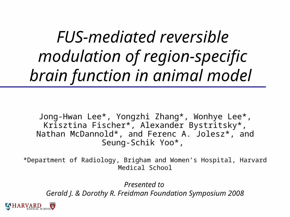
FUS-mediated reversible modulation of region-specific
brain function in animal model
Jong-Hwan Lee*, Yongzhi Zhang*, Wonhye Lee*, Krisztina Fischer*, Alexander Bystritsky*, Nathan McDannold*, and
Ferenc A. Jolesz*, and Seung-Schik Yoo*,
*Department of Radiology, Brigham and Women’s Hospital, Harvard Medical School
Presented to Gerald J. & Dorothy R. Freidman Foundation Symposium 2008

Reversible and non-invasive regional modulation of cortical function
• Needed for short-term/long-term modulation of the brain function.
• Ultimately, it may provide non-pharmaceutical intervention of appetite control !
• Transcranial magnetic stimulation (TMS) suffers from limited penetration depth and lacks spatial specificity.
• Transcranial direct current stimulation (tDCS) lacks spatial specificity.
• Deep brain stimulation (DBS) is invasive.www.biomag.hus.fi/tms/Thesis/dt.html

Idea: Pulsed FUS (FUP)• Motivation and Earlier work
– Trans-skull delivery of FUS (Hynynen et al. 2004)– Reversible modulation of activity in ex vivo brain tissue
(Bachtold et al. 1998) – Previous works on animal sciatic nerves.
• Things to avoid;– Irreversible tissue damage or seizure induction.– BBB disruption.– Excessive heating.
• Proposed method: Instead of continuous application of HIFU, apply the low intensity FUS stimulation with sufficient inter-stimulation intervals as a train of pulses.

Initial excursion: Real-time fMRI during FUP
• Why real-time fMRI?– To monitor the blood-oxygenated-level-dependent
(BOLD) MRI signals that are associated with the activation in real-time, non-invasively.
– Allows the adjustment of the FUP parameters on-fly.
No stimulation stimulation
21 s 21 s12 s
T-1 signal equl
A
Detected activation at p<0.001 level and approximate location of the FUP application in yellow circle.
No stimulation stimulation
15 s 45s12 s
T-1 signal equl
15s
FUP application and intensity adjustment
50 W/cm2
Recovery
TRIAL EXAMPLE

Experimental flow1. Spotting of the FU ‘hot spot’ using phantom
– 690 KHz ultrasound transducer with 8 cm focal depth
2. Anatomical localization
3. Functional localization via fMRI– BOLD-sensitive EPI sequence to detect level of activation
4. Adjustment of positioning system
5. Application of FUP with fMRI
6. fMRI without FUP to monitor
7. MRI Thermometry– Fast SPGR sequence; phase
Dependent thermometry
8. [Group#1] Perfusion of Trypan blue to examine BBB disruption, followed by the histological analysis.
9. [Group #2] Survival for 1 week with behavior observation, followed by the histological analysis
Transducer
Ultrasound beam
Water tank
Water bag
Rabbit
Skull
Plastic plate
MRI Coil
Brain
Sonication target
Positioning system
Control Computer
Function Generator
Amplifier
Power Meter
MRI Room
Matching Network

Experimental setup
Power Monitor
Function Generator
Power Amplifier
Pulse Regulator
MRI Console rtfMRI Processor
Pulse Monitor
3T MRI

No stimulation
stimulation
15 s 9 s12 s 45 s
FUP application
Event-related response and FUP• In order to capture the effect of FUP during short-trial
based visual stimulation, event-related fMRI design was adapted.
• FUP (500 μs duration and 10 msec inter-pulse-interval) at 50W/cm2 intensity was applied for 9, 18, and 27 sec.
Temporal progression of activation
PreFUP
L R
FUP
3min
7min
11min
5min
BOLD Contrast (%) measured from each session
C

Presence of BBB disruption or tissue damage?
• Trypan Blue perfusion after FUP (within 30min) showed no apparent BBB disruption.
• Histological analysis (H&E stain) showed no tissue damage even at 350W/cm2 pulse (10 msec inter-pulse-interval) application of the FUP for 27 sec.
• Survival experiment showed normal post-FUP animal behavior and normal histology results.
McDannold et al. BWH, PNAS 2007
A B C D E

MR Thermometry
• No temperature rise during 27s application of FUP at 500 μs duration and 10 msec inter-pulse-interval at 50W/cm2 intensity. – Virtually no temperature change
• Even at higher level of energy at 350W/cm2 intensity, only 0.95 C rise of temperature was detected after 27s FUP application.
07019
077 sonication 1
Temperatures at 9.9 s.
P7.5
TR:39.2 TE:19.5
bw: 3.57 kHz
Img time: 4.0 s
FOV: 8 cm 256×256 (256×128)
FA: 30° ETL: 1 NEX: 1.00
coef: 0.01000 ppm/°C
Rabbit
Rabbit
08-03-08 2:14:01 PM
R14.8 L25.0
S23.8
I16.0
Tem
pe
ratu
re R
ise
(°C
)
1.8
1.4
1
0.6
0.2
-0.2
-0.6
-1
-1.4
-1.8
27s
18s
9s

Conclusions• Reversible modulation of region-specific brain function
was demonstrated via pulsed application of focused ultrasound energy.
• The given FUP parameter appears to be safe to be used, both short-term and long-term.
• The BOLD signal showed that the visual-stimulation-induced activation was enhanced during the FUP application, followed by the period of non-responsive stages, which suggest the ‘relative refractory period’.– More confirmatory study is needed using different stimulation parameters.
• Potential mechanism ? Temperature-mediation was ruled out. Possibly via mechanical modulation.
• Electrophysiological confirmation will be followed.
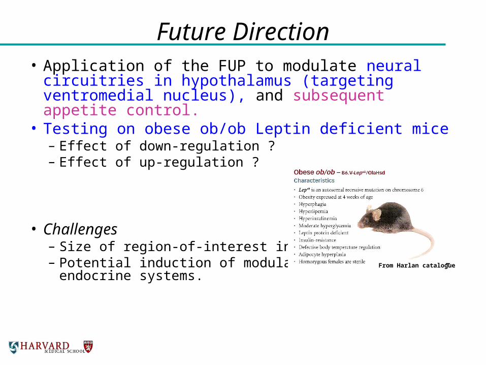
Future Direction• Application of the FUP to modulate neural
circuitries in hypothalamus (targeting ventromedial nucleus), and subsequent appetite control.
• Testing on obese ob/ob Leptin deficient mice– Effect of down-regulation ?– Effect of up-regulation ?
• Challenges– Size of region-of-interest in mouse is small.– Potential induction of modulation in other neuro-
endocrine systems.
From Harlan catalogue

Acknowledgement• Magdalini C. Pilatou, Ph.D.• Lisa Treat, M.S.• Jason White, Ph.D. and with the financial support from
– Gerald J. & Dorothy R. Freidman Foundation– Focused Ultrasound Surgery Foundation– Bystritsky Family Foundation
, and all anonymous rabbits…
Disclaimer: This rabbit was NOT used in the experiment