Further observations on the pathology of cardiac dropsy
37
FURTHER OBSERVATIONS ON THE PATHOLOGY OF CARDIAC DR0PSY.l By CHARLES BOLTON, M.D., D.Sc., F.R.C.P., Physician to U?~iversity College Hospital, London, W.C. From the Graham Research Laboratories, University of Lo9idon. INTRODUCTION. THE present research is a continuatiou of an investigation into the pathological processes occurring in uncompensated heart disease, which was commenced some years ago, the first communication on the subject being published in this Journal in 1903 l. The original idea was to produce experimentally an uncompensated condition of the circulation in order to study the exact alterations which occur in the circulation of the blood, in its volume and composition, in the composition and flow of the lymph, and other problenis connected with this morbid state, iL being quite impossible to obtain precise information on these points by observations on the human subject. Until such information was definitely acquired it was considered that our knowledge of the pathology of cardiac dropsy would advance no further towards a satisfactory solution. RECAPITULATION OF THE AUTHOR’S PREVIOUS RESULTS. The method adopted in my earlier experiments consisted in constricting the perieardium by means of sutures so that the diastolic expansion of the heart was impeded. An uncompensated condition of the circulation resulted, the volume of blood in the veins was augmented and in the arteries diminished, and dropsy supervened (1903 l). I then found that precisely the same circulatory conditions resulted when I narrowed the superior and inferior vena: cave together at their entrance into the heart (1 9 0 7-1 9 09 ”. My experiments both in the case of pericardial constriction and obstruction of the large venous trunks were concerned with the circulatory alterations occurring during the first twenty-four hours following the operation, and the main results which I observed during this period may be briefly expressed as follows. [Received January 10, 1916.1 The expenses of this research have beer defrayed by grants from the Royal Society and the Graham Research Fund.
-
Upload
charles-bolton -
Category
Documents
-
view
215 -
download
2
Transcript of Further observations on the pathology of cardiac dropsy

FURTHER OBSERVATIONS ON THE PATHOLOGY OF CARDIAC DR0PSY.l
By CHARLES BOLTON, M.D., D.Sc., F.R.C.P., Physician to U?~iversity College Hospital, London, W.C.
From the Graham Research Laboratories, University of Lo9idon.
INTRODUCTION.
THE present research is a continuatiou of an investigation into the pathological processes occurring in uncompensated heart disease, which was commenced some years ago, the first communication on the subject being published in this Journal in 1903 l.
The original idea was to produce experimentally an uncompensated condition of the circulation in order t o study the exact alterations which occur in the circulation of the blood, in its volume and composition, in the composition and flow of the lymph, and other problenis connected with this morbid state, i L being quite impossible to obtain precise information on these points by observations on the human subject. Until such information was definitely acquired it was considered that our knowledge of the pathology of cardiac dropsy would advance no further towards a satisfactory solution.
RECAPITULATION OF THE AUTHOR’S PREVIOUS RESULTS. The method adopted in my earlier experiments consisted in
constricting the perieardium by means of sutures so that the diastolic expansion of the heart was impeded. An uncompensated condition of the circulation resulted, the volume of blood in the veins was augmented and in the arteries diminished, and dropsy supervened (1903 l). I then found that precisely the same circulatory conditions resulted when I narrowed the superior and inferior vena: cave together a t their entrance into the heart (1 9 0 7-1 9 09 ”. My experiments both in the case of pericardial constriction and obstruction of the large venous trunks were concerned with the circulatory alterations occurring during the first twenty-four hours following the operation, and the main results which I observed during this period may be briefly expressed as follows.
[Received January 10, 1916.1 The expenses of this research have beer defrayed by grants from the Royal Society and the Graham Research Fund.

FURTHER OBSERVATlONS O N CARDIAC DROPSY. 291
Results of Constriction of Pericnrdkm or iYarrowing of both Superior and Inferior Vencc Cwue.
1. ‘[‘he immediate result is a general rise of pressure throughout the whole venous system extending to the small venous radiclcs, and a marked fall of the mean arterial blood pressure.
2. A gradual swelling of the liver and an increasing engorgement of the splanchnic veins and of the large veins of the trunk, together with their immediate tributaries, rapidly appear. Ascites, hydrothorax, and edema of the mediastinum and retroperitoneal tissue make their appearance, arid in two or three hours the fluid is measurable in amount.
3. The venous pressure, however, does not remain for long at the level i t had immediately assumed. It commences to fall, and eventually reaches the norniirl level or thereabouts in the space of an hour or an hour and a half. The arterial pressure remains at the low level to which it had fallen or even a little lower.
4. When the animal has recovered from the anresthetic and resumed its normal conditions of life the pressures undergo fu,rther changes. The venous pressures in the portal system and in the large veins of the trunk and their immediate tributaries gradually rise until they attain the heights at which they stood immediately after the operation. This rise of pressure may be completed within about four hours, but in many of the cases twenty-four hours later the pressures aro still within normal limits. The venous pressures in the peripheral parts of the circulatory system (periphery of body and extremities), however, after they have once fallen, do not rise again ; in those animals in which the venous pressure in the central portions of the trunk are again raised, the pressures in the extremities and periphery of the trunk are normal ; and in those in which the pressure in the trunk are still normal, the pressures in the extremities are subnormal. The arterial pressure follows the renous pressure in the large veins of the trunk. Those animals in which these venous pressures are raised have normal arterial pressures, and those in which they are a t the normal level have subnormal arterial pressures.
5. The presence and the amount of the dropsy are independent of the height of the venous pressures ; the dropsy commences whilst these pressures are normal, and is present twenty-four hours after the operation whether they are normal or raised.
6. My final interpretation of the circulatory changes occurring up to the end of the first twenty-four hours was, that the distensibility of the liver, portal area, and large veins of the trunk is responsible for the fall in the venous pressures; and that the subsequent rise of pressure in the veins and in the arterial system is due to vasomotor overaction, thc pressure in the peripheral portions of the body and limbs remaining normal or subnormal.
There is, therefore, a depletion of the peripheral parts of the circulatory system and an accumulation of blood in the central portions of the body, the actual height of the pressures depending on the degree of action of the vasomotor centre, so that a relative alteration in the venous pressure levels results, the pressure in the central portion of the trunk being raised relativeZy to that in the peripheral portion and in the extremities.
The amount of blood in the body is definitely limited, and therefore if one portion of the vascular system is increased in capacity the pressure can only be kept at the normal or a raised level by vasomotor overaction.
The vascular constriction occurs automatically by stimulation of the vasomotor centre by the deficient blood supply to the brain. The resulting rise in arterial pressure ensures a fuller supply of blood to the brain, and the rise in venous pressure a fuller supply of blood to the heart.
There is an increased output of lymph with diminished absorption by the
292 CHARLES BOLTON:
capillary vessels in the congested areas, giving rise to dropsy. An increased absorption of lymph occurs in the depleted areas in order to restore the normal volume of blood in these parts.
Cardiac dropsy is thus seen t o be really a form of local dropsy, the locality being determined by the distensibility of a certain portion of the vascular system. Dropsy occurs in this locality only; in other parts of the body increased absorption of lymph takes place. The assumption of the erect posture in man relieves the congestion of the portal system and central portions of the trunk at the expense of the lower part of the body and the legs, so that the dropsy of the peritoneum and central portions of the body is lessened or at a minimum, and cedema of the legs is produced.
With regard to obstruction of the t w o main cenous trunks separutely, I found that, narrowing or even complete ligature of the superior vena cava alone produced no change whatever in the circulatory system of the body as a whole, a local cedema affecting the mediastinum and pleural cavities simply resulting. Narrowing of the inferior vena cava alone, however, produced general circulatory changes similar to those resulting from pericardial constriction or narrowing of both main venous trunks, the only difference being that there was no preliminary venous congestion of the head and fore part of the body, and no hydro- thorax or cedema of the mediastinuni resulted.
Results of Nawovuing of the Inferioy Vena Cava aloiae.
I found that precisely the same adaptive mechanisms as are described above were brought into play as the result of partial obstruction of the inferior vena cava above the diaphragm.
No change whatever was to be noted until the vein was narrowed down to three-fifths of its normal diameter. This is owing to the fact that the veins and capillaries are not filled to distension and will hold a certain amount of blood before the pressure within them is raised. Complete ligature of the vein causes a sudden fall of arterial pressure to a few millimetres above the zero line, and the animal dies in a few hours.
On narrowing the vein to any size below three-fifths of its diameter, the venous pressure behind the obstruction goes up, the rise extending to the small venous radicles of the hind-limbs and intestines, and the general arterial pressure falls. The venous pressure subsequently falls to normal or there- abouts owing to dilatation of the large veins of the trunk and of the liver and portal system. The venous pressure of the trunk and the arterial pressure subsequently rise, but the pressure in the veins of the hind-limbs remains low. Ascites and cedema of the retroperitoneal tissue result. The only difference between the actual circulatory conditions established and those described above is, that the territory drained by the superior vena cava is not included in the congested area, so that blood is abstracted from the whole forepart of the body. The absorptive area, therefore, includes the whole forepart of the body in addition to the peripheral parts of the abdomen and extremities. The mechanisms involved are, therefore, precisely the same, the difference being one of degree only.
RESULTS OF FURTHER EXPERIMENTS. The present series of experiments examines the changes over
several weeks, and the method which I have employed is that of
RURTHER OBSERVATIONS O N CARDIAC DROPS: 293
narrowing of the inferior vena c a w alone. My reasons for adopting this method were, t ha t t he operation is much more easily and qu ick l j performed than tha t of constriction of the pericardiuiu, the degree of obstruction can be accurately measured, and only now and again have 1 found tha t the animal dies frorii the ett'ects of the operation. More- over, the method possesses the distinct advaiitage that hy the establish- ment of anastomoses the animal fiually recovers froin i t s dropsj. One c m , therefore, study the various processes occurring, not only during tlie onset and gradual increase of the dropsy, but also those occurriug during its gradual subsidence.
The subject will be considered under the following head ing :-
1. Method. 2. Blood volume, hzmoglobin percentage, arid vascular
pressures of the normal cat. 3 . Course of the ascites. 4. Output of lymph i n the congested urea. 5. Blood volume of the dropsical animal. 6. Htemoglobin content of t he blood in the dropsical auimal. 7. Vascular pressures of t he dropsical animal. 8. The intake of fluid and the excretion of urine in t,he
9. The occurrence of jaundice. dropsical animal.
10. Summary and conclusions.
1. Method. The q p w a f w n for partial obstruction of the inferior vena cava has been
already described in a former paper (1909 2), to which the reader is referred for the details. The animals used were cats, and in every operation or experi- ment the animal waa anaesthetised with ether. The vein was in all cases obstructed in the chest just below the heart, :irtificial respiration being maintained during the operation through an intuhation tube. The vein was narrowed down to a diameter of 3 mm. I have found the diameter of the vein distended at the normal pressure to vary a little iii different cats, so that the degree of narrowing was not exactly three-fifths of the original diameter in all cases. For the purposes of these experiments it was unnecessary to endeavour to produce the same relative degree of obstruction in each case, and, in fact, such an attempt could not succeed, as one cannot foretell what the exact size of the vein will be in any particular cat.
The percentage'es of hmioglobin and blood corpuscles were estimated with the Haldane-Gowers hamoglobinoxneter aitd the Thoma-Zeiss hamocytometer. It was found that alterations in the amount of hanioglobin corresponded with similar nlterations in the game direction in the number of corpuscles, so that in most of the experiments the haemoglobin alone was estimated in order to save time and labour. Blood from the head was obtained by pricking the marginal vein of the ear ; from the hind-leg by pricking the pad of the paw ; and from the abdominal vessels by making a small incision through the abdominal wall, carefully drying the surface of the liver and pricking it with a needle.
The volume of the blood was obtained by the washing-out method, the 20-JL. O? PATH.-VOL. XX.
294 CHARLES BOLTON
animal being anssthetised with ether. A sample of blood was first removed and diluted a hundredfold with water. The animal was then bled from the carotid artery, and at the same time warm Ringer’s fluid was run into a vein so as to keep up the blood pressure and maintain the action of the heart as long as possible. The blood was collected in citrate solution. By this means the heart would often go on beating until the fluid issuing from the carotid was almost colourless. The next procedure was to insert a cannula into the cl-xending aorta and wash out the abdomen. A certain amount of blood- tained fluid was always obtained from the inferior vena cava by this means,
however thorough had been the original washing-out of the vessels. Similarly, a small amount could be obtained from the head and upper limbs by washing-out through the ascending aorta. The washings were all mixed together and the resulting fluid diluted until it was exactly the same colour as the hundredfold dilution of the sample of the blood. The total volume of the blood was then calculated.
The blood pressures were taken in the carotid artery, the external jugular vein through the posterior auricular vein, the portal vein, the inferior vena cava (lower end), and in a vein of the hind-leg, asdescribed in the papers referred to above.
The rate of output of l!j?rqh was determined at different stages in the course of the ascites by removing all the abdominal fluid with a pipette and absorbent cotton wool, and noting the amount which had accumulated at the end of the next twenty-four hours.
The animals recovered well and lived healthy lives. They were fed on cat’s meat and milk, and the total amount of fluid consumed and of urine passed was carefully noted each day.
2. Blood Volume, Hmntoglobin Percentage, and Vascular Pressures of the Normal Cat.
By the method described above I have found that the blood volume in cubic centimetres of the normal cat is about one twenty-fifth of its body weight in grms.
(a) Blood volume.
The results of ten determinations are given in Table I. Taking the blood volume as one twenty-fifth of the body weight
in grms. expressed in c.c., the greatest error in the above experiments was about 8 C.C.
( b ) The percentage of hmmoglobin is not the same in all eats; for example, cat 4 had 1 0 0 per cent. Hb, cat 6 had 64 per cent. Hb, and cat 8 had 90 per cent. Hb. The percentage of hzmoglobin, therefore, bears no simple relation to the blood volume.
I n all experiments dealing with alterations in the percentage of hamo- globin it is thus necessary to determine in the first place what is the normal percentage of the particular cat under investigation. The blood obtained from an ear vein contains the same amount of hsmoglobin as that obtained by pricking the pad of the foot, but that from the liver appears to contain if any- thing a trifle more ; for example, one determination showed : ear, 90 per cent. ; foot, 90 per cent. ; liver, 92 per cent. The number of red blood corpuscles also varies in different cats, usually being from 10 to 12,000,000 per c.mm.

FURTHER OBSERVAT/ONS ON CARDlAC DROPSY: 295
KO. !
TABLE I.
Weight.
1 . .
2 . .
3 . .
4 . . 5 . .
6 . .
I . . 8 . .
9 . .
10 . .
2085 q n s .
2460 ,, 2500 ,, 2665 ,, 2790 ,,
9800 ,,
2835 :,
3200 ,,
3540 ,, 3776 ,,
Blood Volume determined
by Experirnent.
85 C.C.
91.1 ),
99 I ,
114 ,>
111 ,,
117.6 ,, l I i . 5 ,,
120 ,, 1 4 0 2 ,, i 4 i . n ,,
ilood Volume calculated as one twenty-fifth of the Body Weight in p i s . (expremed in c.c.).
83.4 C.C.
98’4 ,, 100 ,, 106‘6 ,, 111.6 ,,
112 , I
113’4 ,,
128 , I
140.8 ,, 151 ,,
(c) Thc cemu.9 pressure a t the lower end of the inferior vena cava is about 97 mm. MgSO, solution (sp. gr. 1046); in the foot about 1 6 0 mm. MgSO, solution ; in the portal vein about 104 mm. MgSO, solution (usually a few millimetres above that a t the lower end of the inferior vena cava) ; and in the external jugular vein (posterior auricular) about 79 mm. MgSO, solution. The arterial pressure is usually high, and not uncommonly reaches 180 to 200 mm. Hg.
3. Course of the Ascites.
As stated above, the ascites appeaw during thejrs t few hours after the obstruction of the inferior vena cava is produced. It g radudy increases in quantity up to a certain amount, and then slowly disappears. The cause of the disappearance of the ascites is the .establishment of anastomoses. The anastomotic veins of the abdominal wall begin to distend a t once, and they gradually increase in size till fully developed. The portal anastomoses are more slow in their development. For an account of these anastomoses the reader is referred to a recent pztper (1914 3), in which the case of a monkey is described in which the obstruction of the inferior vena cava went on t o complete obliteration. The ascites, which was proved to be present by abdominal section, also disappeared in this case, so that the general statement may be made that to whatever degree the inferior vena cava is narrowed the resulting ascites will disappear with the completion of the establishment of anastomoses.
The length of time thc dropsy persists depends upon the exact degree

296 CHARLES BOLTON.
8th ,,
14th ,, 27th ,, 2 n d I ,
35th ,,
of constriction, and perhaps also upon certain minor and as yet unascertained factors. In most of the cases in which this point was examined the dropsy disappeared from the seventh to the twelfth week. In five cases a trace only was found in the sixth, seventh, seventh, eighth, and eleventh weeks. I n four cases-in the eighth, eighth, tenth, and seventeenth weeks-12 c.c., 3 c.c., 2 c.c., and 15 C.C. were found respectively. The last case is the only one in which the duration of the dropsy extended over a longer period than three months. In four animals killed at the end of the eleventh, thirteenth, sixteenth, and twenty-first weeks no ascites was found. All the anirnals examined a t shorter periods than six weeks showed varying amounts of fluid in the abdomen.
I t is dificult to estimate the time the dropsy takes to attain its maximum height and the time of subsidence, because the amounts of fluid vary in different animals a t the same stage ; in the lesser degrees of constriction smaller amounts of fluid are found and it subsides quicker. I n fourteen animals the amounts of fluid present a t the end of the first twenty-four hours varied from 8 to 35 c.c., in one it was 45 c.c., and in another 72 C.C. Thus, although large amounts may be found, moderate amounts are the rule during the first twenty-four hours. The largest amounts are found after the first twenty-four hours to the end of the fifth week.
In six cases the :mounts were found as detailed in Table IT. :-
100 ,I
50 ,, 100 I ,
90 1,
150 ,,
TABLE i r .
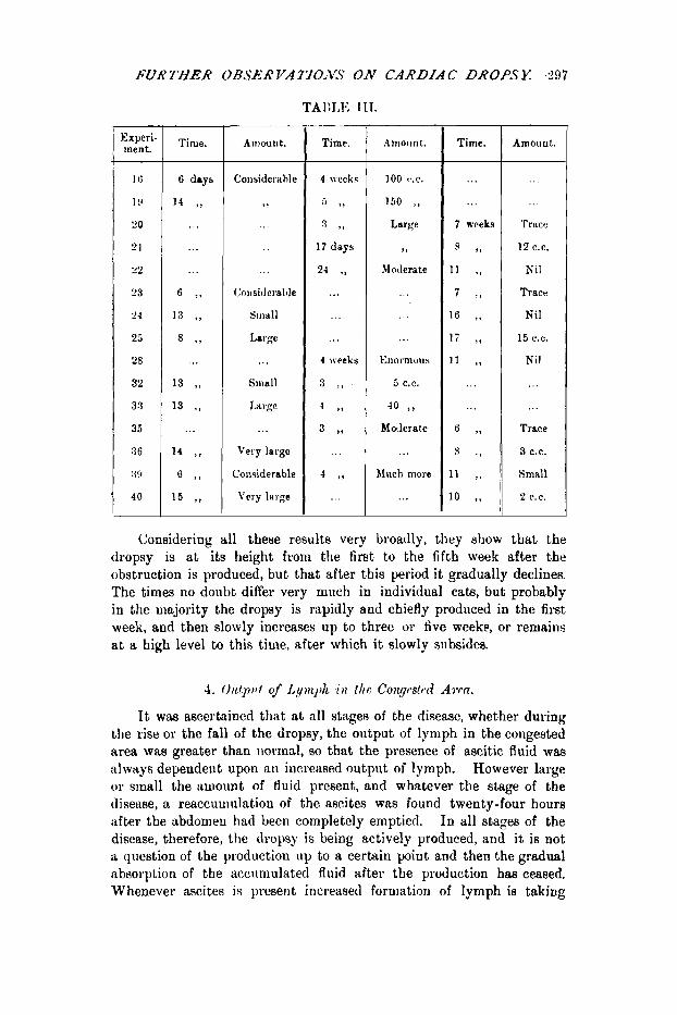
FURirHER UBSER VAT1OiV.S ON CARDIAC DROPSY ,297
Experi- ment.
16
I !I
“0
“ 1
22
23
24
25
28
32
35
35
;3 6
39
40
Tiwe. Atiiwiit.
Cotisidctxhle
...
Coilsi~lcral~le
Ytiin11
Large
... Slllall
J,nrgc
t..
Very large
Considerable
Very large
Time.
4 w c k s
5 ~,
3 ,, 17 days
24 ,, ... ...
...
4 weeks
3 .(
-1 ,,
3 1,
... -1 I ,
...
100 I‘.l’.
150 ,,
Large
,, Moderate
. .
...
... Etiorrnuuh
5 C.C.
40 ,, Moderate
... Milch more
...
Time. Amouut.
. . .
...
errace
12 C.C.
Xi1
Tract?
Xi1
15 C.C.
Nil
...
...
Trace
3 C.C.
Small
2 c.c.
Considering all these results very broadly, they show that the dropsy is at its height from the first to the fifth week after the obstruction is produced, but that after this period it gradually declines. The times no doubt differ very much in individual cats, but probably in the majority the dropsy is rapidly and chiefly prodiiced in the first week, and then slowly increases up to three or five weeke, or remains at a high level to this time, after which i t slowly subsides.
4. O i i t p t ~ t of L.//Y))L,I~L i~ t h e C071.p~tcd Arm.
It was ascertained tlint at all stage8 of the disease, whether during the rise or the fall of the dropsy, the ontput of lymph in the congested area via8 greater than iionnnl, so that the presence of ascitic fluid was always dependeut upon an increiised output of lymph. However large or sinall the amount of tluid present, and whatever the stage of the tlisetise, a reaccuuiuhtion of the ascites was found twenty-four hours after the irbdomeu had been completely emptied. I n all stages of the disease, therefore, the dropsy is being actively produced, and it is not a question of the production up to a certain p i n t and then the gradual absorption of the accuniulated fluid after the production h a s ceased. Whenever ascites is present increased forniatiori of lymph is taking

298 CHARLES BOLTON:
place, but at the point when the ascites commences to disappear the rate of formation has begun to fall off, till finally the lymph is produced in normal amount with the disappearance of the ascites.
The aniount of reaccumulated ascites twenty-four hours after the abdomen has been emptied is shown iu eight cases (Table IV.) :-
, Time of Removal Amount resent of Fluid. 1 Twenty-fonr 8mrs Later.
i 40 ,, i
95 ,)
4 I , 100 ,, 20 , I
! 5 ,* 150 ,, 45 t , I I
I
1 t , I Trace Trace
I Trace Trsce i .- ; , , .- __ I .- - I _-
The amount of reaccumulated fluid during the first five weeks is considerable, but when the dropsy is tiisappearing the rate of accumu- lation is very slow. The rate of formation of lymph semis from the above figures to increase up to the second week 3nd then to diminish, but the number of cases here recorded is of course too small to justify the conclusion that the dropsy is most actively produced in the second or third week and then diniinishes.
The large timounts of lyniph which accumulate in t.he peritoneum in twenty-four hours must be accompanied by a reruoval almost equally rapid, but not sufficiently rapid to prevent R gradual accumulation of ascites. The rate of lymph flow through the thoweic duct psoljably depends on the amount of peritoneal fluid. A rapid circulation of lyriipli thus appears to be constantly in progress from the blood to the p i - toneiim, and thence to the blood again. The rate of circulation is deter- iiiiued by the rate of output, absorption always falling short of output whilst the ascites is accumulating. When the ascites is disappearing the rate of output gradually falls, and closely following it the rate of absorption also falls as the aecites becouies less, so that output fall8 short of absorption during this stage. This theoretical deduction, which I am about to test by experiment, is sdpported by 311
observation of Starling and myself (1910 4), in which we found that the lymph flow of a dog which had devcloped ascites as the rcsult of heart disease was greatly exaggerated.

FURTHER OBSERYATIGENS,.ON CARDIAC DROPSY 299
5. Blood Volume of the Dropsical Animal.
The idea that the volume of the blood is greater than normal in some forms of dropsy has been held for a great number of years, and in the older literature " plethora " played a not unimportant part.
I n the writings of Andral (18366), for example, a form of dropsy due to plethora is described. Oertel (18846) was the first to definitely teach that in cardiac insufficiency hydmmic plethora was present. He held that diminished work of the heart leads to an increased quantity of blood in the body, and that this blood contained an increased percentage of water. He did not, however, provide proof of this supposition. Cohnheim (1883 7) strongly oppoAed the idea of plethora, and considered it as proved that a lasting pathological increase of .the.blood-quantumeould. not occur, since W. Miiller and others h& &own that, when the blood volume of the dog was increased by transfusion of large amounts of blood, the excess was disposed of in a relatively short space of time. He held the same view with regard to hydrsmic plethora, because he and Lichtheim (1877 8) had shown that, after injection of large amounts of salt solution, the.exceas of water was rapidly got rid of by the excretory and secre- tory glands, and that in order, to @uce oedema excessive ambunta' had to be injected. The edema when produced was not distributed a8 in human dropsy, but affected merely the glands in question and the abdominal organs. Cohnheim (1882 i), however, found tha t ,dema of the subcutaneous tissues was produced by hydramic plethora if increased permeability of the vessel walls in the part was also present, as, for example, is occasioned by venous congestion of the leg, or by a slight inflammatory lesion, or even by long continued simple hydrsmia alone. Starling (1896, 1909 9) has maintained on theoretical grounds for many years that an increase in the volume of the blood is a necessary consequence of heart failure, and that without it the high venous pressure necessary to main- tain the increased diaatolic filling of the heart cannot be maintained. He con- siders that the first effect of heart failure is to cause a rise of venous pressure in the large venous trunks, but a fall of venous and capillary pressures in the portal vein and in the limbs. The subsequent vasomotor constriction is unable to extend the rise of venous pressure to the capillaries of the intestines and limbs, so that increased absorption of lymph occurs owing to the low capillary pressure and deficient filling of the arterial system. Plethora is thus brought about. He also considen that the rate of absorption of water from the aliment- ary canal would be increased by splanchnic constriction. Water is retained in the body owing to deficient renal excretion. With regard to the actual cause of cardiac dropsy he is of opinion that it is due to increased capillary pressure in the legs owing to the effect of gravity, acting in conjunction with an alteration in the capillary wall brought about by the stagnation of the blood. The experiments now to be described show that a t a certain stage in the con- dition of circulatory uncompensation an actual increase in the volume of the blood does occur and that it is more dilute than normal. This increase in tho blood volume, which is often very small, is, however, merely a secondary phenomenon in the course of events and is not directly concerned in the initia- tion of the dropsy. My results show five main differences from Starling's deductions.
1. That the initial rise of venous pressure extends through the liver into the portal vein and thence into the venous radicles of the intestines, and also into the venous radicles of the feet. That the capillary pressure in these parts also rises above normal; it falls below normal as the veins dilate, and rises again as the venous pressure goes up but not to its original height, owing to vasomotor constriction, particularly in the feet. I t rises again later when plethora is established.

900 CHARLES BOLTON
2. That a high venow pressure can be maintained by vasomotor constriction and without any increase in the total blood volume.
3. That the increased absorption of lyrnph, largely responsible for the Iiy(1riPmic plethora, does not occur uniformly as a result of the fall of arterial pressure, but only in the depleted areas ; in the congested areas increased ont- put only of lymph occurs. I n other words, fluid is absorbed in one part of the holly and put out in another, both processes occurring concurrently during the whole course of the disease.
4. That the hydraemic plethora occurs later in the course of the disease than i. suggested by Starling.
5. That the dropsy occurs quite independently of t h e actual height of the capillary pressure or of the t o t 1 volume of the blood.
I nlay here mention that the blootl volume of the ascitic clog which I have referred to above was increased. I am aware of one observation only in which the blood volume of a patient with unconipensated heart disease was actually estimated. This was iin observation of Lorrain Smith ( I 902 lo), in which h e found an increase in the 1)lood volume.
TIM blood colzimc in my cxperiincnts was estimated at the different stages in the course of the disease which have been described in Section 3. I t has been shown that the masimum error in the estimation of the blood volume of the normal eats by the method eniployed here was 8 C.C. This amount was, therefore, allowed for in calculating the increase or diminution of blood volume above or below that which was calculated as one twenty-fifth of the body weight in grm. expressed in C.C. The results obtained, therefore, represent values which are as accurate as can be obtained by this method and certainly are not above the actual blood volumee.
The following experiments were performed :-
FIRS WEEK. Experiment ~.--CAI' (weight, 2805 grms.).-Inferior vena c a m constricted
Blood volume, twenty-four hours later = 120.1 C.C.
m n m e n t %--CAT (weight, 2640 grms.).-Inferior Venn cava constricted :Iscites present
Experiment CAT (weight, 3350 grms.).-Inferior rena cava constricted dscites present
hperiment CAT (weight, 35% grms.).-Inferior vena c a w constricted Ascites present =
Experiment CAT (weight, 3420 grnis.).-lnferior vena cava constricted -4scites present =
meriment ~ . - C : A T (weight, 3170 grms.).-Inferior vena cava constricted Ascites present =
Experiment ~ . - - C ' A T (weight, 3340 grins.).-lnferior rena cava constricted Ascites present = 10 C.C.
Experiment &-CAT (weight, 3910 grms.).-Inferior vena cava constricted :Iscites present = 84 C.C.
to a diameter of 3 mm. Ascites present = 33 C.C.
to 3 nini. = 10 C . C .
to 3 mm. = 72 C.C.
to 3 znm. 40 C.C.
to 3 rnm. 40 V.C.
to 3 m n . 27 C.C.
ti, 3 mm.
to 3 mm.
Blood volume, twent.y-four hours later = 121.8 C.C.
Blood volume, twenty-four hours later = 110 C.C.
Rlootl volume, forty-eight hours later = 90 C.C.
Blood volume, forty-eight hours later = 120 C.C.
1:lootl volume, forty-eight hoiirs later = 124 C.C.
Y,lootl voliinic, three days later = 130 C.C.
Rlootl volume, three tl;iya later = 128.2 c.e.

FURTHER 0BSER'VATmN.S ON CARDlAC DROPS: 301
Experiment S.-CAT (weight, 3400 &rins.).--lnferior veiiu cava constricted to 3 mm. Blood volume, tive days later= 133.3 C.C. .Iscites present 30 cc .
Experiment CAT (Wright, 3i53 grins.).-Inferior rena cava con- stricted to 3 nim. Blood roliiiiie, s i r days later = 145.2 V.C. Ascite.: pre-eiit = 15 C.C.
In the first three experiments the aniluals weie killed :it the eud of twenty-four hours. The blood volume of the first was n o n i ~ l , of the second increased by 7 c.c., and of the third decreased by 1 G C.C.
Of those killed at the end of forty-eight hours the blood voluine of the fiiat was decreased by 6 c.c., of the second decreased by 9 c.c., and of the third was normal.
The blood volume of the first animal mliich had lived t1irt.e days was normal, and of the second increased by 4 C.C.
The blood volumes of the two animah which had lived tive arid six days respectively were normal.
During the first week, therefore, ttiere.wns it very slight increase in two cases, a diminution in three, and in five cases the blood volume was normal.
SRCOXI) WEEK. Experiment 11 . -C~a~ (weight, 3360 grnis.).-Infeiioi8 vena c m a coil-
stricted to 3 mrn. Blood volume, eight days later = 116.5 C.C. Ascites preseri t = 100 C.C.
Experiment 12.-Ca~ (weight, 2300 grins.).-Inferior vmin cava con- stricted to 3 rnm. J$lood volume, niiie (lags later ;= 126.4 IX . ;lacites j ~ e s e i i t = 4 C.C.
E x p h e n t 13.-Ca~ (weight, 2920 firms.) -Inferior vena cava coii- stricted to 3 mm. Blood volunie, fourteen days later = 140 C.C. As;cit.es present = 80 c.e.
m r i m e n t 14.-cai (weight, 31 '70 grms.).-Iiiferior vena c:iv:i coii- strictetl to 3 nim. Hlmd volume, fourteeii 11:iys later = 139.5 C.C. :\.wit.es present = 12 C.C.
The aniuual killed on the eighth day showed a diminution i i i its blood volumeof 10 C.C. The other three animals showed :in iuci*e:ise of 28 c.c., 15 c.c., and 6 C.C. respectively.
THIRI) WEEK. Experiment 15.-Ca~ (weight, 3038 gruis.).-lnfciior vena caw coii-
Blood volume, at the end of the third week = 140 O.C. stricted to 3 ruin. Ascites present = 5 C.C.
FOURTH WEEK. Experiment 16.-CAr (weight, 2680 grms.).-Inferior Venn cii\'a coil-
Blood volume, a t the end of the fourth week = 144.6 ('.(*. stricted to 3 mm. Ascites present = 100 C.C.
FIFTH IVEXK.
E.pariment l?.-CaT (weight, 2980 grms.).-Inferior vena c a m con- stricted to 3 mm. Blood volume, a t the end of four weeks and four days = 150.6 C.C.
m r i m e n t 18.-caT (weight, 3060 grms.).--Inferior vcna cava {WII-
etricted to 3 mni. Blood volume, a t the elid of flvc weeks = 148.7 C.C. Ascites present = a trace.
Ascites present = 90 C.C.
302 CHARLES ROLTOX
Erperiment CAT CAT (weight, 3550 grms.).-Inferior vena cava con- Blood volume, at %%e end of five weeks = 156.4 C.C.
The blood volume of each animal in the third, fourth, and fifth weeks was increwd. It w&8 increased in the third week animal by 9 c.c., and in the fourth week animal by 29 C.C. Of the three animals in the fifth week it was increased in the first by 23 c.c., in the second by 18 c.c., and in the third by 46 C.C.
stricted to 3 mm. Ascites present - 160 C.C.
SEVENTH WEEK. Ezperiment ~O.-CAT (weight, 3850 grms.).-Inferior vena cava con-
stricted to 3 mm. Blood volume, at the end of seven weeks = 191'3 C.C. Ascites present = a trace.
EIGHTH WEEE. &Perilllent 21.-Cnr (weight, 3550 grms.).-hferior vena cava con-
Blood volume, a t the end of eight weeks=197 C.C. stricted to 3 mm. Ascites, p e n t = 12 C.C.
ELEVENTH WEEK. Erperiment CAT (weight, 3455 grms.).-Inferior vena cava con-
At the end of three weeks a fair aniount of ascites was KO ascites
stricted to 3 mm. present. present.
Blood volume, at the end of eleven weeks= 160.7 C.C.
THIRTEENTH WEEK.
Experiment CAT (weight, 4025 grms.).-Inferior vena cava con- On the third day after the operation ascites was present,
Blood volume, at the stricted to 3 mm. and at the end of seven weeks only a trace was left. end of thirteen weeks= 176 C.C. No ascites present.
SEVENTEENTH WEEK.
Experiment CAT (weight, 3690 ,arms.).-Inferior vena cava con- At the end of fourteen days ascites was found to be
Blood volume, a t the end of four months and four days= 171 C.C. stricted to 3 mm. present. No ascites present.
EIGHTEEXTH %-EEK.
--me& M CAT (weight, 3410 grins.).-Inferior vena cava cun- Blood volume, at the end of seventeen weeks and two
In all the above experiments from the seventh week onwards the blood volume was increased. The six animals showed increases of 29 C.G., 47 c.c., 14 c.c., 7 c.c., 1 6 c.c., and 37 C.C. respectively.
The general statements are justifiable, that during the first week the blood volume of the dropsical animal is normal in half the cases ; that it is diminished in the majority of the remainder and in the minority slightly increased; and that after this period i t begins to increase and remains high after the disappearance of the dropsy.
Whether the blood volume becomes normal again at a future
stricted to 3 mm. days = 181.3 C.C. Ascites present = 15 C.C.
FUBTHER OBSERVATIONS ON CARDIAC DROPSI.: 303
period of the animal’s existence is not decided by these experiments. Probably it does not fall to the normal limit, because the capacity of the vascular system is increased. The vessels of the congested area are dilated, as may be seen in sections of the liver in which the dilatation goes on to the formation of sinuses ; the anastomotic veins are also very large. Naturally, then, if this state of affairs is permanent the volume of the blood will remain increased owing to increased capacity of the containing system.
During the first two weeks the volume of the blood bears no relation to the amount of the ascites. It will be seen on reference to the previous section that the output of lymph into the peritoneum is very active from the first day and that the fluid accumulates in large qmntities during-the ,firsf-- week. At 4 , h i s m e t h e volrrme of the blood is usually normal or diminished. The amount of ascites in the first twelve experiments quoted is particularly large when the blood volume is small, and vice %emu. There is only one exception to this statement, namely, Experiment 8, and in this experiiment the blood’ volume was only increased by 4 C.C.
The numbers are again quoted in illustration of this statement (Table V.).
TABLE V
Experiment. .
1 . .
2 . .
3 . .
4 . .
5 . .
6 . .
7 . .
8 . .
9 . .
10 . . 11 . . 12 . .
Time. Blood Volume.
Normal
Increase of 7 C.C.
Decrease of 16 ,, ,, 6 t ,
9 , 9 9 ,
Normal
Increase of 4 C.C.
Kormal
7 ,
Decrease of 10 C.C.
Increase of 28 ,,
Ascites.
The increase in the blood volume can in no way, therefore, be said to be the cause of the dropsy, since the primary and essential cause or causes is or are acting during the first twenty-four hours, and later before there is any increase in the blood volume. It appears that when the ascites is profuse the volume of the blood is diminished
304 CHARLES BOLTOA:
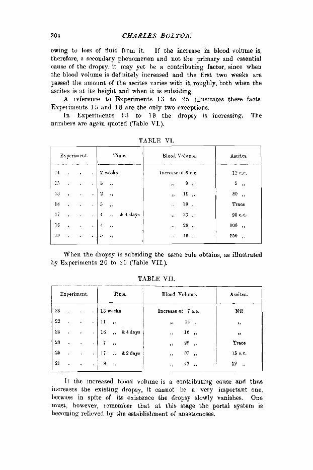
owing to loss of fluid froni it. If the increase in blood volume is, therefore, n secondary phenomenon and not the primary and essential cause of the dropsy. it may yet be a contributing factor, since when the blood volume is defiuitely increased and the first two weeks are passed the amount of the ascites varies with it, roughly, both when the ascites is a t its height and when it is subsiding.
.4 reference to Experiments 13 to 25 illustrates these facts. Experiments 15 and 18 are the only two exceptions.
In Experiments 1:; to 19 the dropsy is increasing. The niimIw~8 are again quoted (Tablc VI.).
TAIILE V T .
1 1 . . .
1.5 . . . 1 3 . . . 1 8 . . . 1 ; . . . 1 6 . . . I!, . . .
Tirile. --
2 weeks
3 .:
3 .:
6 :;
4 .: 8 4 days
-1 . .
.:
Blood ~ ( ~ l ~ l l l l ~ .
Increase of 6 C.C.
) ) 9 .!
:: 15 ~, .. 18 .. ,) 23 .: .. 29 .:
.. 4 c ..
Asci tes.
12 C.C.
5 I ,
80 ,, Trace
90 C.C.
100 ,, 150 ,,
Wheu the dropsy is subsiding the same rule obtains, as illustrated hy Esperirrients 20 to 2 5 (Table VII.).
2 3 . . . 2 2 . . . 2 4 . . .
2 0 . . .
2 6 . . .
2 1 . . .
TABLE VII. --__ __
Time. Blood Volume. Ascites.
I f the increased blood volume is a contributing cause and thus increiises the existing dropsy, it cannot be a very important one, bectrnsc: in spite of its existence the dropsy slowly vanishes. One must, howeve),, remember that a t this stage the portal system is becoming relicvecl by the establishment of anastomoses.

FURTHER ORSERVATZONS ON CARDIAC DROPS: 309
In the eitrly stages, we have seen that tlie abdoniinal vessels distend with blood and, since the volume of the blood is strictly limited, the other portions of the vascular system are depleted. This is particularly the case since the acculriulation of ascites by abstracting fluid from the abdominal vessels may actually diminish the total volume of the blooil. Absorptiou naturally occurs rapidly in the depleted areas, and the coiinective tissue in these regious becomes exceseively dry.
One has, therefore, absorptioii iu the depleted areas slid output from the ;ibdoniin,zl vessels. If the oue tdances the other the total volume of blood remains norinal, i f outpqt overbalances absorption the blood volume will diminish. Both these two conditions actually occur.
As the ascites collects in greater quantity, the flow of lymph through the thoracic duct increases, arrd inore and more lymph is returned to the venous system. The efforts of the aniiiial are thus directed towards increasing its blood volume, but the increase is retarded by the output of increasing ascites. The continual absorption in the depleted areas and the return of the ascitic fluid to the Idood, however, leads finally to an increiise of the blood volunie varying in degree and time of onset i n eiich case. This is rendered possible by the retention in the Lody of fluid, owing to deficient excretory action of the kidneys.
The reason the excess of fluid is not finally got rid of is that, as already stated, the capacity of the vascular system is increased. The condition here is not, therefore, analogous to tlie iLrtificiil1 yro- duction of plethora by injection of blood or saline solution into the normal animal.
6. Hmmoglobin Contetit of the Blood in t he Dropsku1 Aihnal.
During the past twenty years or more many observations, of which the following are the iws t important, have been made upon the condition of the blood in uncompensated heart disease, pirticularly with regard to its content of water and haenloglobin.
The methods employed were either that of weighing the dried residue of t h e blood, of measuring its specific gravity, or of ascertaining tlie percentages of hzmoglobin and red corpuscleh which it contained.
Hammenchlag (1892 11) found that in maiiy cases of cardiac iiisuficiency there was Q slightly higher percentage of water iii the blood than iiormal, but in some cases the concentration was normal.
Stintzing and Guxnprecht (1894 12) found hydrzmia in several cases, and also that as the mdema subsided and compensation was restored, the percent- age of water i n the hydrzmic blood diniinished ; also, conversely, that the blood became diluted a t the onset of dropsy. They considered that the blood volume waR increased.
Grawitz (1895 Is) explained the dilution of the I)lood, which he observed in uncompensated heart disease, 011 the suppositioii that the fall of arterial

306 CHARLES BOLTON;
pressure which occurred, and the consequent relaxation of the smallest vessels, caused a transudation of tissue fluid into them.
Askanazy (1897 14) drew from his observations the conclusions that the blood serum was always more or less diluted, and that in general the dilution varied in proportion to the degree of dropsy, that it increased as the dropsy increased, and diminished and finally disappeared when compensation was restored.
Lakin (1910 1 5 ) investigated twenty-five cases of cardiac edema. I n ten the haernoglobin percentagewas normai, in four it was above normal, Snd in eleven i t was below normal. H e also found in four of the above cases a temporary rise in the hsmoglobin percentage while the edema was subsiding, the percentage finally returning to normal
The hcamoglobin estimations to be now described explain how it is that the blood in uncompensated heart disease may be either of normd concentration or above or below this level.
On some day during the first week the percentage of hzmoglobin in the blood begins to decrease. This never occurs on the first day after the operation, because every case examined a t the end of twenty-four hours showed a normal or slightly increased percentage. It may commence on the second day, but only in about one out of six cases. Two-thirds of tbe cases show a commencing fall of the hsmoglobin percentage on the third day, and the remainder on the fourth, fifth, or sixth days. One case showed a normal percentage on the eighth day. With this last exception every case out of seventy- six estimations, conducted from the seventh day to the end of the fifth week, showed a decrease in the hzmoglobin percentage.
After this period, although the blood volume remains increased, the bwnoglobin percentage rises, becoming normal or slightly above this level in most of the cases during the next few weeks. Of five cases examined after three months, two showed decreases of 6 and S per cent. respectively, one was normal, and the remaining two were increased by 6 and 1 4 per cent. respectively. One hundred and seventy estimations in all were made on forty animals.
The average decreases were observed at the stages mentioned (Table VIII.).
TABLE VIII.
hlaxi 111 urn. Average Decrease in Hb Percentage. Time.
1 s t week . . . . 2nd ), . . . .
5th ), . . . . 7th ~, . . . .
31d ,, . . . . 4th ,, . . . . 6th ,, . . . .
73 16 ,6 19.3 1G.4 15.1 10.1 10.4
25 32 40 36 28 24 18
After this period oiily about four or six eatiniations a week were made, so t h e averages were not re1is;~le enough to quote.
FURTHER OBSERVATIONS ON CARDIAC DROPSY. 307
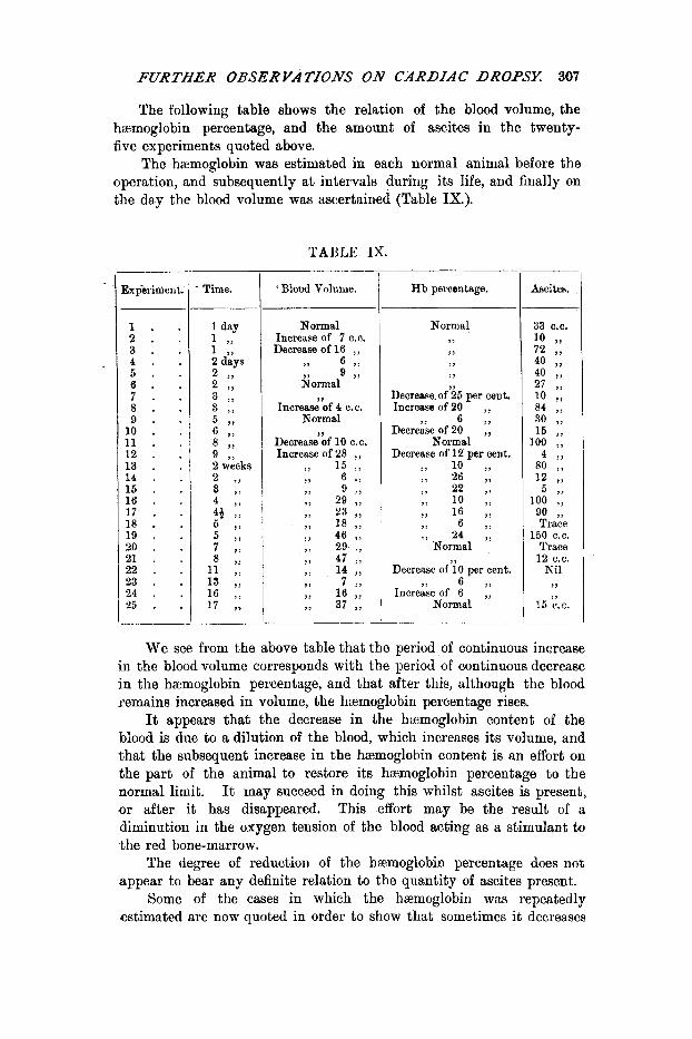
The following table shows the relation of the blood volume, the hcemoglobin percentage, and the amount of ascites in the twenty- five experiments quoted above.
The hemoglobin was estimated in each normal animal before the operation, and subsequently a t intervals during its life, and finally on the day the blood volume was ascertained (Table IX.).
TABLE IX.
Experiment.
1 . . 2 . . 3 . *
4 . . 5 . . 6 . . 7 . . 8 . . 9 . . 10 . . 11 . . 12 . . 13 . . 14 . . 15 . . 16 . . 17 . . 18 . . 19 . . 20 . . 21 . . 22 . . 23 . . 24 . . 25 . .
Time. c BloDd Volume.
Normal Increase of 7 C.C. Decrease of 16 ,,
,, 6 ,, 9 ,,
3ormal
Increase of 4 C.C. Normal
Decrease of 10 c. c. Increase of 28 ,,
9 ,
3 ,
7 , 15 ;,
H b percentage.
Normal > ,, , >,
Decrease.of 25 per cent. Increase of 20 ..
6 7, Decrerse of 20 ..
Normal Decrease of 12 per cent.
3 ,
Decrease of 10 per cent. 3 ) 6 ,,
Normal Increase of 6 ,,
Ascites.
150 C.C. Trace 12 C.C.
Nil ,,
15 e.c.
We see from the above table that the period of continuous increase in the blood volume corresponds with the period of continuous decrease in the hcemoglobin percentage, and that after this, although the blood remains increased in volume, the haemoglobin percentage rises.
It appears that the decrease in the hemoglobin content of the blood is due to a dilution of the blood, which increases its volume, and that the subsequent increase in the htemoglobin content is an effort on the part of the animal to restore its hcemoglobin percentage to the normal limit. It may succeed in doing this whilst ascites is present, or after it has disappeared. This effort may be the result of a diminution in the oxygen tension of the blood acting a8 a stimulant to the red bone-marrow.
The degree of reduction of the hzmoglobin percentage does not appear to bear any definite relation to the quantity of ascites present.
Some of the cases in which the haemoglobin was repeatedly estimated are now quoted in order to show that sometimes it decreases
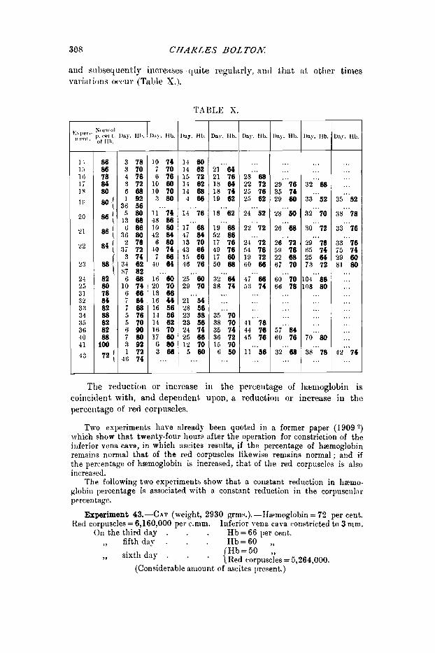
308 CHARLES UOLTOh':
aiiti siibsequeutly i11cre;tses cluite regulurlp, ant1 that at other times variation-; owur (Table X.).
I \ 1 .-, 1 ti 1; 18
1
20
21
22
23
21
31 32 33 3 'I 35 3(; .I 0 4 1
.I 3
') .- -.I
86 86
84 80
78
801
861 84 [
@-I
80 [
82 80 78 84 82 88 82 82 88
100 72 I
3 78 3 70 4 76 3 72 6 8 8 1 92
36 56 5 8 0
-1 3 68 ti 80
36 80 2 70
37 72 3 74
34 02 S i 82 6 8 8
10 74 6 0 6 i 0 4 7 6 8 5 70 5 70 6 9 0 7 8 0 3 92 1 72
4ti 74
10 74 7 70 6 76
10 80 10 70 3 8 0
11."74 48 80 10 80 42 84 6 8 0
10 74 7 8 8
40 64
lti"'60 20 70 13 66 16 4-3 16 56 14 56 14 62 16 70 17 80
hj. HI).
I I 00 14 02 15 72 14 02 14 68 4 0 0
14.. '76
li."68 4 i 84 13 70 43 60 15 66 16 76
2.i"'W 29 70
2 1 " ' M 28 50 23 58 23 58 24 74 25 06
G 8 0 1 1 2 10
3...ML.! 5...80
Da!. €I t )
21"'04 21 76 18 04 18 74 19 62
18'.'62
19-68 52 80 1 i 70 49 70 17 80 50 68
3'L"'M 38 74
...
... 35 ' ' '70 38 70 35 74 36 72 15 70 t i 5 0 ...
1)n.y. lib.
...
29"'68 22 72 25 76 2.5 62
24"'SZ
21"'72
24"'72 54 70 19 72 ti0 66
53 74 4;"'88
...
...
...
4 1 ' ' '78 44 76 45 70
11"'90 ...
)a?. Hh.
...
...
29' ' '70 35 74 29 60
28".54l
26"'68
26'"12 59 10 22 08 67 70
6O""IO 66 78
...
...
...
...
... 5 i 84 60 76
32'"68 ...
hy. IIb.
...
...
32'"68
33"'52
32"'70
so 72 ...
29-78 65 74 25 04 7 3 72
04"'88 08 80
...
...
...
...
...
i o " '80
38-76 ...
A1.Y. Ilb.
...
...
...
... 35-'.!32
88-78
33"'70
33' ' '70 75 74 29 60 81 80
...
...
...
...
...
...
...
...
...
... 42-74
...
The reduct,ion or increase i u the percentage of hltlmoglobin is coincident with, and dependent upon, a reduclioil or increase in the perceiitage of red corpuscles.
Two experiments have already been quoted in a former paper (1909 2) which show that twenty-four hours after the operation for constriction of the inferior vcna cava, in whieli ascites resnlta, if the percentage of haemoglobin remains nornial tha t of the red corpuscles likewise remtlins normal; and if the percentage of hsmoglobin is increased, that of the red corpuscles is also increasccl.
The following two experiments show that a coilstant reduction in harno- globin percentage is associated with a constant reduction in the corpuscul:ir percentage.
Experiment 43.-cAT (weight, 2930 grms.).-lhmoglobin = 72 per cent. Ked corpuscles = 6,160,000 per c.nim. Inferior vena c a m constricted to 3 nim.
(111 the third day . . H b = 6 6 per cent. ,, fifth day . . H b = 6 0 ,,
H b = 5 0 ,, 4 Red corpuscles = 5.264.000. ,, sist.11 day . (Considerable aiiiount of-ascitcs Iksent.) '

FURTHER OBSERVATIONS O N CARDIAC DROPSK 309
Experiment 8 0 . - c A T (weight, 3850 grms.).-Hb=86 per cent. Red corpuscles = 11,152,000. Inferior rena cava confitricted to 3 iiim.
On the fifth day . . f ib = 80 per ccnt. ,, eleventh day . . Hb=74 ,,
Hb=76 ,, Red corpuscles = 8,160,000 perc.mm. Hb = 62 per cent.
,, fourteenth day .
4 Red corpuscles = 5,280,000 perc.mm. ,, eighteenth day . 4
(Abdomen much swollen with a large amount of ascites.)
TLb = 50 per cent. ,, twenty-eighth day . Red corpuscles = 4,912,000 per
c.mm.
Finally, it was thought advisable to find out whether the hzmo- gloloin percentage was the same, whether the blood was drawn from the depleted or the congested area.
The following experiments show that the concentration of the blood is
Experiment 16.-CAr (weight, 2680 grms).-Hb (blood of ear) = 78 per
the same in all the vltscultlr arcas of the body.
cent. Inferior vena cavu constricted to 3 mni. H b (blood of ear) = 76 per cent.
' {IIh ( ,, foot)=76 ,, On the fourth day . ,, sixth day . . H b ( ,, abdominnl wall)= 78
per cent. (Considerable amount of fluid in abclomeii.)
Hb (blood of ear) = 76 per cent. ,, twenty-first day . I. per cent.
Experiment CAT (weight, 3800 grms.).-Hb (blood of ear) = 86 per cent. Inferior vena cttva constricted to 3 mrn.
H b (blood of ear) = 106 per cent. H b ( ,, abdominal wall)=106
I TIb (blood of liver) = 110 per cent. . On the second day . . per cent.
(Ascites 26 C.C. present.)
Erperiment 23.-Ca~ (weight, 4025 grms.).-Hb (blood of ear) = 88 per cent. Inferior vena cava constricted to 3 mni.
On the third day , . H b (blood of ear) = 74 per cent. 1 Ib ( 9 9 9 , >=66 ,, H b ( ,, abdominal wall)=64
per cent. ,, seventhday .
(Ascites present.)
Expeh8nt ASC CAT (weight, 3600 grms.).-Hb (blood of ear) = 100 1ier cent. Inferior vena cava constricted to 3 mm.
On the third day . . Hb (blood of ear) = 92 per cent. ,, sixth day . * H b ( ,, 9 , ) = S O 1,
,, twelfth (lay. . * H b ( 3, ,, )=TO 9,
9 , 9 , ) = 70 ,, * {E ,, liver) = 74 per cent. ,, fifteenth day .
(Considerable amount of ascites present.) 21-JL. OF PAT11.-VOL. TI.

310 CBARLES BOLTON.
7. Vascdar Pressures of the DropskaI Animal.
When my former experiments, in which it was shown that the preliminary fall of arterial pressure wafi recovered from in the animal, were performed, there were no clinical observations to show what the height of the arterial pressure was in cardiac dropsy in the human subject.
When the present instrument for registering the hunian blood pressure came into use, it was soon found that the arterial pressure was normal in cardiac dropsy, thus establishing in the case of the human subject the fact which I had shown experimentally. The first series of observations directly bearing on this point was published by H. J. Starling (1906 16). He showed that in cardiac dropsy the arterial pressure was normal, and that only just before death occurred did a fall of pressure take place. In another group of cases he found the arterial pressure above normal, the latter group containing cases of arterial and kidney disease.
The vascular pressures were measured in thirty-four animals. The pressure at the lower end of the inferior vena cava was above normal in all the cases except four. The pressure was on the whole higher after the first week, when the volume of the blood was increased. I have already shown that the venous pressure after the preliminary fall rose again to the height it stood a t immediately after the inferior vena cava was constricted. This rise is due to vasomotor constriction, and corresponds with a similar rise in the arterial pressure.
When the blood increases in volume, the venous pressure rises higher still, owing to the excessive filling of the veins.
The height of the venous pressure, therefore, in the first instance, depends upon the degree of vasomotor tone, and it is added to by the subsequent increase in volume of the blood. Although the venous pressure is on the whole highest when the blood is increased in volume, there is great variation in the relation between the actual pressure and the degree of increase of the blood volume in individual cases.
The arterial pressure also shows in some cases higher readings when the blood is increased in volume, but in all cases the pressure is within normal limits.
The height of the venous pressure bears no relation to the amount of ascites present. Large amounts of ascites may occur with normal or moderately raised venous pressures, and high pressures are recorded after the ascites has disappeared.
Table XI. shows the blood volumes and amounts of ascitic fluid in the experiments already quoted in relation to the venous and arterial pressures.
The last three cases, together with Experiments 4, 11, 2 2 , 23, and 24, illustrate remarkably well that the venous pressure and the dropsy are quite independent of each other.

FURTHER OBSERVATIONS O N CARDZAC DROPSE: 31 1
...
...
...
__ xperi. nent. __
1 2 3 4 6 i 8 10 11 12 13 14 15 I6 17 18 21 22 23 24 25 -- -
35 C.C. 20 N i i '
TABLE XI.
Time. Inferior Cdva Pressure.
Carotid Pressure. Blood Volume.
Normal Increase of 7 C.C. Decrease ot 16 C.C.
I,
Decrease of 4 c.c. Normal
Decrease of 10 C.C. Increase of 28 ,,
>, 15 , I
1 , 6 > > >, 9 > I
,> 29 ,> ,, 23 7 ,
, i 18 ,, i, 47 ,,
k i t e s .
Ln the following experiments the blood volume was not taken :-
26 27 28
1 day 2 days
11 weeks
In Experiments 4 , 1 1 , 2 6 , and 27 the venous pressures are normal, and yet considerable amounts of ascites are present; in Experiments 22, 23, 24, and 28 the venous pressures are high, and yet the ascites which had been present in the early stages of the disease had disappeared.
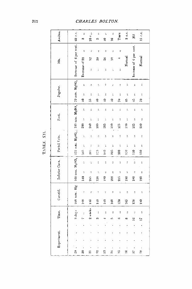
I n Experiments 29-38 (Table XU.) the venous pressures were taken in all parts of the body a t various stages of the disease. It will be seen from this table that the pressure in the portal vein follows the pressure in the inferior vena eava, and is raised to about the same proportionate extent. In some cases it appears to be raised to a rather less extent, but one must remember that, although the portal pressure is in the normal animal usually a little higher than the inferior cava pressure, this is not always the case. There is no correspondence between the pressure in the portal vein and the amount of ascitic fluid.
The venous pressures in the feet and head present the same interesting features which I pointed out in a former paper, dealing with the pressures during the first twenty-four hours after the obstruction was produced.
I n all these cases, whatever the stage of the disease, the venous pressure in the hind-feet is raised, but it is raised to a less relative extent than is the pressure in the inferior vena cava. The jugular
313 CHARLES ROLTON
N c 0 W '3 CI cc
c F
, .. " - ._ I . ., .. .. .. .... -

FURTHER OBSERVATIONS O N CARDIAC DROPSK 313
venous pressure is in all cases below the normal, as it was immediately after the inferior vena cava was obstructed. Table XIII. illustrates these points, and it will be noted that Experiment 30 is the only one in which the pressure in the feet was increased by more than that in the inferior vena cava.
T,iBLE XIII. ~ -- Experi nient. __ 29
30
31
32
33
34
35
36
37
35
Jugular Vein.
Decrease of 9 mm. MgSO,
I , 39 9 3 ,, ,, 24 ,, >, I , 39 ,, 3 3
, I 39 9, I ,
,, 39 ,, ,, ?, 9 I , I ,
9 , 14 >, ,, ,, 34 , I > >
, I 9 9 9 2 ,
There is, therefore, an alteration in the pressure levels, the venous pressure in the trunk being relatively more raised than that in the hind-limbs. The venous pressure in the head is always below what it was before the constriction was applied. These alterations in the pressure relations, which are precisely what they were before the increase in blood volume commenced, indicate an abstraction of blood from the periphery and its accumulation in the central portion of the circulatory system. The increase in the blood volume is unable to restore the normal pressure relations by increasing the amount of blood in the depleted areas, because that of the congested area becomes further increased also. The primary cause of the increased blood volume is to be found in the efforts of the organism to restore the normal amount of blood to the depleted areas, a task which it appears to be unable to accomplish.
8. The Intake of Fluid and the Xxcrction of Urine in the Dmpsical Animal.
The animals were fed on cat’s meat and milk. A certain amount of milk was given twice a day, the leavings on each occasion being measured. The urine passed was collected in the usual manner.

314 CHARLES BOLTON:
The daily amounts of milk consumed and of urine passed were found to vary very considerably in the normal cat. On some days the volume of milk consumed was greater than that of urine passed, and vice vevsa. When the weekly totals were compared it, appeared that on the whole the amount of inilk consumed was greater than the amount of urine passed. This is, of course, a very rough test, because the animal loses water in other ways, and milk contains solid matter.
The same irregularity in consumption and excretion was found to occur in the dropsical cats, but on the whole fairly definite results with regard to retention or loss of fluid from the body were obtained.
When the weekly totals of milk and urine were compared it was found that in sixteen out of twenty-two cases (about 7 3 per cent.) there was definite evidence of retention of fluid in the body during the first two to four weeks. After the third or fourth week there was in sonie a loss of fluid, but in ail equal number there was a mixture of gains and losses, or even a gain during the whole life of the animal, but this was only in the minority.
There is no doubt, however, that in the majority of the cases there was retention of fluid in the body during the period of edema formation and when the blood was becoming dilute and increasing in volume. It would be tedious and unnecessary to give all the details relating to the cases, but one or two examples may be quoted (Table XIV.).
TABLE XIV. - - - - .
I
I Experiment 24. - -~ - I
Asri trb. I
Time. Hb percentage Retention or Loss I Blood olul,,e, (Normal=Y?). 1 ot Flntd. I- I _ _ _ - __ .
l h t neek . .
3rd ,, . . 4 th ,, . . 6th ,, . . 7th ,, . . 8th ,, . . 9th ,, . .
10th ,, . . 11th ,, . . 12th ,, . . 13th ,, . . 14th ,, . . 15th ,, . .
-LXlIl ,) . .
5th ,) . .
16th ), . .
6 8
60 60 64
66
70
...
. . .
...
...
. . .
...
...
...
. . . 88
l i r teiition
Lb’ss
Reten tion Loss
Retell tion
...
. .
...
...
...
...
...
...
...
...
...
...
...
... Increase of 16 C.C.
... Present
...
...
...
...
...
. . .
...
...
...
...
...
... Nil

FURTHBR OBSERVATIONS O N CARDIAC DKOPSY. 315
... 76 I Ret&tion 50 1 Loss
TABLE S IV-continued. - ~~ - ~-
Experiment 35. I I
...
...
...
I i Time. Hb percentage Retention or Loss (Normal= $2). I of Fluid.
' 1st week. . 70 Retention 2nd ,, . . 62 ~
3rd ,, . . ~ 56 Lyss 4th ,, . . . . I > 3
1 5th ,, . . 70 I Retention 7 1 Loss - 1 I 6th > > *
I
Blood Volume.
...
...
...
... ...
...
Ascites.
Fair amount
Slight
I i Experiment 36. -- -
Hb percentage I 1 (Noriiia1=82) 1 I
1 I s t n e e k . . 2nd ,, . . 3rd ,, . .
, 4th ,, . . 5th ) ) . . 6th ,, . . 7th ., . .
I 8th ,, . .
I j ~-
I - -
1st week. . 2nd ,, . . 3rd ., . . 4th ,, . . 5th ), . . 6th ,, . . 7th ,, . . 8th ., . . 9th ,, . . 1 lot11 .) . .
Retention
74 74
...
...
...
...
...
...
...
... ~~~~
Experiment 40.
Hb percentage (Normal=8Y). - 1 -
80 I Retention ... ,> I ... I 60
Lobs f 6 , ... , I
...
...
...
...
...
...
... ~ Large amount
...
...
...
...
... 3 C.C.
1
... Large amount ...
...
...
...
...
... 2 C.C.
1st week. . 2nd ,, . . 3rd ~, . . 4th ,, . . 5th ~, . .
Experiment 19. ~ ~
Hbpercentage
Retention
60 1 Retention
... 62 L A
52 I ,>
...
...
...
...
...
... ... ...
Increase of 46 C.C. 1 45 C.C.
I I I I
Experiment 17.
i 1st week. .
~ 3rd ,, . . I 4th ,, . . I 46 ,f . *
Hb percentage (Normal-84).
Retention 60 64 1 76 ~
,, ,,
ti8 I ,,
...
... I ... ...
... Increase of 23 C.C. 90 C.C.
316 CHARLES BOLTON;
TABLE XIV.-continued. - - __ _ _ _ ~ . -
In the niinority of cases (six out of twenty-two) the tigures showed a loss of flnid from the body during the first week or two, as shown by the following :-
-~
Time.
1st week. . 2nd ,, . . 3rd ,, . . 4th ,, . . 5th ,, . .
1st week. . 2nd ,, . . 3rd ,, . . 5th ,, . . 4th ,, . .
Experiment 34. _-__ - _. -
B,ood Volullle.
~
76 56
58 70
... Loss
Retention
Loss ,
...
...
...
...
... . .............
Experiment 18.
...
... Large amount
10 C.C. ...
.~ -
68 Loss 68 1 Retelltion
...
...
...
... Increase of 18 C.C.
...
...
...
... Trace
Taking into account all sources of error, these observations fairly well support the view that there is retention of fluid in the body during the period of edema formation and increase of water in the blood. The figures do not so well show a loss of fluid during the disappearance of the ascites.
The amount of urine passed a t the different stages of the diseased condition was very similar in all the cases, which agree in showing a definite decrease in the amount passed during the first few days. An increase then occurs, and the excretion becomes normal, with a tendency towards a further increase as the dropsy is disappearing. The amount of urine passed is definitely related to the amount of fluid consumed, and undoubtedly chiefly depends upon this factor.
All the cases agree in this particular, so that it will be necessary to quote the figures of one case only in illustration (Table XV.).
The animals never drink much for the first few days, and therefore pass a diminished amount of urine, which is further diminished by retention of water in the body. Although the excretory power of the kidneys is somewhat lessened by the passive congestion, they act well in response to an increase in the available amount of fluid presented to them to excrete.

FURTHBR OBSERVATIONS ON CARDIAC DROPSY. 317
Time.
- -
1st day 2nd ,, 3rd ,, 4th ,, 5th ,, 6th ,, 7th ,, 8th ,) 9th ,>
10th ,, 11th ), 12th ,, 13th ,, 14th ,, 15th ,, 16th ,, 17th ,, 18th ,, 19th ,, 20th ,, -_
Urine.
TABLE SV. Experiment 35.
...
...
...
... i 0 ... ... ... ... ... ... ... ... 62 ... ... ... ... ... ... -
‘ 21st day I 22nd ,,
23rd ,, 24th ,, 25th ,, 26th ,, ‘Zit11 ,, 28th ,, 29th ,,
’ 30th ,) Ylst ,) 1 321~1 ,,
I 33rd ,, 34th ,, 1 35th ,, 36th ,, 37th ,, 38th .,
1 40th ) )
I 41st ,)
Urine. Milk. 1 Hb percentage ,(Normal = 82).
120 130 ,, ” I ... ” ’
140 ,, I 56 130 ,, ...
... ...
...
...
...
...
...
...
...
...
...
...
... 70 ... ...
9. The Occurrence of Jaz~?adice.
Jaundice occurred in three of the animals only, and was therefore due, probably, to some accidental cause. It occurred during the first week in each case. The tissues were generally stained, and also the ascitic fluid. The gall-bladder was perhaps a little enlarged, but bile could be squeezed out of the papilla readily, and the h c e s were not light in colour. In one of the cases there was “coffee grounds” material in the stomach and small intestine, which gave the guaiacuni and turpentine test for blood.
The only explanation I can offer is, that the condition was one of toxic jaundice resulting from a low grade infection. The animals did not appear to be in any way different from the others, neither did their livers show any difference on microscopical examination. The jaundice which sometimes occurs in uncompensated heart disease may have a similar origin.
1 0. Smamary and Concltuions.
In considering the results of these experiments, in conjunction with those which were announced a t the beginning of this communication, we see that the increased output of lymph into the peritoneum, giving rise to ascites, is merely a link in a very definite chain of events following obstruction of the inferior vena cam or the occurrence of heart failure.

318 CHARLES BOLTON.
The primary cause of this sequence of changes in the circulation is to be found in the fact that the portal area, including the liver, is so large and distensible that it is able to abstract a sufficient quantity of blood froni the general circulation so as to produce a definite depletion of the latter. The above area is the only one in the body which can produce this result. The superior vena cava niay be completely ligatured immediately above the right auricle or the inferior vena cava below the diaphragm, and yet there will be no alteration in the height of the mean blood pressure curve. When, however, the inferior vena cnva above the diaphragm is narrowed down to three-fifths of its normal diameter, an immediate fall in the mean arterial blood pressure curve results, and if the vein is completely occluded that curve promptly falls to within a few millinietres of the zero line.
A sudden narrowing of the vein or constriction of the pericardium allows an appreciable interval to elapse before many of the results have time to manifest themselves, so that we are able to study the sequence of events. In the case of a gradual development of heart failure, however, the circulatory changes are crowded together, so that if we were able to study our clinical cases effectively we could not unravel the series of phenomena so as to determine the various mechanisms a t work.
When the inferior vena cava i s experimentally obstructed aboze the diaphragm the following sequence of events O C C Z G ~ :-
Stage I. Accumulation of blood in-( 1) Abdominal organs : ( 2 ) hinder
Rise of venous pressure to smallest venules in these parts. Capillary pressure in these parts also raised, especiallv in the
Abstraction of blood from head, chest, and fore-limbs. Fall of venous and capillary pressure in these parts. Fall of general arterial blood pressure.
part of body and hind-limbs.
liver.
The accumulation of blood behind the obstruction is the cause of the rise of venous pressure, which in its turn, by increasing the velocity of the blood through the obstruction, enables the maximum amount of blood t o reach the heart. The venous pressure, however, does not rise till a considerable accumulation of bloocl has occurred, since the veins and capillaries are not filled to distension ; it is necessary to constrict the vein to three-fifths of its diameter before a rise of pressure occurs a t all. The rise of venous pressure extends right through the liver to the portal vein, and thence to the fine portal radicles it also extends to the fine venous radicles of the foot. The capillary pressure in all the parts drained by the inferior vena cava rises. In a former paper I found that this rise was from about 4 to S mm. Hg, so that, if the
FURTHER OBSERVATIONS ON CARDIAC DROPSY 319
normal capillary pressure were 30 mni. Hg, after obstruction of the vein the pressure would stand at 34 to 38 mm. Hg. We must, how- ever, remember that the height of the capillary pressure depends not only upon the rate a t which blood leaves the capillaries, but also upon the rate a t which blood is supplied to them. If the arterial pressure remained normal and .these vessels contained the normal arnount of blood, the capillary pressure after obstruction would stand at the height indicated above, but blood is abstracted from the arteries and the pressure in them falls. Less blood than normal is, therefore, supplied to the capillaries, so that for this reason the pressure within them is not raised to the level indicated by the height of the venous pressure.
Exactly what lessening of eflect is produced Ly the fall of arterial pressure it is impossible to say; we can only conclude that the capillary pressure is raised, but to a much smaller extent than one would expect from the height of the venous pressure. Another point to be taken into consideration is, that in the normal condition many capillaries are more or less empty, and that these capillaries have to be filled out with blood after the obstruction is applied before any rise of capillary pressure could occur. I believe this is one of the reasons why the venous pressure does not rise until the vein is narrowed to a considerable extent. The pressure in the liver capillaries is raised to a greater extent because the pressures in both the hepatic vein and the portal vein are raised.
The abstraction of blood from the head, chest, and upper limbs, together with the fall of general arterial pressure, naturally results in a fall of venous and capillary pressure t o these parts.
stage 11.
Dilatation of-( 1) Veins and capillaries of abdoniinal organs and particularly of the liver : (2) inferior vena cava and veins opening directly into it : also of capillaries of retroperitoneal tissue arid surrounding parts.
Fall of venous pressure to normal or thereabouts. Fall of capillary pressure below normal in these parts except
Hind-limbs and posterior part of body wall now contain less
Fall of capillary pressure below normal in these parts. Absorption of lymph in depleted areas of body. Increased output of lymph in plethoric areas leading to-
(1) ascites ; (2) cedema of retroperitoneal tissue.
The distensibility of the abdominal vessels is responsible for a further depletion of the forepart of the body, and the hind-limbs now share in this depletion. Owing to this increased capacity of a part of
in liver when it is normal.
blood than normal, like forepart of the body.

320 CHARLES BOLTON.
the venous system, the pressures in all the parts of the vascular system naturally fall, the blood volume reniaining normal.
The venous pressures in all parts behind the obstruction being normal and the arterial presswe low, it naturally follows that the capillary pressure is subnormal in all parts except the liver, where it is normal. The capillary pressure in the forepart of the body falls lower than i t was before.
The increased output of lymph in the plethoric areas giving rise to ascites usually exactly balances the absorption of lymph in the depleted areas, so that the Mood is neither diluted nor increased in voluine. Occasionally, however, absorption may fall behind and the blood become somewhat concentrated. The increased absorption of lymph is shown by the excessive dryness of the cellular tissue in all the depleted areas.
It is to be noted that the lumbar veins which open directly into the inferior vena cava are dilated and plethoric, and cedema occurs by increased output of lymph from their capillaries (retroperitoneal tissue), whereas the anterior and posterior branches of these veins (abdominal wall) and the veins of the hind-limbs, owing to their much greater distance from the seat of obstruction, actually contain less blood than iiormal, and increased absorption of lymph takes place into their capillaries.
stage 111.
Vasomotor constriction leading to-( 1) Rise of arterial premire
(2) Rise of venous pressure in the central portions of the
(3) IZise of capillary pressure in these parts to normal or above. (4) Venous pressure in the hind-limbs remains a t the normal level. (5) Capillary pressure in these parts remains subnormal.
to the normal.
trunk to its former high level.
The vasomotor constriction automatically occurs by stimulation of the vasomotor centre owing t o deficient blood supply to the brain. If it did not occur it would be impossible for the high venous pressure in the trunk to be maintained, since the vascular system contains its normal amount of blood only. The high venous pressure is necessary so as to increase the velocity of blood through the constriction, in order that the maxiniuin amount possible may reach the heart. Neither would i t be possible without vasomotor constriction to ensure the masimuni supply of blood to the brain.
These alterations in venous and arterial pressures in their turn affect the capillary pressure. The rise of venous pressure results in a rise of capillary pressure in the trunk, but this rise is minimised by the arterial constriction which cuts down the supply of blood to the Capillaries, so that it is doubtful if the capillary pressure here rises

FUKTHEK OBSERVATIONS O N CARDIAC DROPSJ: 521
above its normal level. The veiious pressure in the legs remaining at the nornial level, the capillary pressure in these parts is subuormal.
The venous pressure levels have, therefore, an altered relation, the pressure in the legs being lower relatively to that in the trunk t h m iu the normal animal.
These alterations in lireswre make no difl'erence to the iibsorption aud output of lymph ant1 to the accumulation of dropsical fluid. They occur at variouj timcs according to the power of recovery of tlie aiiimal, and are usually completed within the first twenty-four Iionrs following the atp1)lication of the constriction.
Stage IV. Continuatiou of lymph absorption in depleted :treiis. Continuatiou of lymph output in ldetlioric areiis. Increasing flow of lymph in thoracic duct i is ilscites accuninltttes. Diminished excretion of urine. Increased amount of water in the blood arid fall iii 1)erccntage
of hEmoglobin. Condition of hydrmiic plethora established. Venous arid capillary pressures raised in plethoric area. Venous m d ca1dl;try pressures riiised in hind-limbs, but to ;I
less relative extent than in the trunk. Venous and capillary pressures Iielow noriii:il in the head and
fore1)tti.t of tlie trunk.
This state of affairs is not recognisable in niost cases till the end of the tirst week. It increases and coiitinues till about the end of the third or fifth week, when it gives place to the next stage. Tlie hydrremic plethora varies very miich in auiount and is i~ purely secondary process, depending upon-( 1) Contiuual absorption of lymph from the depleted areas : (2) when the ascites has reached a certain degree, the rapid return to the blood of iiiost of the ascitic fluid which is produced, so that the ascites orily slowly accumulates; and ( 3 ) retention of a certain amount of tluid in the body owing to diminished excretory activity of the kidneys. It is to be noted that although hydrsmic plethora exists, the distribution of the blood in the vascular system is still uneven, the plethoric areas being more markedly plethoric, the hind-limbs coutaiuing a little more than their normal amourit of blood, and the head and forepart of the body still beiiig depleted, as shown by their low venous pressures. The animal is thus seen to be endeavouring to make up the deficiency of blood in its depleted area8, and it does not succeed in doing this except in the hind-limbs, but it produces an increasing plethora of its plethoric areas. The latter are relieved rapidly by the production of ascites, and the ascitic fluid is qnickly returned to the blood, so h a t a constant and rapid circulation of lymph goes on, the ascites gradually increasing as output overbalances absorption.

322 CHARLES B O L T O N
This state of things would undoubtedly go from bad to worse were it not for the gradual establishment of anastomoses. These are rapidly formed in the abdominal wall and gradually increase in size; the anastomoses of the portal circulation are, however, much slower in forming. The height of this morbid condition is perhaps at the fourth or fifth week, when the portal anastomoses begin to effect a reduction in the processes described. The output into the peritoneum becomes less and less rapid, the lymph flow in the thoracic duct also falling as a result, but not quite so rapidly, so that the ascites gradually disappears.
Although a secondary process, the hydrzmic plethora has two results. I t (1) accelerates the output of lymph into the peritoneum, and (2) alters the venous and capillary pressures as described.
Stage V.
The ascites gradually disappears. The hydrzmic plethora becomes a true plethora, the percentage
d true plethora thus becomes established probably because there is an active formation of blood corpuscles owing to stimulation of the red bone-marrow. The reason the blood volume remains high is probably because the total capacity of the vascular system is increased owing to the dilatation of the vessels of the liver, and large veins of the trunk, and the presence of large anastomotic veins. The venous pressures are kept up by the plethora and the altered circulatory conditions.
We thus see that neither plethora nor a high capillary pressure are the primary causes of the increased output of lymph giving rise to the dropsy, because neither is necessary for its production.
The increased venous pressure in the trunk leads directly to enlargement of the liver and dilatation of the other veins, and thus indirectly to abstraction of blood from other parts of the body and increased absorption of lymph in these parts, but the chief r61e played by the various alterations in the vascular pressures, arterial and venous, is that of ensuring as ample a circulation of blood through the brain and other orgaus as is possible under the existing circulatory conditions.
The object of this investigation has been to explain what gross pathological processes are a t work in an uncompensated condition of the circulation and the relation of these processes to each other. I do not, therefore, propose to enter into any theoretical considerations with regard to the ultimate factors which are responsible for causing increased absorption of lymph in the depleted areas, and increased output of lymph in the congested areas. One cannot with profit discuss these obscure factors until all the more gross facts relating to
of hmnoglobin gradually returning to the normal.

FURT'EK OBSZR VATIONS ON CARDIAC DROPSY. 323
the circulatory alterations are found out. These experiments, however, plainly show that purely physical factors, and particularly those which in the past have been held to be direct causes of the dropsy, are not primarily responsible for its presence.
The essential condition for the production of this form of dropsy appears to be one in which an amount of venous blood, above the normal volume, is flowing with a diminished velocity through dilated capillaries a t a pressure either normal in height or raised or perhaps even subnormal. In these circunistances there is naturally a diminished absorption of lymph by the capillaries, but this is no doubt merely a subsidiary factor in the production of the dropsy. We know that the circulatory system contains a definite amount of fluid and that any excess above this amount is rapidly got rid of by the secretory and excretory glands and by the capillary walls, the lymph flow in the thoracic duct being greatly exaggerated. What is true of the whole is true of a part, and if any excess of blood in a territory of the vascular system is not rapidly carried off, owing to the increased velocity of the blood in the veins, it is got rid of by the capillary wall. Simple vaso-dilatation in a part increases, enormously, the velocity of flow of blood in the veins, but does not increase the lymph flow, unless an increased functional activity of the part requires more lymph. In the condition we are studying, the excessive amount of blood is retained in the part owing to the diminished velocity of the venous stream, and, moreover, i t tends gradually to increase in amount, and the plethoric area becomes more plethoric.
Whether the capillary wall actively gets rid of the excessive amount of fluid, or whether the latter leaks out owing to increased permeability of the capillary wall, caused by injury by metabolic products, must remain a subject for speculation. It is significant that the ascites disappears owing to the output of lymph becoming gradually less and less as the surplus of blood is gradually diverted by the growing anastomoses. It appears as if the diminishing output of lymph was conditioned rather by the increasing ease with which the surplus of blood in the part is carried off, than by ablessening degree of damage to or a slow recovery of the capillary wall. The rate of output of lymph in the congested area varies during the course of the disease, and it is difficult to explain this occurrence by the supposition that corresponding variations occur in the degree of damage done to the capillary wall.
I n the condition of uncompensated heart disease the following sequence of events occurs :-
stage I.
Accumulation of blood in veins. Rise of venous pressure to smallest vessels in all parts.

324 CHARLES BOLTON;
Capillary pressure, raised particularly in the liver. Abstraction of blood from arterial system. Fall of arterial blood pressure.
Stage 11.
Dilatation of-(1) Veins of liver and abdominal organs ; (2) large veins of chest and abdomen and their imme- diate tributaries.
Fall of venous pressure to normal or thereabouts. Capillary pressure in all parts below normal except in the
liver where it is normal. Veins of the trunk relieved of their accumulated blood to
some extent a t the expense of those of the legs. Abstraction of blood from head, arms, and upper part of the
trunk (except centraI portion of chest). Absorption of lymph in the depleted areas of the body. Increased output of lymph in the plethoric areas leading to-
(1) Ascites, which is usually not very evident till the cedenia of the legs is marked ; (2) cedema of legs.
Stage 111.
Vasomotor constriction leading to-( 1) Rise of arterial
(2) Rise of venous pressure in central portions of the trunk
(3) Capillary pressure in these parts returns to the noimal or
(4) Venous pressure in upper part of body (except centre
(5) Capillary pressure in these parts subnormal.
pressure to the normal.
and the legs to its former high level.
may be raised ; it is certainly raised in the liver.
part of chest) does not rise.
Stage IV. Continuation of lymph absorption in depleted areas. Continuation of lymph output in plethoric areas. Increased flow of lymph in the thoracic duct as ascites and
dropsy of legs accumulate ; the outflow into the veins is somewhat obstructed by the raised venous pressure in the large veins a t upper part of chest.
Diminished excretion of urine. Increased amount of water in the blood and fall in percentage
of bcemoglobin. Condition of hydremic plethora established. Venous and capillary pressures further raised in the plethoric
areas. ~
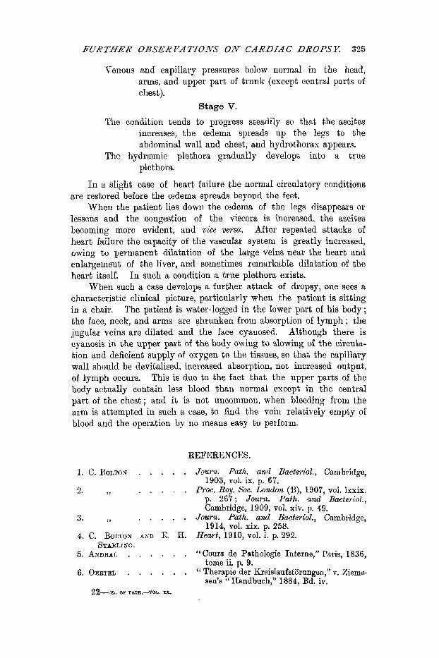
flURTHER OBSZR VATIONS O N CARDIAC DROPSE: 325
Venous and capillary pressures below normal in the head, arms, and upper part of trunk (except central parts of chest).
Stage V.
The condition tends to progress steadily so that the ascites increases, the Edema spreads up the legs to the abdominal wall and chest, and hydrothorax appears.
The hydraemic plethora gradually develops into a true plethora.
I n a slight case of heart failure the normal circulatory conditions are restored before the cedema spreads beyond the feet.
When the patient lies down the edema of the legs disappears or lessens and the congestion of the viscera is increased, the ascites becoming more evident, and vice versa. After repeated attacks of heart failure the capacity of the vascular system is greatly increased, owing to permanent dilatation of the large veins near the heart and enlargenient of the liver, and sometinies remarkable dilatation of the heart itself. In such a condition a true plethora exists.
When such 8 case develops a further attack of dropsy, one sees a characteristic clinical picture, particularly when the patient is sitting in a chair. The patient is water-logged in the lower part of his body ; the face, neck, and arms are shrunken from absorption of lymph ; the jugular veins are dilated and the face cyanoEied. Although there is cyanosis in the upper part of the body owing to slowing of the circula- tion and deficient supply of oxygen to the tissues, so that the capillary wall should be devitalised, increased absorption, not increased output, of lymph occurs. This is due to the fact that the upper parts of the body actually contain less blood than normal except in the central part of the chest; and i t is not uncommon, when bleeding from the arm is attempted in such a case, to find the vein relatively empty of blood and the operation by no means easy to perform.
REFERENCES.
1. C. BOLTON . . . . .
2. . . . . . 3,
5. . . . . . 9 ,
4. C. BOLTON ASD E. H.
5. ANDRAL . . . . . . STARLING.
6. OERTEL . . . . . .
Journ. Path. and Bacteriol., Cambridge, 1903, vol. ix. p. 67.
Proc. Roy. Soc. London (B), 1907, vol. lxxix. p. 267 ; Journ. Path. and Baeteriol., Cambridge, 1909, vol. xiv. p. 49.
Journ. Path. and Baeteriol., Cambridge, 1914, vol. xix. p. 258.
Heart, 1910, vol. i. p. 292.
‘‘ Cours de Pathologie Interne,’’ Paris, 1836,
“ Therapie der Kreislaufstorungen,” v. Ziems- tome ii. p. 9.
sen’s “Handbuch,” 1884, Bd. iv.

326 FURTBER OBSERVATIONS O N CARDIAC DROPSI.:
7.
8. 9.
10.
11.
12.
13. 14. 15.
16.
COHNHEIM . , . . . . COHNHEIM AND LICHTHEIM E. H. STARLING. . . .
LORRAIN SMITH . . . HAMMERSCHLAG . . . . STINTZING AND G~~MPRECHT
GRAWITZ . . . : . . ASKANAZY . . . . . LAKIN . . . . . . . H. J. STARLING . . . .
‘ I Vorlesungen ueber allg. Pathologie,” Berlin,
Viirchow’s Archiv, 1877, Bd. lxix. S. 106. ‘‘ Arris and Gale Lectures,” Lancet, London,
1896, vol. i. pp. 1267-1407 and 1897, vol. i. pp. 569-723; also “The Fluids of the Body,” 1909, pp. 150 and 172.
Tram. Path. SOC. Lodon, 1902, vol. 1%.
1882, Aufl. 2, Bd. i.
p. 136. Ztschr. f. klin. Med., Berlin, 1892, Ed. xxi.
s. 475. Deutsches Arch. f. klin. Metl., 1894, Bd. liii.
Ibid., 1895, Bd. liv. S. 588. Ibid., 1897, Bd. lix. S. 385. Med. Chron., Manchester, 1910, Ser. IV. vol.
Lancet, London, 1906, vol. ii. p. 846.
S. 265.
xx. p. 5.