Fungi between extremotolerance and opportunistic ... · F between extremotolerance and...
19
Fungi between extremotolerance and opportunistic pathogenicity on humans Cene Gostinc ˇar 1,2 • Janja Zajc 1,3 • Metka Lenassi 4 • Ana Plemenitas ˇ 4 • Sybren de Hoog 5,6 • Abdullah M. S. Al-Hatmi 5,6,7 • Nina Gunde-Cimerman 1 Received: 1 June 2018 / Accepted: 22 October 2018 / Published online: 9 November 2018 Ó The Author(s) 2018 Abstract Numerous agents of infections in humans and other mammals are found among fungi that are able to survive extreme environmental conditions and to quickly adapt to novel habitats. Nevertheless, the relationship between opportunistic potential and polyextremotolerance was not yet studied systematically in fungi. Here, the link between polyextremotol- erance and opportunistic pathogenicity is shown in a kingdom-wide phylogenetic analysis as a statistically significant co- occurrence of extremotolerance (e.g. osmotolerance and psychrotolerance) and opportunism at the level of fungal orders. In addition to extremotolerance, fungal opportunists share another characteristic—an apparent lack of specialised virulence traits. This is illustrated by a comparative genomic analysis of 20 dothideomycetous and eurotiomycetous black fungi. While the genomes of specialised fungal plant pathogens were significantly enriched in known virulence-associated genes that encode secreted proteases, carbohydrate active enzyme families, polyketide synthases, and non-ribosomal peptide synthetases, no such signatures were observed in human opportunists. Together the presented results have several implications. If infection of human hosts is a side effect of fungal stress tolerance and adaptability, the human body is most likely neither the preferred habitat of such species, nor important for their evolutionary success. This defines opportunism as opposed to pathogenicity, where infection is advantageous for the species’ fitness. Since opportunists are generally incapable of the host-to-host transmission, any host-specific adaptations are likely to be lost with the resolution of the infection, explaining the observed lack of specialised virulence traits. In this scenario opportunistic infections should be seen as an evolutionary dead end and unlikely to lead to true pathogenicity. Keywords Virulence factor Á Extremotolerance Á Stress tolerance Á Mycosis Á Opportunistic infection Á Black yeast Introduction Fungi form an integral part of biodiversity of many extreme environments. Here, the maintenance of costly stress tolerance and adaptability mechanisms pays off in decreased competition with other microbes. Two modes of adaptation can be recognised in fungi. On the one hand Cene Gostinc ˇar, Janja Zajc and Metka Lenassi contributed equally as first authors. Electronic supplementary material The online version of this article (https://doi.org/10.1007/s13225-018-0414-8) contains supplementary material, which is available to authorized users. & Cene Gostinc ˇar [email protected] 1 Department of Biology, Biotechnical Faculty, University of Ljubljana, Jamnikarjeva 101, 1000 Ljubljana, Slovenia 2 Department of Molecular and Biomedical Sciences, Joz ˇef Stefan Institute, Jamova 39, 1000 Ljubljana, Slovenia 3 National Institute of Biology, Vec ˇna pot 111, 1000 Ljubljana, Slovenia 4 Institute of Biochemistry, Faculty of Medicine, University of Ljubljana, Vrazov trg 2, 1000 Ljubljana, Slovenia 5 Westerdijk Fungal Biodiversity Institute, Utrecht, The Netherlands 6 Center of Expertise in Mycology of RadboudUMC/Canisius Wilhelmina Ziekenhuis, Nijmegen, The Netherlands 7 Directorate General of Health Services, Ibri, Oman 123 Fungal Diversity (2018) 93:195–213 https://doi.org/10.1007/s13225-018-0414-8
-
Upload
phungthien -
Category
Documents
-
view
224 -
download
0
Transcript of Fungi between extremotolerance and opportunistic ... · F between extremotolerance and...

Fungi between extremotolerance and opportunistic pathogenicityon humans
Cene Gostincar1,2 • Janja Zajc1,3 • Metka Lenassi4 • Ana Plemenitas4 • Sybren de Hoog5,6 •
Abdullah M. S. Al-Hatmi5,6,7 • Nina Gunde-Cimerman1
Received: 1 June 2018 / Accepted: 22 October 2018 / Published online: 9 November 2018� The Author(s) 2018
AbstractNumerous agents of infections in humans and other mammals are found among fungi that are able to survive extreme
environmental conditions and to quickly adapt to novel habitats. Nevertheless, the relationship between opportunistic
potential and polyextremotolerance was not yet studied systematically in fungi. Here, the link between polyextremotol-
erance and opportunistic pathogenicity is shown in a kingdom-wide phylogenetic analysis as a statistically significant co-
occurrence of extremotolerance (e.g. osmotolerance and psychrotolerance) and opportunism at the level of fungal orders. In
addition to extremotolerance, fungal opportunists share another characteristic—an apparent lack of specialised virulence
traits. This is illustrated by a comparative genomic analysis of 20 dothideomycetous and eurotiomycetous black fungi.
While the genomes of specialised fungal plant pathogens were significantly enriched in known virulence-associated genes
that encode secreted proteases, carbohydrate active enzyme families, polyketide synthases, and non-ribosomal peptide
synthetases, no such signatures were observed in human opportunists. Together the presented results have several
implications. If infection of human hosts is a side effect of fungal stress tolerance and adaptability, the human body is most
likely neither the preferred habitat of such species, nor important for their evolutionary success. This defines opportunism
as opposed to pathogenicity, where infection is advantageous for the species’ fitness. Since opportunists are generally
incapable of the host-to-host transmission, any host-specific adaptations are likely to be lost with the resolution of the
infection, explaining the observed lack of specialised virulence traits. In this scenario opportunistic infections should be
seen as an evolutionary dead end and unlikely to lead to true pathogenicity.
Keywords Virulence factor � Extremotolerance � Stress tolerance � Mycosis � Opportunistic infection � Black yeast
Introduction
Fungi form an integral part of biodiversity of many
extreme environments. Here, the maintenance of costly
stress tolerance and adaptability mechanisms pays off in
decreased competition with other microbes. Two modes of
adaptation can be recognised in fungi. On the one hand
Cene Gostincar, Janja Zajc and Metka Lenassi contributed
equally as first authors.
Electronic supplementary material The online version of thisarticle (https://doi.org/10.1007/s13225-018-0414-8) containssupplementary material, which is available to authorizedusers.
& Cene Gostincar
1 Department of Biology, Biotechnical Faculty, University of
Ljubljana, Jamnikarjeva 101, 1000 Ljubljana, Slovenia
2 Department of Molecular and Biomedical Sciences, Jozef
Stefan Institute, Jamova 39, 1000 Ljubljana, Slovenia
3 National Institute of Biology, Vecna pot 111, 1000 Ljubljana,
Slovenia
4 Institute of Biochemistry, Faculty of Medicine, University of
Ljubljana, Vrazov trg 2, 1000 Ljubljana, Slovenia
5 Westerdijk Fungal Biodiversity Institute, Utrecht, The
Netherlands
6 Center of Expertise in Mycology of RadboudUMC/Canisius
Wilhelmina Ziekenhuis, Nijmegen, The Netherlands
7 Directorate General of Health Services, Ibri, Oman
123
Fungal Diversity (2018) 93:195–213https://doi.org/10.1007/s13225-018-0414-8(0123456789().,-volV)(0123456789().,-volV)

specialised extremophilic and extremotolerant species
(‘‘monoextremophilic’’ and ‘‘monoextremotolerant’’ spe-
cies) evolved to efficiently cope with a specific stress factor
and are limited in their capacity for habitat shifts. Such
species can tolerate some of the most extreme conditions
on the planet, with one important exception—very few
fungi are truly thermophilic. Polyextremotolerant species
on the other hand tolerate many different types of stress
and are often extremely adaptable (Gostincar et al. 2010).
This makes them good candidates for colonising novel
habitats that are considered as suboptimal for microbial
growth (Gostincar et al. 2011). Hot and wet indoor habitats,
for example, uncovered a surprising diversity of polyex-
tremotolerant, oligotrophic fungi (Zalar et al. 2011;
Hamada and Abe 2010; Lian and de Hoog 2010). The
human-shaped environment is additionally characterised by
factors such as hygienic measures, chemical pollution, and
limited water availability, which all provide novel extreme
conditions for specialized microbes and have been sug-
gested to increase the risks of harmful fungus-human
interactions (Gostincar et al. 2011; Robert and Casadevall
2009; Casadevall et al. 2011; Gostincar et al. 2015).
For fungi to successfully infect a human body, they have
to overcome several obstacles, such as high temperature
(Robert and Casadevall 2009), low water activity and low
pH in case of skin penetration (Elias 2007), oxidative
bursts of human phagocytes and severe iron limitation
(Hamad 2008; Kumamoto 2008). In the case of the few true
fungal pathogens (also named primary pathogens), which
can infect healthy individuals, specialised mechanisms to
counter the above described immune defences possibly
evolved as a response to selection pressures during an
infection. The infection potential of these species enhances
their fitness and is therefore considered as an essential part
of their natural lifestyle. However, such adaptive evolution
seems improbable in the case of opportunists, a much
longer list of species limited to sporadic infections of often
immunocompromised hosts. Compared to the large popu-
lations of opportunistic pathogens outside the host the
infection events are extremely rare and it is unlikely that
they would noticeably contribute to the biological success
of the species. Therefore, traits promoting virulence in
opportunists have likely evolved for purposes other than
survival within the host (Song et al. 2017). Published lit-
erature suggests various selection pressures driving the
emergence of such pre-existing adaptations (exaptations),
among them adaptations to stress encountered outside the
mammalian host. These traits would primarily promote the
survival of the fungus in the environment, but (as an
unintentional side-effect) also allow its establishment in the
host (van Burik and Magee 2001; Casadevall 2007). For
example, in the prominent pathogen Cryptococcus neo-
formans the mechanisms that enable its survival during
infection are thought to have evolved in response to stress
in its primary ecological niche, bird manure (Brown et al.
2007). Several examples of polyextremotolerant fungi that
are also opportunistic pathogens are found among doth-
ideomycetous and eurotiomycetous black yeasts, a group of
melanised ascomycetous species, which are investigated
here in more detail in a comparative genomic analysis.
If opportunistic infections are indeed accidental
colonisations enabled by adaptability and stress tolerance,
opportunistic potential should be phylogenetically linked to
polyextremotolerance. Opportunists should contain few if
any traits important in virulence compared to their non-
opportunistic relatives. In this study we investigate the
distribution of stress tolerant and opportunistic species
across all orders of the fungal kingdom, and use a com-
parative genome analysis of 11 dothideomycetous and 9
eurotiomycetous fungi in search of genomic signatures of
extremotolerance and opportunism.
Materials and methods
Phylogenetics
Large subunit ribosomal RNA (LSU) sequences were
obtained from GenBank following the Catalogue of Life,
the fungal taxonomic tree, the index of fungi and Dic-
tionary of Fungi. The sequences were initially aligned
using Clustal X 1.8 (Thompson et al. 1997). The alignment
was subsequently checked visually and corrected for
obviously misaligned positions to maximize primary
sequence homology utilizing BioEdit 7.0.0 (Hall 1999).
This final dataset was used for the construction of a phy-
logenetic tree including 1214 LSU sequences spanning 5
phyla, 40 classes and 141 orders, with Chytridiales as an
outgroup. Maximum-likelihood (ML) analyses were per-
formed using RaxML 8.2.9 on the CIPRES Science Gate-
way portal (Miller et al. 2010). For ML analyses, the rapid
bootstrapping (BS) algorithm with the automatic halt
option and the default parameters were used. The resulting
trees were plotted using FigTree 1.4.2 and MEGA 6.06.
Maximum likelihood bootstrap values (BS) equal or
greater than 80% are given above each node (Fig. 1).
Four approximate ecological categories were plotted on
the tree. (1) Surface colonisers, mainly belonging to rock-
inhabiting fungi and epiphytes, which are subjected in their
natural habitat to varying temperatures, dryness, and solar
irradiation. Most are recognizable by compacted pheno-
types and survival of external conditions of their habitat
during the most hostile season. Lichens, the algal-fungus
combination enhancing survival of environmental stress,
and lichenicolous fungi, species subjected to similar irra-
diation and tolerating the antimicrobial toxins produced by
196 Fungal Diversity (2018) 93:195–213
123

Osmotolerant Clinical0 20 40 60 80 100
96
96
96
100
100
97
99
100
100
100
98
10088
95
100
100
100100
100
10010010098
94
100100
100100
100
10010098100
88
100
95
Rock inhabiting Psychrophilic Plant
Capnodiales 73306
Myriangiales 3699
Valsariales 35Arthoniales 460
Strigulales 1Dyfrolomycetales 4Acrospermales 49Eremithallales 2
Abrothallales 37Jahnulales 35
Not assigned 3036 Asterotexiales 3
Phaeotrichales 11
Laboulbeniales 2325
Trypetheliales 148
Mytilinidiales 156
64 Pleosporales 9410Hysteriales 169
Patellariales 134Botryosphaeriales 4239
Tubeufiales 369Lichenotheliales 28Asterinales 1605Ostropales 1358Lichinales 161
Natipusillales 4
Microthyriales 872
64
20 Microascales 813Xylariales 2640
Trichosphaeriales 144Pisorisporiales 4
Not assigned 889Meliolales 2509
Melanosporales 2509Coronophorales 257
Savoryellales 12Chaetosphaeriales 517
Lulworthiales 588 Ophiostomatales 354
10 Diaporthales 3373Magnaporthales 277Coniochaetales 95
Boliniales 4841 Sordariales 1398
Mycocaliciales 68
60 Eurotiales 1364
Arachnomycetales 14
Ajellomycetaceae 138
Onygenales 41178
Coryneliales 55Phaeomoniellales 13
Verrucariales 364
40 Chaetothyriales 610
Hypocreales 4779
Pyrenulales 188
Dothideales 780
*
*
*
*
**
*
Venturiales 545
Fig. 1 RAxML phylogenetic
tree of fungal 140 orders based
on the sequences of large
subunits ribosomal RNA (LSU).
The order tree was estimated by
a maximum likelihood analysis.
All branches received at least
80% bootstrap support (numbers
above respective branches).
Collapsed parts of the tree are
shaded in pink and correspond
to orders listed in the right
panel. The right panel depicts
the relative number of species in
each order assigned to different
categories (grey: rock
inhabiting, blue:
psychrotolerant, yellow:
osmotolerant, red or red
asterisk: clinical; green: plant
surface). Numbers on the left
hand side of the chart indicate
the number of medically
relevant species and of all
species contained within each
order (according to the Index
Fungorum, catalogue of life).
The tree was rooted with
Chytridiales as an outgroup
Fungal Diversity (2018) 93:195–213 197
123

lichen thalli, as well as fungi tolerating xenobiotics, were
also placed in this category. (2) Osmotolerant/osmophilic
fungi, including fungi of the dry fraction of the indoor
mycobiome, from desert soil, and other similar habitats. (3)
Psychrotolerant/psychrophilic fungi, represented mainly by
species from deep ocean waters and polar regions. (4)
Clinically relevant fungi covering opportunists and true
pathogens; the fungi colonising the healthy mammal body
usually without causing infection, such as Malassezia
(cutaneous) and Pneumocystis (pulmonary) were also
added to this category. These are the species involved in a
minimum of ten proven cases of infection and listed in the
Atlas of Clinical Fungi (de Hoog et al. 2015). Nearly all are
able to grow at 37 �C. The number of strictly thermotol-
erant/thermophilic fungi never reported from mammalian
infection are extremely rare, their number being too low to
be included as a separate category for the statistical com-
parison. A special subcategory of animal-associated com-
mensals, which typically survive without tissue invasion
but cause occasional infections, is included in the medical
Thelebolales 133Phacidiales 20
Erysiphales 991Helotiales 7410
Geoglossales 110
Umbilicariales 75
Lecanorales 3991Peltigerales 668
Agyriales 1Lecanorales 3991
Orbiliales 401Pezizales 2660Taphrinales 150
>5
14
Kriegeriales 6Heterogastridiales 17Sporidiobolales 91
Naohideales 1Not assigned 22
Erythrobasidiales 9Cystobasidiales 22
Pucciniales 8014Helicobasidiales 36Platygloeales 69Gloeophyllales 42
Hymenochaetales 1019Amylocorticiales 46
Agaricales 23225Cantharellales 750Auriculariales 288
Geastrales 115Phallales 172
Gomphales 488Hysterangiales 145
Sebacinales 93Trechisporales 114
Cystofilobasidiales 28
Holtermanniales 11
17 Tremellales 435
Trichosporonales 7810
Mucorales 28730
Entomophthorales 326Basidiobolales 5
Mortierellales 122Endogonales 36
Monoblepharidales 34Blastocladiales 215
Asellariales 14Dimargaritales 14
Zoopagales 221Olpidiales 57
Harpellales 271Rhizophlyctidales 35Spizellomycetales 24Rhizophydiales 191Lobulomycetales 6
Chytridiales-outgroup 607
Baeomycetales 97
Kickxellales 37
Cyttariales 14
Rhytismatales 769
Acarosporales 106
Xylonales 1Candelariales 45
Ustilaginales 1088Malasseziales 22
Microstromatales 44
Polyporales 42
Filobasidiales 27
Boletales 2173Atheliales 110
Russulales 3137
Harpellales 271
Entylomatales 273
Geminibasidiales 5
Neolectales 5Schizosaccharomycetales 5Saccharomycetales 1110
Ceraceosorales 3Exobasidiales 166
Wallemiales 9Tilletiales 236
Dacrymycetales 171
Pneumocystidales >5
Atractiellales 51
99100
100
100
100100100
100100
100
96
100
100100
97
100100
99100
100100
100
100100
100
100
100100
10095
100100
9499 100
100100100
9495
87100
100100
10081 100
96
87100
99
83
100
100
90
100100
100
100
100
100100
93 98100100
85
0.05
60
21
*
*
Fig. 1 continued
198 Fungal Diversity (2018) 93:195–213
123
category. (5) Plant pathogens. Plant pathogenicity has been
principally defined as causing recurrent disease on specific
plant hosts, but this category is difficult to delimit from
growth on plant debris and endophytic life styles. We
included only those groups where a significant number of
species is involved in well-known plant diseases.
Phylogenomics
The proteomes of 11 dothideomycetous and 9 euro-
tiomycetous fungi, were used in this and other performed
comparative genomics analyses. Among the Doth-
ideomycetes, representatives of the orders Dothideales (A.
pullulans, A. subglaciale, A. namibiae, A. melanogenum)
(Gostincar et al. 2014), Capnodiales [H. werneckii (Lenassi
et al. 2013; Sinha et al. 2017), Baudoinia compniacensis
(Ohm et al. 2012), Mycosphaerella graminicola (Goodwin
et al. 2011)), Hysteriales (Hysterium pulicare (Ohm et al.
2012)), Pleosporales (Cochliobolus heterostrophus (Ohm
et al. 2012)] and Botrysphaeriales [Botryosphaeria doth-
idea (Grigoriev et al. 2014)] were included. Among the
Eurotiomycetes, representatives of the orders Eurotiales
[Aspergillus nidulans (Arnaud et al. 2010), A. niger (Pel
et al. 2007), A. fumigatus (Cerqueira et al. 2014), Eurotium
rubrum (Kis-Papo et al. 2014)], Onygenales [Coccidioides
immitis (Black Yeasts Sequencing Project, Broad Institute
of Harvard and MIT), Histoplasma capsulatum (Black
Yeasts Sequencing Project, Broad Institute of Harvard and
MIT)], and Chaetothyriales (E. aquamarina (Black Yeasts
Sequencing Project, Broad Institute of Harvard and MIT),
E. dermatitidis (Chen et al. 2014), Cladophialophora
yegresii [Black Yeasts Sequencing Project, Broad Institute
of Harvard and MIT) and Coniosporium apollinis (NCBI
BioProject PRJNA245130)], were included. Neurospora
crassa (Sordariales, Sordariomycetes) (Galagan et al.
2003) and Sclerotinia sclerotiorum (Helotiales, Leo-
tiomycetes) (Amselem et al. 2011) were used as outgroups.
In case of H. werneckii, which contains two nearly iden-
tical copies of most genes, one copy from each pair was
randomly selected for the phylogenomic analysis. The
fungi were assigned to various ecological categories as
colour-coded in the tree (Fig. 2).
A super alignment of the proteomes was constructed
with the Hal pipeline (Robbertse et al. 2011), without
allowing for missing data. After removing the poorly
aligned positions and positions with gaps using Gblocks
0.91b (Talavera and Castresana 2007), this resulted in a
328 552-amino-acid-long alignment. The maximum num-
ber of allowed contiguous non-conserved positions was set
to five amino acids, and the minimum length of a block to
15 amino acids. The best protein evolution model was
determined using ProtTest 3.2.1 (Darriba et al. 2011). The
PhyML 3.1 software (Guindon et al. 2010) was used to
generate the species tree with the approximate Bayes
branch support values. The LG model of evolution with the
ProtTest-estimated a-parameter of the g-distribution of six
substitution rate categories (1.155), and the ProtTest-de-
termined proportion of invariable sites (0.215), were used.
The tree was calibrated with the r8 s software (Sanderson
2003), by assigning the root of the tree to an arbitrary value
of 1.
CAFE analysis
The analysis of the protein family expansion and contrac-
tion was performed with the CAFE 3.0 software (Han et al.
2013). Three tables of protein families/clusters were used
as input: (i) the numbers of proteins with a specific Pfam
domain in each proteome, for each Pfam motif found by
the stand-alone Pfam scanner and a database downloaded
on 8 July, 2015 (Punta et al. 2012); (ii) the numbers of blast
hits in each proteome (e-value cut-off, 1 9 e-20) where
proteins from the Database of Fungal Virulence Factors
[downloaded on 7 July, 2015; (Lu et al. 2012)] were used
as queries; (iii) the numbers of blast hits in each proteome
(e-value cut-off, 1 9 e-20) where proteins from the core
set of the Fungal Stress Response Database [received from
the authors on 4 March, 2015; (Karanyi et al. 2013)] were
used as queries. The resulting list of protein groups with
significant predicted expansion/contraction was manually
checked with a focus on the branches leading to A. mela-
nogenum, H. werneckii and E. dermatitidis.
Secreted proteins
Predicted secreted proteins were identified by searching
through the whole proteomes for signal peptides with
SignalP (Petersen et al. 2011), using the default cut-off
D-value of 0.43. Proteins containing predicted transmem-
brane regions using TMHMM (Krogh et al. 2001) were
then removed from the output. The functions of putative
secreted proteins were predicted by the CAZymes Analysis
Toolkit for CAZymes (Park et al. 2010) and MEROPS for
proteases (Rawlings et al. 2012). The respective heatmaps
were produced with the package heatmap.2 in R (R
Development Core Team 2015) from scaled data and using
the default parameters. For CAZymes, only families with
more than five representatives are shown, and for proteases,
only families with more than two representatives are
shown.
Polyketide synthases and non-ribosomal peptidesynthetases
Polyketide synthases (PKS) were identified using NaPDoS
(Ziemert et al. 2012). The ketosynthase domains identified
Fungal Diversity (2018) 93:195–213 199
123

by the programme, together with the representative PKSs
for each previously defined PKS cluster (HR-PKSs (lo-
vastatin—AAD39830.1, T toxin—AAB08104.3, fumon-
isin—AAD43562.2), PR-PKSs (6-MSAS—BAA20102.2),
NR-PKSs [group I—XP_681178.1, AGC95321.1; group
II—AAD31436.3, CAM35471.1; group III—AAC39471.1,
Q03149.2; group IV—Q12397.2, BAE71314.1; group V—
XP_664675.1, XP_746435.1; group VI—XP_681652.1,
XP_664052.1; group VII—XP_658638.1, XP_658127.1)]
and fatty-acid synthase (outgroup, AN9407), were used for
the phylogenetic analysis with the PhyML software (Liu
et al. 2015; Varga et al. 2003).
The protein representatives of the melanin synthesis
pathway according to (Chen et al. 2014) were identified by
running local blastp against the proteomes of all of the
listed fungi, with A. fumigatus homologues used as queries,
and with an e-value cut-off of 1 9 e-20.
Non-ribosomal peptide synthetases (NRPSs) were iden-
tified by analysing the whole proteomes with a stand-alone
version of antiSMASH, using the default parameters (Blin
et al. 2013). Adenylation (A) domains as determined by the
programme, together with A domains from previously
characterised NRPSs from A. nidulans, A. fumigatus and C.
heterostrophus were used for the phylogenetic analysis
(Cramer et al. 2006; vonDohren 2009; O’Hanlon et al. 2012;
Condon et al. 2013; Bushley and Turgeon 2010) (A. nidu-
lans, A. fumigatus: SidC [XP_753088.1], SidD
[XP_748662.1], GliP [XP_750855.1], FtmA [XP_747
187.1], Pes3 [XP_753380.1], PesL [XP_751084.1], Pes1
[XP_752404.1], ACVS [XP_660225.1], and C. heterostro-
phus: NPS2 [AAX09984.1], NPS10 [AAX09992.1], NPS4
[AAX09986.1], NPS6 [AAX09988.1]) and several adeno-
sine monophosphate (AMP)-binding proteins (outgroup;
ACS [XP_751720.1], AAL [AAG53991.2], LCFAL
[XP_753087.1], AAR [XP_751705.1]. As one NRPS can
contain several A domains, all of them were included in the
analysis, but the numbers discussed here were given for the
abundance of proteins in the species, and not for the number
of A domains.
The phylogenies of keto-synthase (in PKSs) and
adenylation (in NRPSs) domains were estimated by first
aligning the protein sequences with the MAFFT software in
the ‘-auto’ mode (Katoh and Toh 2008). Then the model of
protein evolution, the a-parameter, and the proportion of
Fig. 2 Phylogenetic tree of the selected fungal species. The genomes
of the listed species were used here for comparative genomics studies.
Fungal lifestyle: blue, extremotolerant; red, animal opportunists;
green, plant pathogens. The phylogenetic tree was constructed using
PhyML (Guindon et al. 2010), from super alignment of the whole
proteomes produced by the Hal pipeline (Robbertse et al. 2011). The
branch supports are calculated as approximate Bayes values. Right:
Genome sizes and number of predicted proteins
200 Fungal Diversity (2018) 93:195–213
123

invariable sites were estimated using ProtTest 3.2.1 (Dar-
riba et al. 2011). Finally, the trees were generated with the
PhyML 3.1 software (Guindon et al. 2010), and the branch
supports were calculated as approximate Bayes values.
Statistics
T test implemented in The Gnumeric (http://www.gnu
meric.org/), an open-source spreadsheet programme
(Keeling and Pavur 2011), was used to test the significance
of differences between the abundances of selected protein
families for the following pairs of species groups:
extremotolerant versus non-extremotolerant, plant and
animal opportunists versus non-opportunists, Euro-
tiomycetes versus Dothideomycetes. Significant differences
(P\ 0.05) are reported and/or indicated in the figures with
an asterisk (*). For phylogeny, statistical analyses were
performed using the SPSS statistical software, version 20
(SPSS Inc., Chicago, IL, USA). The significance of the
associations among fungal presence and the environmental
variables and the strength of the association was deter-
mined using Chi square test (v2). Chi square was used for
examining the ecological characteristics correlation
between medical fungi and surface colonisers, between
medical relevance and general extremotolerance, between
medical fungi and osmotolerance, and between medical
relevance and psychrotolerance. Differences with P\ 0.05
were considered to be statistically significant.
Results
Distribution of extremotoleranceand pathogenicity in the fungal kingdom
An LSU tree was reconstructed on the basis of represen-
tatives of 140 fungal orders or ordinal groups, distin-
guished according to recent taxonomy of the fungal
kingdom (Fig. 1). The backbone of the tree remained lar-
gely unresolved, which interfered with classification above
the ordinal level. Most of the orders, however, were sta-
tistically supported (bootstrap[ 80%), particularly in
basal taxonomic lineages. In the Basidiomycota a number
of groups were discernible of which the Ustilaginomy-
cotina were in ancestral position to the Ascomycota, and
Pucciniomycotina and Agaricomycotina formed recognis-
able clusters. The lower fungi were all found at large
phylogenetic distances from each other. The adjacent
clusters of Ascomycota were generally closer together.
Some groups were found in unexpected positions, such as
the family Ajellomycetaceae, currently classified in the
Onygenales but in our tree forming a distinct cluster. The
small orders Calosphaeriales and Togniniales were united
under the overarching order Diaporthales. Protoventuria
was clearly different from Venturiales.
We subsequently plotted broad ecological categories
[surface colonisers (1), osmotolerant/osmophilic fungi (2),
psychrotolerant/psychrophilic fungi (3), clinically relevant
fungi covering primary pathogens, opportunistic pathogens
and mammal colonisers (4), plant pathogens (5)], expressed
as the number of species with a particular ecology, relative
to the number of species described in that order. Orders
containing lichenised fungi (1) showed a preponderance of
rock-inhabiting lifestyles. Lichens are known to be highly
diverse (Schoch et al. 2009) and are found in Arthoniales
and adjacent orders of Arthoniomycetes, in Lecanor-
omycetes and in Ostropomycetes. Non-lichenised rock-in-
habiting fungi are associated to e.g. Verrucariales and
Chaetothyriales. Other types of extremotolerant ecology (2,
3) are more difficult to define and categorise over the entire
fungal kingdom. Rock-inhabiting lifestyles involve multi-
ple stress factors such as limited nutrient availability, and
osmotic and temperature stress, which are linked to sur-
vival under arid conditions. Desert-fungi (3) are common
e.g. among Sordariales and Eurotiales. These fungi are also
prevalent in the dry fraction of indoor fungal biomes. For
this reason, we separated osmotolerance (2) from rock-in-
habiting lifestyles and analysed data in combination as well
as separate. An entirely different type of osmotolerance is
found in numerous yeasts (Saccharomycetales) which live
in flowers and fruit juices. In ocean water, basidiomycetous
yeasts tend to be prevalent. The latter category is further
characterized by low temperature, which otherwise is noted
in the small groups of polar fungi. Plant-associated life-
styles (5) are widely distributed in Ascomycota. Several
orders (e.g. Asterinales, Erysiphales, Meliolales, Myrian-
giales) contain almost exclusively plant-associated species
with very similar patterns of infection, while major plant
pathogens are also found in Botryosphaeriales, Dia-
porthales, Hypocreales, Magnaporthales, Pleosporales,
Venturiales and others. Numerous strict plant pathogens
are located in basidiomycetous orders belonging to Usti-
laginomycotina and Pucciniomycotina.
Most fungi that are able to grow at 37 �C have also been
encountered in human infection (4) (red bars in Fig. 1).
Infection of mammal hosts requires tolerance of body
temperature at or close to 37 �C. Only a small number of
fungi are thermophilic without having any apparent inva-
sive ability, e.g. species of Myceliophthora, Byssochlamys
or Thermoascus (the group is too small to be displayed in
Fig. 1). The main order comprising species considered as
pathogens [i.e. causing transmissible disease; (de Hoog
et al. 2018)] is Onygenales. The order contains agents of
systemic disease (i.e. exhibiting a specialised invasive
phase in tissue) in the Ajellomycetaceae and Onygenaceae,
in addition to the dermatophytes classified in
Fungal Diversity (2018) 93:195–213 201
123

Arthrodermataceae. Members of Pneumocystidales are
pulmonary colonisers. As only a very small number of
species has been described in this order so far, numerous
additional mammal host-specific taxa are likely to exist
showing parallel evolution with their hosts (Guillot et al.
2001). Members of the small order Malasseziales are
lipophilic fungi associated with mammals as superficial
commensals, but can be involved in infection, e.g. in
patients receiving lipid-rich parenteral nutrition (Baker
et al. 2016). In total, about 100 species may have some
kind of advantage of the use of a mammal host, via
infection or colonisation. All remaining fungi (approxi-
mately 550 species) listed in the Atlas of Clinical Fungi are
categorised here as being opportunists, i.e. having infec-
tious ability but showing no sign of specialisation for the
mammal host and having an environmental primary habitat
(de Hoog et al. 2015).
Opportunistic fungi are found scattered all over the fungal
tree (Fig. 1), distributed over 21 orders (15.0% of all dis-
cerned orders): three out of 18 orders of lower fungi (16.7%),
three out of 42 orders of Basidiomycota (7.1%) and 15 out of
80 orders of Ascomycota (18.8%) contain potentially
infectious fungi. In absolute numbers the Eurotiales (60),
Hypocreales (64), Onygenales (78), and Pleosporales (64)
contain large numbers of clinically relevant species. Rela-
tively, compared to the number of currently known species in
the order, Chaetothyriales (6.6% opportunists), Onygenales
(19.0%) in Ascomycota, Trichosporonales (7.8%) in
Basidiomycota, andMucorales (10.5%) inMucoromycotina
show the largest infectious potential.
An association of clinical relevance (4) with surface
colonising life-style (1) is found in five orders, with
osmotolerance/osmophily (2) in six orders, psychrotoler-
ance/psychrophily (3) in five orders, and with preponder-
antly plant-inhabiting (mostly saprobe or opportunist) life
styles (5) in four orders, and without clear association with
any of the listed parameters in three orders; this includes
the pulmonary and cutaneous colonisers of Pneumocysti-
dales and Malasseziales. Statistically significant correlation
was confirmed between medical relevance and general
extremotolerance/osmophily (P = 0.0001), and between
medical relevance and osmotolerance (P = 0.0007). There
was also a significant correlation with psychrotolerance
(P = 0.0043). However, there was no significant associa-
tion between medical relevance and surface colonising
lifestyle (P = 0.324).
Genomic signatures of extremotoleranceand opportunistic pathogenicity
To search for possible genomic signatures of opportunism
in fungi that are able to cause human infections, we per-
formed the genomic comparison of 11 dothideomycetous
and 9 eurotiomycetous species, selected here to represent
black fungi, a group which harbours many examples of
polyextremotolerant species that are also able to switch to
opportunism. The investigated fungi cover a range of
ecological strategies, colour coded on Fig. 2 (extremotol-
erant in blue, animal opportunists and plant pathogens in
red and green, respectively). As explained in the Methods,
we focused on the predicted proteins (primarily their copy
numbers), known to be involved in virulence and stress
tolerance and searched for common patterns in distantly
related species with similar lifestyles.
Secretome
The fungal secretome was previously implicated in pro-
moting pathogenicity (Ranganathan and Garg 2009). In this
study we show that the numbers of proteases are signifi-
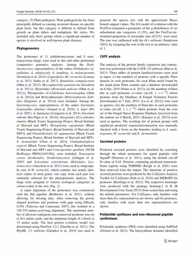
cantly increased in the plant pathogens (Fig. 3c1). There
are fewer proteases seen in the extremotolerant species,
although this is mainly due to the protease-rich plant
pathogens in the non-extremotolerant group, and the dif-
ference did not reach statistical significance. When com-
paring the most abundant individual enzyme families
(Fig. 3c2), the plant pathogens have significantly more
aspartic endopeptidases or pepsin family A1 proteases
(Fig. 3c2, A01A) and serine endopeptidases or subtilisin
family proteases (Fig. 3c2, S08A), while the extremotol-
erant species have significantly fewer serine exopeptidases
(Fig. 3c2, S28).
The differences in carbohydrate-active enzymes
(CAZymes) abundance between plant pathogens and non-
plant pathogenic species did not reach statistical signifi-
cance. Nevertheless, three out of four plant pathogens
analysed here have higher numbers of CAZymes than seen
for 16 non-plant pathogenic species (except A. nidulans
and H. werneckii; Fig. 3b1). Moreover, the plant patho-
genic fungi are significantly enriched with members of the
CAZymes auxiliary activity family (AA7), carbohydrate-
binding module families (CBM13, CBM18, CBM35,
CBM50), carbohydrate esterase family (CE5), glycoside
hydrolase families (GH2, GH12, GH92), and glycosyl
transferase family (GT34) (Fig. 3b2). The extremotolerant
species have significantly more members of the CAZymes
GH32 family, which comprises invertases and other
enzymes that are involved in energy storage and recovery.
Despite these patterns, no lifestyle-linked clustering of the
species was observed based on their enzyme profiles
(Fig. 3b2, c2).
Polyketide synthases
Polyketide synthases (PKSs) are responsible for the syn-
thesis of secondary metabolite polyketides. Our
202 Fungal Diversity (2018) 93:195–213
123
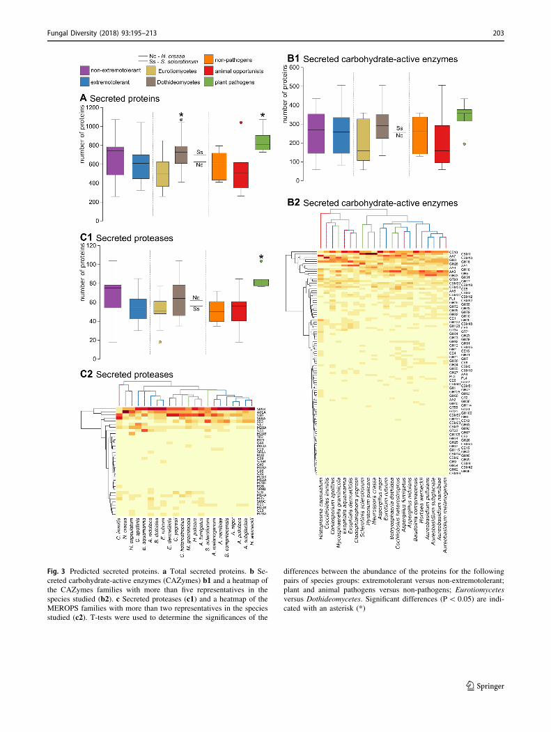
Fig. 3 Predicted secreted proteins. a Total secreted proteins. b Se-
creted carbohydrate-active enzymes (CAZymes) b1 and a heatmap of
the CAZymes families with more than five representatives in the
species studied (b2). c Secreted proteases (c1) and a heatmap of the
MEROPS families with more than two representatives in the species
studied (c2). T-tests were used to determine the significances of the
differences between the abundance of the proteins for the following
pairs of species groups: extremotolerant versus non-extremotolerant;
plant and animal pathogens versus non-pathogens; Eurotiomycetes
versus Dothideomycetes. Significant differences (P\ 0.05) are indi-
cated with an asterisk (*)
Fungal Diversity (2018) 93:195–213 203
123

phylogenetic analysis of ketosynthase domains character-
istic of PKSs, produces four well-defined clusters that
correspond to fatty-acid synthases (FAS), non-reducing
(NR)-PKSs, highly reducing (HR)-PKSs, and partially
reducing (PR)-PKSs (Fig. 4a). All of the major subgroups
of NR-PKSs (groups I-VII) and HR-PKSs (lovastatins,
fumonisins, T-toxins) were identified in the phylogenetic
tree. The total number of PKSs analysed is significantly
higher for the plant pathogens compared to the non-
pathogenic fungi and animal opportunists (Fig. 4b), which
is due to the significant enrichment of NR-PKSs and HR-
PKSs (Fig. 4d, e). There is no difference in the PKS
numbers between Dothideomycetes and Eurotiomycetes.
Genes encoding 1,3,6,8-tetrahydroxynaphthalene (THN)
synthases involved in the synthesis of melanin, as well as
genes involved in the 3,4-dihydroxyphenylalanine
Fig. 4 Predicted polyketide
synthases. a Phylogeny of the
ketosynthase domains
determined using the Natural
Product Domain Seeker. b–eAbundance of the PKS
subgroups in the studied fungal
species. FAS fatty-acid
synthases (c), PR-PKS partially
reducing PKS, NR-PKS non-
reducing PKS (d), HR-PKSfully reducing PKS (e). T-testswere used to test the
significances of the differences
between the abundances of
these proteins for the following
pairs of species groups:
extremotolerant versus non-
extremotolerant; plant and
animal pathogens versus non-
pathogens; Eurotiomycetes
versus Dothideomycetes.
Significant differences
(P\ 0.05) are indicated with an
asterisk (*)
204 Fungal Diversity (2018) 93:195–213
123
(DOPA)-melanin synthesis pathway and pyomelanin syn-
thesis (L-tyrosine degradation) pathway are present in the
majority of the 22 analysed species (Supplemental
Table S1).
Non-ribosomal peptide synthetases
The here reconstructed phylogeny of the A domains from
the non-ribosomal peptide synthetases (NRPSs) or NRPS-
like proteins identified by antiSMASH software reveals
two major groups: the NRPSs and the adenylating
enzymes, such as long-chain fatty-acyl-CoA synthases
(Fig. 5a). Where possible, the NRPS phylogenetic groups
based on the A domains were named after the previously
characterised homologues (Fig. 5a). Different A domains
from single NRPS genes with several A domains mostly
occur in the same clade, with the exceptions being dike-
topiperazine synthetases and Pes1 homologues, the A
domains of which are placed in two or more well-separated
clusters (Fig. 5a).
The total number of NRPSs are significantly higher in
the plant pathogens, both with the inclusion (data not
shown) and the exclusion (Fig. 5b) of the large group of
adenylating enzymes, although no lifestyle-associated
patterns are observed for the adenylating enzymes alone
(Fig. 5c). The numbers of NRPSs involved in the synthesis
of extracellular (but not intracellular) siderophores are
higher in most of the animal opportunists and plant
pathogens compared to the non-pathogens, although the
differences reached statistical significance only for the
plant pathogens (Fig. 5e).
Selected fungal virulence factors
Several protein families that experienced significant
expansion or contraction in various parts of the phyloge-
netic tree were investigated in more detail. When the
expansion and contraction of these protein families are
compared within and between the groups of species with
similar lifestyles, there are no significant differences in
most cases, although there are some exceptions, a selection
of which is shown in Fig. 6. We show that the number of
fungal TP-binding cassette (ABC) type multidrug resis-
tance (MDR) efflux transporters is higher in the plant
pathogens than in most other fungi, but the between-group
difference is not significant. The number of siderophore-
iron transporters does not differ between the fungal groups
with different lifestyles (Fig. 6b). In 12 extremotolerant
species the number of siderophore genes is higher than in
five non-extremophile species, the exceptions being B.
compniacensis, C. apollinis and B. dothidea. However, the
between-group differences did not reach statistical signifi-
cance. Tripeptidyl-peptidases (TPPs), secreted serine
proteases capable of protein degradation at acidic pH differ
significantly in number between Eurotiomycetes and
Dothideomycetes (Fig. 6c) as well as in the animal
opportunists versus plant pathogens. Finally, proteins
similar to the effector Ecp6, a scavenger of chitin frag-
ments, are enriched in the plant pathogens (Fig. 6d). No
significant differences were observed between Doth-
ideomycetes and Eurotiomycetes.
Discussion
Extremotolerance and opportunism are linkedat the level of fungal orders
Infection of mammal hosts is a rare ability in the fungal
kingdom, as shown by our study. Of the 140 orders dis-
tinguished in this paper based on current taxonomy
(Fig. 1), only 21 (15.0%) contain species that repeatedly
show this infectious potential. Among these, most oppor-
tunistic orders and the main order containing true patho-
gens causing transmissible disease [Onygenales (de Hoog
et al. 2018)] belong to Ascomycota (14 out of 80 orders,
17.3%). Of the much fewer opportunistic orders in Basid-
iomycota (3 out of 42, 7.1%) Malasseziales are generally
regarded as asymptomatic cutaneous commensals/pul-
monary colonisers and the same is true for the ascomyce-
tous Pneumocystidales (Baker et al. 2016). Members of the
remaining 18 orders are able to cause infection in specific
circumstances but typically have an environmental habitat.
Our results show that opportunism in mammals is cor-
related with extremotolerance. Association of the 21 orders
with all categories of medical relevance and general
extremotolerance was found to be significant (P = 0.0001).
However, while some large melanised and extremotolerant
groups within the fungal kingdom are only rarely
encountered in human infection, others (such as
Chaetothyriales) have a particularly high number of
opportunists. This apparent discrepancy can be explained
by distinguishing between two modes of extremotolerance:
‘polyextremotolerance’, where tolerance to a variety of
stress types is coupled with large adaptability and resulting
in great potential for habitat shifts, and ‘monoextremotol-
erance’ describing a specialisation for a particular type of
stress and with low potential for habitat shifts. While the
first type of extremotolerance was significantly associated
with opportunism in our analysis (as described above) the
second (surface colonising lifestyle) was not. A similar
observation was made by Prenafeta-Boldu et al. (2006),
introducing dual ecology as a mechanism enhancing
habitat shifts. Other factors besides stress tolerance may
play a role, such as the ability to assimilate monoaromatic
hydrocarbons, linked to the tendency of fungi to infect the
Fungal Diversity (2018) 93:195–213 205
123

central nervous system (Prenafeta-Boldu et al. 2006), but
this was not tested here due to the limited available data.
In addition to almost complete association of thermo-
tolerance with clinical occurrence, osmotolerance/os-
mophily was also identified as a major factor correlating
with opportunism (P = 0.0007), confirming the connection
proposed by de Hoog et al. (2005). Interestingly, despite
the ability to grow at 37 �C being the main virulence factor
in fungi (Robert and Casadevall 2009), opportunism also
correlated to tolerance to low temperatures (P = 0.0043).
In 97 orders no obvious extremotolerance of any kind is
known; of these, only five (5.2%) contain species with an
infectious potential. Two of these concern the lower fungi
in Entomophthoromycotina, where opportunism has been
hypothesised to be enhanced by intestinal occurrence in
cold-blooded tropical animals (Gugnani and Okafor 1980).
The large and species-rich classes of Pezizomycetes,
Fig. 5 Predicted non-ribosomal
peptide synthetases.
a Phylogeny of the adenylation
domains (A domains)
determined using the antibiotics
and secondary metabolite
analysis shell. The dots mark
the positions of the reference
proteins from A. fumigatus and
A. nidulans (purple) and C.
heterostrophus (pink) used to
infer the functions of the protein
clusters (Cramer et al. 2006;
von Dohren 2009; O’Hanlon
et al. 2012; Condon et al. 2013;
Bushley and Turgeon 2010).
The names of the reference
proteins are in round brackets,
while the inferred products of
the NRPSs are in square
brackets. Note that different
domains from the same proteins
similar to the GliP and NPS4/
Pes1 proteins are included in
more than one cluster. b–eAbundance of the NRPS
proteins in the studied fungal
species, without the adenylating
enzymes (b), and for the
selected protein subfamilies of
the adenylating enzymes (c),and intracellular (d) andextracellular (e) siderophores.T-tests were used to test the
significances of the differences
between the abundances of
these proteins for the following
pairs of species groups:
extremotolerant versus non-
extremotolerant; plant and
animal pathogens versus non-
pathogens; Eurotiomycetes
versus Dothideomycetes.
Significant differences
(P\ 0.05) are indicated with an
asterisk (*)
206 Fungal Diversity (2018) 93:195–213
123

Orbiliomycetes, Lecanoromycetes, and Leotiomycetes do
not contain any clinical representatives.
An association between human opportunism and plant
pathogenicity that has been put forward earlier on the basis
of similar virulence factors (Kempf et al. 2002; Kirzinger
et al. 2011; Melotto et al. 2014) is not supported by our
results. Of the 26 fungal orders containing major plant
pathogens, only three also contain a few or more clinical
species. One of these is Pleosporales, an order showing a
wide ecological diversity including extremotolerance
(Ahmed et al. 2014). The Hypocreales contains species
causing both human infections (only 64 out of 4479
described species) as well as major plant diseases, as an
example of cross-kingdom pathogenicity (van Baarlen
et al. 2007; Segorbe et al. 2017) or more accurately, cross-
kingdom opportunism. Other such examples include spe-
cies of Fusarium, where both plant and human infection is
frequent and strains have an unusually wide host range (Al-
Hatmi et al. 2016; Sisic et al. 2018). Similarly, Sharma
et al. (2014) noted the plant-virulence of clinical strains of
Exserohilum rostratum (Pleosporales), but this species is
not host-specific, it has a wide host range on diverse
grasses, and is also found in soil and on rotten materials
(Ellis 1971). However, in orders where host-specific plant-
pathogenicity is among the prevalent life styles (e.g.
Myriangiales, Asterinales, Meliolales, Magnaporthales,
Erysiphales, Taphrinales, Tilletiales, Exobasidiales, Usti-
laginales, Microstromatales, Pucciniales), no human
opportunistic species are encountered. In the genus Al-
ternaria mammal opportunism is found almost exclusively
in the few saprobic representatives (de Hoog and Horre
2002).
Few genomic signatures reflect the lifestylesof black fungi
If, as we suggest, fungal opportunism is a side effect of
polyextremotolerance and not a specialised lifestyle,
opportunists are expected to have few differences from
their non-opportunistic counterparts. This is supported by
the results of our analysis of genomic signatures in 11
dothideomycetous and 9 eurotiomycetous fungi. We
focused on enzymes for secondary metabolite production,
carbohydrate-active enzymes (CAZymes), and small
secreted proteins and peptidases, proteins with documented
or proposed roles as effectors in virulence.
Although proteases are believed to be important for
animal pathogenesis (Monod et al. 2002), in our dataset
they were not enriched in opportunistic species compared
to strictly non-opportunistic species (Fig. 3c1). The same
was true for individual protease families (Fig. 3c2) and
also for CAZyme families. In contrast, CAZyme family
GH32 (invertases and other enzymes involved in energy
storage and recovery) was significantly enriched in
extremotolerant species, possibly reflecting their energeti-
cally demanding life in extreme conditions (Oren 2011).
Fig. 6 Abundance of the
selected fungal virulence factors
in the fungal species studied.
a Multidrug resistance (MDR)
efflux transporters.
b Siderochrome–iron
transporters. c Tripeptidyl
peptidases. d Extracellular
proteins 6. The protein groups
were selected based on
significant protein family
expansion or contraction, as
determined by analysis using
CAFE 3. T-tests were used to
test the significances of
differences between the
abundances of the proteins for
the following pairs of species
groups: extremotolerant versus
non-extremotolerant; plant and
animal pathogens versus non-
pathogens; Eurotiomycetes
versus Dothideomycetes.
Significant differences
(P\ 0.05) are indicated with an
asterisk (*)
Fungal Diversity (2018) 93:195–213 207
123
Generally, however, no lifestyle-linked clustering of the
species was observed based on their CAZyme profiles
(Fig. 3b2, c2), confirming the observations of Krijger et al.
(2014).
Secondary metabolites such as polyketides are important
for interactions of fungi with other organisms (Fischbach
and Walsh 2006), yet no enrichment in PKSs catalysing
their synthesis was observed in human opportunists
(Fig. 4). One of the PKS products, melanin, plays an
important role in virulence and resistance to clinically used
antifungal agents (van Baarlen et al. 2007; Feng et al. 2001;
Nosanchuk et al. 2015; Schnitzler et al. 1999) [although
these roles have been disputed by some authors (Song et al.
2017)] and protection against abiotic stress (Slepecky and
Starmer 2009; Gostincar et al. 2012; Kogej et al. 2007;
Kejzar et al. 2013), but again we found no significant
differences between the opportunistic and other species in
numbers of enzymes involved in the PKS melanin syn-
thesis pathway and also in the alternative DOPA-melanin
synthesis pathway (Langfelder et al. 2003), and pyome-
lanin synthesis/L-tyrosine degradation pathway (Schmaler-
Ripcke et al. 2009) (Supplemental Table S1). The genes
known to be involved in the three different melanin syn-
thesis pathways are present in the majority of the 22 spe-
cies studied here (Supplemental Table S1).
Similarly to all above discussed genes, genes for non-
ribosomal peptide synthetases (NRPSs) were not enriched
in animal opportunists, although they are involved in the
synthesis of important virulence determinants, such as iron-
chelating siderophores (Bushley and Turgeon 2010; Silva
et al. 2011), and toxins, such as enniatin, victorin, HC-toxin
and AM-toxin (Walton 1996; Haese et al. 1993). Iron
overload in a host is known to exacerbate many infectious
diseases, such as cryptococcosis in people infected with
HIV, and conversely, iron withholding is an important
defence strategy for mammalian hosts (Jung et al. 2006).
There were no lifestyle-associated differences in the
number of siderophore–iron transporters, which are
responsible for the uptake of siderophore–iron chelates
(Philpott 2006) and linked to stress response and virulence
in Cryptococcus neoformans (Singh et al. 2015; Jung et al.
2006), or the number of multidrug resistance (MDR) efflux
transporters (Fig. 6a), which provide fungi with protection
against antibiotics, plant defence compounds, and fungi-
cides (de Waard et al. 2006). The differences in the number
of secreted serine proteases [previously linked to virulence
in A. fumigatus (Reichard et al. 2006)] between animal
opportunists and plant pathogens can be attributed to their
phylogenetic history, as the same difference was observed
between Eurotiomycetes and Dothideomycetes—most here
studied animal opportunists are Eurotiomycetes and most
plant pathogens are Dothideomycetes (Fig. 6c).
While no genomic traits of analysed black fungi were
linked to their opportunism, several traits were significantly
associated with their plant pathogenicity. Proteases,
believed to be involved in fungal signalling, nutrition,
degradation of host tissues, and digestion of plant defence
proteins (Ohm et al. 2012), were significantly enriched in
the plant pathogens (Fig. 3c1). Some of the differences are
in line with previous observations, for example in the case
of A01 and S08A proteases (ten Have et al. 2004; Armijos
Jaramillo et al. 2013), additionally validating our general
approach. Similarly, certain CAZyme families (AA7,
CBM13, CBM18, CBM35, CBM50, CE5, GH2, GH12,
GH92, GT34; Fig. 3b2) were shown to be enriched in plant
pathogens, likely linked to the role of enzymes from these
families in breaking down the barrier of the plant cell-wall
polysaccharides and using plant polysaccharides as a car-
bon source (Ohm et al. 2012; Lowe et al. 2015). Addi-
tionally, plant pathogens differed from other analysed
species in the total number of PKSs, in having a signifi-
cantly higher abundance of extracellular siderophore
NRPSs (Fig. 5e) and in the enrichment of the effector Ecp6
(Fig. 6d). The latter is easily explained as the Ecp6 is a
known scavenger of chitin fragments that are released by
chitinases, thereby preventing recognition of the fungus by
the host immune receptors for chitin (Sanchez-Vallet et al.
2013; de Jonge et al. 2010).
We can conclude that while our analysis successfully
identified genomic traits linked to plant pathogenicity, we
could find no such genomic signatures connected to the
fungal opportunistic lifestyle—supporting our hypothesis
that virulence traits of opportunistic fungi are exaptations,
which can also be found in other, non-opportunistic
species.
Implications of the overlap between adaptationto environmental stress and opportunism
The importance of stress tolerance acquired out-of-host in
pathogenesis was first proposed for the well-known
pathogen C. neoformans (Brown et al. 2007; van Burik and
Magee 2001). This interpretation is even more plausible in
the case of emerging opportunistic pathogens such as black
yeasts, which cause substantially fewer infections than C.
neoformans. If the adaptations that allow opportunistic
fungi to survive within a human host are indeed exaptations
(i.e., mechanisms that originally evolved for different
purposes, such as tolerance to environmental stress) and
that are only later found to be useful during an infection,
there should be few (if any) traits that can be directly
linked to the opportunistic potential of the fungal species.
Indeed, unlike bacteriologists, mycologists have largely
been unsuccessful in finding classical virulence factors,
even in notorious human pathogens like Candida albicans
208 Fungal Diversity (2018) 93:195–213
123

and A. fumigatus (Casadevall and Pirofski 2014). Similarly,
in this study we show that apart from growth at 37 �C there
appear to be few other traits that can distinguish oppor-
tunistic human pathogens from their non-pathogenic rela-
tives. No such traits were found through the comparative
genomics investigations, while in contrast, plant pathogens
were distinguished in several aspects. Taken together, these
observations indicate that most generalistic polyextremo-
tolerant fungi can be seen as potential opportunistic
pathogens as long as they can grow at the temperatures in
the mammalian body. Fortunately, this is a trait that (for
now) most fungi lack (Robert and Casadevall 2009).
The observation that polyextremotolerance and oppor-
tunistic pathogenesis repeatedly share a common phylo-
genetic history supports the hypothesis that traits important
for fungal pathogenicity are shaped by selection pressures
outside of the host. This corresponds to the concept of
‘‘accidental virulence’’ as postulated by Casadevall and
Pirofski (2007). However, care should be taken not to view
the infection events from a pan-adaptationist perspective.
On the one hand, it is unclear whether the opportunist
causing the infection is able to escape from the host back
into the environment—if it is not, any adaptations to host
acquired during the infection are meaningless from an
evolutionary perspective. On the other hand, if the oppor-
tunist is able to return to the environment, any newly
acquired adaptations will be either beneficial, neutral or
detrimental for its survival in the environment. Evidently,
adaptations detrimental to environmental fitness would be
selected against once outside the host. Adaptations with a
neutral effect would likely be drowned in the much larger
gene pool of environmental strains. Finally, for adaptations
beneficial to both survival in the host and in the out-of-host
environment it is difficult to envisage why they would need
to arise in the small populations during rare and time-
limited infection events rather than in much larger out-of-
host populations. The speculation of Casadevall and
Pirofski (2007) that a passage in animal hosts might
increase the fitness of environmental microbes in their out-
of-host environment therefore appears unlikely except for
species for which animals are a major and regular habitat.
An accidental infection is thus likely a (literal or evolu-
tionary) dead end, since the persistence of hypothetical
adaptations acquired during infection is improbable. For
this reason the gradual evolution of true pathogens through
a series of repeated opportunistic infections (in the absence
of host-to-host transmission) would be expected to happen
extremely rarely, if at all. Our hypothesis of polyex-
tremotolerance (coupled with the ability to grow at 37 �C)as the background of opportunism explains why oppor-
tunistic species are relatively rare, and why evolution
towards true pathogenicity hardly ever happens at all.
Nevertheless, many species of black fungi are now
increasingly being recognised as a medical issue (Silveira
and Nucci 2001; Chowdhary et al. 2015). Besides greater
numbers of susceptible hosts and improved diagnostics,
changes of our lifestyle have been proposed as another
reason for this trend (Casadevall et al. 2011; Gostincar
et al. 2011, 2015). In addition to the well-known dry
fraction of indoor environments, wet cells have recently
revealed a gamut of opportunistic species. Both A. mela-
nogenum and E. dermatitidis were found to be common in
tap water, bathrooms, steam-baths and dishwashers (Novak
Babic et al. 2016; Hamada and Abe 2010; Zalar et al.
2011). Changes that render indoor habitats inhospitable to a
majority of microbes frequently make conditions favour-
able for other, more resilient and more adaptable species—
polyextremotolerant fungi with greater potential to cause
opportunistic human infections (Gostincar et al.
2011, 2015). These are problematic especially if they are
also enriched for thermotolerance, such as E. dermatitidis,
Saprochaete clavata and Magnusiomyces capitatus in
dishwashers (Zalar et al. 2011; Zupancic et al. 2016) or for
the ability to metabolise phenols and hydrocarbons (Pre-
nafeta-Boldu et al. 2006, 2012).
This study shows that complex phenotypes such as the
ability to cause opportunistic infections of mammals do not
evolve from scratch. The genomic toolkit that is the basis
of selection is neither unlimited in quantity nor infinitely
malleable. It can differ substantially between different
taxonomic groups, resulting in repeated emergence of
opportunistic pathogens in some groups, but not in others.
Our results support the hypothesis that opportunistic fungi
do not specialise for pathogenicity as such but that their
invasive potential is tightly linked to their polyextremo-
tolerant ecology and most likely uncoupled from their
hosts. As a consequence, tackling the emerging problem of
opportunistic fungi will require an epidemiological
approach very different from the one applied to true
pathogens.
Acknowledgements The authors acknowledge the financial support
from the state budget of the Slovenian Research Agency (Research
Programmes P1-0170 and P1-0207, Infrastructural Centre Mycosmo,
MRIC UL, Postdoctoral Project Z7-7436 to J. Zajc). The authors
would like to thank Chris Berrie for language editing assistance.
Open Access This article is distributed under the terms of the Creative
Commons Attribution 4.0 International License (http://creative
commons.org/licenses/by/4.0/), which permits unrestricted use, dis-
tribution, and reproduction in any medium, provided you give
appropriate credit to the original author(s) and the source, provide a
link to the Creative Commons license, and indicate if changes were
made.
Fungal Diversity (2018) 93:195–213 209
123
References
Ahmed SA, van de Sande WWJ, Stevens DA, Fahal A, van
Diepeningen AD, Menken SBJ, de Hoog GS (2014) Revision
of agents of black-grain eumycetoma in the order Pleosporales.
Persoonia 33:141–154. https://doi.org/10.3767/
003158514x684744
Al-Hatmi AMS, Meis JF, de Hoog GS (2016) Fusarium: molecular
diversity and intrinsic drug resistance. PLoS Pathog. https://doi.
org/10.1371/journal.ppat.1005464
Amselem J, Cuomo CA, van Kan JAL, Viaud M, Benito EP, Couloux
A, Coutinho PM, de Vries RP, Dyer PS, Fillinger S, Fournier E,
Gout L, Hahn M, Kohn L, Lapalu N, Plummer KM, Pradier JM,
Quevillon E, Sharon A, Simon A, ten Have A, Tudzynski B,
Tudzynski P, Wincker P, Andrew M, Anthouard V, Beever RE,
Beffa R, Benoit I, Bouzid O, Brault B, Chen ZH, Choquer M,
Collemare J, Cotton P, Danchin EG, Da Silva C, Gautier A,
Giraud C, Giraud T, Gonzalez C, Grossetete S, Guldener U,
Henrissat B, Howlett BJ, Kodira C, Kretschmer M, Lappartient
A, Leroch M, Levis C, Mauceli E, Neuveglise C, Oeser B,
Pearson M, Poulain J, Poussereau N, Quesneville H, Rascle C,
Schumacher J, Segurens B, Sexton A, Silva E, Sirven C, Soanes
DM, Talbot NJ, Templeton M, Yandava C, Yarden O, Zeng QD,
Rollins JA, Lebrun MH, Dickman M (2011) Genomic analysis of
the aecrotrophic fungal pathogens Sclerotinia sclerotiorum and
Botrytis cinerea. PLoS Genet. https://doi.org/10.1371/journal.
pgen.1002230
Armijos Jaramillo VD, Vargas WA, Sukno SA, Thon MR (2013)
Horizontal transfer of a subtilisin gene from plants into an
ancestor of the plant pathogenic fungal genus Colletotrichum.
PLoS ONE 8(3):e59078. https://doi.org/10.1371/journal.pone.
0059078
Arnaud MB, Chibucos MC, Costanzo MC, Crabtree J, Inglis DO,
Lotia A, Orvis J, Shah P, Skrzypek MS, Binkley G, Miyasato
SR, Wortman JR, Sherlock G (2010) The Aspergillus Genome
Database, a curated comparative genomics resource for gene,
protein and sequence information for the Aspergillus research
community. Nucleic Acids Res 38:420–427. https://doi.org/10.
1093/Nar/Gkp751
Baker RM, Stegink RJ, Manaloor JJ, Schmitt BH, Stevens JC,
Christenson JC (2016) Malassezia pneumonia: a rare complica-
tion of parenteral nutrition therapy. JPEN J Parenter Enteral Nutr
40(8):1194–1196. https://doi.org/10.1177/0148607115595224
Blin K, Medema MH, Kazempour D, Fischbach MA, Breitling R,
Takano E, Weber T (2013) antiSMASH 2.0—a versatile
platform for genome mining of secondary metabolite producers.
Nucleic Acids Res 41(1):204–212. https://doi.org/10.1093/nar/
gkt449
Brown SM, Campbell LT, Lodge JK (2007) Cryptococcus neofor-
mans, a fungus under stress. Curr Opin Microbiol
10(4):320–325. https://doi.org/10.1016/j.mib.2007.05.014
Bushley KE, Turgeon BG (2010) Phylogenomics reveals subfamilies
of fungal nonribosomal peptide synthetases and their evolution-
ary relationships. BMC Evol Biol. https://doi.org/10.1186/1471-
2148-10-26
Casadevall A (2007) Determinants of virulence in the pathogenic
fungi. Fungal Biol Rev 21(4):130–132. https://doi.org/10.1016/j.
fbr.2007.02.007
Casadevall A, Pirofski LA (2007) Accidental virulence, cryptic
pathogenesis, Martians, lost hosts, and the pathogenicity of
environmental microbes. Eukaryot Cell 6(12):2169–2174.
https://doi.org/10.1128/Ec.00308-07
Casadevall A, Pirofski LA (2014) Microbiology: ditch the term
pathogen. Nature 516(7530):165–166. https://doi.org/10.1038/
516165a
Casadevall A, Fang FC, Pirofski LA (2011) Microbial virulence as an
emergent property: consequences and opportunities. PLoS
Pathog. https://doi.org/10.1371/journal.ppat.1002136
Cerqueira GC, Arnaud MB, Inglis DO, Skrzypek MS, Binkley G,
Simison M, Miyasato SR, Binkley J, Orvis J, Shah P, Wymore F,
Sherlock G, Wortman JR (2014) The Aspergillus Genome
Database: multispecies curation and incorporation of RNA-Seq
data to improve structural gene annotations. Nucleic Acids Res
42(D1):705–710. https://doi.org/10.1093/nar/gkt1029
Chen ZH, Martinez DA, Gujja S, Sykes SM, Zeng QD, Szaniszlo PJ,
Wang Z, Cuomo CA (2014) Comparative genomic and tran-
scriptomic analysis of Wangiella dermatitidis, a major cause of
phaeohyphomycosis and a model black yeast human pathogen.
G3 Genom Genet 4(4):561–578. https://doi.org/10.1534/g3.113.
009241
Chowdhary A, Perfect J, de Hoog GS (2015) Black molds and
melanized yeasts pathogenic to humans. Cold Spring Harb
Perspect Med. https://doi.org/10.1101/cshperspect.a019570
Condon BJ, Leng YQ, Wu DL, Bushley KE, Ohm RA, Otillar R,
Martin J, Schackwitz W, Grimwood J, MohdZainudin N, Xue
CS, Wang R, Manning VA, Dhillon B, Tu ZJ, Steffenson BJ,
Salamov A, Sun H, Lowry S, LaButti K, Han J, Copeland A,
Lindquist E, Barry K, Schmutz J, Baker SE, Ciuffetti LM,
Grigoriev IV, Zhong S, Turgeon BG (2013) Comparative
genome structure, secondary metabolite, and effector coding
capacity across Cochliobolus pathogens. PLoS Genet. https://doi.
org/10.1371/journal.pgen.1003233
Cramer RA, Stajich JE, Yamanaka Y, Dietrich FS, Steinbach WJ,
Perfect JR (2006) Phylogenomic analysis of non-ribosomal
peptide synthetases in the genus Aspergillus. Gene 383:24–32.
https://doi.org/10.1016/j.gene.2006.07.008
Darriba D, Taboada GL, Doallo R, Posada D (2011) ProtTest 3: fast
selection of best-fit models of protein evolution. Bioinformatics
27(8):1164–1165. https://doi.org/10.1093/bioinformatics/btr088
de Hoog GS, Horre R (2002) Molecular taxonomy of the Alternaria
and Ulocladium species from humans and their identification in
the routine laboratory. Mycoses 45(8):259–276. https://doi.org/
10.1046/j.1439-0507.2002.00747.x
de Hoog GS, Zalar P, Gerrits van den Ende AHG, Gunde-Cimerman
N (2005) Relation of halotolerance to human-pathogenicity in
the fungal tree of life: an overview of ecology and evolution
under stress. In: Gunde-Cimerman N, Oren A, Plemenitas A
(eds) Adaptation to life at high salt concentrations in Archaea,
Bacteria, and Eukarya. Springer, Dordrecht, pp 373–395
de Hoog GS, Guarro J, Gene J, Figueras MJ (2015) Atlas of clinical
fungi. The ultimate benchtool for diagnostics. https://doi.org/10.
1111/j.1439-0507.1996.tb00148.x
de Hoog G, Ahmed S, Danesi P, Guillot J, Graser Y (2018)
Distribution of pathogens and outbreak fungi in the fungal
kingdom. In: Seyedmousavi S, de Hoog G, Guillot J, Verweij PE
(eds) Emerging and epizootic fungal infections in animals.
Springer, Dordrecht, pp 3–16
de Jonge R, van Esse HP, Kombrink A, Shinya T, Desaki Y, Bours R,
van der Krol S, Shibuya N, Joosten MHAJ, Thomma BPHJ
(2010) Conserved fungal LysM effector Ecp6 prevents chitin-
triggered immunity in plants. Science 329(5994):953–955.
https://doi.org/10.1126/science.1190859
de Waard MA, Andrade AC, Hayashi K, Schoonbeek HJ, Ster-
giopoulos I, Zwiers LH (2006) Impact of fungal drug trans-
porters on fungicide sensitivity, multidrug resistance and
virulence. Pest Manag Sci 62(3):195–207. https://doi.org/10.
1002/ps.1150
Elias PM (2007) The skin barrier as an innate immune element.
Semin Immunopathol 29(1):3–14. https://doi.org/10.1007/
s00281-007-0060-9
Ellis MB (1971) Dematiaceous Hyphomycetes. CMI, Kew
210 Fungal Diversity (2018) 93:195–213
123

Feng B, Wang X, Hauser M, Kaufmann S, Jentsch S, Haase G, Becker
JM, Szaniszlo PJ (2001) Molecular cloning and characterization
of WdPKS1, a gene involved in dihydroxynaphthalene melanin
biosynthesis and virulence in Wangiella (Exophiala) dermati-
tidis. Infect Immun 69(3):1781–1794
Fischbach MA, Walsh CT (2006) Assembly-line enzymology for
polyketide and nonribosomal peptide antibiotics: logic, machin-
ery, and mechanisms. Chem Rev 106(8):3468–3496. https://doi.
org/10.1021/cr0503097
Galagan JE, Calvo SE, Borkovich KA, Selker EU, Read ND, Jaffe D,
FitzHugh W, Ma LJ, Smirnov S, Purcell S, Rehman B, Elkins T,
Engels R, Wang S, Nielsen CB, Butler J, Endrizzi M, Qui D,
Ianakiev P, Bell-Pedersen D, Nelson MA, Werner-Washburne
M, Selitrennikoff CP, Kinsey JA, Braun EL, Zelter A, Schulte U,
Kothe GO, Jedd G, Mewes W, Staben C, Marcotte E, Greenberg
D, Roy A, Foley K, Naylor J, Stange-Thomann N, Barrett R,
Gnerre S, Kamal M, Kamvysselis M, Mauceli E, Bielke C, Rudd
S, Frishman D, Krystofova S, Rasmussen C, Metzenberg RL,
Perkins DD, Kroken S, Cogoni C, Macino G, Catcheside D, Li
W, Pratt RJ, Osmani SA, DeSouza CP, Glass L, Orbach MJ,
Berglund JA, Voelker R, Yarden O, Plamann M, Seiler S,
Dunlap J, Radford A, Aramayo R, Natvig DO, Alex LA,
Mannhaupt G, Ebbole DJ, Freitag M, Paulsen I, Sachs MS,
Lander ES, Nusbaum C, Birren B (2003) The genome sequence
of the filamentous fungus Neurospora crassa. Nature
422(6934):859–868
Goodwin SB, Ben M’Barek S, Dhillon B, Wittenberg AHJ, Crane CF,
Hane JK, Foster AJ, Van der Lee TAJ, Grimwood J, Aerts A,
Antoniw J, Bailey A, Bluhm B, Bowler J, Bristow J, van der
Burgt A, Canto-Canche B, Churchill ACL, Conde-Ferraez L,
Cools HJ, Coutinho PM, Csukai M, Dehal P, De Wit P, Donzelli
B, van de Geest HC, Van Ham RCHJ, Hammond-Kosack KE,
Henrissat B, Kilian A, Kobayashi AK, Koopmann E, Kourmpetis
Y, Kuzniar A, Lindquist E, Lombard V, Maliepaard C, Martins
N, Mehrabi R, Nap JPH, Ponomarenko A, Rudd JJ, Salamov A,
Schmutz J, Schouten HJ, Shapiro H, Stergiopoulos I, Torriani
SFF, Tu H, de Vries RP, Waalwijk C, Ware SB, Wiebenga A,
Zwiers LH, Oliver RP, Grigoriev IV, Kema GHJ (2011) Finished
genome of the fungal wheat pathogen Mycosphaerella gramini-
cola reveals dispensome structure, chromosome plasticity, and
stealth pathogenesis. PLoS Genet. https://doi.org/10.1371/jour
nal.pgen.1002070
Gostincar C, Grube M, de Hoog GS, Zalar P, Gunde-Cimerman N
(2010) Extremotolerance in fungi: evolution on the edge. FEMS
Microbiol Ecol 71(1):2–11
Gostincar C, Grube M, Gunde-Cimerman N (2011) Evolution of
fungal pathogens in domestic environments? Fungal Biol
115(10):1008–1018. https://doi.org/10.1016/j.funbio.2011.03.
004
Gostincar C, Muggia L, Grube M (2012) Polyextremotolerant black
fungi: oligotrophism, adaptive potential, and a link to lichen
symbioses. Front Microbiol 3:390. https://doi.org/10.3389/
fmicb.2012.00390
Gostincar C, Ohm RA, Kogej T, Sonjak S, Turk M, Zajc J, Zalar P,
Grube M, Sun H, Han J, Sharma A, Chiniquy J, Ngan CY,
Lipzen A, Barry K, Grigoriev IV, Gunde-Cimerman N (2014)
Genome sequencing of four Aureobasidium pullulans varieties:
biotechnological potential, stress tolerance, and description of
new species. BMC Genomics 15(1):549. https://doi.org/10.1186/
1471-2164-15-549
Gostincar C, Gunde-Cimerman N, Grube M (2015) Polyextremotol-
erance as the fungal answer to changing environments. In:
Bakermans C (ed) Microbial evolution under extreme condi-
tions. Life in extreme environments, vol 2. de Gruyter, Berlin,
pp 185–208. https://doi.org/10.1515/9783110340716.185
Grigoriev IV, Nikitin R, Haridas S, Kuo A, Ohm R, Otillar R, Riley
R, Salamov A, Zhao X, Korzeniewski F, Smirnova T, Nordberg
H, Dubchak I, Shabalov I (2014) MycoCosm portal: gearing up
for 1000 fungal genomes. Nucleic Acids Res 42(1):699–704.
https://doi.org/10.1093/nar/gkt1183
Gugnani HC, Okafor JI (1980) Mycotic flora of the intestine and other
internal organs of certain reptiles and amphibians with special
reference to characterization of Basidiobolus isolates. Mykosen
23(5):260–268
Guillot J, Demanche C, Hugot JP, Berthelemy M, Wakefield E, Dei-
Cas E, Chermette R (2001) Parallel phylogenies of Pneumocystis
species and their mammalian hosts. J Eukaryot Microbiol
48(1):113–115
Guindon S, Dufayard JF, Lefort V, Anisimova M, Hordijk W,
Gascuel O (2010) New algorithms and methods to estimate
maximum-likelihood phylogenies: assessing the performance of
PhyML 3.0. Syst Biol 59(3):307–321. https://doi.org/10.1093/
sysbio/syq010
Haese A, Schubert M, Herrmann M, Zocher R (1993) Molecular
characterization of the enniatin synthetase gene encoding a
multifunctional enzyme catalyzing N-Methyldepsipeptide for-
mation in Fusarium scirpi. Mol Microbiol 7(6):905–914. https://
doi.org/10.1111/j.1365-2958.1993.tb01181.x
Hall TA (1999) BioEdit: a user-friendly biological sequence align-
ment editor and analysis program for Windows 95/98/NT.
Nucleic Acids Symp Ser 41:95–98
Hamad M (2008) Antifungal immunotherapy and immunomodula-
tion: a double-hitter approach to deal with invasive fungal
infections. Scand J Immunol 67(6):533–543. https://doi.org/10.
1111/j.1365-3083.2008.02101.x
Hamada N, Abe N (2010) Comparison of fungi found in bathrooms
and sinks. Biocontrol Sci 15(2):51–56
Han MV, Thomas GWC, Lugo-Martinez J, Hahn MW (2013)
Estimating gene gain and loss rates in the presence of error in
genome assembly and annotation using CAFE 3. Mol Biol Evol
30(8):1987–1997. https://doi.org/10.1093/molbev/mst100
Jung WH, Sham A, White R, Kronstad JW (2006) Iron regulation of
the major virulence factors in the AIDS-associated pathogen
Cryptococcus neoformans. PLoS Biol 4(12):2282–2295. https://
doi.org/10.1371/journal.pbio.0040410
Karanyi Z, Holb I, Hornok L, Pocsi I, Miskei M (2013) FSRD: fungal
stress response database. Database. https://doi.org/10.1093/
database/bat037
Katoh K, Toh H (2008) Recent developments in the MAFFT multiple
sequence alignment program. Brief Bioinform 9(4):286–298
Keeling KB, Pavur RJ (2011) Statistical accuracy of spreadsheet
software. Am Stat 65(4):265–273. https://doi.org/10.1198/tas.
2011.09076
Kejzar A, Gobec S, Plemenitas A, Lenassi M (2013) Melanin is
crucial for growth of the black yeast Hortaea werneckii in its
natural hypersaline environment. Fungal Biol 117(5):368–379.
https://doi.org/10.1016/j.funbio.2013.03.006
Kempf VAJ, Hitziger N, Riess T, Autenrieth IB (2002) Do plant and
human pathogens have a common pathogenicity strategy?
Trends Microbiol 10(6):269–275. https://doi.org/10.1016/
S0966-842x(02)02372-7
Kirzinger MWB, Nadarasah G, Stavrinides J (2011) Insights into
cross-kingdom plant pathogenic Bacteria. Genes 2(4):980–997.
https://doi.org/10.3390/genes2040980
Kis-Papo T, Weig AR, Riley R, Persoh D, Salamov A, Sun H, Lipzen
A, Wasser SP, Rambold G, Grigoriev IV, Nevo E (2014)
Genomic adaptations of the halophilic Dead Sea filamentous
fungus Eurotium rubrum. Nat Commun. https://doi.org/10.1038/
ncomms4745
Kogej T, Stein M, Volkmann M, Gorbushina AA, Galinski EA,
Gunde-Cimerman N (2007) Osmotic adaptation of the halophilic
Fungal Diversity (2018) 93:195–213 211
123

fungus Hortaea werneckii: role of osmolytes and melanization.
Microbiology 153(Pt 12):4261–4273
Krijger JJ, Thon MR, Deising HB, Wirsel SGR (2014) Compositions
of fungal secretomes indicate a greater impact of phylogenetic
history than lifestyle adaptation. BMC Genomics. https://doi.org/
10.1186/1471-2164-15-722
Krogh A, Larsson B, von Heijne G, Sonnhammer ELL (2001)
Predicting transmembrane protein topology with a hidden
Markov model: application to complete genomes. J Mol Biol
305(3):567–580. https://doi.org/10.1006/jmbi.2000.4315
Kumamoto CA (2008) Niche-specific gene expression during C.
albicans infection. Curr Opin Microbiol 11(4):325–330. https://
doi.org/10.1016/j.mib.2008.05.008
Langfelder K, Streibel M, Jahn B, Haase G, Brakhage AA (2003)
Biosynthesis of fungal melanins and their importance for human
pathogenic fungi. Fungal Genet Biol 38(2):143–158. https://doi.
org/10.1016/S1087-1845(02)00526-1
Lenassi M, Gostincar C, Jackman S, Turk M, Sadowski I, Nislow C,
Jones S, Birol I, Gunde-Cimerman N, Plemenitas A (2013)
Whole genome duplication and enrichment of metal cation
transporters revealed by de novo genome sequencing of
extremely halotolerant black yeast Hortaea werneckii. PLoS
ONE 8(8):e71328. https://doi.org/10.1371/journal.pone.0071328
Lian X, de Hoog GS (2010) Indoor wet cells harbour melanized
agents of cutaneous infection. Med Mycol 48(4):622–628.
https://doi.org/10.3109/13693780903405774
Liu L, Zhang Z, Shao CL, Wang JL, Bai H, Wang CY (2015)
Bioinformatical analysis of the sequences, structures and func-
tions of fungal polyketide synthase product template domains.
Sci Rep. https://doi.org/10.1038/srep10463
Lowe RGT, McCorkelle O, Bleackley M, Collins C, Faou P,
Mathivanan S, Anderson M (2015) Extracellular peptidases of
the cereal pathogen Fusarium graminearum. Front Plant Sci.
https://doi.org/10.3389/fpls.2015.00962
Lu T, Yao B, Zhang C (2012) DFVF: database of fungal virulence
factors. Database. https://doi.org/10.1093/database/bas032
Melotto M, Panchal S, Roy D (2014) Plant innate immunity against
human bacterial pathogens. Front Microbiol. https://doi.org/10.
3389/fmicb.2014.00411
Miller M, Pfeiffer W, Schwartz T (2010) Creating the CIPRES
Science Gateway for inference of large phylogenetic trees. In:
Proceedings of the Gateway Computing Environments Work-
shop (GCE), vol 1 (1), pp 1–8
Monod M, Capoccia S, Lechenne B, Zaugg C, Holdom M, Jousson O
(2002) Secreted proteases from pathogenic fungi. Int J Med
Microbiol 292(5–6):405–419. https://doi.org/10.1078/1438-
4221-00223
Nosanchuk JD, Stark RE, Casadevall A (2015) Fungal melanin: what
do we know about structure? Front Microbiol. https://doi.org/10.
3389/fmicb.2015.01463
Novak Babic M, Zalar P, Zenko B, Dzeroski S, Gunde-Cimerman N
(2016) Yeasts and yeast-like fungi in tap water and groundwater,
and their transmission to household appliances. Fungal Ecol
20:30–39. https://doi.org/10.1016/j.funeco.2015.10.001
O’Hanlon KA, Gallagher L, Schrettl M, Jochl C, Kavanagh K, Larsen
TO, Doyle S (2012) Nonribosomal peptide synthetase genes
pesL and pes1 are essential for fumigaclavine C production in
Aspergillus fumigatus. Appl Environ Microbiol
78(9):3166–3176. https://doi.org/10.1128/Aem.07249-11
Ohm RA, Feau N, Henrissat B, Schoch CL, Horwitz BA, Barry KW,
Condon BJ, Copeland AC, Dhillon B, Glaser F, Hesse CN, Kosti
I, Labutti K, Lindquist EA, Lucas S, Salamov AA, Bradshaw
RE, Ciuffetti L, Hamelin RC, Kema GH, Lawrence C, Scott JA,
Spatafora JW, Turgeon BG, de Wit PJ, Zhong S, Goodwin SB,
Grigoriev IV (2012) Diverse lifestyles and strategies of plant
pathogenesis encoded in the genomes of eighteen
Dothideomycetes fungi. PLoS Pathog 8(12):e1003037. https://
doi.org/10.1371/journal.ppat.1003037
Oren A (2011) Thermodynamic limits to microbial life at high salt
concentrations. Environ Microbiol 13(8):1908–1923. https://doi.
org/10.1111/j.1462-2920.2010.02365.x
Park BH, Karpinets TV, Syed MH, Leuze MR, Uberbacher EC (2010)
CAZymes Analysis Toolkit (CAT): web service for searching
and analyzing carbohydrate-active enzymes in a newly
sequenced organism using CAZy database. Glycobiology
20(12):1574–1584. https://doi.org/10.1093/glycob/cwq106
Pel HJ, de Winde JH, Archer DB, Dyer PS, Hofmann G, Schaap PJ,
Turner G, de Vries RP, Albang R, Albermann K, Andersen MR,
Bendtsen JD, Benen JA, van den Berg M, Breestraat S, Caddick
MX, Contreras R, Cornell M, Coutinho PM, Danchin EG, Debets
AJ, Dekker P, van Dijck PW, van Dijk A, Dijkhuizen L,
Driessen AJ, d’Enfert C, Geysens S, Goosen C, Groot GS, de
Groot PW, Guillemette T, Henrissat B, Herweijer M, van den
Hombergh JP, van den Hondel CA, van der Heijden RT, van der
Kaaij RM, Klis FM, Kools HJ, Kubicek CP, van Kuyk PA,
Lauber J, Lu X, van der Maarel MJ, Meulenberg R, Menke H,
Mortimer MA, Nielsen J, Oliver SG, Olsthoorn M, Pal K, van
Peij NN, Ram AF, Rinas U, Roubos JA, Sagt CM, Schmoll M,
Sun J, Ussery D, Varga J, Vervecken W, van de Vondervoort PJ,
Wedler H, Wosten HA, Zeng AP, van Ooyen AJ, Visser J, Stam
H (2007) Genome sequencing and analysis of the versatile cell
factory Aspergillus niger CBS 513.88. Nat Biotechnol
25(2):221–231
Petersen TN, Brunak S, von Heijne G, Nielsen H (2011) SignalP 4.0:
discriminating signal peptides from transmembrane regions. Nat
Methods 8(10):785–786. https://doi.org/10.1038/nmeth.1701
Philpott CC (2006) Iron uptake in fungi: a system for every source.
BBA Mol Cell Res 1763(7):636–645. https://doi.org/10.1016/j.
bbamcr.2006.05.008
Prenafeta-Boldu FX, Summerbell R, de Hoog GS (2006) Fungi
growing on aromatic hydrocarbons: biotechnology’s unexpected
encounter with biohazard? FEMS Microbiol Rev 30(1):109–130.
https://doi.org/10.1111/j.1574-6976.2005.00007.x
Prenafeta-Boldu FX, Guivernau M, Gallastegui G, Vinas M, de Hoog
GS, Elias A (2012) Fungal/bacterial interactions during the
biodegradation of TEX hydrocarbons (toluene, ethylbenzene and
p-xylene) in gas biofilters operated under xerophilic conditions.
FEMS Microbiol Ecol 80(3):722–734. https://doi.org/10.1111/j.
1574-6941.2012.01344.x
Punta M, Coggill PC, Eberhardt RY, Mistry J, Tate J, Boursnell C,
Pang N, Forslund K, Ceric G, Clements J, Heger A, Holm L,
Sonnhammer ELL, Eddy SR, Bateman A, Finn RD (2012) The
Pfam protein families database. Nucleic Acids Res 40:290–301.
https://doi.org/10.1093/Nar/Gkr1065
R Development Core Team (2015) R: a language and environment for
statistical computing. R Foundation for Statistical Computing,
Vienna
Ranganathan S, Garg G (2009) Secretome: clues into pathogen
infection and clinical applications. Genome Med. https://doi.org/
10.1186/gm113
Rawlings ND, Barrett AJ, Bateman A (2012) MEROPS: the database
of proteolytic enzymes, their substrates and inhibitors. Nucleic
Acids Res 40(D1):343–350. https://doi.org/10.1093/Nar/Gkr987
Reichard U, Lechenne B, Asif AR, Streit F, Grouzmann E, Jousson O,
Monod M (2006) Sedolisins, a new class of secreted proteases
from aspergillus fumigatus with endoprotease or tripeptidyl-
peptidase activity at acidic pHs. Appl Environ Microbiol
72(3):1739–1748. https://doi.org/10.1128/Aem.72.3.1739-1748.
2006
Robbertse B, Yoder RJ, Boyd A, Reeves J, Spatafora JW (2011) Hal:
an automated pipeline for phylogenetic analyses of genomic
212 Fungal Diversity (2018) 93:195–213
123
data. PLoS Curr 3:RRN213. https://doi.org/10.1371/currents.
rrn1213
Robert VA, Casadevall A (2009) Vertebrate endothermy restricts
most fungi as potential pathogens. J Infect Dis
200(10):1623–1626. https://doi.org/10.1086/644642
Sanchez-Vallet A, Saleem-Batcha R, Kombrink A, Hansen G,
Valkenburg DJ, Thomma BPHJ, Mesters JR (2013) Fungal
effector Ecp6 outcompetes host immune receptor for chitin
binding through intrachain LysM dimerization. Elife. https://doi.
org/10.7554/elife.00790
Sanderson MJ (2003) r8s: inferring absolute rates of molecular
evolution and divergence times in the absence of a molecular
clock. Bioinformatics 19(2):301–302. https://doi.org/10.1093/
bioinformatics/19.2.301
Schmaler-Ripcke J, Sugareva V, Gebhardt P, Winkler R, Kniemeyer
O, Heinekamp T, Brakhage AA (2009) Production of pyome-
lanin, a second type of melanin, via the tyrosine degradation
pathway in Aspergillus fumigatus. Appl Environ Microbiol
75(2):493–503. https://doi.org/10.1128/Aem.02077-08
Schnitzler N, Peltroche-Llacsahuanga H, Bestier N, Zundorf J,
Lutticken R, Haase G (1999) Effect of melanin and carotenoids
of Exophiala (Wangiella) dermatitidis on phagocytosis, oxida-
tive burst, and killing by human neutrophils. Infect Immun
67(1):94–101
Schoch CL, Crous PW, Groenewald JZ, Boehm EWA, Burgess TI, de
Gruyter J, de Hoog GS, Dixon LJ, Grube M, Gueidan C, Harada
Y, Hatakeyama S, Hirayama K, Hosoya T, Huhndorf SM, Hyde
KD, Jones EBG, Kohlmeyer J, Kruys A, Li YM, Lucking R,
Lumbsch HT, Marvanova L, Mbatchou JS, Mcvay AH, Miller
AN, Mugambi GK, Muggia L, Nelsen MP, Nelson P, Owensby
CA, Phillips AJL, Phongpaichit S, Pointing SB, Pujade-Renaud
V, Raja HA, Plata ER, Robbertse B, Ruibal C, Sakayaroj J, Sano
T, Selbmann L, Shearer CA, Shirouzu T, Slippers B, Suetrong S,
Tanaka K, Volkmann-Kohlmeyer B, Wingfield MJ, Wood AR,
Woudenberg JHC, Yonezawa H, Zhang Y, Spatafora JW (2009)
A class-wide phylogenetic assessment of Dothideomycetes. Stud
Mycol 64:1–15. https://doi.org/10.3114/sim.2009.64.01
Segorbe D, Di Pietro A, Perez-Nadales E, Turra D (2017) Three
Fusarium oxysporum mitogen-activated protein kinases
(MAPKs) have distinct and complementary roles in stress
adaptation and cross-kingdom pathogenicity. Mol Plant Pathol
18(7):912–924. https://doi.org/10.1111/mpp.12446
Sharma K, Goss EM, Dickstein ER, Smith ME, Johnson JA,
Southwick FS, van Bruggen AHC (2014) Exserohilum rostra-
tum: characterization of a cross-kingdom pathogen of plants and
humans. PLoS ONE. https://doi.org/10.1371/journal.pone.
0108691
Silva MG, Schrank A, Bailao EFLC, Bailao AM, Borges CL, Staats
CC, Parente JA, Pereira M, Salem-Izacc SM, Mendes-Giannini
MJS, Oliveira RMZ, Silva LKRE, Nosanchuk JD, Vainstein
MH, Soares CMD (2011) The homeostasis of iron, copper, and
zinc in Paracoccidioides brasiliensis, Cryptococcus neoformans
var. grubii, and Cryptococcus gattii: a comparative analysis.
Front Microbiol. https://doi.org/10.3389/fmicb.2011.00049
Silveira F, Nucci M (2001) Emergence of black moulds in fungal
disease: epidemiology and therapy. Curr Opin Infect Dis
14(6):679–684
Singh A, Rella A, Schwacke J, Vacchi-Suzzi C, Luberto C, Del Poeta
M (2015) Transmembrane transporter expression regulated by
the glucosylceramide pathway in Cryptococcus neoformans.
BMC Res Notes 8(1):681. https://doi.org/10.1186/s13104-015-
1613-y
Sinha S, Flibotte S, Neira M, Formby S, Plemenitas A, Cimerman
NG, Lenassi M, Gostincar C, Stajich JE, Nislow C (2017) Insight
into the recent genome duplication of the halophilic yeast
Hortaea werneckii: combining an improved genome with gene
expression and chromatin structure. G3 Genes Genom Genet
7(7):2015–2022. https://doi.org/10.1534/g3.117.040691
Sisic A, Bacanovic-Sisic J, Al-Hatmi AMS, Karlovsky P, Ahmed SA,
Maier W, de Hoog GS, Finckh MR (2018) The ‘forma specialis’
issue in Fusarium: a case study in Fusarium solani f. sp. pisi. Sci
Rep. https://doi.org/10.1038/s41598-018-19779-z
Slepecky RA, Starmer WT (2009) Phenotypic plasticity in fungi: a
review with observations on Aureobasidium pullulans. Mycolo-
gia 101(6):823–832. https://doi.org/10.3852/08-197
Song YG, Laureijssen-van de Sande WWJ, Moreno LF, van den Ende
BG, Li RY, de Hoog S (2017) Comparative ecology of capsular
Exophiala species causing disseminated infection in humans.
Front Microbiol. https://doi.org/10.3389/fmicb.2017.02514
Talavera G, Castresana J (2007) Improvement of phylogenies after
removing divergent and ambiguously aligned blocks from
protein sequence alignments. Syst Biol 56(4):564–577. https://
doi.org/10.1080/10635150701472164
ten Have A, Dekkers E, Kay J, Phylip LH, van Kan JAL (2004) An
aspartic proteinase gene family in the filamentous fungus
Botrytis cinerea contains members with novel features. Micro-
biology 150:2475–2489. https://doi.org/10.1099/mic.0.27058-0
Thompson JD, Gibson TJ, Plewniak F, Jeanmougin F, Higgins DG
(1997) The ClustalX windows interface: flexible strategies for
multiple sequence alignment aided by quality analysis tools.
Nucleic Acid Res 24:4876–4882
van Baarlen P, van Belkum A, Summerbell RC, Crous PW, Thomma
BPHJ (2007) Molecular mechanisms of pathogenicity: how do
pathogenic microorganisms develop cross-kingdom host jumps?
FEMS Microbiol Rev 31(3):239–277. https://doi.org/10.1111/j.
1574-6976.2007.0065.x
van Burik JAH, Magee PT (2001) Aspects of fungal pathogenesis in
humans. Annu Rev Microbiol 55:743–772
Varga J, Rigo K, Kocsube S, Farkas B, Pal K (2003) Diversity of
polyketide synthase gene sequences in Aspergillus species. Res
Microbiol 154(8):593–600. https://doi.org/10.1016/S0923-
2508(03)00169-4
von Dohren H (2009) A survey of nonribosomal peptide synthetase
(NRPS) genes in Aspergillus nidulans. Fungal Genet Biol
46:S45–S52. https://doi.org/10.1016/j.fgb.2008.08.008
Walton JD (1996) Host-selective toxins: agents of compatibility.
Plant Cell 8(10):1723–1733
Zalar P, Novak M, De Hoog GS, Gunde-Cimerman N (2011)
Dishwashers—a man-made ecological niche accommodating
human opportunistic fungal pathogens. Fungal Biol
115(10):997–1007. https://doi.org/10.1016/j.funbio.2011.04.007
Ziemert N, Podell S, Penn K, Badger JH, Allen E, Jensen PR (2012)
The Natural Product Domain Seeker NaPDoS: a phylogeny
based bioinformatic tool to classify secondary metabolite gene
diversity. PLoS ONE. https://doi.org/10.1371/journal.pone.
0034064
Zupancic J, Babic MN, Zalar P, Gunde-Cimerman N (2016) The
black yeast Exophiala dermatitidis and other selected oppor-
tunistic human fungal pathogens spread from dishwashers to
kitchens. PLoS ONE. https://doi.org/10.1371/journal.pone.
0148166
Fungal Diversity (2018) 93:195–213 213
123


![A Ballad for Metka Krasovec [excerpt]](https://static.fdocuments.in/doc/165x107/5517e760497959a97a8b459d/a-ballad-for-metka-krasovec-excerpt.jpg)