FUNGAL TINEA CAPITIS AND ASSOCIATED RISK FACTORS IN …
90
FUNGAL TINEA CAPITIS AND ASSOCIATED RISK FACTORS IN SCHOOL GOING CHILDREN AGED 3-14 YEARS IN KAKAMEGA CENTRAL SUB- COUNTY BY RONALD WAMALWA A THESIS SUBMITTED IN PARTIAL FULFILLMENT OF THE REQUIREMENTS FOR THE DEGREE OF MASTER OF SCIENCE MEDICAL MICROBIOLOGY SCHOOL OF PUBLIC HEALTH AND COMMUNITY DEVELOPMENT MASENO UNIVERSITY © 2019
Transcript of FUNGAL TINEA CAPITIS AND ASSOCIATED RISK FACTORS IN …

FUNGAL TINEA CAPITIS AND ASSOCIATED RISK FACTORS IN SCHOOL GOING
CHILDREN AGED 3-14 YEARS IN KAKAMEGA CENTRAL SUB- COUNTY
BY
RONALD WAMALWA
A THESIS SUBMITTED IN PARTIAL FULFILLMENT OF THE REQUIREMENTS
FOR THE DEGREE OF MASTER OF SCIENCE MEDICAL MICROBIOLOGY
SCHOOL OF PUBLIC HEALTH AND COMMUNITY DEVELOPMENT
MASENO UNIVERSITY
© 2019

ii
DECLARATION
(This thesis is my original work and has not been presented for a degree in any other university.)
Signature …………………………............... Date……………………………
Mr. Ronald Wamalwa
PG/MSC/00038/2013
This thesis has been submitted for examination with our approval as University supervisors.
1. Signature ………………………...... Date…………………………….
Dr. Bernard Guyah, PhD
Maseno University, School of Public Health and Community Development
2. Signature …….................................. Date…………….........….............
Dr. Christine Bii, PhD
Kenya Medical Research Institute, Nairobi,

iii
ACKNOWLEDGEMENT
I am grateful to people who supported me intellectually, morally, spiritually and financially in
the process of writing this thesis. Above all, I thank the Almighty God for giving me strength,
peace of mind and wisdom. My deep gratitude to my supervisors and advisors, Dr Bernard
Guyah and Dr Christine Bii who provided enlightenment, timely guidance and wise counsel in
every step of this study. Thanks to my parents Mr. Peter Wamalwa and Mrs. Electina Nanyama
Wamalwa who painstakingly gave their moral and financial support so that I may achieve my
goals. Thanks for your endless prayers. Thanks to my wife, Mrs. Diana Akhabosa Wanyonyi and
our children Chris, Nebat, Nathanael and Princess Asenath for being there for me at all times,
especially the prayers they offered me and encouragement when writing my thesis. I also
appreciate my brothers and Sisters: Janerose, Chris, Martin, Eunince, Cyrus, Fred, Protus, Alex
and Ruth for their consistent encouragement despite the many challenges I encountered.
My friends; Agnes Amugamwa, Nathan Shaviya, Samuel Kibaru, Mustafa Barasa, Sarah
Muyonga and Beatrice Chumba for their words of encouragement. More so, am indebted to Dr.
Nathan Shaviya for the technical support he offered me and his immense contribution during my
thesis writing. The journey was not easy but you made it enjoyable. I give glory to God.
Furthermore, I m really indebted to my brother Chris Wanjala Peter for the financial support he
offered me so as to continue with my education.

iv
DEDICATION
I dedicate this work to my wife Diana Salome Akhabosa Wanyonyi.

v
ABSTRACT
Tinea capitis (ring worm) is a superficial fungal infection of the scalp and hair of the head.
According to World Health Organization (WHO), the prevalence rate of superficial mycotic
infection worldwide has been found to be 20-25% and the infection presents a major public
health problem in primary school-age children especially those from low and middle income
countries like Kenya. Tinea capitis inflicts a lot of psychosocial trauma in children due to
attached stigma, ulceration and irritation that hampers pupil’s concentration and performance in
class. Moreover, rural and urban/peri-urban settings in Kenya have varied access to social
amenities including clean tap water, schools with poor environmental sanitation and
overcrowded classrooms, limited playing grounds and communal barber shops which contribute
to Tinea capitis disease burden. It is therefore important to look at the distribution in these
settings. Additionally, the infection is common in children particularly those of pre-pubertal age.
These children are susceptible to dermatophytic infections because of their poor personal
hygiene habits and poor environmental sanitation. Many children in developing countries attend
public schools which are overcrowded. In Kenya, it is estimated 9.6% primary school going
children are infected by Tinea capitis, Tanzania 13%, Nigeria 31.2%, Germany 0.1% and
London 0.1% children are infected respectively. A significant number of primary school-going
children in Kakamega Central Sub-County have observable clinical symptoms suggestive of
Tinea capitis infection. However, the Tinea capitis causing fungal species among these children
have not been identified. Fungal agents causing Tinea capitis vary in virulence and clinical
presentation. In addition, their distribution varies depending on geographical location. This study
investigated the etiological agents of Tinea capitis and associated risk factors among primary
school-going children in Kakamega Central Sub-County. The specific objectives were; to
determine the prevalence of Tinea capitis among primary school going children in Kakamega
Central Sub-County, to characterize the fungal species causing Tinea capitis in primary school
going children in Kakamega Central Sub-County and to determine the risk factors associated
with Tinea capitis infection in primary school going children in Kakamega central Sub-County.
A cross-sectional study design was used where 375 primary school-going children from four
public schools, two rural and two urban primary schools in Kakamega Central Sub-County were
sampled from a population of 4611 pupils. The children were examined for fungal agents using
microscopy and laboratory culture techniques. This study established a prevalence of 17.4%
Tinea capitis infection in Kakamega Central Sub-County. The causative species isolated were T.
tonsurans spp constituting 51.9%, M. canis 13.5%, T. rubrum 3.8%, M. auduoinii 5.8%, A. niger
5.8%, T. mentagrophytes 5.8%, A. flavus, C. glugosa and E. flocosum had 1.9% each while co-
infections were reported at 7.7%. Risk factors associated with Tinea capitis were found to be age
(OR; 2.79, 95% CI; 1.43-5.17, P=0.002), number of baths per week (OR; 4.65, 95% CI; 2.03-
5.91, P<0.0001), sharing of bed (OR; 1.96, 95% CI; 1.27-3.74, P=0.021), sharing of combs (OR;
3.82 95% CI; 1.93-6.77, P<0.0001) and number of occupants in a bedroom (OR; 6.01, 95% CI;
2.01-8.36, P<0.0001). These finding show a high prevalence of tinea capitis with Trichophyton
genus as the most prevalent fungal etiological agent causing Tinea capitis among school-going
children in Kakamega Central Sub-County and is associated strongly with environmental
sanitation and personal hygiene practices. Prophylactic measures on Trichophyton genus can
significantly reduce Tinea capitis burden. The results are useful in advocacy for proper
environmental sanitation and personal hygiene practices which may offer solutions to reduce the
Tinea capitis prevalence in Kakamega Central Sub-County.

vi
TABLE OF CONTENTS
DECLARATION ............................................................................................................................ ii
ACKNOWLEDGEMENT ............................................................................................................. iii
DEDICATION ............................................................................................................................... iv
ABSTRACT .................................................................................................................................... v
TABLE OF CONTENTS ............................................................................................................... vi
LIST OF ABBREVIATIONS AND ACRONYSMS .................................................................... ix
LIST OF TABLES .......................................................................................................................... x
LIST OF FIGURES ....................................................................................................................... xi
LIST OF PLATES ........................................................................................................................ xii
LIST OF APPENDICES ............................................................................................................... xii
CHAPTER ONE: INTRODUCTION ......................................................................................... 1
1.1 Background Information ....................................................................................................... 1
1.2 Epidemiology of Tinea capitis .............................................................................................. 3
1.3 Statement of the Problem ..................................................................................................... 5
1.4 General Objective .................................................................................................................. 6
1.4.1 Specific Objectives ......................................................................................................... 6
1.4.2 Research Questions......................................................................................................... 6
1.5 Significance of the Study ...................................................................................................... 7
CHAPTER TWO: LITERATURE REVIEW ............................................................................ 8
2.1 Introduction ........................................................................................................................... 8
2.2 Prevalence of Tinea capitis ................................................................................................... 8
2.3 Fungal Species Causing Tinea capitis ................................................................................. 11
2.4 Risk Factors Associated with Tinea capitis Infection ......................................................... 14
2.5 Summary of Knowledge Gaps ............................................................................................ 16
CHAPTER THREE: MATERIALS AND METHODS........................................................... 17
3.1 Study Area ........................................................................................................................... 17
3.2 Study Design ....................................................................................................................... 18
3.3 Study Population ................................................................................................................. 18

vii
3.3.1 Inclusion Criteria .......................................................................................................... 19
3.3.2 Exclusion Criteria ......................................................................................................... 19
3.4 Sample Size ......................................................................................................................... 19
3.5 Sampling Techniques .......................................................................................................... 20
3.6 Data Collection Process ...................................................................................................... 21
3.6.1 Recruitment of research assistants ................................................................................ 21
3.6.2 Training of research assistants ...................................................................................... 21
3.6.3 Pre-testing ..................................................................................................................... 22
3.6.4 Data Collection ............................................................................................................. 22
3.6.5 Specimen Collection ..................................................................................................... 22
3.7 Laboratory Analyses .......................................................................................................... 23
3.9 Ethical Considerations......................................................................................................... 25
CHAPTER FOUR: RESULTS .................................................................................................. 27
4.1 Prevalence of Tinea capitis among primary school going children in Kakamega Central
Sub-County................................................................................................................................ 27
4.2 Characterization of the fungal species causing Tinea capitis in school going children in
Kakamega Central Sub-County ................................................................................................. 28
4.2.1 Characterization of the fungal species causing Tinea capitis ....................................... 28
4.2.2 Prevalence of Fungal Agents Isolated from the Cultures ............................................. 30
4.3 The risk factors associated with Tinea capitis infection in primary school going children in
Kakamega central Sub-County.................................................................................................. 32
4.3.1 Socio demographic and anthropometric characteristics of study participants ............. 32
4.3.2 Risk Factors Associated with the Children Participating in the Study ......................... 35
4.3.3 Association of Tinea capitis with Risk Factors ............................................................ 36
CHAPTER FIVE: DISCUSSION .............................................................................................. 38
5.1 Prevalence of Tinea capitis among primary school going children in Kakamega Central . 38
Sub-County................................................................................................................................ 38
5.2. Characterization of the fungal species causing Tinea capitis in school going children in . 41
Kakamega Central Sub-County ................................................................................................. 41

viii
5.3 Risk factors associated with Tinea capitis in school going children in Kakamega Central 44
Sub-County................................................................................................................................ 44
CHAPTER SIX: SUMMARY OF FINDINGS, CONCLUSIONS
ANDRECOMMENDATIONS ................................................................................................... 48
6.1 Summary of the Findings .................................................................................................... 48
6.2 Conclusion ........................................................................................................................... 48
6.3 Recommendations from the Study ...................................................................................... 49
6.4 Recommendations for Future Studies ................................................................................. 49
REFERENCES ............................................................................................................................ 50
APPENDICES ............................................................................................................................. 59
ix
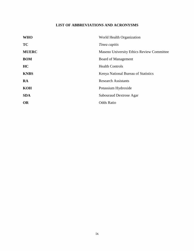
LIST OF ABBREVIATIONS AND ACRONYSMS
WHO World Health Organization
TC Tinea capitis
MUERC Maseno University Ethics Review Committee
BOM Board of Management
HC Health Controls
KNBS Kenya National Bureau of Statistics
RA Research Assistants
KOH Potassium Hydroxide
SDA Sabouraud Dextrose Agar
OR Odds Ratio

x
LIST OF TABLES
Table 3.1 Number of study participarts per school. ...................................................................... 21
Table 4.1 Prevalence of Tinea capitis in school going children in Kakamega Central Sub-County.
....................................................................................................................................................... 28
Table 4.2 Characterization of fungal species in children presenting with Tinea capitis .............. 29
Table 4.3 Summary of isolated fungal agents…………………………………...……………….30
Table 4.4 : Socio demographic and anthropometric characteristics of children and their aregivers
participating in the study ............................................................................................................... 33
Table 4.5 Risks factors associated with children participating in study…………………….…...36
Table 4.6 Association of Tinea capitis with risk factors……………….……………….………..47

xi
LIST OF FIGURES
Figure 3.1 A map showing Kakamega Central Sub-County………….………………………….18

xii
LIST OF PLATES
Plate No 1: Microsporum canis surface view………………………………………………...... 31
Plate No 2: Microsporum canis reverse………………………………………...................…….31
Plate No 3: Candida rugosa………………………………………………………………..……31
Plate No 4: Aspergillus niger surface view…………………………………………………………….31
Plate No 5: Aspergillus niger reverse view……………………………………………………...31
Plate No 6: Trichophyton tonsurans surface view………………………………………………31
Plate No 7: Trichophyton tonsurans reverse view………………………………………………31
Plate No 8: Epidermophyton floccosum surface view…………………………………………..31
Plate No 9: Epidermophyton floccosum reverse view………………………………………..…31

xiii
LIST OF APPENDICES
Appendix 1 Informed Consent by parent/guardian of the participants……………………….....64
Appendix 2 Kibali kilichofahamishwa na mzazi / mlezi wa washiriki………………………….67
Appendix 3 Informed Assent for the participants…………………………………………….....69
Appendix 4 Results for Pre-test of Socio demographic and anthropometric characteristics of
children and care givers participating in the study………………………...…………………….71
Appendix 5 Semi structured Questionnaire (English version) . …………………………………73
Appendix 6 Hojaji funge…………………………………………………………………………75
Appendix 7 Ethical approval letter.........................................................................................…...77
Appendix 8 Permission letter by Headmaster Mwiyala Primary School…………………..……78
Appendix 9 Permission letter by Headmaster Mahiakalo Primary School……………...………79
Appendix 10 Permission letter by Headmaster Elukho Primary School…………………….…..80
Appendix 11 Permission letter by Headmaster Emusala Primary School………………….……81
Appendix 12 SGS proposal approval letter-…………………………………..…….………….. 82

1
CHAPTER ONE
INTRODUCTION
1.1 Background Information
Tinea capitis (ring worm) is a superficial fungal infection of the scalp and hair of the head.
Globally, it is common in school children aged between three and thirteen years (Guerrant et al.,
2011; Terragni et al., 1991). Fungal agents responsible for Tinea capitis can live on the dead
tissue of the scalp which is caused by mold-like fungi that belong to a group of fungi called
dermatophytes (cause infections of the skin, hair and nails) that entails three genera:
Microsporum, Epidermophyton and Trichophyton. According to World Health Organization
(WHO), prevalence of superficial mycotic including Tinea capitis infection is estimated to be
20-25% of the world’s population (Coulibaly et al., 2018; Lakshmanan et al., 2015). Africa
being among the settings mostly affected, it is estimated that the range of Tinea capitis infection
ranges between 10-30% among primary school aged children (Moto et al., 2015).
Dermatophytes commonly cause skin disease in animals and humans. Up to date, there are about
40 species causing dermatophytosis, though only 8 species are associated with Tinea capitis.
However, epidemiological distribution of fungal agents causing Tinea capitis varies depending
on the geographical location and seasonal factors that includes emigration, immigration, life
style, type of population and climatic conditions (Moto et al., 2015). Additionally, research by
reviewers have indicated that as much as some species of etiologic agents causing TC have been
reported from every continent, others have geographically limited areas (Balci et al., 2014). For
instance, Trichophyton infections are common in Central America and parts of Western Europe
while Microsporum species have been identified as the key etiological agents of Tinea capitis in
South America, Southern and Central Europe, Africa and Middle East. However, studies have
revealed that, Tinea capitis is more common in Africa than in European and Asian countries

2
and this is corroborated by another study in India which observed that in developing countries
TC is high because of factors like poverty, overcrowding, improper hygiene and illiteracy. Tinea
capitis causing agents usually colonize the keratin tissues of the scalp hence inflammation occurs
around the infected area due to host response to metabolic by-products (Tainwala & Sharma,
2011) hence causing discomfort to infected children due to itching and this could affect their
concentration in class. The causative agents are generally restricted to non-living cornified layer
of the epidermis due to their inability to penetrate viable tissues of an immune competent host
(Lee & Hsu, 1992).
Previous studies have associated Tinea capitis with a number of factors including socio-
demographic, geographical regions, species virulence and environmental (Olutoyin et al., 2017;
Philpot, 1978). For instance, antropophilic fungal agents that causes Tinea capitis like
Trichophyton tonsurans, violaceum sudanense and Microsporum audouinii are most prominent
in tropical areas (Elewski, 2000; Fuller, 2009; Hay et al., 2001). Also, Trichophyton tonsurans
has been found to be the common cause of out breaks of Tinea capitis in children and it’s the
main cause of endothrix (inside the hair follicles ) infections. Additionally, Trichophyton rubrum
has been shown to be the common cause of favusin, (crusts are seen on scalp) (Michaels & Del
Rosso, 2012). A recent cross-sectional study in the Mathare slums, informal settlement in
Nairobi City reported an overall Tinea capitis prevalence of 81.3% (Moto et al., 2015). These
findings in Mathare study implies that TC is still a disease of concern in Kenya, though unlike
the current study, it had a smaller sample size and few risk factors associated with
dermatophytosis.
Kakamega County is the second most populous county in Kenya (KNBS, 2009), it comprise of
12 sub-counties. Kakamega Central Sub-County; my study site, has an estimated population of

3
100, 000 people where 57% of the population stays below poverty lines. Primary school going
children comprise 36,918 (KNBS, 2009). A significant number of Primary school going children
in this Sub-County have observable clinical signs of Tinea capitis and most of the primary
schools are characterized by overcrowding of pupils in classes (Barasa, 2016). Etiological agents
and associated risk factors of Tinea capitis among primary school going children in Kakamega
Central Sub- County have not been identified. Currently, there is very limited data on the
etiology and associated risk factors of Tinea capitis in school going children in Kakamega
Central Sub-County. Therefore, this study sought to find out the specific fungal causative agents
of Tinea capitis and their associated risk factors in primary school going children and the finding
of this study can help Kakamega County plan on how to treat the disease and prevent further
spreading of the disease and by so doing improving the concentration and performance of
children in school.
1.2 Epidemiology of Tinea capitis
Dermatophytosis is one of the most commonly encountered cutaneous fungal infections
worldwide. The higher prevalence in tropical and subtropical areas is supposedly due to the hot
and humid climatic condition. Other factors like personal hygiene habits and prevalence of
virulent species in the soil are also known to influence the infection.
Tinea capitis can occur in three distinctly different forms, "gray patch," "black dot" and favus.
Black dot Tinea capitis, most often caused by Trichophyton tonsurans, is the form
predominantly seen in the United States. Tinea favosa due to Trychophyton Schoenleinii is an
important anhtropophilic dermatophytes that is endemic in Africa, for example in Tunisia and
Nigeria T. Schoenleinii is significantly observed in primary school going children (Khaled et al.,
2007). However researchers have observed that improvement in hygiene practices and living

4
standards has reduced favus infection significantly. Immigration and travel may contribute to the
occurrence of favus in patients who live in non-endemic areas. Gray-patch ringworm
(Microsporosis) is an ectothrix infection or pre-pubertal Tinea capitis (is common in an African-
American male child). Hairs in the involved areas assume a characteristic dull, grayish,
discolored appearance. Infected hairs are broken and shorter. Papular lesions around hair shafts
spread and form typical patches of ring form.
Tinea capitis occurs primarily in children and occasionally in other age groups. It is seen most
commonly in children younger than 10 years. Most commonly affected range is children aged 3-
7 years (Ginter-Hanselmayer et al., 2007) In adults, women are infected more frequently than
men, possibly because of women having greater exposure to infected children and possibly
because of hormonal factors . Microsporum audouinii is also a common fungal agent that causes
TC in primary school children and a study in Swiss primary schools observed that M. audouinii
is more common in boys than in girls (Donghi, Hauser, & Bosshard, 2011). More recently, the
most prevalent organisms causing Tinea capitis infections in children are anthropophilic
organisms, which spread directly from person to person.
Severity of Tinea capitis depends on the site of formation of their arthroconidia. Some mild
ringworm or pre-pubertal Tinea capitis infections are of the ectothrix (fragmentation of
mycelium into conidia around the hair shaft) type. It is also termed the grey-patch type
(microsporosis). Depending on the extent of associated inflammation, lesions may heal with
scarring. Endothrix infections are noted by the arthrospores present within the hair shaft, this
contributes to the chronicity of the infections which tend to progress and may last into adult life.
Since the organisms usually remain superficial, little potential for mortality exists. However, TC
5
caused by T. tonsurans has been reported to cause lesions in form of concentric rings known as
tinea imbracata, which eventually causes disseminated systemic disease that has been observed
in patients who are severely immune compromised (Narang et al., 2012).
A survey done in primary schools in Kisumu District (western Kenya) in 1993 had a prevalence
rate of superficial mycoses of 10.1%. Three-quarters of the affected children suffered from Tinea
capitis with a prevalence rate 7.8% and the common causative agents were: Microsporum
audouinii, Trichophyton violaceum and Microsporum canis (Schmeller et al., 1997). Another
study was done in Eldoret which found a significant Tinea capitis infection of 33.3%, (Ayaya et
al., 2001) while another study in Kibera slums in Nairobi, Kenya had a prevalence of incidence
of 11.2% Tinea capitis (Chepchirchir et al., 2009) and a more recent study was done in Mathare,
informal settlement in Nairobi reported a much higher prevalence of 81.2%. (Moto et al., 2015).
Superficial mycoses have neither been the focus of intensive study nor of active control
programmes in the sub-Saharan Africa, including Kenya. Consequently, there is scarcity of
information on the epidemiology of superficial mycoses in Kenya and this lack of scientific
information has negatively affected development of adequate patient diagnosis, disease control
and antifungal drug resistance surveillance. The findings of this study can assist to address these
gaps in school going children in Kakamega Central Sub-County.
1.3 Statement of the Problem
Studies elsewhere have shown that scalp ringworm infections are endemic among school
children in tropical Africa. These infections have been known to inflict a lot of psychosocial
trauma on children due to attached social stigma, ulceration, and sometimes irritation which
hampers pupil’s concentration in class. A significant number of primary school going children in
Kakamega Central Sub-County has observable clinical symptoms suggestive of Tinea capitis

6
infection. Although the prevalence, fungal species causing Tinea capitis among primary school
going children and risk factors associated with TC infection have been identified elsewhere in
Kenya, this has not been done in Kakamega Central Sub-County, and this remains a problem
because, fungal agents causing Tinea capitis vary in virulence, clinical presentation and they are
geographically limited of greater or less extent, this study sought to find out the prevalence,
fungal etiologic agents causing TC and risk factors associated with the TC in Kakamega central
Sub-County.
1.4 General Objective
To determine the prevalence, characterize fungal agents causing Tinea capitis and associated risk
factors among primary school going children aged 3 – 14 years in Kakamega Central Sub-
County.
1.4.1 Specific Objectives
1. To determine the prevalence of Tinea capitis among primary school going children in
Kakamega Central Sub-County.
2. To characterize the fungal species causing Tinea capitis in primary school going children in
Kakamega Central Sub-County.
3. To determine the risk factors associated with Tinea capitis infection in primary school going
children in Kakamega central Sub-County.
1.4.2 Research Questions
1. What is the prevalence of Tinea capitis among primary school going children in Kakamega
Central Sub-County?
2. What are the fungal species causing Tinea capitis in primary school going children in
Kakamega Central Sub-County?

7
3. What are the risk factors associated with Tinea capitis infection in primary school going
children in Kakamega Central Sub-County.
1.5 Significance of the Study
This study has established a prevalence of 17.3% of TC infection in primary school going
children in Kakamega Central Sub-County hygiene practices and age are among the major risk
factors associated with prevalence of Tinea capitis in Kakamega Central Sub-County; children at
the age group of 3-8 years were three times at risk of having Tinea capitis compared to those in
the aged 9-14 years. Also, it was revealed that, children that did not take baths daily, over-
crowding in schools, shared beddings, combs and many bed occupants are strongly associated
with having Tinea capitis than those who take bath daily, don’t share beddings, combs and bed.
These results can help County government of Kakamega, to plan on how to curb the risk factors
and hygiene practices associated with Tinea capitis infection in Kakamega Central Sub-County.
By alleviating such factors in primary schools and homes, it will assist in eradicating Tinea
capitis in the county and this will eventually improve the overall well-being of children in
school.

8
CHAPTER TWO
LITERATURE REVIEW
2.1 Introduction
Tinea capitis is a common superficial fungal infection of the scalp and hair. It is an exogenous
infection that is characterized by invasion of dermatophytes into hair follicles and keratinized
layer of hairy skin leading to hair loss, scaling, kerion agminate folliculitis, favus black dot, grey
patch type, erthema or impetigo-like grey patch type, erythema or impetigo-like lesions. It is
commonly caused by dermatophytes in the Epidermophyton, Trichophyton, and Microsporum
genera. Dermatophytes metabolize keratin and cause a range of pathologic clinical presentations,
including Tinea capitis, tinea pedis, tinea corporis, tinea cruris, and Majocchi's granuloma
(Boral, Durdu, & Ilkit, 2018). T. rubrum is the main cause of Majocchi's granuloma, though
other fungal species like T. mentagrophytes, T.violaceum and T. tonsurans also causes the
disease. Tinea capitis is a worldwide infection occurring in both rural and urban areas; it has
commonly been reported in children of Afro-Caribbean origin in North America, Central
America, and South America. In the United States T. tonsurans now causes more than 95% of
TC infections (Magill et al., 2007). Africa being among the settings mostly affected the rate of
TC infection ranges between 10-70% among school aged children (Coulibaly et al., 2018).
2.2 Prevalence of Tinea capitis
The prevalence of superficial mycosis infections has globally risen to such a level that skin
mycoses now affect more than 20-25% of the world's population, (Bassiri-Jahromi & Khaksari,
2009), in Europe, Tinea capitis (scalp ringworm) is the most common dermatophytes infection of
the scalp affecting mainly children and rarely adults. Studies have established that, prevalence of
Tinea capitis varies within different geographical areas throughout the world hence it can occur
sporadically or epidemically and an increase in its incidence has been noted over the last few

9
decades. In Europe, the countries reporting the highest incidence of Tinea capitis infections are
mainly in the Mediterranean but also bordering countries like Austria, Hungary, Germany and
Poland. Research done in São Paulo, Brazil in 1996, in Private and Public Pediatrics Service
involving children from 0 to 15 years established a prevalence of 85% (Moraes et al., 2006)
which is quite similar to the TC prevalence in African set up. In South-East Amsterdam, research
established prevalence of 7% TC infection (Timen et al.,1999). This was much lower than that of
São Paulo, Brazil. In Nablus district in Palestine, a study reported a much decreased prevalence
of 1% Tinea capitis infection, the infection occurred both in rural and urban school going
children, although the incidence was higher in schools in rural areas (1.9%) than urban areas
(0.4%) where boys were more commonly affected than girls (Ali-Shtayeh et al., 1998).
In Africa, Tinea capitis disease is a public health burden. It is the mostly affected continent with
infection ranging between 10 and 70% among school-aged children of below 15years (Coulibaly
et al., 2018).
The predominant clinical forms and causative agents vary from one region to another. Poor
socioeconomic status, high population densities, and poor sanitary conditions are some of the
factors accountable for the high prevalence of dermatophytosis in many developing countries,
which include countries in southern and eastern Africa. A Study carried out to review the
prevalence of Tinea capitis in countries including Kenya, Ethiopia, Tanzania, South Africa,
Mozambique, Madagascar, Malawi, Rwanda, Burundi, Uganda, Zambia, Zimbabwe and
Botswana had a prevalence ranging from 56.7% to 95%. For instance, a study done in Nigeria,
Nok community of Kaduna State, reported a prevalence of 45% and girls had higher prevalence
(51.4%) than boys (41.5%) but not significantly different. Another study done in Gabon,
established Tinea capitis infection rate that varied according to the school studied, the prevalence

10
ranged from 20.4% in the urban school with a higher socioeconomic status to 26.3% in the rural
school with a lower socioeconomic status ( Hogewoning et al., 2011). In India, Kolkata, West
Bengal state 10% TC infection rate was reported which was slightly lower that in Gabon. This
trend is similar to that of United Kingdom and North America.
In Kenya, a study done in Mathare slums, informal settlement in Nairobi, Kenya established a
prevalence rate of 81.3% infection rate of TC in primary school going children, Trichophyton
species (61.3%) were the common fungal agents detected with T. tonsurans being the most
prevalent, children in age groups 3-5 and 6-8years were the most affected (Moto et al., 2015). In
another study carried out in Eldoret town in primary school going children aged 3-14years,
reported a prevalence of 33.3% TC infection with a pick age of 10years (Ayaya et al, 2001),
Similarly, T. tonsurans was identified as the most common fungal agent causing TC like in
Mathare research. In Kisumu town, a study reported 10.1% prevalence of dermatophyto-mycoses
infection in primary school going children with three-quarter of the infected children suffering
from TC infection (7.8%), and the commonly isolated fungal agent was M. audouinii
var.langeronii (Schmeller et al., 1997).
Additionally, research done in primary school children in Kibera slums in Nairobi established
11.2% prevalence of dermatophytes infection with TC being the most common (Chepchirchir et
al., 2009). In these studies carried out in Kenya, most of the fungal agents species causing TC
disease are different, for instance, in Kisumu TC infection was mainly caused by Microsporum
audouinii var. langeronii, Trichophyton violaceum and Microsporum canis while in Kibera
slums Nairobi City T. violecium was the predominant species isolated followed by T.
mentagrophytes; although Mathare and Eldoret researches reported T. tonsurans as the most
prevalent species causing TC infection in primary school children. There is no research that has

11
been done in Kakamega County to establish the prevalence of Tinea capitis infection despite the
observable clinical presentation of primary school going children with ring worms of the scalp,
lack of this scientific knowledge that can help the Kakamega County plan how to eradicate the
disease motivated this current study to research to establish the extent of TC infection.
2.3 Fungal Species Causing Tinea capitis
Tinea capitis is a superficial fungal infection of the scalp and hair that is caused by
dermatophytes commonly in the Epidermophyton, Trichophyton, and Microsporum genera. Up
to date, we have about 40 species causing dermatophytosis (Pai et al., 2013), though only 8
species are associated with Tinea capitis. Epidemiological distribution of Tinea capitis causing
agent varies depending on the geographical location. For instance, Trichophyton infections are
common in Central America and parts of Western Europe while Microsporum species have been
identified as the key etiological agents of Tinea capitis in South America, Southern and Central
Europe, Africa and Middle East. Trichophyton tonsurans is an anthropophilic dermatophyte,
with a worldwide distribution, although its prevalence varies considerably between different
geographical regions. Whereas in North America T. tonsurans is the main fungal species causing
TC infections in children below 15 years in the European continent they appear relatively rare.
However, T. tonsurans is primarily associated with Tinea capitis (Hryncewicz et al., 2011).
Similarly, a study done in Irish pediatric population reported T. tonsurans to be the predominant
fungal etiological agent causing TC disease in Ireland, same results were reported in
Netherlands, southeastern Amsterdam, and Cleveland. Contrary to other studies which indicates
T. tonsurans as a dominant fungal species causing TC in Europe, another study by (Ginter-
Hanselmayer et al., 2007) reported Microsporum canis, (zoophilic dermatophytes,) as the most
common causative agent of Tinea capitis in Europe. Another study has observed that T. rubrum

12
is a common cause of superficial mycosis in developing countries in Europe, it’s a predominant
pathogen in Germany, Finland and Russia regions (Tietz et al., 1999). However there is a
significant shift towards anthropophilic Tinea capitis mainly in urban areas in Europe, with
exception of France that reported Trichophyton soudanense and Microsporum audouinii as
common species causing TC infection (Ginter-Hanselmayer et al., 2007).
Tinea capitis is more common in Africa than in Europe and Asia (Ali et al., 2009). Poor
socioeconomic status, high population densities, and poor sanitary conditions are some of the
factors responsible for the high prevalence of dermatophytosis in many developing countries
(Nweze & Eke, 2017). By contrast, species such as T. violaceum and T.Soudanense were
reported to be the most common fungal species causing TC in primary school going children in
parts of Africa and West Asia (Magill, 2007), have rarely been isolated from patients in the
United States. But studies in Abidjan, Cote d'Ivoire, Gabon and Ivory Coast identified
Trichophyton soudanense as the most prominent fungal etiological agents causing TC infection
(Adou-Bryn et al., 2004; Fulgence et al., 2013; Hogewoning et al., 2011) while among school
children in Nok community of Kaduna State, Nigeria, studies reported Trichophyton rubrum
followed by Microsporum canis as the most prevalent dermatophytes isolated. Notably, in Egypt
recent studies reveals a shift of trend of TC infection, where M. canis is replacing T. violaceum
as a prominent fungal species casing TC disease in primary school going children (Bassyouni et
al., 2017).
Studies done in Kenya have similarly reported same trend like other parts of the world, where
TC infection is associated with different geographical location though, most of the infections are
caused by etiologic agents commonly from the three genera that causes TC; Trichophyton,
Microsporum and Epidermophyton. For instance, a study done in primary school children in

13
Eldoret town found out that, T. tonsurans was predominantly isolated as TC causative fungal
agent (Ayaya et al., 2001). This finding was different from a study done in Kisumu town which
found that Microsporum audouinii var. langeronii, was the most predominant species causing
TC in primary school going children however, Trichophyton violaceum and Microsporum canis
were also isolated (Schmeller et al., 1997). In Kibera slums of Nairobi, a study reported T.
violaceum as the predominant fungal species causing TC (Chepchirchir et al., 2009). A recent
study that was done in school going children from Mathare informal settlement in Nairobi
Kenya, reported T. tonsurans as the common dermatophyte causing TC (Moto et al., 2015).
Previous studies done found that more than 90% of the Tinea capitis infection in children
worldwide is caused by T. tonsurans and less than 5% infections are caused by Microsporum,
species especially in Europe and some parts of Africa (Abd Elmegeed et al., 2015). But
Trichophyton soudanense and M.audouinii has been reported to be the most common fungal
agents causing TC in France (Donghi et al., 2011). Similar studies of T. soudanense being most
prevalent etiologic agent of TC has been observed in, Gabon (Hogewoning et al., 2011) and
Ivory Coast.
However, as much as some fungal etiological agents causing TC have been reported from every
continent, studies have shown that most of the species have geographically limited areas of
greater or less extent (Ginter-Hanselmayer et al., 2007) or can change overtime (Pai et al., 2013;
Soyinka, 1978; Fuller, 2009). The diversity in geographical and seasonal variation of fungal
agent causing TC can be attributed to factors such as; widespread use of antifungal agents like
griseofulvin which is more effective against M. auduoinii than T. tonsurans, changes in
immigration patterns and increase in internal travel (Pai et al., 2013). Additionally, other studies
have corroborated this findings by attributing the geographical diversity of TC fungal causing

14
agents to life style, type of population, endemicity, type of animal reservoirs and climatic
conditions (Dogo et al., 2016; Elewski, et al., 2000; Soyinka, 1978). Other studies have shown
that Tinea capitis is more endemic to tropical and subtropical African regions because
dermatophytes grow best in warm and moist (humid) conditions (Dogo et al., 2016).
Furthermore, there is progressively changing patterns in etiology and chemical manifestation of
Tinea capitis infections (Chokoeva et al., 2016). This therefore may mean that even with well-
known diseases, there could be other facts still hidden for future revelations. Studies have
revealed that, apart from the known dermatophytes that causes Tinea capitis, molds also have
been reported to be on the rise in causing TC infections (Chokoeva et al., 2016). For example,
genus Apergillus is significantly being reported as an emerging mould-induced fungal etiological
agent causing TC. For instance, in a retrospective study done in Brazil reported that A. niger and
A. flavus species are responsible for causing TC (Chokoeva et al., 2016).
The presence of clinically observable signs of Tinea capitis in a significant number of primary
school going children in Kakamega Central Sub-County and also considering factors that
influence the presence of fungal agent causing TC like geographical and seasonal variation,
endemicity of the disease, changes in immigration patterns and increase in internal travels and
limited information in this setting about characterization of fungal agents causing Tinea capitis
and risk factor associated with Tinea capitis infection motivated me to carry out this study at
Kakamega Central Sub-County.
2.4 Risk Factors Associated with Tinea capitis Infection
Tinea capitis infection has remained a public health problem worldwide and some of the risk
factors that are attributed to predispose a population to an infection include poor hygiene, sharing
of formites, overcrowding and low socio economic factors (Moto et al., 2015). Growth of
15
dermatophytes including TC causing agents are supported by warmth and moist environment
(Dogo et al., 2016). Also prevalence of TC disease can be attributed to Lack of frequent bathing,
having damp skin for extended periods of time for instance, not showering and drying off
completely, sweating, minor skin and injuries, close contact with others who have ringworm,
such as sharing of combs or a room or sitting in close conduct in an overcrowded classroom with
infected classmates (Enendu & Ibe, 2005). A study done in Turkey, established that school
settlement is a risk factor for Tinea capitis infection and spread (Balci et al., 2014). The same
study of Turkey reported that TC was more frequent in children under the age of 12 years and
more common in boys than girls. Also, prevalence of Tinea capitis varies within different
geographical areas and climatic conditions, these are some of the major risk factors that
determines the fungal agents that cause TC throughout the world, (Hibstu & Kebede, 2017) .
In Africa, Tinea capitis is common in humid countries and its spread is promoted by poor-living,
poor sanitary conditions with overcrowding, sharing of combs, towels, barbers clippers, house
hold pets e.g cats (Enendu & Ibe, 2005), the same associated factors were reported in Gabon and
Ivory coast (Adou-Bryn et al., 2004; Fulgence et al., 2013; Hogewoning et al., 2011).
In Kenya, risk factors associated with the infection and spread of TC were partially mentioned
in studies done in Kibera, Narobi city, and in Mathare informal settlement in Nairobi
(Chepchirchir et al., 2009; Moto et al., 2015). There are no studies that have been done to find
out the risk factors associated with TC infection in Kakamega Central Sub-County to assist in
prevention and treatment of Tinea capitis.

16
2.5 Summary of Knowledge Gaps
Kakamega Central Sub-County is warm and humid area hence conducive for Tinea capitis. In as
much as primary school going children present with Tinea capitis, the fungal etiological agents
causing this infection have not been characterized. Additionally, the burden of Tinea capitis in
Kakamega Central Sub-County has not been examined in Kakamega Central Sub-County. This
study, established the prevalence of Tinea capitis in the school going children aged 3-14 years,
identified the fungal etiologic agents causing the Tinea capitis disease and the relationship
between the disease and personal hygiene practices and environmental sanitation.

17
CHAPTER THREE
MATERIALS AND METHODS
3.1 Study Area
The study was done between January and June 2016 among primary school going children aged
3-14 years attending public primary schools in Kakamega Central Sub-County, Kakamega
County. The Sub-County has an estimated population of 100,000 of whom 36,918 are primary
school going children; 18,268 (49%) boys and 18,852 (51%) girls. In addition, the Sub-County
has a total of 65 public primary schools, of which 26 are urban and 39 rural (Ogola, 2010).
Kakamega Central Sub-County is approximately 415 KMs west of Nairobi located within
Kakamega County latitude: 0°16'60.00"N and longitude: 34° 44' 59.99" E (Figure 3.1). There are
two distinct climatic seasons; the wet and dry seasons. The former has long and short rainfall;
long rains in April to July and short rains in September to November, while the latter season is
between December and April (Ndetei, 2013).
The average atmospheric temperature is 31°C. The study site is encompassed by Kakamega
forest and Lake Victoria which is around 68kms south of Kakamega town, these contributes to
humidity and warm weather that is conducive for growth and multiplication of dermatophytes
including ring worm of the scalp presently under investigation. Furthermore, census 2009
indicates that, an estimated 57% of the populations lives below the poverty line (KNBS, 2009).
The main economic activities are: large scale farming of sugar cane, mixed farming, commercial
businesses and boda boda transport which is popular among youths aged 18-35 years. Kakamega
Central Sub-County has both urban and rural settings both of which vary in access to social
amenities including clean tap water, healthcare services.

18
Figure 3.1: A map showing Kakamega Central Sub-County
3.2 Study Design
The study adopted cross-sectional study design.
3.3 Study Population
Study population of 4,611 children from the four selected schools constituted the study
population (Table 3.1). A total of 375 children were randomly recruited to participate in the
study: 65 children with T. capitis were the subjects and 310 who were Tinea capitis negative as
control.

19
All the schools selected for this study were public schools. Two schools were from urban setting
and the other two from rural.
3.3.1 Inclusion Criteria
All pupils aged 3 – 14 years present in school at the time of study, whose parents had consented
(Appendix 1) for English and (Appendix 2) for Kiwahili version, and those who had assented
(Appendix 3) were included in the study.
3.3.2 Exclusion Criteria
Children on treatment for Tinea capitis and those whose parents did not consent or those children
who did not give assent were exempted from the study.
3.4 Sample Size
Sample size determination was calculated using the following formula:
n = z²pq/d² (Fisher et al., 1978)
Where n = sample size
z = the standard normal deviate (1.96 for a 95% confidence level)
p = prevalence- Prevalence of 33.3% from Eldoret study (Ayaya et al., 2001) was used due to
lack of prevalence information in these settings.
q = 1-p
d = 0.05 as the level of statistical significance.
Therefore: n=1.962x0.0333 (1-0.333)/0.05
2=341
10% of the 341 participants were included to cater for non-response.

20
= 10% X 341 = 34 participants.
Therefore, 375 study participants were recruited.
The sample size of 375 school going children randomly selected from the 4611 is a
representative of the population of primary school going children in Kakamega Central Sub-
County. Prevalence of 33.3% of a study in Eldoret was used (Ayaya et al., 2001). This is
corroborated by other studies where, if the prevalence is unknown, then the prevalence of
previous studies published within the study domain can be used (Pourhoseingholi et al., 2013).
Present study settled on sample size of 375 primary school going children based on the formula
used in 3.2 above, and also as corroborated by study done in Kayseri city in Turkey, which had
a total 139,422, primary school children, out of which 8122 children were selected to participate
in the study (Balci et al., 2014). Another study in Mathare slums informal settlement in Nairobi,
Kenya, 150 primary school going children were selected to participate in the study (Moto
Maingi, & Nyamache., 2015). Also, another study in Ebonyi state, in Nigeria had a sample size
of 279 primary school going children randomly selected from four schools to participate in the
study (Anosike et al., 2005).
3.5 Sampling Techniques
The number of study participants per primary school is shown in Table 3.1. Purposive sampling
technique was used to select the four schools in the Sub-County that was used as a representative
sample of Kakamega Central Sub-County primary schools; this technique was used so that to get
both rural and urban schools to be included in the study. Two schools were from urban set up
and two from rural. Systematic random sampling method was then used to select the participants.
The desired sample size (n) was 375 while the study population (p) was 4611. The interval of
study subjects` selection was given by p /n (4611/375) which was 12. Therefore, every 12th

21
pupil was selected. Same sampling technique was used to identify children presenting clinically
with scalp ring worms. The sample size (n) in this case was proportionately distributed to each of
the four primary school; Mwiyala primary school 71, Mahiakalo 123, Elukho 102 and Emusala
79 participants. Study participants were then selected with the predetermined sampling interval.
This was done until the required sample size was reached. This was replicated in all the four
schools. The total number of participants included in the study was sum of participants sampled
from all the four schools.
Table 3 .1 Number of study participants per school
School Children population Formula Sample
Mwiyala (Urban) 876 876/4611*375 71
Mahiakalo (Urban) 1515 1515/4611*375 123
Elukho (Rural) 1250 1250/4611*375 102
Emusala (Rural) 970 970/4611*375 79
Total population 4,611 Total Sample Size 375
3.6 Data Collection Process
3.6.1 Recruitment of research assistants
Two research assistants (RAs) with a qualification with at least certificate in Medical Laboratory
Technology with working experience in a Mycology laboratory were recruited. The RAs were
hired for two months to assist in sample collection.
3.6.2 Training of research assistants
The RA were trained for two weeks on standard operating procedures of how to screen, collect
and transport samples from the schools to Masinde Muliro University Microbiology laboratory
for processing and analysis.

22
3.6.3 Pre-testing
The pre-testing study was done in the neighboring Kisumu County to test the research tools
where 10% of the sample size was used. A Questionnaire was tested for collection of socio-
demographic data (Appendix 4).
3.6.4 Data Collection
There was two types of data collected: 1) Clinical samples; scalp scrapings and hair for fungal
identification was collected in sterile white envelopes; 2) Socio-demographic and anthropometric
data was obtained through semi-structured questionnaires (Appendix 5) for English version or
(Appendix 6) for Kiswahili version. Information both from children participating in the study on
the age, gender, weight, education level was collected, Also, socio-demographic data from
parents/ care givers on age, age of the mother when giving birth, marital status, education level,
occupation and monthly income, of parents was collected too.
The 375 pupils examined to the study went through the following procedure: physical
assessment- was carried out to screen the presence of Tinea capitis (clinical symptoms)
infections. The examination took place at a designated room in each of the selected schools. The
skin scalp, eyebrows, and eyelashes of each child was carefully examined for characteristic
features of ring worm of the scalp as before collection, thereafter the pupils were interviewed
to collect socio-demographic data.
3.6.5 Specimen Collection
Each child was examined in a room with sufficient light; the scalp was examined for scaly grey
patches, lusterless hair strands and purulent lesions. Affected areas were cleansed with 70% v/v
ethanol, allowed to dry and light scrapings (skin scales, crusts, hair pieces) were taken from the
active edge of lesion using a disposable blunt sterile scalpel blade. The samples were collected in

23
a sterile brown envelopes with each participant’s sample handled separately to avoid scrapings
getting mixed and code labeled, samples were then transported to the laboratory within 2 hours
at room temperature for microscopic and culture analysis.
3.7 Laboratory Analyses
Preliminary mycological analysis of specimens was carried out at Masinde Muliro University of
Science and Technology in Microbiology Labs, Kakamega. Direct microscopy as a preliminary
test was done to get an impression of the presence of fungal cells prior to culture. A portion of
each specimen was placed on a clean sterile slide and a drop of about 20% potassium hydroxide
(KOH) was added to the sample and incubated for 30 minutes for the digestion of keratin to
occur; then examined for the presence of hyphae and/or arthroconidia under low (x10) and high
(x40) power objective. However, direct microscopy method is not an identification method for
fungal agents but it augments’ culture during characterization of fungal agents. Modified
Sabourauds Dextrose Agar (SDA) is the preferred selective medium primarily used for the
isolation of dermatophytes (Rijal, 2015).
Scrapings were inoculated onto modified SDA plates with 0.05g/mL chloramphenical and
cycloheximide 0.5mg/mL (Weitzman & Summerbell, 1995) and on another set of SDA media
plates without cycloheximide (Chepchirchir et al., 2009). The media was prepared according to
the standard procedures ((Rijal, 2015) . Chloramphenical inhibits the growth of bacterial
contaminants while cycloheximide suppresses saprophytic fungi. Sample were separately
inoculated onto the prepared SDA using a sterile wire loop then incubated the plates at 30°C in
an inverted position with humidity. Samples were examined daily to capture pathogens as per
their rate of growth (Attal, 2016). Cultures were kept for a maximum of eight weeks before being
ruled out as negative for growth. The cultural characteristics of the isolates were noted and

24
identified based on duration of growth, surface morphology, pigment production on the reverse
,the texture, whether fluffy, powdery cottony or floccose, buff, whether the hyphae was radiating
at the margins or whether their colony were folded. Microscopic examination was done by
making a thin preparation of the fungal culture with a drop of lacto phenol cotton blue stain on a
glass slide, covered with a coverslip and observed under a microscope using (x10) and (x40)
objectives. Identification was based on macroscopic (growth characteristics and pigmentation)
and microscopic morphology (formation of macroconidia and microconidia or other typical
elements).
3.8 Data Management and Analysis
Each study questionnaire had a coded study identity that is uniquely associated with each study
participant and clinical and laboratory tests. Biological samples were coded in line with study
participant’s codes. Socio-demographic, clinical and laboratory information was entered into
Microsoft Office Access data bases, dated and time-stamped with an electronic signature. The
database was configured such that an audit trial is created to track any changes made to the
record(s), with time- and date-stamping, and an electronic signature appended. Electronic data
was backed-up daily in google drive, a web-based storage device. Scanned images of study
participant questionnaires were saved on google drive while source documents were placed in
lockable cabinets. Data was summarized as numbers and percentages and presented in tables.
Categorical variables were analyzed using Chi square test of homogeneity. Two by two
contingency tables were analyzed using the Fischer’s exact test and three by two tables were
analyzed using Chi-square test. Continuous variables such as weight and height were analyzed
using Mann Whitney U test. Binary logistic regression was used to determine the associations
between variables for objective three. Statistical analysis was set at P ≤ 0.05.

25
3.9 Ethical Considerations
Scientific approval was sought from Maseno University Ethics Review Committee (MUERC)
School of Graduate Studies (Appendix 7). Permission was also sought from the Board of
Management (BOM) through the Head masters of the selected schools for the study (Appendix
8, 9, 10 and 11). Before samples were taken and socio demographic data collected,
parents/caregivers consented for the children below 13 years (Appendix 1) for English version,
(Appendix 2) for Kiswahili version and assent was obtained (Appendix 3) from participants who
were above 13 years.
Participation into the study was on voluntary basis and the participants were encouraged to feel
free to withdraw from the study at any stage. The approval was on the agreement that
participant’s anonymity and confidentiality shall be maintained. Interviews were one-to-one
interaction and no information was given to any other unauthorized person. No names were
recorded; only serial numbers were entered into the questionnaire. Also, any information
concerning this study was stored in password protected computers that are accessible only by the
principal investigator.
Additionally, good laboratory practice/quality control was observed to ensure materials
collected from participants containing infectious materials do not infect laboratory workers.
Those participants that were infected by TC were treated by Ketoconazole cream, they were
instructed by a pharmaceutical Technologist to apply twice (morning and evening) daily for 7-14
days. Thin layer of the ointment is applied at the affected area then rub gently, for those whose
ring worm would not have cleared by end of 2 weeks or experiencing side effects like irritation,
itching, reddening of the area being treated, they were advised to visit the nearest health facility

26
for further assistance. Additionally safety measures were observed during the disposal of
laboratory wastes to prevent contaminating the environment and in turn the public get infected.
There were no foreseeable risks attached to this study.

27
CHAPTER FOUR
RESULTS
4.1 Prevalence of Tinea capitis among primary school going children in Kakamega Central
Sub-County
This study had a total of 375 participants randomly selected from the four primary schools. Up
on screening of the participants based on clinical observation, 65 children presented clinically
with symptoms of Tinea capitis hence revealing a prevalence of 17.3% of Tinea capitis infection
in primary school going children in Kakamega Central Sub-County [Table 4.1]. Of the infected
population 33 (50.8%) were male and 32 (49.2%) female. Children in these primary schools
were clustered into four categories; 3-5years, 6-8years, 9-11years and 12-14years. Children in
these clusters have more less the same behavior in terms of playing and personal hygiene
practices. The clusters have some common characteristics both at school and at home like
playing together and similar hygiene practices.

28
Table 4.1 Prevalence of Tinea capitis in school going children in Kakamega Central Sub-
County.
Age group
(years)
Schools
Mwiyala
(n=71)
Mahiakalo
(n=123)
Elukho
(n=102)
Emusala
(n=79)
Total n
(%)
M
(n=36)
F
(n=35)
M
(n=64)
F
(n=59)
M
(n=52)
F
(n=50)
M
(n=38)
F (n=41)
3-5 1 3 3 3 3 4 1 2 20
6-8 3 5 4 2 4 3 4 3 28
9-11 1 1 2 0 3 2 1 2 12
12-14 0 1 1 0 1 0 1 1 5
Totals 5 10 10 5 11 9 7 8 65/375
(17.3)
Data are presented as numbers (n) or as prevalence (%). M, male children within an age bracket.
F, female children within an age bracket. The data represents the overall prevalence of children
presenting physically with Tinea capitis in the four schools in Kakamega Central Sub-County,
Kakamega County.
4.2 Characterization of the fungal species causing Tinea capitis in school going children in
Kakamega Central Sub-County
4.2.1 Characterization of the fungal species causing Tinea capitis
After culture, 52 (80%) samples out of the 65 collected samples had growth and 13 (20%) had
no growth after eight weeks of incubation at 30°C. Lack of growth for the 13 samples, could be
inferred that, some of the dry lesions had almost healed hence the lack of growth and recovery of
the arthroconidia. The cultural characteristics of the isolates were used to identify fungal agents;
surface morphology, pigment production on the reverse, the texture, whether fluffy, powdery
cottony or floccose, buff, whether the hyphae was radiating at the margins or whether their

29
colony were folded (Table 4.2). Microscopic examination was done using lacto phenol blue to
reveal the arrangement of hyphae (Table 4.2).
Table 4.2 Characterization of fungal isolates grown on SDA at 30°C
Incubation
period
Cultural characteristics Microscopic
description of
lactophenol blue
Isolates
Surface Reverse
5-9 days Brown to tan waxy colony
with brownish powdery
aerial mycelia colonies often
with radial grooves.
Orange to Cream Broad hyphae,
irregular , much
branched and
mulitiseptate
T.
tonsurans
2-4 days Woolly or cottony white ,
beige with flat colonies and
radiating edges
Pale white Rough thick walled
and multiseptate
macroconidia
M. canis
3-5 days White to cream colony of
closely matted mycelia.
Central knob with radiating
and unfolded surface
Yellow-brown to
reddish-brown
Short segmented and
relative hyphae
present
M.
audouinii
3-5 days Red flat granular colonies
with clear tinted center.
Rose brown Numerous
microconidia ,
conidia, rounded
and pea born singly
or in clusters spiral
hyphae present.
T.
mentagroph
yte
5-7 days Heaped up white to reddish
cottony colonies
Cherry red T. rubrum
2-7 days Initially white then Green
brown or black velvety or
cottony
Whitish golden or
brown
Septate hyphae,
enlarged
conidiophore
A .niger
2-7 days Dark green hyaline Coarsely roughend
conidiophore
A. flav
us
5-12 days Greenish-brown/
khakicoloured with
suedelike surface. Raised &
folded in the centre, flat
periphery and submerged
growth.
Deep yellow-brown
pigment
Smooth, thin-walled
macroconidia in
clusters
E.
floccosum
3-5 days White to cream flat glabrous
with submerged edge
Cream to white Elongated cells with
multilateral budding
and pseudohyphae
Candida
rugosa
Data presented as incubation period; time taken before the growth is noticed, Surface is front
appearance of the colonies, reverse, the back appearance of the culture plates, Microscopy of
colonial morphology in lacto phenol blue preparation, isolates; fungal agents identified from the
cultures

30
4.2.2 Prevalence of Fungal Agents Isolated from the Cultures
Out of the 52 isolates, 31 (60%) were of the genus Trichophyton. In the genus trichophyton, the
most prevalent fungal agent was Trichophyton tonsurans which accounted for 51.9% of the
fungal agents identified and 84.9% of the genus Trichophyton [Table 4.3]. Other fungal species
in the genus Trichophyton identified were, Trichophyton mentagrophyte 3 (5.8%) and
Trichophyton rubrum 2 (3.8%). The other fungal agents identified were of the genus
Microsporum which accounted for 23.9% of all the fungal agents. In the genus Microsporum;
Micropsporum canis was the most prevalent accounting for 13.6% of the entire fungal agents
identified. The other fungal agent in this genus identified was Micropsporum audouinii
accounted for 5.8% of all fungal agents identified in study participants. Another genus identified
was Aspergillus in which two species were identified; Aspergillus niger 3 (5.8%) and Aspergillus
flavus 1 (1.9%) [Table 4.3]. Additionally, Epidermophyton flocosum was also identified
accounting for 1.9% of the total fungal agents. Interestingly, 1 isolate of Candida rugosa was
identified among the fungal species. It accounted for 1.9% of all the fungal agents identified.
Among the children presenting with Tinea capitis, 4 (7.7%) had co-infection of two or more
fungal causing agents [Table 4.3].
Table 4.3: Summary of isolated fungal agents
Fungal agents Number identified (n/65) Prevalence (%)
Trichophyton tonsurans 27 51.9%
Microsporum audouinii 3 5.8%
Trichophyton mentagrophyte 3 5.8%
Micropsporum canis 7 13.5%
Aspergillus niger 3 5.8%
Aspergillus flavus 1 1.9%
Candida rugosa 1 1.9%
Epidermophyton flocosum 1 1.9%
Trichophyton rubrum 2 3.8%
Co-infections 4 7.7%
Data are presented as numbers (n) and prevalence (%). Co-infections, two or more fungal agents
were identified in a sample.

31
4.2.3 Sampled culture plates of fungal isolates after 2-7 days growth at 30°C on SDA
Key: Plate 1: M. canis-surface; woolly or cottony white flat colonies, plate 2: M. canis-Reverse;
pale white, plate 3: C. rugosa- surface; white to cream colonies, plate 4: A. flavus- surface; dark
green colonies, plate 5: A. niger- surface; green to brown velvety colonies, plate 6: M. audouinii
– surface; white to cream colony with central knob with radiating surface, plate 7: M. audouinii-
reverse yellow-brown. Plate 8: E. flocossum -surface; greenish to brown/khaki colonies with
suede like surface, plate 9: : E. flocossum- reverse; deep yellow to brown pigment.
Plate 1
Plate 2 Plate 3
Plate 4 Plate 5
Plate 6
Plate 7 Plate 8 Plate 9

32
4.3 The risk factors associated with Tinea capitis infection in primary school going children
in Kakamega central Sub-County
4.3.1 Socio demographic and anthropometric characteristics of study participants
A total of 375 children aged between 3 and 14 years were enrolled in the study. 194 (51.7%)
were male children and 181 (48.3%) accounted for female children. 65 (17.3%) children
presented clinically with Tinea capitis while 310 (82.6%) children were without clinical
manifestation hence regarded as Tinea capitis negative.
Gender of those participants who had Tinea capitis disease and those who were negative was
comparable (P=0.391) [Table 4.4]. Male children accounted for 55.2% of the Tinea capitis
negative and 50.8% of the Tinea capitis positive while female accounted for 44.8% Tinea capitis
negative and 49.2% Tinea capitis positive. Similarly, age did not vary significantly between the
groups (P=0.907) with the children ages in both groups ranging from 3 to 14 years. Additionally,
the anthropometric measures of weight and height of the children participating in the study did
not differ (P= 0.941 and P=0.849) respectively [Table 4.4]. Likewise, the religion and ethnicity
of children with TC and those without did not differ.
The age of the mother and (or) caregiver was not significantly different between those children
with the disease and those who were negative (P=0.828, 0.896 and 0.543), respectively.
Contrastingly, the age of the mother when giving birth to the child was significantly different
between the two clinical groups (P=0.0001) with mothers in the Tinea capitis negative group
being older than those of the cases. Birth order of the child also varied significantly (P=0.0001)
with the cases being of a higher birth order. Marital status of the caregivers did not differ
between the groups (P=0.457). Education level of the caregivers varied significantly (P=0.002)
with the Tinea capitis negative being better schooled than the cases. Similarly, income of the

33
caregiver differed significantly (P=0.0001) between the groups. Occupation of the caregivers did
not vary (P=0.996). However, the meal frequency per day differed significantly between the
groups (P=0.0001) with majority of participants in the Tinea capitis negative group having more
than 3 meals in a day compared with the Tinea capitis positive cases.
Table 4.4 Socio demographic and anthropometric characteristics of children and their
caregivers participating in the study
Variable Category Tinea capitis Status P
T. capitis [-]
(n=310)
T. capitis [+] (n=65)
Gender Male
Female
171 (55.2)
139(44.8)
33 (50.8)
32(49.2)
0.391b
Age 3-5 yrs.
6-8 yrs.
9-11 yrs.
12-14 yrs.
93 (30.0)
126 (40.6)
76 (24.6)
15 (4.8)
20 (30.8)
28 (43.1)
12 (18.5)
5 (7.6)
0.546a
Weight, kg - 18.5 (10.3) 18.0 (10.3) 0.941
Height, cm - 63.0 (18.0) 63.0 (15.3) 0.849
Religion Christian
Muslim
230 (74.2)
80 (25.8)
47 (72.3)
18 (27.7)
0.828b
Ethnicity Luhya
Kalenjin
Luo
Teso
Other Bantus
146 (47.1)
44 (14.2)
18 (5.8)
22 (7.1)
80 (25.8)
30 (46.1)
10 (15.4)
4(6.2)
5 (7.7)
16 (24.6)
0.896a

34
Age of caregiver 20-35 yrs.
36-45 yrs.
>45 yrs.
273 (88.2)
37 (11.8)
0 (0.0)
55 (84.6)
9 (13.8)
1 (1.5)
0.543a
Age of mother
when giving
birth to the child
<20 yrs.
20-30 yrs.
>30 yrs.
21 (6.8)
171 (55.2)
107 (34.5)
12 (18.5)
52 (80.0)
1 (0.9)
<0.0001a
Birth order of
the child
≤2
>2
230 (74.2)
80 (25.8)
17 (26.2)
48 (73.8)
<0.0001b
Marital status of
caregiver
Married
Divorced
Widowed
Unmarried
241 (77.7)
18 (5.9)
37 (11.9)
14 (4.5)
53(81.5)
5 (7.7)
3.5 (5.4)
3.5 (5.4)
0.457a
Education level
of caregiver
Primary
Secondary
College and
above
18 (5.8)
113 (36.5)
178 (57.4)
22 (32)
19 (28)
26 (40)
0.002a
Income of the
caregiver
<5000Kshs
5000-29999Kshs
>30000Kshs
22 (7.1)
219 (70.6)
69 (22.3)
31 (47.6)
30 (46.2)
4 (6.2)
<0.0001a
Occupation of
the caregiver
Employed
Self-employed
Peasant farmer
Unemployed
139 (44.8)
73 (23.5)
80 (25.8)
18 (5.8)
23 (33.3)
31 (46.6)
14 (20.5)
0 (0.0)
0.996a

35
Meal frequency
per day
≤3
>3
69(22.3)
241 (77.7)
32 (49.2)
33 (50.8)
<0.0001b
Type of house
floor
Cemented/Tile
Soil/Mud
204 (65.8)
106 (34.2)
21 (32.3)
44 (67.7) <0.0001
b
Source of energy
for cooking
Electricity
Gas
Wood/Kerosene
11(3.6)
156 (50.3)
143 (46.1)
3 (4.6)
23 (35.4)
40 (61.5)
0.030a
Number of
baths per week
Daily
≥3
≤2
175 (56.5)
91 (29.4)
44 (14.2)
3 (4.6)
10 (15.4)
52 (80)
<0.0001a
Sharing bed Yes
No
87 (28.1)
223 (71.9)
51 (78.5)
14 (21.5) <0.0001
b
Sharing clothing Yes
No
77 (24.8)
233 (75.2)
57 (87.7)
8 (12.3) <0.0001
b
Occupants in a
bedroom
1
2
˃2
208 (67.1)
91 (29.4)
11 (3.5
6 (9.2)
6 (9.2)
53 (81.5)
<0.0001a
Sharing combs Yes
No
211 (68.1)
99 (31.9)
61 (93.8)
4 (6.2) <0.0001
b
Data are presented as numbers (percentage) or as median (IQR). Tinea capitis (-); children who
did not present with TC; T. capitis [+], children presenting with Tinea capitis. Data are analyzed
using aChi square test and
bFischer’s exact test. Continuous data for weight and height was
analyzed using Mann Whitney U test. P values < 0.05 have been bolded.
4.3.2 Risk Factors Associated with the Children Participating in the Study
Risk factors associated by the study participants were analyzed. Type of the house floor varied
significantly (P< 0.0001) between the two groups with the cases having houses with more of
soil/mud floors while the Tinea capitis negative group having more of cement/tile floors [Table
4.5]. Similarly, the source of energy for cooking differed significantly (P=0.030) with the Tinea
capitis group using more of fire wood and kerosene while the Tinea capitis negative using more
of gas. The frequency of baths per week also significantly (P=0.0001) differed between the
groups with the cases less frequently taking a bath compared to the Tinea capitis negative [Table
4.5]. Moreover, children presenting clinically with Tinea capitis were more likely to share

36
bedding and clothes (P< 0.0001). Likewise, most of the children with Tinea capitis had more
occupants in the rooms they were sleeping in (P=0.0001). Also, children who had TC more
likely to share combs unlike the Tinea capitis negative [Table 4.5]. All variables describing risk
factors associated significantly with Tinea capitis.
Table 4.5 Risk factors associated with children participating in the study.
Category T. capitis Status P
Variable T. capitis (–)
(n=310)
T. capitis
[+](n=65)
Age <8 yrs
≥8 yrs
219
91
48
17 0.017
a
Type of house floor Cement/Tile
Soil/mud
204
106
21
44 0.0001
b
Source of energy for
cooking
Electricity
Gas
Wood/Kerosene
11
156
143
3
23
40
0.030a
Number of baths per week Daily
≥3
≤2
175
91
44
3
10
52
0.0001a
Sharing bed Yes
No
87
223
51
14 0.0001
b
Sharing clothing Yes
No
77
233
57
8 0.0001
b
Occupants in a bedroom One
Two
More than Two
208
91
11
6
6
53
0.0001a
Sharing combs Yes
No
211
99
61
4 0.0001
b
Data are presented as numbers (n). Tinea capitis negative is children without Tinea capitis; T.
capitis [+], children presenting with TC. Data are analyzed using the aChi square and
bFisher’s
exact test. P values < 0.05 have been bolded.
4.3.3 Association of Tinea capitis with Risk Factors
Clinical presentation of Tinea capitis was associated with some risk factors. Age was associated
with Tinea capitis with children aged ˂8 years being three times at risk (odds ratio 2.793;
P=0.002) compared to those in the aged ˃8 years [Table 4.6]. Marital status of the parents did
not associate with clinical presentation of Tinea capitis (odds ratio 0.466; P=0.700). On the
contrary, the number of baths was correlated with presentation of Tinea capitis were children
taking a bath daily were almost five times protected (odds ratio 4.646; P<0.0001) [Table 4.6].

37
Likewise, sharing of beds was associated with clinical presentation of Tinea capitis with those
sharing beds being two times more likely to present with Tinea capitis (odds ratio 1.959;
P=0.021). Additionally, sharing of combs was strongly correlated with Tinea capitis. Children
sharing combs were four times more at risk (odds ratio 3.815; P<0.0001) [Table 4.6]. The
number of occupants also was strongly associated with Tinea capitis with children who have a
bedroom to themselves being six times protected (odds ratio 6.001; P<0.0001).
Table 4.6: Association of Tinea capitis with risk factors
Risk OR 95% CI P
Age
<8 yrs
≥8 yrs
2.79
Ref
1.43-5.17
-
0.002
Number of baths/week
<2
≥3
4.65
Ref
2.03-5.91
-
<0.0001
Sharing of bed
Yes
No
1.96
Ref
1.42-3.74
-
0.021
Sharing of combs
Yes
No
3.82
Ref
1.93-6.77
-
<0.0001
Sharing clothing
Yes
No
1.74
Ref
1.34-3.49
-
0.033
Occupants of a bedroom
>1
1
6.01
Ref
2.01-8.36
-
<0.0001
Type of house floor
Soil/Mud
Cement/Tile
1.53
Ref
1.23-3.38
-
0.045
Source of energy for cooking
Wood/Kerosene
Gas/Electricity
1.61
Ref
1.16-3.41
-
0.031
Association of Tinea capitis with risk factors. Odds ratios (OR) and 95% confidence intervals
(CI) were determined using binary logistic regression controlling for gender. Values in bold are
significant P values at a cut-off of P<0.05.

38
CHAPTER FIVE
DISCUSSION
5.1 Prevalence of Tinea capitis among primary school going children in Kakamega Central
Sub-County
The prevalence rate of Tinea capitis in school going children in Kakamega Central Sub-County
was observed to be 17.3%. The success growth rate of culture was 80% (52/65) of samples; no
growth was recorded in 13 samples (20%), this could be attributed to difficulty in sample
collection and also it could be samples were collected from dry lesions that were healing from
the treatment administered by parents of the affected children (Enendu & Ibe, 2005).
The prevalence rate of this study was found to be higher than some of the previous studies done
worldwide including those in Kenya. A study in Cleveland had an estimated prevalence of 13%
(Ghannoum et al., 2003), in London, (Hay et al., 2001) observed a prevalence rate of
approximately 12%. However, some studies have observed higher prevalence rates than the
findings in this study.
A study in Ethiopian School children reported a prevalence rate of 59% (Woldeamanuel et al.,
2005), in South-Western Nigeria a prevalence of 35% was reported (Oke et a., l2014) and in
Madagascar a study found a prevalence of 20.5 % (Contet-Audonneau et al., 2006). Some
studies in Kenya have reported much higher prevalences than the current study; In Eldoret a
prevalence rate of 33.3% was reported (Ayaya et al., 2001). The most recent study in Mathare
which is an informal settlement in Nairobi City Kenya, reported significantly higher prevalence
of 81.2% (122/150) among school children (Moto et al., 2015). In the recent years, the
prevalence of Tinea capitis has increased in children worldwide especially in Africa which has a
prevalence rate of 14-86% among children (Guerrant et al., 2011). The marked difference in
prevalence of Tinea capitis infection between countries and continents can be attributed to

39
variation in geographic distribution and occurrence of dermatophytes that causes TC worldwide.
In addition, risk factors like endemicity migration, type of animal reservoirs, local climatic
conditions can influence the variation of prevalence rates (Soyinka, 1978). Furthermore,
previous studies have also found out that , factors like, population growth temperature, relative
humidity economic status and close contact among infected children at home and school, and
poor personal hygiene are also associated with prevalence of Tinea capitis infection in primary
school going children (Fathi & al-Samarai, 2000; Ginter-Hanselmayer et al., 2007; Moraes et al.,
2006). The prevalence of Tinea capitis infection was lower in children schooling in urban
schools (16.7%) than their counterparts in rural schools (18.7%), though the variation was not
significantly different, but this variation is alluded to the fact that, children in urban areas come
from a higher socioeconomic status than those from rural schools with a lower socioeconomic
status. These findings corroborates with a study done in Gabon which found out that, Tinea
capitis infection was 20.4% in the urban schools and 26.3% in rural schools (Hogewoning et al.,
2013).
This study has also observed that, children aged below 10 years are more likely to have Tinea
capitis than those above 10 years. Low prevalence of TC in children above 10 years appears to
be as a result of presence of protective fatty acids in their scalp and also it is attributed to the
fact that as the level of education increases , the level of awareness and knowledge of students
about health matters also increases hence increasing the level of personal hygiene consequently
improving the effectiveness of health behavior (Hibstu & Kebede, 2017). High prevalence in
children below 10 years is alluded to some predisposing factors like poor personal hygiene,
crowded living conditions, and low socioeconomic status. This is consistent with previous
40
reports that Tinea capitis is mostly exclusive to children and rarely occurs after puberty (Ali-
Shtayeh et al., 1998; Oyeka, 1990; Sun et al., 2017).
In the contrary, adults are rarely infected and the tendency of scalp ringworm to clear
spontaneously at puberty is believed to be due to the change in sebum composition at this age,
hence this disease is normally regarded as “disease of the primary school going children”. This is
corroborated with a study carried out in San Diego California, USA (Chen & Friedlander, 2001)
which found out that TC is a common disease in childhood and it is an age-old problem that
continues to be a burden to affected families . The prevalence of Tinea capitis in boys (50.8 %)
and girls (49.2%) was not different. This is contrary to some findings from previous studies that
found out a statistical difference in TC infection with gender. For example, a study conducted
among primary school children in Amsterdam, Holland (Timen et al., 1999), Tikrit, Iraq (Fathi
& al-Samarai, 2000) and Nablus Palestine (Ali-Shtayeh et al., 1998). In addition, a more recent
study that was done among school children in Kolkata in India (Kundu et al., 2012) found out
that the prevalence rate was high among boys than girls, the same was also reported in a study
carried out in Eldoret town Kenya (Ayaya et al., 2001) which indicated that, there were more
male school children in Africa that were infected with Tinea capitis than female.
However, just like the present study, a study in Milwaukee USA did not find sex of children to
be associated with Tinea capitis in univariate and multivariate analysis (Pomeranz et al., 1999).
In Kenya, gender related studies on the prevalence of ringworm is varied, a study in Kibera
slums in Nairobi (Chepchirchir et al., 2009) did not find sex a significant risk factor, but
findings of studies done in Eldoret and Mathare slum, an informal settlement in Nairobi indicates
that Sex is an associated risk factor to TC infection (Ayaya et al., 2001; Moto et al., 2015). Lack
of statistical difference of Tinea capitis infection in boys and girls in the present study probably

41
suggests that, the infection is related to personal hygiene and its prevalence can be reduced by
adequate health education and good personal hygiene practices.
5.2. Characterization of the fungal species causing Tinea capitis in school going children in
Kakamega Central Sub-County
Direct microscopy (KOH Preparation) and culture techniques were used to identify etiological
agents causing Tinea capitis; 39 (60 %) Of the 65 cases collected had yeast cells and or fungal
elements in KOH preparation while 26 (40%) had no yeast cells and or fungal elements. Fungal
agents were phenotypically identified after culture, 31 (59.6%) isolates were of the genus
Trichophyton and the most prevalent fungal agent was Trichophyton tonsurans which accounted
for 51.1% of the fungal agents identified. Predominance of T. tonsurans could be attributed to its
anthropophilic nature and abundance among human carriers as observed by Kalinowska (2012).
These findings were similar to those obtained from previous studies in Kenya by Ayaya et al
(2001) in Eldoret town and in the recent study in Mathare slum Nairobi by Moto et al (2015).
Quite a number of studies in other countries have reported similar findings regarding T.
tonsurans being a predominant species causing Tinea capitis in children. A study in the United
Kingdom reported a prevalence range of 50-90% of Tinea capitis cases being caused by T.
tonsurans (Fuller, 2009). This was further observed in another study in Irish pediatric population
where T. tonsurans accounted for 81.9% of the Tinea capitis infection (Nasir et al., 2013). Other
fungal species identified in genus Trichophyton were: Trichophyton mentagrophyte 3 (5.8%) and
Trichophyton rubrum 2 (3.8%). Trichophyton mentagrophyte was the second commonest
Trichophyton spp isolated in this study, these findings revealed similar results as those
previously conducted in other sub-Saharan countries that recorded 7.3–15.7% (Hogewoning et
al., 2013; Hryncewicz-Gwóźdź et al., 2011) ring worm infection by Trichophyton

42
mentagrophyte. However, among this genus, T. rubrum (3.8%), was the least isolated, this result
was similar to previous studies that have shown lower than 10% prevalence trend of T. rubrum
in Kenya and Nigeria. However some studies have reported much higher prevalence of T.
rubrum than the current study; Gulbarga Hospital in India a study reported T.rubrum infection
of 46.87% as the predominant fungal etiological agent isolated (Jayanthi et al., 2016).
The other fungal agents identified were of the genus Microsporum, which accounted for 23.9%
of all the fungal agents. In this genus, Micropsporum canis was the most prevalent accounting
for 13.5% of the entire fungal agents identified. The other fungal agent in this genus identified
was Micropsporum audouinii which accounted for 5.8% of all fungal agents identified in study
participants. Previous studies have observed that Tinea capitis disease is primarily caused by
dermatophytes in the genera of Trichophyton and Microsporum (Burgdorf, 2004). Some
Previous studies have reported lower prevalence than the present study, a research Ebony State
in Nigeria reported prevalence of 11.9 % of M. canis. However, some studies have reported
higher prevalence of M. cans than the one in present study. A study in Kaduna State in Nigeria
22.7% infection (Dogo et al., 2016). Another study in Brazil, reported a much higher prevalence
of 70.5 % (Moraes et al., 2006). M. canis infection is attributed to the fact that, being a zoophilic
etiologic agent it might have originated from cats, dogs cow, and other animals since people
share the residential houses with domestic animals. Microsporum audouinii (5.8%) was the third
common Species isolated. Some previous study has reported much lower prevalence of 1.1%
in Western Europe ( Pai et al., 2013). However studies that was done in Kenya and Egypt
reported higher prevalence of M. audouinii (7.8% and 36%) than the present study (Schmeller et
al., 1997; Bassyouni et al., 2017). Studies have observed that etiological agents of Tinea capitis
in a given geographical area can change over time (Pai et al., 2013). For the case of

43
Microsporum audouinii, previous studies indicates that, it was the predominant etiological agent
in North America and Europe until the 1950s (Pai et al., 2013). But currently, Trichophyton
tonsurans is more common in the USA, and becoming more predominant in Europe and the
United Kingdom. The decline in prevalence of M. audounii infection in most part of the world is
thought to be due to the widespread use of griseofulvin, which is more effective against M.
audounii than other species like T. tonsurans (Ginter-Hanselmayer et al., 2007).
Two species of genus Aspergillus were identified; Aspergillus niger 3 (5.8%) and Aspergillus
flavus 1 (1.9%). Isolation of these non-dermatophyte molds is in concurrence with recent
etiological and epidemiological studies carried out globally. A retrospective analysis done in a 11
year child in 2004–2014 and etiology of TC in 0–18 year-old children from Bulgaria, revealed
surprisingly high incidence of molds majorly A. niger and A. flavus as etiological agent of Tinea
capitis. More and more research indicates that not only the main causative agent among separate
groups of dermatophytes is different in the different geographical areas throughout the world but
also, there is an increasing alteration in etiological subgroups.
Therefore, A. niger and A. flavus could be a possible new etiopathogenic agents in Tinea
capitis, and this is alluded to risk factors including; increasing resistance in some species,
evolutionary reduction of other causative agents, global alterations in different geographical
regions, differences in social-economic factors associated with the patient, the age, and even the
gender (Chokoeva et al., 2016). These observations suggest non-dermatophyte molds to be new
pathogenic etiologic agent for TC in children, especially in patients with poor living conditions
or social deprivation.
Additionally, Epidermophyton flocosum was also identified accounting for 1.9% of the total
fungal agents isolated. Interestingly, 1(1.9%) isolate of Candida rugosa was identified among

44
the fungal species causing TC. Among the children presenting with Tinea capitis, 4 (7.7%) had
co-infection of two or more fungal agents. According to the studies done globally, Tinea capitis
is commonly widespread in African countries (Hogewoning et al., 2013; Fulgence et al, 2013 &
Thakur et al., 2013; Pai et al., 2013) and in children of Afro-Caribbean extraction, in North
America, Central America, and South America.
5.3 Risk factors associated with Tinea capitis in school going children in Kakamega Central
Sub-County
Tinea capitis is currently a disease of global importance and a public health burden. It is caused
by dermatophytes, which attack and grow on dead animal keratin. The predominant clinical
forms and causative agents vary from one region of the world to another. This study focused on
determining the prevalence, etiological agents and risk factors associated with the infection of
Tinea capitis in school going children aged between 3 and 14 in primary school going children in
Kakamega Central Sub-County. Kakamega County is one the most densely populated Counties
in Kenya (KNBS, 2009). Poor socioeconomic status, high population densities, and poor sanitary
conditions are some of the factors responsible for the high prevalence of Tinea capitis in many
developing countries including Kenya.
This study found no statistical difference in the gender and age of study participants. The study
participants were therefore homogeneously distributed in terms of age and gender in the clinical
groups eliminating any bias based on these two variables. Likewise, the current study did not
find significant difference in anthropometric measures of weight and height between the two
groups, there is limited information in previous studies about weight and height being factors
associated with TC infection in children below 14vyears of age. This present study also observed
that majority of the participants were of the Christian faith with the rest being of Muslim faith,

45
however religion did not vary significantly between the groups and this finding is consistent with
other previous studies which did not find any difference in religion among the various study
groups. Ethnicity and the age of the caregiver also did not differ between the groups. Kakamega
County is predominantly occupied by Bantu of Luhya origin and were the majority of the study
participants. It borders Nandi, Siaya and Busia Counties which are occupied by Kalenjin, Luo
and Teso ethnic groups respectively hence a number of the study participants were from these
ethnic groups. Age of the mother when giving birth to child participating in the study varied
significantly between the groups. Children presenting with Tinea capitis were from young
mothers in age compared to the healthy children suggesting that the age of the mother is a
determinant of Tinea capitis status. Generally, older mothers are more experienced in child care
compared to younger mothers.
This is the most plausible explanation to this finding. Similarly, birth order of the child in the
family differed significantly between the two groups. Children presenting with Tinea capitis
were of a higher birth order compared with the healthy controls suggesting that crowding in a
family increases the chance of a child getting Tinea capitis infection. Marital status of the
mothers or care givers did not differ significantly between healthy controls and cases implying
that this socio-demographic factor did not influence Tinea capitis status. Their education level of
the mothers or care giver varied significantly between the groups. Children presenting with Tinea
capitis clinically had mothers who had mostly primary or secondary school level of education
unlike the healthy controls whose mothers’ level of education was mostly secondary and college.
This finding concur with study done in Egypt which associated level of education with Tinea
capitis infection (Bassyouni et al., 2017). However some studies have found contrary results;
Turkey (Balci et al., 2014), Nigeria (Dogo et al., 2016) and Kenya (Chepchirchir et al., 2009)

46
reported that the level of education of parents is not associated with TC infection in school
going children. The finding of the present study can be alluded to lack of knowledge on hygienic
practices for mothers with a lower level of education and hence high likelihood of their children
getting Tinea capitis infection. In addition, the income of the mothers and care givers differed
significantly between the groups. Children in the Tinea capitis group had mothers and or care
givers with a low income compared to the healthy controls. This result was supported with the
findings in Mathare informal settlement slum in Nairobi Kenya ( Moto et al., 2015) and in
Turkey (Balci et al., 2014). This is alluded to the fact that, parents’ income determines the
affordability of basic amenities necessary for proper hygiene practices. On the other hand, the
number of times the children had a meal in a day varied between the groups. Healthy children
seemingly ate more frequently in a day compared to the children presenting with Tinea capitis.
This study is in agreement with the finding in South Western Nigeria (Olutoyin et al., 2017).
It is alluded to the fact that adequate nutrition and avails the necessary requirements to build a
better immunity for the children hence they are better protected from the infection. In the present
study, the occupation of the mothers and care givers did not vary between the groups.
Analysis of hygiene practices from the study participants revealed that, there is an association
between hygiene practices and prevalence of Tinea capitis; number of baths per week PV
(0.0001), Sharing of combs PV (0.0001), sharing of clothes PV (0.0001) and sharing of combs
PV (0.0001). These finding is corroborated with previous studies which have associated Tinea
capitis infection with risk factors including hygiene practices. A study in Turkey (Balci et al.,
2014) observed that some of the factors that affect the prevalence rate of Tinea capitis infection
are, geographical differences, poor hygiene and sanitary condition. Some of the dermatophytes
causing TC infection vary with geographic location, since some species are widely distributed in

47
the world whereas others are geographically restricted. Similar findings were found in another
study done in Iraq (Fathi & al-Samarai, 2000). Furthermore, the finding of this study is also
corroborated with findings in a recent study done in Mathare slums an informal settlement in
Nairobi, Kenya which noted that Tinea capitis infection is also associated with haircuts
especially boys go for shaves in barber shops where they use universal shaving machines and
clippers which are not properly cleaned and sterilized accordingly; sharing of combs and the
heavy mingling with friends without conscious on personal hygiene are also noted factors
associated with TC infection in Mathare slums (Moto et al., 2015). Studies in Kibera Slums
Nairobi, Kenya (Chepchirchir et al., 2009) and (Nweze and Eke, 2018) in Eastern and Southern
part of Africa also found out that poor socioeconomic status, poor sanitary conditions are factors
that are equally associated with the spread of Tinea capitis infection in primary school children
globally.
In relation to gender, there was no significant difference of TC infection. The frequency of baths
per week also significantly (P=0.0001) differed between the groups with 80.9% of cases less
frequently taking a bath compared to 14.1% of Tinea capitis negative that frequently too bath.
Moreover, children presenting clinically with T. capitis were more likely to share bedding,
clothes and combs (P< 0.0001). These findings were in consistence with research done in many
part of developing countries including Kenya and Nigeria, and some parts of European countries.
The prevalence rate of Tinea capitis increased with increase in the number of occupants per
bedroom in the children's homes, and those sharing items like combs and clothes unlike the
Tinea capitis negative group. More occupants in a bedroom is associated with a low
socioeconomic profile, i.e. low standard of living, poor hygiene and overcrowded living
conditions and these facilitate the transmission of Tinea capitis

48
CHAPTER SIX
SUMMARY OF FINDINGS, CONCLUSIONS ANDRECOMMENDATIONS
6.1 Summary of the Findings
This study has established a prevalence of 17.3% Tinea capitis infection in Kakamega Central
Sub-County. In Africa, the infection rate ranges between 10 and 30% among school-aged
children of below 15years, hence this is a public health burden. . Trichophyton genus was
identified as the most prevalent fungal etiological agent causing Tinea capitis among school-
going. T.tonsorans spp was the most common species isolated (51.9%) followed by M. canis
13.5% and T. rubrum. Additionally, the study established a significant association between Tinea
capitis disease and poor personal hygiene habits and poor environmental sanitation both in
schools and homes.
6.2 Conclusion
1. The prevalence of Tinea capitis among primary school going children in Kakamega Central
Sub-County is 17.3%.
2. Trichophyton tonsorans is the most common etiological agent of Tinea capitis among
primary school children presenting with Tinea capitis in Kakamega Central Sub-County.
3. Aspergillus niger and Aspergillus flavus are possible new etiopathogenic agents causing
Tinea capitis pre-pubertal age in Kakamega Central Sub-County.
4. Tinea capitis infection among primary school going children in Kakamega Central Sub-
County was significantly associated with risk factors like personal hygiene practices
including frequency of taking baths, sharing of beds, combs, number of occupants of a
bedroom, and environmental sanitation of the children aged 3-14 years.

49
6.3 Recommendations from the Study
1. Routine surveillance and evaluation of superficial mycoses in primary schools in Kakamega
Central Sub-County as a public health measure for asymptomatic carriers of Tinea capitis
should be carried out and treated, since they are the continuous source of infection.
2. Prophylactic measures on Trichophyton tonsorans should be taken since it’s the most
common fungal etiological agent among children presenting with Tinea capitis in
Kakamega Central Sub-County.
3. Siblings and playmates of patients should avoid close physical contact and sharing of toys or
other personal objects, such as combs and hairbrushes, since organisms can spread from one
person to another and infectious agents can be transported to different classrooms within the
same or in different schools.
6.4 Recommendations for Future Studies
1. Studies should be carried out to determine the variation in virulence of fungal etiological
agents causing Tinea capitis in Kakamega Central Sub-County.
2. Study can be done to establish the phylogeny and molecular signatures of the different fungal
agents causing Tinea capitis in Kakamega Central Sub-County.

50
REFERENCES
Abd Elmegeed, A. S. M., Ouf, S. A., Moussa, T. A. A., & Eltahlawi, S. M. R. (2015).
Dermatophytes and other associated fungi in patients attending to some hospitals in
Egypt. Brazilian Journal of Microbiology, 46(3), 799–805.
https://doi.org/10.1590/S1517-838246320140615
Adou-Bryn, K. D., Assoumou, A., Haddad, R. N., Aka, B. R., & Ouhon, J. (2004).
[Epidemiology of Tinea capitis in Abidjan, Cote d’Ivoire]. Medecine Tropicale: Revue
Du Corps De Sante Colonial, 64(2), 171–175.
Ali-Shtayeh, M. S., Arda, H. M., & Abu-Ghdeib, S. I. (1998). Epidemiological study of Tinea
capitis in schoolchildren in the Nablus area (West Bank). Mycoses, 41(5–6), 243–248.
Anosike, J. C., Keke, I. R., Uwaezuoke, J. C., Anozie, J. C., Obiukwu, C. E., Nwoke, B. E.
B., & Amajuoyi, O. U. (2005). Prevalence and distribution of ringworm infections in
primary school children in parts of Eastern, Nigeria. Journal of Applied Sciences and
Environmental Management, 9(3), 21–26. https://doi.org/10.4314/jasem.v9i3.17347
Attal, R. (2016). Tinea capitis among primary school children: A clinicomycological study in A
rural hospital in central india.
Ayaya, S. O., Kamar, K. K., & Kakai, R. (2001). Aetiology of Tinea capitis in school children.
East African Medical Journal, 78(10), 531–535.
Balci, E., Gulgun, M., Babacan, O., Karaoglu, A., Kesik, V., Yesilkaya, S., … Nedret Koc,
A. (2014). Prevalence and risk factors of Tinea capitis and tinea pedis in school children
in Turkey. JPMA. The Journal of the Pakistan Medical Association, 64, 514–518.
Barasa, F. M. (2016). State of sanitation and hygiene of public primary schools in Kakamega
municipality, western Kenya. https://doi.org/10.4172/2329-6879.C1.029

51
Bassiri-Jahromi, S., & Khaksari, A. A. (2009). Epidemiological survey of dermatophytosis in
Tehran, Iran, from 2000 to 2005. Indian Journal of Dermatology, Venereology and
Leprology, 75(2), 142–147.
Bassyouni, R. H., El-Sherbiny, N. A., Abd El Raheem, T. A., & Mohammed, B. H. (2017).
Changing in the Epidemiology of Tinea capitis among School Children in Egypt. Annals
of Dermatology, 29(1), 13–19. https://doi.org/10.5021/ad.2017.29.1.13
Boral, H., Durdu, M., & Ilkit, M. (2018). Majocchi’s granuloma: current perspectives.
Infection and Drug Resistance, 11, 751. https://doi.org/10.2147/IDR.S145027
Burgdorf, W. H. C. (2004). Fitzpatrick’s dermatology in general medicine. 6th edition: Irwin
M. Freedberg, MD, Arthur Z. Eisen, MD, Klauss Wolff, MD, K. Frank Austen, MD,
Lowell A. Goldsmith, MD, and Stephen I. Katz, MD, PhD, editors, New York, 2003,
McGraw-Hill. 2 volumes, 3328 pages. $495.00. ISBN: 0-07-138076-0. Journal of the
American Academy of Dermatology, 51(2), 325–326.
https://doi.org/10.1016/j.jaad.2003.12.039
Chen, B. K., & Friedlander, S. F. (2001). Tinea capitis update: a continuing conflict with an
old adversary. Current Opinion in Pediatrics, 13(4), 331–335.
Chepchirchir, A., Bii, C., & Ndinya-Achola, J. O. (2009). Dermatophyte infections in primary
school children in Kibera slums of Nairobi. East African Medical Journal, 86(2), 59–68.
Chokoeva, A. A., Zisova, L., Chorleva, K., & Tchernev, G. (2016). Aspergillus niger – a
possible new etiopathogenic agent in Tinea capitis? Presentation of two cases. The
Brazilian Journal of Infectious Diseases, 20(3), 303–307.
https://doi.org/10.1016/j.bjid.2016.01.002

52
Contet-Audonneau, N., Grosjean, P., Razanakolona, L.-R., Andriantsinjovina, T., &
Rapelanoro, R. (2006). [Tinea capitis in Madagascar: a survey in a primary school in
Antsirabe]. Annales De Dermatologie Et De Venereologie, 133(1), 22–25.
Coulibaly, O., L’Ollivier, C., Piarroux, R., & Ranque, S. (2018). Epidemiology of human
dermatophytoses in Africa. Medical Mycology, 56(2), 145–161.
https://doi.org/10.1093/mmy/myx048
Dogo, J., Afegbua, S. L., & Dung, E. C. (2016). Prevalence of Tinea capitis among School
Children in Nok Community of Kaduna State, Nigeria. Journal of Pathogens, 2016,
9601717. https://doi.org/10.1155/2016/9601717
Donghi, D., Hauser, V., & Bosshard, P. P. (2011). Microsporum audouinii Tinea capitis in a
Swiss school: assessment and management of patients and asymptomatic carriers.
Medical Mycology, 49(3), 324–328. https://doi.org/10.3109/13693786.2010.522602
Elewski, B. E. (2000). Tinea capitis: a current perspective. Journal of the American Academy of
Dermatology, 42(1 Pt 1), 1–20; quiz 21–24.
Enendu, N. E., & Ibe, S. N. (2005). Prevalence of Tinea capitis among primary school pupils In
uli, anambra state, nigeria. African Journal of Applied Zoology and Environmental
Biology, 7(1), 1–4. https://doi.org/10.4314/ajazeb.v7i1.41136
Fathi, H. I., & al-Samarai, A. G. (2000). Prevalence of Tinea capitis among schoolchildren in
Iraq. Eastern Mediterranean Health Journal = La Revue De Sante De La Mediterranee
Orientale = Al-Majallah Al-Sihhiyah Li-Sharq Al-Mutawassit, 6(1), 128–137.
Fisher et al, et al. (1978). Fisher Equation - Overview, Formula and Example. Retrieved March
26, 2019, from Corporate Finance Institute website:
https://corporatefinanceinstitute.com/resources/knowledge/economics/fisher-equation/

53
Fulgence, K. K., Abibatou, K., Vincent, D., Henriette, V., Etienne, A. K., Kiki-Barro, P. C.,
… Hervé Menan, E. I. (2013). Tinea capitis in schoolchildren in southern Ivory Coast.
International Journal of Dermatology, 52(4), 456–460. https://doi.org/10.1111/j.1365-
4632.2012.05733.x
Fuller, L. C. (2009). Changing face of Tinea capitis in Europe. Current Opinion in Infectious
Diseases, 22(2), 115–118. https://doi.org/10.1097/QCO.0b013e3283293d9b
Ghannoum, M., Isham, N., Hajjeh, R., Cano, M., Al-Hasawi, F., Yearick, D., Elewski, B.
(2003). Tinea capitis in Cleveland: survey of elementary school students. Journal of the
American Academy of Dermatology, 48(2), 189–193.
https://doi.org/10.1067/mjd.2003.109
Ginter-Hanselmayer, G., Weger, W., Ilkit, M., & Smolle, J. (2007). Epidemiology of Tinea
capitis in Europe: current state and changing patterns. Mycoses, 50 Suppl 2, 6–13.
https://doi.org/10.1111/j.1439-0507.2007.01424.x
Guerrant, R. L., Walker, D. H., & Weller, P. F. (2011). Tropical Infectious Diseases:
Principles, Pathogens and Practice E-Book. Elsevier Health Sciences.
Guto, J. A., Bii, C. C., & Denning, D. W. (2016). Estimated burden of fungal infections in
Kenya. Journal of Infection in Developing Countries, 10(8), 777–784.
Hay, R. J., Robles, W., Midgley, G., & Moore, M. K. (2001). Tinea capitis in Europe: new
perspective on an old problem. Journal of the European Academy of Dermatology and
Venereology, 15(3), 229–233. https://doi.org/10.1046/j.1468-3083.2001.00214.x
Hibstu, D. T., & Kebede, D. L. (2017). Epidemiology of Tinea capitis and Associated Factors
among School Age Children in Hawassa Zuria District, Southern Ethiopia, 2016. Journal
of Bacteriology & Parasitology, 8(2), 1–5. https://doi.org/10.4172/2155-9597.1000309

54
Hogewoning, A. A., Adegnika, A. A., Bavinck, J. N. B., Yazdanbakhsh, M., Kremsner, P.
G., Raaij‐Helmer, E. M. H. van der, … Lavrijsen, A. P. M. (2011). Prevalence and
causative fungal species of Tinea capitis among schoolchildren in Gabon. Mycoses,
54(5), e354–e359. https://doi.org/10.1111/j.1439-0507.2010.01923.x
Hogewoning, A., Amoah, A., Bavinck, J. N. B., Boakye, D., Yazdanbakhsh, M., Adegnika,
A., … Lavrijsen, A. (2013). Skin diseases among schoolchildren in Ghana, Gabon, and
Rwanda. International Journal of Dermatology, 52(5), 589–600.
https://doi.org/10.1111/j.1365-4632.2012.05822.x
Hryncewicz-Gwóźdź, A., Beck-Jendroschek, V., Brasch, J., Kalinowska, K., & Jagielski, T.
(2011). Tinea capitis and tinea corporis with a severe inflammatory response due to
Trichophyton tonsurans. Acta Dermato-Venereologica, 91(6), 708–710.
https://doi.org/10.2340/00015555-1153
J Tietz, H., Czaika, V., M Ulbricht, H., & Sterry, W. (1999). Tinea capitis in Germany. A
Survey in 1998. Mycoses, 42 Suppl 2, 73–76. https://doi.org/10.1111/j.1439-
0507.1999.tb00017.x
Jayanthi, R. S., Nagamani, R., Lavanya, M., & Rao, R. (2016). Incidence and Culture
Characteristics of Dermatophytes Isolated at a Tertiary Care Hospital in the Outskirts of
Hyderabad, India. International Journal of Current Microbiology and Applied Sciences,
5(8), 372–380. https://doi.org/10.20546/ijcmas.2016.508.039
Khaled, A., Ben Mbarek, L., Kharfi, M., Zeglaoui, F., Bouratbine, A., Fazaa, B., & Ridha
Kamoun Barek, M. (2007). Tinea capitis favosa due to Trichophyton schoenleinii. Acta
Dermatovenerologica Alpina, Panonica, et Adriatica, 16, 34–36.

55
KNBS. (2009). Kenya National Bureau of Statistics, Nairobi, Kenya. Retrieved July 18, 2018,
from Kenya National Bureau of Statistics website: https://www.knbs.or.ke/
Kundu, D., Mandal, L., & Sen, G. (2012). Prevalence of Tinea capitis in school going children
in Kolkata, West Bengal. Journal of Natural Science, Biology, and Medicine, 3(2), 152–
155. https://doi.org/10.4103/0976-9668.101894
Lakshmanan, A., Ganeshkumar, P., Mohan, S. R., Hemamalini, M., & Madhavan, R.
(2015). Epidemiological and clinical pattern of dermatomycoses in rural India. Indian
Journal of Medical Microbiology, 33(5), 134. https://doi.org/10.4103/0255-0857.150922
Lee, J. Y., & Hsu, M. L. (1992). Pathogenesis of hair infection and black dots in Tinea capitis
caused by Trichophyton violaceum: a histopathological study. Journal of Cutaneous
Pathology, 19(1), 54–58.
Magill, S. S., Manfredi, L., Swiderski, A., Cohen, B., & Merz, W. G. (2007). Isolation of
Trichophyton violaceum and Trichophyton soudanense in Baltimore, Maryland. Journal
of Clinical Microbiology, 45(2), 461–465. https://doi.org/10.1128/JCM.02033-06
Michaels, B. D., & Del Rosso, J. Q. (2012). Tinea capitis in infants: recognition, evaluation,
and management suggestions. The Journal of Clinical and Aesthetic Dermatology, 5(2),
49–59.
Moraes, M. S., Godoy-Martínez, P., Alchorne, M. M. A., Boatto, H. F., & Fischman, O.
(2006). Incidence of <Emphasis Type="Italic">Tinea capitis</Emphasis> in São Paulo,
Brazil. Mycopathologia, 162(2), 91–95. https://doi.org/10.1007/s11046-006-0031-z
Moto, J. N., Maingi, J. M., & Nyamache, A. K. (2015). Prevalence of Tinea capitis in school
going children from Mathare, informal settlement in Nairobi, Kenya. BMC Research
Notes, 8, 274. https://doi.org/10.1186/s13104-015-1240-7

56
Narang, K., Pahwa, M., & Ramesh, V. (2012). Tinea capitis in the form of Concentric Rings in
an HIV Positive Adult on Antiretroviral Treatment. Indian Journal of Dermatology,
57(4), 288–290. https://doi.org/10.4103/0019-5154.97672
Ndetei, C. J., Opere, A. O., & Mutua, F. M. (). Flood frequency analysis in Lake Victoria
basin based on tail behaviour of distributions. 10.
Nweze, E. I., & Eke, I. E. (2017). Dermatophytes and dermatophytosis in the eastern and
southern parts of Africa. Medical Mycology. https://doi.org/10.1093/mmy/myx025
Ogola, F. O. (2010). Free education in Kenya’s public primary schools: addressing the
challenges. Addis Ababa: Organisation for Social Science Research in Eastern and
Southern Africa.
Oke, O. O., Onayemi, O., Olasode, O. A., Omisore, A. G., & Oninla, O. A. (2014). The
Prevalence and Pattern of Superficial Fungal Infections among School Children in Ile-Ife,
South-Western Nigeria [Research article]. https://doi.org/10.1155/2014/842917
Olutoyin, O. O., Onayemi, O., & Gabriel, A. O. (2017). Risk factors associated with acquiring
superficial fungal infections in school children in South Western Nigeria: a comparative
study. African Health Sciences, 17(2), 330–336. https://doi.org/10.4314/ahs.v17i2.6
Oyeka, C. A. (1990). Tinea capitis in Awka local government area of Anambra State. West
African Journal of Medicine, 9(2), 120–123.
Pai, V. V., Hanumanthayya, K., Tophakhane, R. S., Nandihal, N. W., & Kikkeri, N. S. N.
(2013). Clinical study of Tinea capitis in Northern Karnataka: A three-year experience at
a single institute. Indian Dermatology Online Journal, 4(1), 22–26.
https://doi.org/10.4103/2229-5178.105461

57
Philpot, C. M. (1978). Geographical distribution of the dermatophytes: a review. The Journal of
Hygiene, 80(2), 301–313.
Pomeranz, A. J., Sabnis, S. S., McGrath, G. J., & Esterly, N. B. (1999). Asymptomatic
dermatophyte carriers in the households of children with Tinea capitis. Archives of
Pediatrics & Adolescent Medicine, 153(5), 483–486.
Pourhoseingholi, M. A., Vahedi, M., & Rahimzadeh, M. (2013). Sample size calculation in
medical studies. Gastroenterology and Hepatology From Bed to Bench, 6(1), 14–17.
Rijal, N. (2015, July 5). Sabouraud Dextrose Agar (SDA): Principle, composition, uses and
colony morphology. Retrieved May 22, 2019, from Microbeonline website:
https://microbeonline.com/sabouraud-dextrose-agar-sda-principle-composition-uses-
colony-morphology/
Schmeller, W., Baumgartner, S., & Dzikus, A. (1997). Dermatophytomycoses in children in
rural Kenya: The impact of primary health care. Mycoses, 40, 55–63.
https://doi.org/10.1111/j.1439-0507.1997.tb00171.x
Soyinka, F. (1978). Epidemiologic study of dermatophyte infections in Nigeria (clinical survey
and laboratory investigations). Mycopathologia, 63(2), 99–103.
https://doi.org/10.1007/BF00441255
Sun, P.-L., Lin, Y.-C., Wu, Y.-H., Kao, P.-H., Ju, Y.-M., & Fan, Y.-C. (2017). Tinea
folliculorum complicating tinea of the glabrous skin: an important yet neglected entity.
Medical Mycology. https://doi.org/10.1093/mmy/myx086
Tainwala, R., & Sharma, Y. (2011). PATHOGENESIS OF DERMATOPHYTOSES. Indian
Journal of Dermatology, 56(3), 259–261. https://doi.org/10.4103/0019-5154.82476
58
Terragni, L., Lasagni, A., Oriani, A., & Gelmetti, C. (1991). Pityriasis versicolor in the
pediatric age. Pediatric Dermatology, 8(1), 9–12.
Timen, A., Bovée, L., Leentvaar-Kuijpers, A., Peerbooms, P. G., & Coutinho, R. A. (1999).
[Tinea capitis in primary school age children in southeastern Amsterdam: primarily due
to Trichophyton tonsurans]. Nederlands Tijdschrift Voor Geneeskunde, 143(1), 24–27.
Woldeamanuel, Y., Leekassa, R., Chryssanthou, E., Menghistu, Y., & Petrini, B. (2005).
Prevalence of Tinea capitis in Ethiopian schoolchildren. Mycoses, 48(2), 137–141.
https://doi.org/10.1111/j.1439-0507.2004.01081.x

59
APPENDICES
Appendix 1 Informed Consent by parent/guardian of the participants.
(English Version)
Consent by-------------------- of --------------- Primary School.
Title of Research: Fungal Tinea capitis and Associated Risk Factor in School going Children
Aged 3-14 years in Kakamega Central sub- County
Researcher: Ronald Wamalwa, Masters student, Maseno University
Purpose of the Study
You (parent/guardian of the child participating in the study) are being asked to allow your
children to participate in this research entitled above. For them to be able to decide whether to
participate in this project, they should understand what the project is about, as well as the
possible risks and benefits in order to make an informed decision. This process is known as
informed consent. This describes the purpose, procedures, possible benefits, and risks. It also
explains how their personal information will be used and protected. Once you have read this
form and your questions about the study are answered, you will be asked to sign it. This will
allow your children to participation in this study.
Explanation of the study
Tinea capitis is a dermatological disease of the scalp that has caused a lot of psychosocial trauma
to primary school going children in Kakamega Central Sub-County. This is due to attached
social stigma like, ulceration and irritation around the infected area that hampers pupil’s
concentration in class.

60
The results of this study can help County government of Kakamega, to plan on how to curb the
risk factors and hygiene practices associated with Tinea capitis infection in Kakamega Central
Sub-County.
Risks and Benefits
There is minimal discomfort felt when collecting the sample from the affected area of the scalp
of the participant when scraping is being done. However it is a bearable process. Once the
sample is collected, the participants will each be given a tube of anti-fungal ointment for
application at the infected area under the guidance of a Pharmaceutical Technologist.
Confidentiality
The information we get from your children is purposely for research and will not be relayed to
anybody. We will keep the confidentiality of every participant by use of serial numbers on the
questionnaire. Names will not be used at any point and is of no purpose for this study
Contact Information
If you have any questions regarding this study, please contact Ronald Wamalwa, on phone
number 0720469159 or the Secretary MUERC , P.O BOX Private Bag-40105, Maseno, Kenya.
Telephone number: +254 057 622, Fax: +254 057 221.

61
Declaration
Having read and understood the purpose of the study, on behalf of my child, I willingly allow
Mr. Ronald Wamalwa to collect samples from my child.
Name----------------------------------------------------- Signature ---------------- Date------------

62
Appendix 2 Kibali kilichofahamishwa na mzazi / mlezi wa washiriki.
(Toleo la Kiswahili)
Ruhusa kwa -------------------- ya --------------- Shule ya Msingi
Kichwa cha Utafiti: Ugomjwa wa mashiling (TC) na Mambo yanayohusiana na Hatari yake
katika Shule kwenda Watoto wenye umri wa miaka 3-14 katika kaunti ndogo ya Kakamega
Kati
Mtafiti: Ronald Wamalwa , Mwanafunzi wa masters, Chuo kikuu cha Maseno University
Kusudi la Utafiti:
Wewe (mzazi / mlezi wa mtoto anayeshiriki katika utafiti) wanaombwa kuruhusu watoto wako
kushiriki katika utafiti huu ulio juu hapo. Kwao kuwa na uwezo wa kuamua kama kushiriki
katika mradi huu, wanapaswa kuelewa ni nini mradi huo unahusu,. amoja na uwezekano wa
hatari na faida ili kufanya uamuzi sahihi. Utaratibu huu unajulikana kama kibali cha habari. Hii
inaelezea kusudi, taratibu, faida iwezekanavyo, na hatari. Pia inafafanua jinsi maelezo yao ya
kibinafsi yatatumika na kulindwa. Mara baada ya kusoma fomu hii na maswali yako kuhusu
utafiti yanaswaliwa, utaulizwa kuisaini. Hii itawawezesha watoto wako kushiriki katika utafiti
huu.
Maelezo ya utafiti
Ugonjwa mashiliiingi ni ugonjwa wa ngozi wa kichwa unaosababisha maumivu mengi ya
kisaikolojia kwa watoto wa shule ya msingi kwa kaunti ndogo ya Kakamega ya kati. Hii ni
kutokana na unyanyapaa wa kijamii kama vile, ulceration na hasira karibu na eneo la
kuambukizwa ambalo huzuia mkusanyiko wa wanafunzi katika darasa. Matokeo ya utafiti huu

63
yanaweza kusaidia serikali ya kata ya Kakamega, kupanga jinsi ya kukabiliana na sababu za
hatari na usafi unaohusishwa na maambukizi ya Tinea capitis katika kata ndogo ya Kakamega.
Hatari na Faida:
Kuna usumbufu mdogo waliona wakati wa kukusanya sampuli kutoka eneo lililoathiriwa la
kamba la mshiriki wakati uchunguzi unafanyika. Hata hivyo ni mchakato wa kubeba. Mara
sampuli itakusanywa, washiriki watapewa kila mmoja tube la mafuta ya kupambana na vimelea
kwa ajili ya matumizi katika eneo la kuambukizwa chini ya uongozi wa Teknolojia ya Madawa.
Usiri:
Taarifa tunayopata kutoka kwa watoto wako ni kwa makusudi ya utafiti na haitatumiwa kwa mtu
yeyote. Tutaweka siri ya kila mshiriki kwa kutumia namba za serial kwenye swali la maswali.
Majina hayatatumiwa wakati wowote na hayana madhumuni ya utafiti huu.
Maelezo ya Mawasiliano:
Ikiwa una maswali yoyote kuhusu utafiti huu, tafadhali wasiliana na Ronald Wamalwa, kwenye
namba ya simu 0720469159 au Katibu MUERC, sanduku la posta la kibinafsi -40105, Maseno,
Kenya.
Nambari ya simu : +254 057 622, Fax: +254 057 221.
Azimio:
Baada ya kusoma na kuelewa madhumuni ya utafiti, kwa niaba ya mtoto wangu, mimi kwa hiari
kuruhusu Mheshimiwa Ronald Wamalwa kukusanya sampuli kutoka kwa mtoto wangu.
Jina ------------------------------------------------- ---- Saini ---------------- Tarehe ------------

64
Appendix 3 Informed Assent for the participants.
Title of Research: Fungal Tinea capitis and Associated Risk Factor in School going Children
Aged 3-14 years in Kakamega Central sub- County
Researcher: Ronald Wamalwa, Masters Candidate, Maseno University
Purpose of the Study
You are being asked to participate in this research entitled above. For you to be able to decide
whether to participate in this project, you should understand what the project is about, as well as
the possible risks and benefits in order to make an informed decision. This describes the purpose,
procedures, possible benefits, and risks. It also explains how your personal information will be
used and protected. Once you have read this form and your questions about the study are
answered, you will be asked to sign it. This will allow you to participate in this study.
Explanation of the study
Tinea capitis is a dermatological disease of the scalp that has caused a lot of psychosocial trauma
to primary school going children in Kakamega Central Sub-County. This is due to attached
social stigma like, ulceration and irritation around the infected area that hampers pupil’s
concentration in class.
The results of this study can help County government of Kakamega, to plan on how to curb the
risk factors and hygiene practices associated with Tinea capitis infection in Kakamega Central
Sub-County
Risks and Benefits
There is minimal discomfort you will feel when the sample will be collected by scrapping gently
from the affected area of the scalp. However it is a bearable process. Once the sample is

65
collected, you will be given a tube of anti-fungal ointment for application at the infected area
under the guidance of a Pharmaceutical Technologist.
Confidentiality
The information we get from your children is purposely for research and will not be relayed to
anybody. We will keep the confidentiality of every participant by use of serial numbers on the
questionnaire. Names will not be used at any point and is of no purpose for this study
Contact Information
If you have any questions regarding this study, please contact Ronald Wamalwa, on phone
number 0720469159 or the Secretary MUERC , P.O BOX Private Bag-40105, Maseno, Kenya.
Telephone number: +254 057 622, Fax: +254 057 221.
Declaration
Having read and understood the purpose of the study, I willingly accept to participate in the
study
Signature …………………….. Date ………………………

66
Appendix 4 Results for Pre-test of Socio demographic and anthropometric characteristics
of children and care givers participating in the study:
Variable Category Participants interviewed
(n=40)
Gender Male
Female
24 (60)
16 (40)
Age 3-8 yrs.
9-11 yrs.
12-14 yrs.
21 (52.5)
11 (27.5)
8 (20)
Weight, kg - 18.5
Height, cm - 55.0
Religion Christian
Muslim
35 (87.5)
5 (12.5)
Ethnicity Luhya
Kalenjin
Luo
Teso
Other Bantus
10 (25)
4 (10)
24 (60)
0 (0)
2 (5)
Age of caregiver 20-35 yrs.
36-45 yrs.
>45 yrs.
26 (65)
14 (35)
0 (0)
Age of mother
when giving
birth to the child
<20 yrs.
20-30 yrs.
>30 yrs.
7 (17.5)
19 (47.5)
14 (35)
Birth order of
the child
≤2
>2
27 (67.5)
13 (32.5)
Marital status of
caregiver
Married
Divorced
Widowed
Unmarried
23 (57.5)
5 (12.5)
7 (17.5)
5 (12.5)
Education level
of caregiver
Primary
Secondary
College and
above
8 (20)
17 (42.5)
15 (37.5)
Income of the
caregiver
<5000Kshs
5000-29999Kshs
>30000Kshs
13 (32.5)
18 (45)
9 (22.5)
Occupation of
the caregiver
Employed
Self-employed
Peasant farmer
Unemployed
10 (25)
16 (40)
9 (22.5)
5 (12.5)
Meal frequency ≤3 15 (37.5)

67
per day >3 25 (62.5)
Type of house
floor
Cemented/Tile
Soil/Mud
16 (40)
24 (60)
Source of energy
for cooking
Electricity
Gas
Wood/Kerosene
5 (12.5)
10 (25)
25 (62.5)
Number of
baths per week
Daily
≥3
≤2
12 (30)
21 (52.5)
7 (17.5)
Sharing bed Yes
No
21 (52.5)
19 (47.5)
Sharing clothing Yes
No
17 (42.5)
23 (57.5)
Occupants in a
bedroom
1
2
˃2
10 (25)
14 (35)
16 (40)
Sharing combs Yes
No
28 (70)
12 (30)
Data are presented as numbers (percentage) or as median (IQR).

68
Appendix 5 Semi structured Questionnaire (English version)
Socio-demographic information for school children in -------------- primary school
participating in the study.
Semi structured Questionnaire for the Study on socio-demographic information for school going
children between ages 3 years to 14 years in Kakamega Sub-County was used.
Participant Study Number:
Tick all answers of respondent (let her/him respond to the question).
1. Date of Birth: (dd/mm/yyyy)
2. Sex: Male Female
3. Religion: Muslim Catholic Anglican
SDA Others (specify)
4 .a) Weight in Kg b) Height in Cm
5. Children’s Level of Education: Nursery to class 3 4-6 7-8
6. Birth order of the child ≤ 2 ≥ 2
7. Does the child have clinical presentation of Tinea capitis? Yes No
8. If yes for 5 above, has child ever been treated on the same disease before? Yes No
9. Meal frequency per day: ≤ 3 ˃3
10. Number of baths per week: Daily ≥3 ≤2
11. Sharing of bed: Yes No
12. Sharing clothing: Yes No

69
13. Sharing combs: Yes No
14. Occupants in a bed room: One two more than two
Socio-demographic information for parents/care giver participating in the study.
1. Age of the parent/caregiver 20-35yrs 36-45yrs ˃45yrs
2. Age of the mother when giving birth ˂ 20yrs 20-30yrs ˂30yrs
3. Marital Status: Married Widowed Divorced unmarried
4. How many children are in primary schools?
5. Approximate monthly income of parents? ˂ 5000 KShs 5000-29999 KShs
˃ 30,000 KShs
6. Education level of caregiver: Primary Secondary College & above
7. Occupation of caregiver: employed self employed peasant farmer
Unemployed
8. Type of house floor: Cement/Tile Soil/mud
9. Source of energy for cooking: Electricity Gas Woo/Kerosene
Thank you for your participation.

70
Appendix 6 Hojaji funge. (Toleo la Kiswahili)
Habari ya wanafunzi wa shule ya musingi ya-------------- waliyo shiriki kwa utafiti
Hojaji funge kutafuta tarifa ya kijamii na idadi ya watu kwa wanafunzi wa shule ya musingi
ambao wako kati ya umri wa mwaka 3 hadi 14 kwa kaunti ndogo ya Kakamega Central.
Namba ya mushiriki ……………………………..
1. Tarehe ya kuzaliwa: (siku/mwezi/waka)
2. Jinsia: Kiume Kike
3. Dini: Muisilamu Mukatoliki Muangilikan
SDA ingine(Taja)
4. a) Uzito kwa kilo b) Urefu kwa Sentimita
5. Kiwango cha masomo cha wanafunzi: Chekechea hadi darasa la 3 4-6 7-8
6. Mtoto wangapi kwa familia ≤ 2 ≥ 2
7. Je, wanafunzi ako na dalili ya ungojwa wa mashilingi (TC)? Ndio La
8. Kama ndio kwa namba 5 hapo juu, je mwanafunzi, amewai tibiwa ? Ndio La
9. Mzunguko wa chakula kwa siku: ≤ 3 ˃3
10. Uoga mara ngapi kwa wiki? Kila siku ˃3 ≤2
11. kugawana kitanda Ndio La
12. Kushirikiana nguo Ndio La
13. Kushirikiana Vichana: Ndio La
14. Idadi ya watoto wanao lala chumba kimoja: Mmoja wawili zaidi ya wawili

71
Maelezo ya jamii na idadi ya watu kwa ajili ya wazazi / mlezi mshiriki katika utafiti.
1. Umri wa mzazi / mlezi 20-35yrs 36-45yrs ˃45yrs
2. Umri wa mama wakati wa kuzaa ˂ 20yrs 20-30yrs ˂30yrs
3. Hali ya ndoa: Alioa mjane Talaka Wasiooa
4. Ni watoto wangapi katika shule za msingi?
5. Mapato ya kila mwezi ya wazazi ˂ 5000 KShs 5000-29999 KShs
˃ 30,000 KShs
6. Ngazi ya elimu ya mlez i: Msingi Upili chuo na juu
7. Kazi ya mlezi: Walioajiriwa kazi binafsi mukulima Wasio na kazi
8. Aina ya sakafu ya nyumba: Saruji/Tile udongo / matope
9. chanzo cha nishati kwa kupikia: Umeme gesi kuni/mafuta taa
Asante kwa ushiriki wako.

72
Appendix 7 Ethical approval letter

73
Appendix 8 Permission letter by Headmaster Mwiyala Primary School

74
Appendix 9 Permission letter by Headmaster Mahiakalo Primary School

75
Appendix 10 Permission letter by Headmaster Elukho Primary School

76
Appendix 11 Permission letter by Headmaster Emusala Primary School

77
Appendix 12 SGS proposal approval letter


![Ringworm Disease- Causes, Diagnosis and Treatment: AMYCOT ... · Tinea capitis, Tinea pedis, and . Tinea unguium . or onychomycosis [1]. Ringworm is the most common type of fungal](https://static.fdocuments.in/doc/165x107/5ca4e5e688c993a3308c5db0/ringworm-disease-causes-diagnosis-and-treatment-amycot-tinea-capitis.jpg)