Fungal Peritonitis Caused Lecythophora mutabilis · Fungal peritonitis caused byLecythophora...
5
JOURNAL OF CLINICAL MICROBIOLOGY, Aug. 1985, p. 182-186 0095-1137/85/080182-05$02.00/0 Copyright © 1985, American Society for Microbiology Fungal Peritonitis Caused by Lecythophora mutabilis SUHAIL AHMAD,'* RICHARD J. JOHNSON,' SHARON HILLIER,2 WILLIAM R. SHELTON,3 AND MICHAEL G. RINALDI4 Divisions of Nephrologyl and Laboratory Medicine,2 Departments of Medicine and Microbiology, University of Washington, Seattle, Washington 98175; Microbiology Laboratory, Laboratory of Pathology, Swedish Hospital Medical Center, Seattle, Washington 981043; and Department of Pathology, University of Texas Health Science Center and Clinical Microbiology Laboratories, Audie L. Murphy Memorial Veterans Hospital, San Antonio, Texas 782844 Received 15 November 1984/Accepted 26 April 1985 Fungal peritonitis caused by Lecythophora mutabilis, a mold rarely isolated from humans, is described. A patient on continuous peritoneal dialysis developed clinical, microbiological, and serological evidence for peritonitis due to this fungus. In vitro susceptibility testing of the fungus revealed marked differences in the activities of various antifungal agents. Although initially responding to treatment with oral ketoconazole, intraperitoneal miconazole, and catheter replacement, the patient had a documented relapse. The patient was eventually cured following the removal of a second catheter in association with prolonged imidazole treatment. Peritoneal dialysis patients, and especially those on con- tinuous ambulatory peritoneal dialysis (CAPD), are at risk for fungal peritonitis (R. J. Johnson, P. G. Ramsey, N. Gallagher, and S. Ahmad, Am. J. Nephrol., in press). Most infections are caused by yeast fungi, with species of the genus Candida Berkhout being most commonly implicated (1, 8; Johnson et al., in press). However, peritonitis may be caused by saprobic fungi such as Fusarium sp. strain Link, Trichosporon sp. strain Behrend, Rhodotorula sp. strain Harrison, Acremonium sp. strain Link, Penicillium sp. strain Link, and Drechslera sp. strain Ito (3, 7-10, 13; Johnson et al., in press; M. T. Moulsdale, J. M. Harper, and G. N. Thatcher, Letter, Med. J. Aust. 1:88, 1981). Recently, several taxonomically problematic fungi have been studied, reevaluated, and described, and new combina- tions have been proposed (4). Among these molds was Lecythophora mutabilis (van Beyma) W. Gams et McGinnis, a new combination for the organism known previously as Phialophora mutabilis (van Beyma) Schol-Schwarz. L. mutabilis has been reported as a rare cause of endocarditis (19; C. A. Pierach; G. Gulmen, G. J. Dhar, and J. C. Kiser, Letter, Ann. Intern. Med. 79:900-901, 1973). This species is morphologically similar to other fungi classified in the genera Phialophora sp. strain Medlar and Phialemonium sp. strain W. Gams et McGinnis (4, 16). This communication describes a patient on CAPD who had clinical, microbiological, and serological evidence for peritonitis caused by L. mutabilis. The clinical course is discussed and mycological investiga- tions, in vitro susceptibility studies, and response to antifungal therapy are emphasized. CASE REPORT A 61-year-old woman with polycystic kidney disease was switched from hemodialysis to CAPD in January 1981. Her course on peritoneal dialysis was uneventful except for the development of insulin-dependent diabetes mellitus. In mid-July 1982, she developed abdominal discomfort, cloudy dialysate, and low grade fever (38°C). A peritoneal fluid sample revealed 333 leukocytes, with 72% neutrophils, 16% monocytes, 11% lymphocytes, and no eosinophils. Intraperitoneal methicillin (200 mg/liter of dialysate) was administered for 5 days. Cultures remained negative, there * Corresponding author. was no clinical improvement, and tobramycin treatment (8 mg/liter of dialysate) was started. Although peritoneal fluid cultures for bacteria remained negative, a beige-colored mold grew in both the brain heart infusion broth and the blood agar 3 days after the initial culture. The patient was admitted to the Swedish Hospital Medical Center, Seattle, Wash., on 8 August 1981. She was in moderate distress; her oral temperature was 38°C. There was marked tenderness without rebound over her abdomen which was maximal in the left lower quadrant. The results of the pelvic examination were normal. Hematocrit was 28%; a leukocyte count was 8,000/mm3, with a differential of 70%, neutrophils, 1% bands, 20% lymphocytes, 6% monocytes, and 3% eosinophils. Blood urea nitrogen was 20 mg/dl, serum creatinine was 4.8 mg/dl, and serum albumin was 1.9 g/dl. Peritoneal cell count showed only 18 leukocytes, with 72% neutrophils and no eosinophils. The tobramycin was discontinued, and the patient was started on continuous peritoneal lavage with miconazole added to the dialysate at a concentration of 2 p.g/ml. Keto- conazole was also given orally in doses of 200 mg twice daily. The patient continued to experience abdominal pain, but two further diagnostic tests, abdominal ultrasound and computerized tomography, were both negative for intra- abdominal pathology. Of the three peritoneal cultures obtained before the insti- tution of antifungal therapy, only the initial culture was positive for fungal growth. The remaining two cultures were sterile. The mold was initially incorrectly identified as Aureobasidium pullulans (de Bary) Arnaud. However, sub- sequent studies by several mycologists led to agreement that the fungus represented a typical isolate of L. mutabilis. In vitro susceptibility studies generated results which reflected a significant discrepancy between institutions as to values obtained for amphotericin B and miconazole (Table 1). Two weeks after the commencement of therapy with miconazole and ketoconazole, the patient was markedly improved. Because of in vitro susceptibility data, the dosage of miconazole was increased to 6 ,ug/ml of the dialysate. After the increase in dosage, the patient experienced wors- ening abdominal discomfort. Peritoneal fluid remained ab- normal with a leukocyte count of 940 with 69% neutrophils and 16% eosinophils. Due to continued abdominal pain and fever, the patient 182 Vol. 22, No. 2 on May 8, 2019 by guest http://jcm.asm.org/ Downloaded from
Transcript of Fungal Peritonitis Caused Lecythophora mutabilis · Fungal peritonitis caused byLecythophora...
JOURNAL OF CLINICAL MICROBIOLOGY, Aug. 1985, p. 182-1860095-1137/85/080182-05$02.00/0Copyright © 1985, American Society for Microbiology
Fungal Peritonitis Caused by Lecythophora mutabilisSUHAIL AHMAD,'* RICHARD J. JOHNSON,' SHARON HILLIER,2 WILLIAM R. SHELTON,3 AND
MICHAEL G. RINALDI4Divisions ofNephrologyl and Laboratory Medicine,2 Departments of Medicine and Microbiology, University of
Washington, Seattle, Washington 98175; Microbiology Laboratory, Laboratory ofPathology, Swedish Hospital MedicalCenter, Seattle, Washington 981043; and Department of Pathology, University of Texas Health Science Center and
Clinical Microbiology Laboratories, Audie L. Murphy Memorial Veterans Hospital, San Antonio, Texas 782844
Received 15 November 1984/Accepted 26 April 1985
Fungal peritonitis caused by Lecythophora mutabilis, a mold rarely isolated from humans, is described. Apatient on continuous peritoneal dialysis developed clinical, microbiological, and serological evidence forperitonitis due to this fungus. In vitro susceptibility testing of the fungus revealed marked differences in theactivities of various antifungal agents. Although initially responding to treatment with oral ketoconazole,intraperitoneal miconazole, and catheter replacement, the patient had a documented relapse. The patient waseventually cured following the removal of a second catheter in association with prolonged imidazole treatment.
Peritoneal dialysis patients, and especially those on con-
tinuous ambulatory peritoneal dialysis (CAPD), are at riskfor fungal peritonitis (R. J. Johnson, P. G. Ramsey, N.Gallagher, and S. Ahmad, Am. J. Nephrol., in press). Mostinfections are caused by yeast fungi, with species of thegenus Candida Berkhout being most commonly implicated(1, 8; Johnson et al., in press). However, peritonitis may becaused by saprobic fungi such as Fusarium sp. strain Link,Trichosporon sp. strain Behrend, Rhodotorula sp. strainHarrison, Acremonium sp. strain Link, Penicillium sp. strainLink, and Drechslera sp. strain Ito (3, 7-10, 13; Johnson etal., in press; M. T. Moulsdale, J. M. Harper, and G. N.Thatcher, Letter, Med. J. Aust. 1:88, 1981).
Recently, several taxonomically problematic fungi havebeen studied, reevaluated, and described, and new combina-tions have been proposed (4). Among these molds wasLecythophora mutabilis (van Beyma) W. Gams et McGinnis,a new combination for the organism known previously asPhialophora mutabilis (van Beyma) Schol-Schwarz. L.mutabilis has been reported as a rare cause of endocarditis(19; C. A. Pierach; G. Gulmen, G. J. Dhar, and J. C. Kiser,Letter, Ann. Intern. Med. 79:900-901, 1973). This species ismorphologically similar to other fungi classified in the generaPhialophora sp. strain Medlar and Phialemonium sp. strainW. Gams et McGinnis (4, 16). This communication describesa patient on CAPD who had clinical, microbiological, andserological evidence for peritonitis caused by L. mutabilis.The clinical course is discussed and mycological investiga-tions, in vitro susceptibility studies, and response toantifungal therapy are emphasized.
CASE REPORT
A 61-year-old woman with polycystic kidney disease wasswitched from hemodialysis to CAPD in January 1981. Hercourse on peritoneal dialysis was uneventful except for thedevelopment of insulin-dependent diabetes mellitus.
In mid-July 1982, she developed abdominal discomfort,cloudy dialysate, and low grade fever (38°C). A peritonealfluid sample revealed 333 leukocytes, with 72% neutrophils,16% monocytes, 11% lymphocytes, and no eosinophils.Intraperitoneal methicillin (200 mg/liter of dialysate) was
administered for 5 days. Cultures remained negative, there
* Corresponding author.
was no clinical improvement, and tobramycin treatment (8mg/liter of dialysate) was started. Although peritoneal fluidcultures for bacteria remained negative, a beige-coloredmold grew in both the brain heart infusion broth and theblood agar 3 days after the initial culture.The patient was admitted to the Swedish Hospital Medical
Center, Seattle, Wash., on 8 August 1981. She was inmoderate distress; her oral temperature was 38°C. There wasmarked tenderness without rebound over her abdomenwhich was maximal in the left lower quadrant. The results ofthe pelvic examination were normal. Hematocrit was 28%; a
leukocyte count was 8,000/mm3, with a differential of 70%,neutrophils, 1% bands, 20% lymphocytes, 6% monocytes,and 3% eosinophils. Blood urea nitrogen was 20 mg/dl,serum creatinine was 4.8 mg/dl, and serum albumin was 1.9g/dl. Peritoneal cell count showed only 18 leukocytes, with72% neutrophils and no eosinophils.The tobramycin was discontinued, and the patient was
started on continuous peritoneal lavage with miconazoleadded to the dialysate at a concentration of 2 p.g/ml. Keto-conazole was also given orally in doses of 200 mg twicedaily. The patient continued to experience abdominal pain,but two further diagnostic tests, abdominal ultrasound andcomputerized tomography, were both negative for intra-abdominal pathology.Of the three peritoneal cultures obtained before the insti-
tution of antifungal therapy, only the initial culture waspositive for fungal growth. The remaining two cultures weresterile. The mold was initially incorrectly identified asAureobasidium pullulans (de Bary) Arnaud. However, sub-sequent studies by several mycologists led to agreement thatthe fungus represented a typical isolate of L. mutabilis. Invitro susceptibility studies generated results which reflecteda significant discrepancy between institutions as to valuesobtained for amphotericin B and miconazole (Table 1).Two weeks after the commencement of therapy with
miconazole and ketoconazole, the patient was markedlyimproved. Because of in vitro susceptibility data, the dosageof miconazole was increased to 6 ,ug/ml of the dialysate.After the increase in dosage, the patient experienced wors-
ening abdominal discomfort. Peritoneal fluid remained ab-normal with a leukocyte count of 940 with 69% neutrophilsand 16% eosinophils.Due to continued abdominal pain and fever, the patient
182
Vol. 22, No. 2
on May 8, 2019 by guest
http://jcm.asm
.org/D
ownloaded from

FUNGAL PERITONITIS 183
TABLE 1. Susceptibility of L. mutabilis to antifungal antimicrobial agents in vitro
Laboratory Inoculum Medium Type of test MIC/MLC (,ug/mI)a(conidia/ml) AMB 5-FC MON KETO
Montana State 104 Synthetic amino Macrobroth 1.16/2.31b >322.75/- >19.16/- >12.8/-University, Bozeman, acid medium- dilutionMont. fungal
University of -c Yeast nitrogen Agar dilution 25/NDd 100/ND 3.1/ND NDWashington, Seattle, base agarWash.a Abbreviations: AMB, Amphotericin B; 5-FC, 5-fluorocytosine; MON, miconazole; KETO, ketoconazole.b MICs and MLCs were read at 24 and 72 h, respectively, at Montana State University.c Measured by photometrically comparing inoculum to a laboratory strain of C. albicans adjusted to a 0.5 McFarland standard.d MICs were read at 48 h at the University of Washington; MLCs were not done (ND).
underwent a limited laparotomy 17 days after admission.The findings included a small amount of purulent materialadherent to the deep cuff of the Tenckhoff catheter, as wellas peritoneal adhesions. Cultures grew no microorganisms.The catheter was replaced, and oral ketoconazole (200 mgtwice daily) and intraperitoneal miconazole (4 ktg/liter ofdialysate) were continued. Over the next 2 weeks thepatient's symptoms abated, and she was discharged onCAPD on 10 September 1982. Her ketoconazole treatmentwas discontinued 2 weeks later (total treatment duration, 6weeks), and the intraperitoneal miconazole was completedin early November 1982 (total treatment duration, 14 weeks).Six weeks after completion of antifungal therapy, she againdeveloped abdominal pain, cloudy dialysate, and a tempera-ture of 38°C. Peritoneal fluid revealed 550 leukocytes, with20% neutrophils and 70% eosinophils. Although a routineperitoneal culture was negative, culture of a 0.45-,u.m (poresize) filter (Gelman Sciences, Inc., Ann Arbor, Mich.)through which 100 ml of dialysate had been filtered waspositive for L. mutabilis. Gram stain was negative. Thepatient was restarted on ketoconazole orally, 200 mg twicedaily, and miconazole intraperitoneally at a dose of 1 ,ug/ml.Despite some clinical improvement, peritoneal cultures werepositive 1 month later. The catheter was removed on 3February 1983, and the patient was placed on hemodialysis.The ketoconazole treatment was continued for an additional6 months. The patient has now been on hemodialysis forover 2 years and has had no further evidence of mycoticdisease.
MATERIALS AND METHODS
Culture technique. Peritoneal fluid was inoculated, eitherdirectly or after concentration, by passing 100 ml of thedialysate through a 0.45-,um (pore size) filter (17) onto thefollowing media: brain heart infusion broth, MacConkeyagar, phenylethyl acetate agar with 5% human blood, andTrypticase soy agar (Difco Laboratories, Detroit, Mich.)with 5% sheep blood. After incubation for 48 h at 37°C, theblood agar and brain heart infusion broth were further incu-bated for 2 weeks at room temperature. After the initialisolation of a mold, the following media were added to allcultures: Sabouraud agar plate, Sabouraud agar plate withchloramphenicol (50 ,ug/ml, and Mycosel agar plate (BBLMicrobiology Systems, Cockeysville, Md.).
Procedures for fungal identification. The organism wasplated on 2% malt-extract agar, asparagine-yeast agar, oat-meal agar, or potato flakes agar (15). All cultures wereincubated at 30 to 35°C for as long as 6 weeks. Fragments
taken directly from mold growth on the various media andScotch tape preparations were mounted in lactophenol cot-ton blue and studied by transmitted-light microscopy.
Immunoelectrophoresis. (i) Preparation of crude extracts.The fungal extract of L. mutabilis was prepared by grinding,with a mortar and pestle, the growth from a 14-daySabouraud agar slant in 0.1 M sodium barbital buffer (pH8.6). The resultant suspension was centrifuged at 3,000 x gfor 10 min to remove disrupted fungal elements. An extractof Candida albicans (Robin) Berkhout was similarly pre-pared for use as a control.
(il) Preparation of electrophoresis slides. Glass microscopeslides (2.5 by 7.5 cm) were precoated with 1 ml of 0.6%agarose (Bio-Rad Standard; Bio-Rad Laboratories, Rich-mond, Calif.), dried overnight, and stored at room tempera-ture. The agarose well bed was prepared by mixing 1.5 ml of1.2% agarose with 1.5 ml of sodium barbital buffer to yield afinal agarose concentration of 0.6%. After the agarose solidi-fied, a razor blade was used to remove the agarose from 5.0cm of the slide, leaving an agarose bed of approximately 2.5by 2.5 cm at one end of the slide. A sample (0.2 ml) of thepatient's acute or convalescent serum was added to 1.8 ml of0.6% agarose to yield a 10% serum concentration. Thissuspension was poured onto the shaved section of theprecoated slide, forming an agarose bed containing serum of2.5 by 5.0 cm.
(iii) Rocket electrophoresis. A sample (10 ,ul) of the C.albicans control was also run against the patient's serum.The agar slides were subjected to electrophoresis at 3 V/cmfor 12 h at room temperature. The slides were washedovernight in 1 x phosphate-buffered saline (pH 7.2), followedby a 4-h rinse in distilled water. After they were stained for4 min in Coomassie brillant blue (Sigma Chemical Co., St.Louis, Mo.), the slides were destained in ethanol, distilledwater, and acetic acid (9:9:2) until precipitin lines wereclearly visible.
Antifungal susceptibility testing. Susceptibility studiesagainst antifungal antimicrobial agents were carried out attwo institutions. Studies performed at one institution (theUniversity of Washington) utilized inoculum concentrationsas determined by diluting a suspension of the fungal isolateto the same optical density as a 0.5 McFarland standard(prepared using an isolate of C. albicans maintained in thelaboratory as a standard susceptibility testing strain exhibit-ing in-house reproducibility). An agar dilution methodologywas used with yeast nitrogen base (Difco) as the testmedium, and MICs were determined at 48 h (18). Testsperformed at the other institution (Montana State Univer-sity) involved the inoculation of 104 conidia per ml, as
VOL. 22, 1985
on May 8, 2019 by guest
http://jcm.asm
.org/D
ownloaded from
184 AHMAD ET AL.
determined with a hemocytometer and verified by platecounts, into a liquid medium, synthetic amino acid medium-fungal (GIBCO Laboratories, Grand Island, N.Y.), incuba-tion at 35°C, and visible determination of MIC and minimumlethal concentrations (MLCs) (6). The antifungal antimicro-bial agents tested at the first institution were amphotericin B,5-fluorocytosine, and miconazole, while those tested at thesecond institution were amphotericin B, 5-fluorocytosine,miconazole, and ketoconazole. Antifungal agents were dis-solved in the medium used for testing; appropriate controlorganisms were included with each test run.
RESULTSDescription of the causative fungus. Macroscopic and mi-
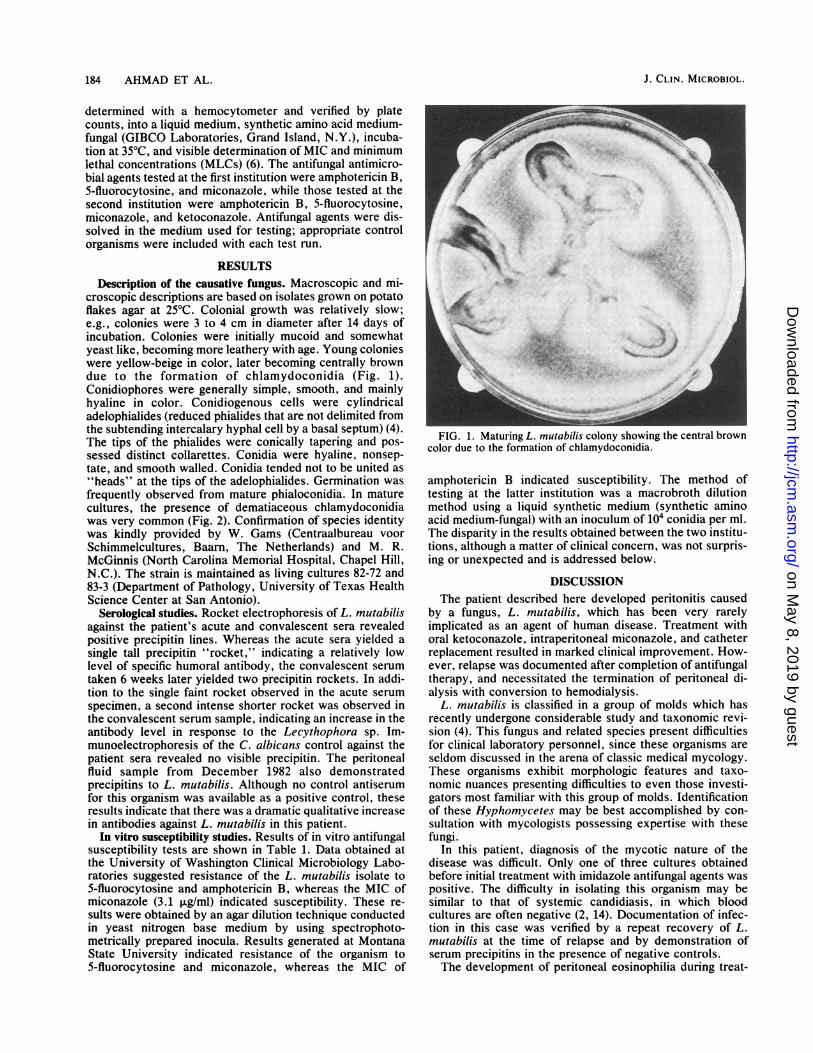
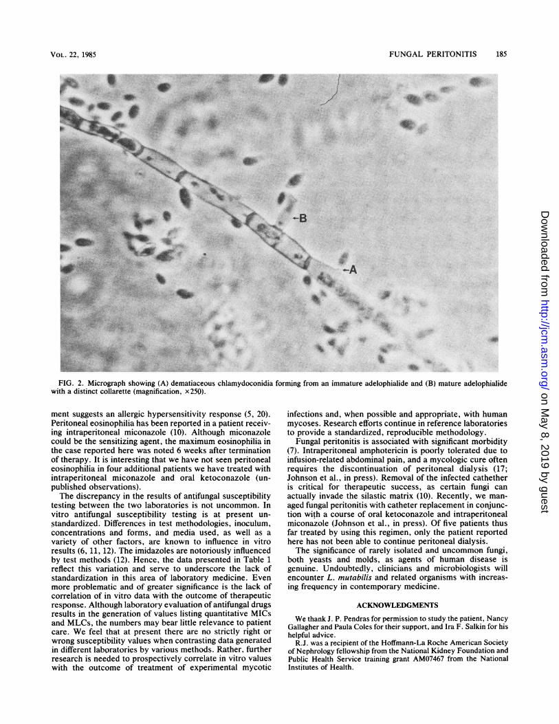
croscopic descriptions are based on isolates grown on potatoflakes agar at 25°C. Colonial growth was relatively slow;e.g., colonies were 3 to 4 cm in diameter after 14 days ofincubation. Colonies were initially mucoid and somewhatyeast like, becoming more leathery with age. Young colonieswere yellow-beige in color, later becoming centrally browndue to the formation of chlamydoconidia (Fig. 1).Conidiophores were generally simple, smooth, and mainlyhyaline in color. Conidiogenous cells were cylindricaladelophialides (reduced phialides that are not delimited fromthe subtending intercalary hyphal cell by a basal septum) (4).The tips of the phialides were conically tapering and pos-sessed distinct collarettes. Conidia were hyaline, nonsep-tate, and smooth walled. Conidia tended not to be united as"heads" at the tips of the adelophialides. Germination wasfrequently observed from mature phialoconidia. In maturecultures, the presence of dematiaceous chlamydoconidiawas very common (Fig. 2). Confirmation of species identitywas kindly provided by W. Gams (Centraalbureau voorSchimmelcultures, Baarn, The Netherlands) and M. R.McGinnis (North Carolina Memorial Hospital, Chapel Hill,N.C.). The strain is maintained as living cultures 82-72 and83-3 (Department of Pathology, University of Texas HealthScience Center at San Antonio).
Serological studies. Rocket electrophoresis of L. mutabilisagainst the patient's acute and convalescent sera revealedpositive precipitin lines. Whereas the acute sera yielded asingle tall precipitin "rocket," indicating a relatively lowlevel of specific humoral antibody, the convalescent serumtaken 6 weeks later yielded two precipitin rockets. In addi-tion to the single faint rocket observed in the acute serumspecimen, a second intense shorter rocket was observed inthe convalescent serum sample, indicating an increase in theantibody level in response to the Lecythophora sp. Im-munoelectrophoresis of the C. albicans control against thepatient sera revealed no visible precipitin. The peritonealfluid sample from December 1982 also demonstratedprecipitins to L. mutabilis. Although no control antiserumfor this organism was available as a positive control, theseresults indicate that there was a dramatic qualitative increasein antibodies against L. mutabilis in this patient.
In vitro susceptibility studies. Results of in vitro antifungalsusceptibility tests are shown in Table 1. Data obtained atthe University of Washington Clinical Microbiology Labo-ratories suggested resistance of the L. mutabilis isolate to5-fluorocytosine and amphotericin B, whereas the MIC ofmiconazole (3.1 ,ug/ml) indicated susceptibility. These re-sults were obtained by an agar dilution technique conductedin yeast nitrogen base medium by using spectrophoto-metrically prepared inocula. Results generated at MontanaState University indicated resistance of the organism to5-fluorocytosine and miconazole, whereas the MIC of
FIG. 1. Maturing L. mutabilis colony showing the central browncolor due to the formation of chlamydoconidia.
amphotericin B indicated susceptibility. The method oftesting at the latter institution was a macrobroth dilutionmethod using a liquid synthetic medium (synthetic aminoacid medium-fungal) with an inoculum of 104 conidia per ml.The disparity in the results obtained between the two institu-tions, although a matter of clinical concern, was not surpris-ing or unexpected and is addressed below.
DISCUSSIONThe patient described here developed peritonitis caused
by a fungus, L. mutabilis, which has been very rarelyimplicated as an agent of human disease. Treatment withoral ketoconazole, intraperitoneal miconazole, and catheterreplacement resulted in marked clinical improvement. How-ever, relapse was documented after completion of antifungaltherapy, and necessitated the termination of peritoneal di-alysis with conversion to hemodialysis.
L. mutabilis is classified in a group of molds which hasrecently undergone considerable study and taxonomic revi-sion (4). This fungus and related species present difficultiesfor clinical laboratory personnel, since these organisms areseldom discussed in the arena of classic medical mycology.These organisms exhibit morphologic features and taxo-nomic nuances presenting difficulties to even those investi-gators most familiar with this group of molds. Identificationof these Hyphomycetes may be best accomplished by con-sultation with mycologists possessing expertise with thesefungi.
In this patient, diagnosis of the mycotic nature of thedisease was difficult. Only one of three cultures obtainedbefore initial treatment with imidazole antifungal agents waspositive. The difficulty in isolating this organism may besimilar to that of systemic candidiasis, in which bloodcultures are often negative (2, 14). Documentation of infec-tion in this case was verified by a repeat recovery of L.mutabilis at the time of relapse and by demonstration ofserum precipitins in the presence of negative controls.The development of peritoneal eosinophilia during treat-
J. CLIN. MICROBIOL.
2%.
.l
.e
1'W4
on May 8, 2019 by guest
http://jcm.asm
.org/D
ownloaded from
FUNGAL PERITONITIS 185
O
i.v~~~~
.A_
*..0-
vr
'À _0i.
4 `S:
t'
.;MF:,
leW..
te; 1b
qAm
p
4*
FIG. 2. Micrograph showing (A) dematiaceous chlamydoconidia forming from an immature adelophialide and (B) mature adelophialidewith a distinct collarette (magnification, x250).
ment suggests an allergic hypersensitivity response (5, 20).Peritoneal eosinophilia has been reported in a patient receiv-ing intraperitoneal miconazole (10). Although miconazolecould be the sensitizing agent, the maximum eosinophilia inthe case reported here was noted 6 weeks after terminationof therapy. It is interesting that we have not seen peritonealeosinophilia in four additional patients we have treated withintraperitoneal miconazole and oral ketoconazole (un-published observations).The discrepancy in the results of antifungal susceptibility
testing between the two laboratories is not uncommon. Invitro antifungal susceptibility testing is at present un-standardized. Differences in test methodologies, inoculum,concentrations and forms, and media used, as well as avariety of other factors, are known to influence in vitroresults (6, 11, 12). The imidazoles are notoriously influencedby test methods (12). Hence, the data presented in Table 1reflect this variation and serve to underscore the lack ofstandardization in this area of laboratory medicine. Evenmore problematic and of greater significance is the lack ofcorrelation of in vitro data with the outcome of therapeuticresponse. Although laboratory evaluation of antifungal drugsresults in the generation of values listing quantitative MICsand MLCs, the numbers may bear little relevance to patientcare. We feel that at present there are no strictly right orwrong susceptibility values when contrasting data generatedin different laboratories by various methods. Rather, furtherresearch is needed to prospeçtively correlate in vitro valueswith the outcome of treatment of experimental mycotic
infections and, when possible and appropriate, with humanmycoses. Research efforts continue in reference laboratoriesto provide a standardized, reproducible methodology.
Fungal peritonitis is associated with significant morbidity(7). Intraperitoneal amphotericin is poorly tolerated due toinfusion-related abdominal pain, and a mycologic cure oftenrequires the discontinuation of peritoneal dialysis (17;Johnson et al., in press). Removal of the infected cathetheris critical for therapeutic success, as certain fungi canactually invade the silastic matrix (10). Recently, we man-aged fungal peritonitis with catheter replacement in conjunc-tion with a course of oral ketoconazole and intraperitonealmiconazole (Johnson et al., in press). Of five patients thusfar treated by using this regimen, only the patient reportedhere has not been able to continue peritoneal dialysis.The significance of rarely isolated and uncommon fungi,
both yeasts and molds, as agents of human disease isgenuine. Undoubtedly, clinicians and microbiologists willencounter L. mutabilis and related organisms with increas-ing frequency in contemporary medicine.
ACKNOWLEDGMENTS
We thank J. P. Pendras for permission to study the patient, NancyGallagher and Paula Coles for their support, and Ira F. Salkin for hishelpful advice.
R.J. was a recipient of the Hoffmann-La Roche American Societyof Nephrology fellowship from the National Kidney Foundation andPublic Health Service training grant AM07467 from the NationalInstitutes of Health.
VOL. 22, 19$5
ii
,,7
lk
on May 8, 2019 by guest
http://jcm.asm
.org/D
ownloaded from
186 AHMAD ET AL.
LITERATURE CITED1. Bayer, A. S., M. J. Blumenkrantz, J. Z. Montgomerie, J. E.
Galpin, J. W. Coburn, and L. B. Guze. 1976. Candidaperitonitis: report of 22 cases and review of the English litera-ture. Am. J. Med. 61:832-840.
2. Bodey, G. P. 1966. Fungal infections complicating acute leuke-mia. J. Chronic Dis. 19:667-668.
3. Eisenberg, E. S., B. E. Alpert, R. A. Weiss, N. Mittman, and R.Soeiro. Rhodotorula rubra peritonitis in patients undergoingcontinuous peritoneal dialysis. Am. J. Med. 75:349-352.
4. Gams, W., and M. R. McGinnis. 1983. Phialemonium, a newanamorph genus intermediate between Phialophora andAcremonium. Mycologia 75:977-987.
5. Gokal, R., J. M. Ramos, M. K. Ward, and D. N. S. Kerr. 1981."Eosinophilic" peritonitis in continuous ambulatory peritonealdialysis (CAPD). Clin. Nephrol. 15:328-330.
6. Hoeprich, P. D., and P. D. Fihn. 1972. Obfuscation of theactivity of antifungal antimicrobics by culture media. J. Infect.Dis. 126:353-361.
7. Kerr, C. M., J. R. Perfect, P. C. Craven, J. H. Jorgensen, D. J.Drutz, J. H. Shelburne, H. A. Gallis, and R. A. Gutman. 1983.Fungal peritonitis in patients on continuous ambulatory perito-neal dialysis. Ann. Intern. Med. 99:334-337.
8. Khanna, R., D. G. Oreopoulos, S. Vas, W. McCready, and N.Donibros. 1980. Fungal peritonitis in patients undergoingchronic intermittent or continuous ambulatory peritoneal dialy-sis. Proc. Eur. Dial. Transplant Assoc. 17:291-296.
9. Landy, M. E., J. H. Greenwald, A. A. Steiner, and D. L.Ashbach. 1982. Cephalosporium (Acremonium) in dialysis-con-nected peritonitis. J. Indiana State Med. Assoc. 75:391.
10. McNeely, D. J., S. Vas, N. Dombros, and D. G. Oreopoulos.1981. Fusarium peritonitis: an uncommon complication of con-
tinuous ambulatory peritoneal dialysis. Perit. Dial. Bull.1:94-96.
11. Odds, F. C. 1979. Problems in the laboratory assessment ofantifungal activity. Postgrad. Med. J. 55:677-680.
12. Odds, F. C. 1980. Laboratory evaluation of antifungal agents: acomparative study of five imidazole derivatives of clinicalimportance. J. Antimicrob. Chernother. 6:749-760.
13. Pearson, J. G., T. D. McKinney, and W. J. Stone. 1983.Penicillium peritonitis in a continuous ambulatory peritonealdialysis patient. Perit. Dial. Bull. 3:20-21.
14. Pizzo, P. A., K. J. Robichaud, F, A. Gill, and G. F. Witebsky.1982. Empiric antibiotic and antifungal therapy for cancerpatients with prolonged fever and granulocytopenia. Am. J.Med. 72:101-111.
15. Rinaldi, M. G. 1982. Use of potato flakes agar in clinicalmycology. J. Clin. Microbiol. 15:1159-1160.
16. Rinaldi, M. G., E. L. McCoy, and D. F. Winn. 1982. Glutealabscess caused by Phialophora hoffimannii and review of therole of this organism in human mycoses. J. Clin. Microbiol.16:181-185.
17. Rubinf J., W. A. Rogers, H. M. Taylor, E. D. Everett, B. G.Prowant, L. V. Fruto, and K. D. Nolph. 1980. Peritonitis duringcontinuous peritoneal dialysis. Ann. Intern. Med. 92:7-13.
18. Shadoiny, S., and A. Espinel-Ingroff. 1980. Susceptibility testingwith antifungal drugs, p. 647-653. In E. H. Lennette, A.Balows, W. H. Hausler, Jr., and J. P. Truant (ed.), Manual ofclinical microbiology, 3rd ed. American Society for Microbiol-ogy, Washington, D.C.
19. Slifkin, M., and H. M. Bowers. 1975. Phialophora mutabilisendocarditis. Am. J. Clin. Pathol. 63:120-130.
20. Steiner, R. W. 1982. Clinical observations in the pathogenesis ofperitoneal dialysate eosinophilia. Perit. Dial. Bull. 2:118-119.
J. CLIN. MICROBIOL'.
on May 8, 2019 by guest
http://jcm.asm
.org/D
ownloaded from