Fundamentals of biomedical applications of biomorphic SiC 14
50
Fundamentals of biomedical applications of biomorphic SiC 297 X Fundamentals of biomedical applications of biomorphic SiC Mahboobeh Mahmoodi 1,2 and Lida Ghazanfari 2 1 Material Group, Faculty of Engineering, Islamic Azad University of Yazd, Yazd, Iran 2 Biomaterial Group, Faculty of Biomedical Engineering, Amirkabir University of Technology Tehran, Iran 1. Introduction In recent years, silicon carbide (SiC) has become an increasingly important material in numerous applications including high frequency, high power, high voltages, and high temperature devices. It is used as a structure material in applications which require hardness, stiffness, high temperature strength (over 1000° C), high thermal conductivity, a low coefficient of thermal expansion, good oxidation and corrosion resistance, some of which are characteristic of typical covalently bonded materials. It seems that SiC can create many opportunities for chemists, physicists, engineers, health professional and biomedical researches (Presas et al., 2006; Greil, 2002; Feng et al.2003). Silicon carbides are emerging as an important class of materials for a variety of biomedical applications. Examples of biomedical applications discussed in this chapter include bioceramic scaffolds for tissue engineering, biosensors, biomembranes, drug delivery, SiC-based quantum dots and etc. Although several journals exist that cover selective clinical applications of SiC, there is a void for a monograph that provides a unified synthesis of this subject. The main objective of this chapter is to provide a basic knowledge of the biomedical applications of SiC so that individuals in all disciplines can rapidly acquire the minimal necessary background for research. A description of future directions of research and development is also provided. 2. Properties of Biomorphic SiC Structural ceramics play a key role in modern technology because of their excellent density, strength relationship and outstanding thermo-mechanical properties. Crystalline silicon carbide is well known as a chemically inert material that is suitable for worst chemical environments even under high temperatures. The same is true for the amorphous modification although the thermal stability is limited to 250 °C. Corrosion resistance under normal biological conditions (neutral pH, body temperature) is excellent. The dissolution rate is well below 30 nm per year (Bolz, 1995; Harder et al., 1999). The properties that make this material particularly promising for biomedical applications are: 1) the wide band gap that increases the sensing capabilities of a semiconductor; 2) the chemical inertness that suggests the material resistance to corrosion in harsh environments such as body; 3) the high 14 www.intechopen.com
Transcript of Fundamentals of biomedical applications of biomorphic SiC 14
Fundamentals of biomedical applications of biomorphic SiC 297
X
Fundamentals of biomedical applications of biomorphic SiC
Mahboobeh Mahmoodi1,2 and Lida Ghazanfari2
1Material Group, Faculty of Engineering, Islamic Azad University of Yazd, Yazd, Iran 2Biomaterial Group, Faculty of Biomedical Engineering, Amirkabir University of Technology
Tehran, Iran
1. Introduction
In recent years, silicon carbide (SiC) has become an increasingly important material in numerous applications including high frequency, high power, high voltages, and high temperature devices. It is used as a structure material in applications which require hardness, stiffness, high temperature strength (over 1000° C), high thermal conductivity, a low coefficient of thermal expansion, good oxidation and corrosion resistance, some of which are characteristic of typical covalently bonded materials. It seems that SiC can create many opportunities for chemists, physicists, engineers, health professional and biomedical researches (Presas et al., 2006; Greil, 2002; Feng et al.2003). Silicon carbides are emerging as an important class of materials for a variety of biomedical applications. Examples of biomedical applications discussed in this chapter include bioceramic scaffolds for tissue engineering, biosensors, biomembranes, drug delivery, SiC-based quantum dots and etc. Although several journals exist that cover selective clinical applications of SiC, there is a void for a monograph that provides a unified synthesis of this subject. The main objective of this chapter is to provide a basic knowledge of the biomedical applications of SiC so that individuals in all disciplines can rapidly acquire the minimal necessary background for research. A description of future directions of research and development is also provided.
2. Properties of Biomorphic SiC
Structural ceramics play a key role in modern technology because of their excellent density, strength relationship and outstanding thermo-mechanical properties. Crystalline silicon carbide is well known as a chemically inert material that is suitable for worst chemical environments even under high temperatures. The same is true for the amorphous modification although the thermal stability is limited to 250 °C. Corrosion resistance under normal biological conditions (neutral pH, body temperature) is excellent. The dissolution rate is well below 30 nm per year (Bolz, 1995; Harder et al., 1999). The properties that make this material particularly promising for biomedical applications are: 1) the wide band gap that increases the sensing capabilities of a semiconductor; 2) the chemical inertness that suggests the material resistance to corrosion in harsh environments such as body; 3) the high
14
www.intechopen.com
Properties and Applications of Silicon Carbide298
hardness (5.8 GPa), high elastic modulus (424 GPa), and low friction coefficient (0.17) that make it an ideal material for smart-implants (Coletti et al., 2007). Mechanical properties of SiC are altered by changing the sintering additives. At elevated temperature, SiC ceramics with boron and carbon additions, which are free from oxide grain-boundary phases, exhibit high-strength and relatively high-creep resistance. These properties of boron- and carbon-doped SiC originate from the absence of grain-boundary phases and existence of covalent bonds between SiC grains (Zawrah & Gazery, 2007). Biomimetics is one such novel approach, the purpose of which is to advance man-made engineering materials through the guidance of nature. Following biomimetic approach, synthesis of ceramic composites from biologically derived materials like wood or organic fibres has recently attained particular interest. Plants often possess natural composite structures and exhibit high mechanical strength, low density, high stiffness, elasticity and damage tolerance. These advantages are because of their genetically built anatomy, developed and matured during different hierarchical stages of a long-term evolutionary process. Development of novel SiC materials by replication of plant morphologies, with tailored physical and chemical properties has a tremendous potential (Chakrabarti, 2004). Biological performs from various soft woods and hard woods can be used for making different varieties of porous SiC ceramics. A wide variety of non-wood ingredients of plant origin commonly used in pulp and paper making can also be employed for producing porous SiC ceramics by replication of plant morphologies (Sieber, 2000). Wood-based biomorphic SiC has been a matter of consideration in the last decade. There has been a great deal of interest in utilizing biomimetic approaches to fabricate a wide variety of silicon-based materials (Gutierrez-Mora et al., 2005; Greil, 2001; Martinzer et al., 2001; Sieber et al., 2001; Varela-Feria et al., 2002). A number of these fabrication approaches have utilized natural wood or cellulosic fiber to produce carbon performs. Biomorphic SiC is manufactured by a two step process: a controlled pyrolyzation of the wood followed by a rapid controlled reactive infiltration of the carbon preform with molten Si. The result is a Si/SiC composite that replicates the highly interconnected microstructure of the wood with SiC, while the remaining unreacted Si fills most of the wood channels. The diversity of wood species, including soft and hard, provides a wide choice of materials, in which the density and the anisotropy are the critical factors of the final microstructure and hence of the mechanical properties of the porous SiC ceramics (Presas et al., 2006; Galderon et al., 2009). Ceramics mimicking the biological structure of natural developed tissue has attracted increasing interest. The mechanical properties of this material not only depend on the component and porosity, but are also highly dependent on the sizes, shapes, and orientation of the pores as well as grains. The lightweight, cytocompatible for human fibroblasts and osteoblasts (Naji and Harmand, 1991) and open porosity of these materials make them great candidates for biomedical applications.
3. Biomedical applications of SiC
Silicon carbides are emerging as an important class of materials for a variety of biomedical applications, including the development of stents, membranes, orthopedic implant, imaging agents, surface modification of biomaterials, biosensors, drug delivery, and tissue engineering. In the coming chapter, we will discuss our experimental studies and some
practical issues in developing SiC for biomedical applications. Hence, we will review some proof-of-concept studies that highlight the unique advantages of SiC in biomedical research.
4. Biocompatibility
Biocompatibility is related to the behavior of biomaterials in various contexts. The term may refer to specific properties of a material without specifying where or how the material is used, or the more empirical clinical success of a whole device in which the material or materials feature. The ambiguity of the term reflects the ongoing development of insights into how biomaterials interact with the human body and eventually how those interactions determine the clinical success of a medical device (such as pacemaker, hip replacement or stent). Modern medical devices and prostheses are often made of more than one material so it might not always be sufficient to talk about the biocompatibility of a specific material. Cell-semiconductor hybrid systems represent an emerging topic of research in the biotechnological area with intriguing possible applications. To date, very little has been known about the main processes that govern the communication between cells and the surfaces they adhere to. When cells adhere to an external surface, an eterophilic binding is generated between the cell adhesion proteins and the surface molecules. After they adhere, the interface between them and the substrate becomes a dynamic environment where surface chemistry, topology, and electronic properties have been shown to play important roles. (Maitz et al., 2003). Coletti et al. studied single-crystal SiC biocompatibility by culturing mammalian cells directly on SiC substrates and by evaluating the resulting cell adhesion quality and proliferation (Coletti et al., 2006). The crystalline SiC is indeed a very promising material for bio-applications, with better bio-performance than crystalline Si. 3C-SiC, which can be directly grown on Si substrates, appears to be an especially promising bio-material. The Si substrate used for the epi-growth would in fact allow for cost-effective and straightforward electronic integration, while the SiC surface would constitute a more biocompatible and versatile interface between the electronic and biological world. The main factors that have been shown to define SiC biocompatibility are its hydrophilicity and surface chemistry. The identification of the organic chemical groups that bind to the SiC surface, together with the calculation of SiC zeta potential in media, could be used to better understand the electronic interaction between cell and SiC surfaces. Using an appropriate cleaning procedure for the SiC samples before their use as substrates for cell cultures is also important. The cleaning chemistry may affect cell proliferation and emphasize the importance of the selection of an appropriate cleaning procedure for biosubstrates. SiC has been shown to be significantly better than Si as a substrate for cell culture, with a noticeably reduced toxic effect and enhanced cell proliferation. One of the possible drawbacks that may be associated with the use of SiC in vivo is related to the unclear and highly debated cytotoxic level of SiC particles. Nonetheless, the potential cytotoxicity of SiC particles does not represent a dramatic issue as much as it does for Si, since the great tribological properties of SiC make it less likely to generate debris. Several studies have discussed testing SiC in vitro. In one study, the researchers tested SiC deposited from radiofrequency sputtering using alveolar bone osteoblasts and gingival fibroblasts for 27 days (Kotzara et al., 2002). The investigators reported that ‘‘Silicon carbide looks cytocompatible both on basal and specific cytocompatibility levels. However, fibroblast and osteoblast attachment is not highly satisfactory, and during the second phase
www.intechopen.com
Fundamentals of biomedical applications of biomorphic SiC 299
hardness (5.8 GPa), high elastic modulus (424 GPa), and low friction coefficient (0.17) that make it an ideal material for smart-implants (Coletti et al., 2007). Mechanical properties of SiC are altered by changing the sintering additives. At elevated temperature, SiC ceramics with boron and carbon additions, which are free from oxide grain-boundary phases, exhibit high-strength and relatively high-creep resistance. These properties of boron- and carbon-doped SiC originate from the absence of grain-boundary phases and existence of covalent bonds between SiC grains (Zawrah & Gazery, 2007). Biomimetics is one such novel approach, the purpose of which is to advance man-made engineering materials through the guidance of nature. Following biomimetic approach, synthesis of ceramic composites from biologically derived materials like wood or organic fibres has recently attained particular interest. Plants often possess natural composite structures and exhibit high mechanical strength, low density, high stiffness, elasticity and damage tolerance. These advantages are because of their genetically built anatomy, developed and matured during different hierarchical stages of a long-term evolutionary process. Development of novel SiC materials by replication of plant morphologies, with tailored physical and chemical properties has a tremendous potential (Chakrabarti, 2004). Biological performs from various soft woods and hard woods can be used for making different varieties of porous SiC ceramics. A wide variety of non-wood ingredients of plant origin commonly used in pulp and paper making can also be employed for producing porous SiC ceramics by replication of plant morphologies (Sieber, 2000). Wood-based biomorphic SiC has been a matter of consideration in the last decade. There has been a great deal of interest in utilizing biomimetic approaches to fabricate a wide variety of silicon-based materials (Gutierrez-Mora et al., 2005; Greil, 2001; Martinzer et al., 2001; Sieber et al., 2001; Varela-Feria et al., 2002). A number of these fabrication approaches have utilized natural wood or cellulosic fiber to produce carbon performs. Biomorphic SiC is manufactured by a two step process: a controlled pyrolyzation of the wood followed by a rapid controlled reactive infiltration of the carbon preform with molten Si. The result is a Si/SiC composite that replicates the highly interconnected microstructure of the wood with SiC, while the remaining unreacted Si fills most of the wood channels. The diversity of wood species, including soft and hard, provides a wide choice of materials, in which the density and the anisotropy are the critical factors of the final microstructure and hence of the mechanical properties of the porous SiC ceramics (Presas et al., 2006; Galderon et al., 2009). Ceramics mimicking the biological structure of natural developed tissue has attracted increasing interest. The mechanical properties of this material not only depend on the component and porosity, but are also highly dependent on the sizes, shapes, and orientation of the pores as well as grains. The lightweight, cytocompatible for human fibroblasts and osteoblasts (Naji and Harmand, 1991) and open porosity of these materials make them great candidates for biomedical applications.
3. Biomedical applications of SiC
Silicon carbides are emerging as an important class of materials for a variety of biomedical applications, including the development of stents, membranes, orthopedic implant, imaging agents, surface modification of biomaterials, biosensors, drug delivery, and tissue engineering. In the coming chapter, we will discuss our experimental studies and some
practical issues in developing SiC for biomedical applications. Hence, we will review some proof-of-concept studies that highlight the unique advantages of SiC in biomedical research.
4. Biocompatibility
Biocompatibility is related to the behavior of biomaterials in various contexts. The term may refer to specific properties of a material without specifying where or how the material is used, or the more empirical clinical success of a whole device in which the material or materials feature. The ambiguity of the term reflects the ongoing development of insights into how biomaterials interact with the human body and eventually how those interactions determine the clinical success of a medical device (such as pacemaker, hip replacement or stent). Modern medical devices and prostheses are often made of more than one material so it might not always be sufficient to talk about the biocompatibility of a specific material. Cell-semiconductor hybrid systems represent an emerging topic of research in the biotechnological area with intriguing possible applications. To date, very little has been known about the main processes that govern the communication between cells and the surfaces they adhere to. When cells adhere to an external surface, an eterophilic binding is generated between the cell adhesion proteins and the surface molecules. After they adhere, the interface between them and the substrate becomes a dynamic environment where surface chemistry, topology, and electronic properties have been shown to play important roles. (Maitz et al., 2003). Coletti et al. studied single-crystal SiC biocompatibility by culturing mammalian cells directly on SiC substrates and by evaluating the resulting cell adhesion quality and proliferation (Coletti et al., 2006). The crystalline SiC is indeed a very promising material for bio-applications, with better bio-performance than crystalline Si. 3C-SiC, which can be directly grown on Si substrates, appears to be an especially promising bio-material. The Si substrate used for the epi-growth would in fact allow for cost-effective and straightforward electronic integration, while the SiC surface would constitute a more biocompatible and versatile interface between the electronic and biological world. The main factors that have been shown to define SiC biocompatibility are its hydrophilicity and surface chemistry. The identification of the organic chemical groups that bind to the SiC surface, together with the calculation of SiC zeta potential in media, could be used to better understand the electronic interaction between cell and SiC surfaces. Using an appropriate cleaning procedure for the SiC samples before their use as substrates for cell cultures is also important. The cleaning chemistry may affect cell proliferation and emphasize the importance of the selection of an appropriate cleaning procedure for biosubstrates. SiC has been shown to be significantly better than Si as a substrate for cell culture, with a noticeably reduced toxic effect and enhanced cell proliferation. One of the possible drawbacks that may be associated with the use of SiC in vivo is related to the unclear and highly debated cytotoxic level of SiC particles. Nonetheless, the potential cytotoxicity of SiC particles does not represent a dramatic issue as much as it does for Si, since the great tribological properties of SiC make it less likely to generate debris. Several studies have discussed testing SiC in vitro. In one study, the researchers tested SiC deposited from radiofrequency sputtering using alveolar bone osteoblasts and gingival fibroblasts for 27 days (Kotzara et al., 2002). The investigators reported that ‘‘Silicon carbide looks cytocompatible both on basal and specific cytocompatibility levels. However, fibroblast and osteoblast attachment is not highly satisfactory, and during the second phase
www.intechopen.com
Properties and Applications of Silicon Carbide300
of osteoblast growth, osteoblast proliferation is very significantly reduced by 30%’’ (Naji et al., 1991). According to another paper, in a 48 h study using human monocytes, SiC had a stimulatory effect comparable to polymethacrylate (Nordsletten et al., 1996). Cytotoxicity and mutagenicity has been performed on SiC-coated tantalum stents. Amorphous SiC did not show any cytotoxic reaction using mice fibroblasts L929 cell cultures when incubated for 24 h or mutagenic potential when investigated using Salmonella typhimurium mutants TA98, TA100, TA1535, and TA1537 (Amon et al., 1996). An earlier study by the same authors of a SiC-coated tantalum stent reported similar results (Amon et al., 1995). Cogan et al. (Cogan et al., 2003) utilized silicon carbide as an implantable dielectric coating. a-SiC films, deposited by plasma-enhanced chemical vapour deposition, have been evaluated as insulating coatings for implantable microelectrodes. Biocompatibility was assessed by implanting a-SiC-coated quartz discs in animals. Histological evaluation showed no chronic inflammatory response and capsule thickness was comparable to silicone or uncoated quartz controls. The a-SiC was more stable in physiological saline than silicon nitride (Si3N4) and well tolerated in the cortex. Kotzar et al. (Kotzar et al, 2002) evaluated materials used in microelectromechanical devices for biocompatibility. These included single crystal silicon, polysilicon (coating, chemical vapor deposition, CVD), single crystal cubic SiC (3CSiC or β-SiC, CVD), and titanium (physical vapor deposition). They concluded that the tested Si, SiC and titanium were biocompatible. Other studies have also confirmed the good tissue biocompatibility of SiC, usually tested as a coating made by CVD (Bolz & Schaldach, 1990; Naji & Harmand, 1991; Santavirta et al., 1998). Even though crystalline SiC biocompatibility has not been investigated in the past, information exists concerning the biocompatibility of the amorphous phase of this material (a-SiC).
5. Haemocompatibility
The interaction between blood proteins and the material is regarded as an important source of thrombogenesis. The adsorption of proteins is explained, from the thermodynamic point of view, in terms of the systems free energy or surface energy. However, adsorption itself does not induce thrombosis. Theories regarding correlations between thrombogenicity of a material and its surface charge or its binding properties proved not to be useful (Bolz, 1993). Thrombus formation on implant materials is one of the first reactions after deployment and may lead to acute failure due to occlusion as well as a trigger for neointimal formation. Next to the direct activation by the intrinsic or extrinsic coagulation cascade, thrombus formation can also be initiated directly by an electron transfer process, while fibrinogen is close to the surface. The electronic nature of a molecule can be defined as either a metal , a semiconductor, or an insulator. Contact activation is possible in the case of a metal since electrons in the fibrinogen molecule are able to occupy empty electronic states with the same energy (Rzany et al., 2000). Therefore, the obvious way to avoid this transfer is to use a material with a significantly reduced density of empty electronic states within the range of the valence band of the fibrinogen. This is the case for the used silicon carbide coating (Schmehl, 2008). Haemocompatibility leads to the following physical requirements (Bolz, 1995): (1) to prevent the electron transfer the solid must have no empty electronic states at the transfer level, i.e., deeper than 0.9 eV below Fermi's level. This requirement is met by a semiconductor with a
sufficiently large band gap (precisely, its valence band edge must be deeper than 1.4 eV below Fermi´s level) and a low density of states inside the band gap. (2) To prevent electrostatic charging of the interface (which may interfere with requirement 1) the electric conductivity must be higher than 10-3 S/cm. A material that meets these electronic requirements is silicon carbide in an amorphous, heavily n-doped, hydrogen-rich modification (a-SiC:H). The amorphous structure is required in order to avoid any point of increased density of electronic states, especially at grain boundaries (Harder, 1999). At present, a-SiC:H is known for its high thromboresistance induced by the optimal barrier that this material presents for protein (and therefore platelet) adhesion(Starke et al., 2006). These properties may translate into less protein biofouling and better compatibility for intravascular applications rather than Si. SiC has relatively low levels of fibrinogen and fibrin deposition when contacting blood (Takami et al., 1998). These proteins promote local clot formation; thus, the tendency not to adsorb them will resist blood clotting. It is now well established that SiC coatings are resistant to platelet adhesion and clotting both in vitro and in vivo. In a study by Bolz et al. (Bolz & Schaldach, 1993), the a-SiC:H films were deposited using the glow discharge technique or plasma-enhanced chemical vapour deposition (PECVD), because it provides the most suitable coating process owing to the high inherent hydrogen concentration which satisfies the electronically active defects in the amorphous layers. They used fibrinogen as an example model for thrombogenesis at implants although most haemoproteins are organic semiconductors. a-SiC:H coatings showed no time-dependent increase in the remaining protein concentration, confirming that no fibrinogen activation and polymerisation had taken place. These results support the electrochemical model for thrombogenesis at artificial surfaces and prove that a proper tailoring of the electronic properties leads to a material with superior haemocompatibility. The in vitro test showed that the morphology of the cells was regular. The a-SiC:H samples showed the same behaviour as the control samples. Blood and membrane proteins have similar band-gaps because the electronic properties depend mainly on the periodicity of the amino acids, and the proteins differ only in the acid sequence, not in their structural periodicity. A-SiC: H has a superior haemocompatibility; its clotting time is 200 percent longer compared with the results of titanium and pyrolytic carbon. Furthermore, it has been shown that small variations in the preparation conditions cause a significant change in haemocompatibility. Therefore, it is of paramount importance to know exactly the physical properties of the material in use, not only the name. Amorphous silicon carbide can be deposited on any substrate material which is resistant to temperatures of about 250 °C. This property makes amorphous silicon carbide a suitable coating material for all hybrid designs of biomedical devices. The substrate material can be fitted to the mechanical needs, disregarding its haemocompatibility, whereas the coating ensures the haemocompatibility of the device. Possible applications are catheters or sensors in blood contact and implants, especially artificial heart valves. Bolz and Schaldach (Bolz & Schaldach, 1990) evaluated PECVD amorphous SiC for use on prosthetic heart valves. They showed a decreased thrombogenicity of an amorphous layer of SiC compared to titanium. Several other studies showed that hydrogen-rich amorphous SiC coating on coronary artery stents is anti-thrombogenic (Bolz et al., 1996; Bolz & Schaldach, 1990; Carrie et al., 2001; Monnink et al., 1999). Three studies (on 2,125 patients) showed a benefit that was attributed to the SiC-coated stent (Elbaz et al., 2002; Hamm et al., 2003;
www.intechopen.com
Fundamentals of biomedical applications of biomorphic SiC 301
of osteoblast growth, osteoblast proliferation is very significantly reduced by 30%’’ (Naji et al., 1991). According to another paper, in a 48 h study using human monocytes, SiC had a stimulatory effect comparable to polymethacrylate (Nordsletten et al., 1996). Cytotoxicity and mutagenicity has been performed on SiC-coated tantalum stents. Amorphous SiC did not show any cytotoxic reaction using mice fibroblasts L929 cell cultures when incubated for 24 h or mutagenic potential when investigated using Salmonella typhimurium mutants TA98, TA100, TA1535, and TA1537 (Amon et al., 1996). An earlier study by the same authors of a SiC-coated tantalum stent reported similar results (Amon et al., 1995). Cogan et al. (Cogan et al., 2003) utilized silicon carbide as an implantable dielectric coating. a-SiC films, deposited by plasma-enhanced chemical vapour deposition, have been evaluated as insulating coatings for implantable microelectrodes. Biocompatibility was assessed by implanting a-SiC-coated quartz discs in animals. Histological evaluation showed no chronic inflammatory response and capsule thickness was comparable to silicone or uncoated quartz controls. The a-SiC was more stable in physiological saline than silicon nitride (Si3N4) and well tolerated in the cortex. Kotzar et al. (Kotzar et al, 2002) evaluated materials used in microelectromechanical devices for biocompatibility. These included single crystal silicon, polysilicon (coating, chemical vapor deposition, CVD), single crystal cubic SiC (3CSiC or β-SiC, CVD), and titanium (physical vapor deposition). They concluded that the tested Si, SiC and titanium were biocompatible. Other studies have also confirmed the good tissue biocompatibility of SiC, usually tested as a coating made by CVD (Bolz & Schaldach, 1990; Naji & Harmand, 1991; Santavirta et al., 1998). Even though crystalline SiC biocompatibility has not been investigated in the past, information exists concerning the biocompatibility of the amorphous phase of this material (a-SiC).
5. Haemocompatibility
The interaction between blood proteins and the material is regarded as an important source of thrombogenesis. The adsorption of proteins is explained, from the thermodynamic point of view, in terms of the systems free energy or surface energy. However, adsorption itself does not induce thrombosis. Theories regarding correlations between thrombogenicity of a material and its surface charge or its binding properties proved not to be useful (Bolz, 1993). Thrombus formation on implant materials is one of the first reactions after deployment and may lead to acute failure due to occlusion as well as a trigger for neointimal formation. Next to the direct activation by the intrinsic or extrinsic coagulation cascade, thrombus formation can also be initiated directly by an electron transfer process, while fibrinogen is close to the surface. The electronic nature of a molecule can be defined as either a metal , a semiconductor, or an insulator. Contact activation is possible in the case of a metal since electrons in the fibrinogen molecule are able to occupy empty electronic states with the same energy (Rzany et al., 2000). Therefore, the obvious way to avoid this transfer is to use a material with a significantly reduced density of empty electronic states within the range of the valence band of the fibrinogen. This is the case for the used silicon carbide coating (Schmehl, 2008). Haemocompatibility leads to the following physical requirements (Bolz, 1995): (1) to prevent the electron transfer the solid must have no empty electronic states at the transfer level, i.e., deeper than 0.9 eV below Fermi's level. This requirement is met by a semiconductor with a
sufficiently large band gap (precisely, its valence band edge must be deeper than 1.4 eV below Fermi´s level) and a low density of states inside the band gap. (2) To prevent electrostatic charging of the interface (which may interfere with requirement 1) the electric conductivity must be higher than 10-3 S/cm. A material that meets these electronic requirements is silicon carbide in an amorphous, heavily n-doped, hydrogen-rich modification (a-SiC:H). The amorphous structure is required in order to avoid any point of increased density of electronic states, especially at grain boundaries (Harder, 1999). At present, a-SiC:H is known for its high thromboresistance induced by the optimal barrier that this material presents for protein (and therefore platelet) adhesion(Starke et al., 2006). These properties may translate into less protein biofouling and better compatibility for intravascular applications rather than Si. SiC has relatively low levels of fibrinogen and fibrin deposition when contacting blood (Takami et al., 1998). These proteins promote local clot formation; thus, the tendency not to adsorb them will resist blood clotting. It is now well established that SiC coatings are resistant to platelet adhesion and clotting both in vitro and in vivo. In a study by Bolz et al. (Bolz & Schaldach, 1993), the a-SiC:H films were deposited using the glow discharge technique or plasma-enhanced chemical vapour deposition (PECVD), because it provides the most suitable coating process owing to the high inherent hydrogen concentration which satisfies the electronically active defects in the amorphous layers. They used fibrinogen as an example model for thrombogenesis at implants although most haemoproteins are organic semiconductors. a-SiC:H coatings showed no time-dependent increase in the remaining protein concentration, confirming that no fibrinogen activation and polymerisation had taken place. These results support the electrochemical model for thrombogenesis at artificial surfaces and prove that a proper tailoring of the electronic properties leads to a material with superior haemocompatibility. The in vitro test showed that the morphology of the cells was regular. The a-SiC:H samples showed the same behaviour as the control samples. Blood and membrane proteins have similar band-gaps because the electronic properties depend mainly on the periodicity of the amino acids, and the proteins differ only in the acid sequence, not in their structural periodicity. A-SiC: H has a superior haemocompatibility; its clotting time is 200 percent longer compared with the results of titanium and pyrolytic carbon. Furthermore, it has been shown that small variations in the preparation conditions cause a significant change in haemocompatibility. Therefore, it is of paramount importance to know exactly the physical properties of the material in use, not only the name. Amorphous silicon carbide can be deposited on any substrate material which is resistant to temperatures of about 250 °C. This property makes amorphous silicon carbide a suitable coating material for all hybrid designs of biomedical devices. The substrate material can be fitted to the mechanical needs, disregarding its haemocompatibility, whereas the coating ensures the haemocompatibility of the device. Possible applications are catheters or sensors in blood contact and implants, especially artificial heart valves. Bolz and Schaldach (Bolz & Schaldach, 1990) evaluated PECVD amorphous SiC for use on prosthetic heart valves. They showed a decreased thrombogenicity of an amorphous layer of SiC compared to titanium. Several other studies showed that hydrogen-rich amorphous SiC coating on coronary artery stents is anti-thrombogenic (Bolz et al., 1996; Bolz & Schaldach, 1990; Carrie et al., 2001; Monnink et al., 1999). Three studies (on 2,125 patients) showed a benefit that was attributed to the SiC-coated stent (Elbaz et al., 2002; Hamm et al., 2003;
www.intechopen.com

Properties and Applications of Silicon Carbide302
Kalnins et al., 2002). In a direct comparison of silicon wafers and SiC-coated (PECVD) silicon wafers for blood compatibility, both appeared to provoke clot formation to a greater extent than diamond-like coated silicon wafers; silicon was worse than SiC-coated silicon (Nurdin et al., 2003). In conclusion, the haemocompatibility of SiC was demonstrated.
6. Biosensors
In the last decade, there has been a tremendous development in the field of miniaturization of chemical and biochemical sensor devices (Berthold et al., 2002). This is because it is expected that miniaturization will improve the speed and reliability of the measurements and will dramatically reduce the sample volume and the system costs. There is a need for the introduction of a semiconducting material that displays both biocompatibility and great sensing potentiality. Most of the studies conducted in the past on single-crystal SiC provide evidence of the attractive bio-potentialities of this material and hence suggest similar properties for crystalline SiC. The availability of SiC single crystal substrates and epitaxial layers with different dopings and conductivities (n-type, p-type and semi-insulating) makes it possible to fully explore the impressive properties of this semiconductor. In the past, the fact that cells could be directly cultured on Si crystalline substrates led to a widespread use of these materials for biosensing applications. The studies report the significant finding that SiC surfaces are a better substrate for mammalian cell culture than Si in terms of both cell adhesion and proliferation (Coletti et al., 2007). In (bio)-chemical sensor applications, the establishment of a stable organic layer covalently attached to the semiconductor surface is of central importance (Yakimova et al., 2007; Botsoa et al., 2008; Frewin et al., 2009). Recent interest has arisen in employing these materials, tools and technologies for the fabrication of miniature sensors and actuators and their integration with electronic circuits to produce smart devices and systems. This effort offers the promise of: (1) increasing the performance and manufacturability of both sensors and actuators by exploiting new batch
fabrication processes developed including micro stereo lithographic and micro molding
techniques; (2) developing novel classes of materials and mechanical structures not possible previously, such as diamond-like carbon, silicon carbide and carbon nanotubes, micro-turbines and micro-engines; (3) development of technologies for the system level and wafer level integration of micro components at the nanometer precision, such as self-assembly techniques and robotic manipulation; (4) development of control and communication systems for microelectromechanical systems (MEMS), such as optical and radio frequency wireless, and power delivery systems, etc. The integration of MEMS, nanoelectromechanical systems, interdigital transducers and required microelectronics and conformal antenna in the multifunctional smart materials and composites results in a smart system suitable for sending and controlling a variety of functions in automobile, aerospace, marine and civil
strutures and food and medical industries (Varadan, 2003). The emerging field of monitoring biological signals generated during nerve excitation, synaptic transmission, quantal release of molecules and cell-to-cell communication, stimulates the development of new methodologies and materials for novel applications of bio-devices in basic science, laboratory analysis and therapeutic treatments. The electrochemical gradient results in a membrane potential that can be measured directly with an intracellular electrode. Extracellular signals are smaller than transmembrane potentials, depending on the distance of the signal source to the electrode. Over the last 30 years, non-
invasive extracellular recording from multiple electrodes has developed into a widely-used standard method. A microelectrode array is an arrangement of several (typically more than 60) electrodes allowing the targeting of several sites for stimulation and extracellular recording at once. One can plan the realisation of four activities with the following tasks: Task 1. Development of new biocompatible substrates favoring neuronal growth along
specific pathways. Task 2. Monitoring of electrical activity from neuronal networks. Task 3. Resolution of cellular excitability over membrane micro areas. Task 4. Detection of quantal released molecules by means of newly designed biosensors. Task number 1 can be realized by means of SiC substrates, by plating the cells directly on the substrate or eventually with an additional proteic layer. For this purpose, 3C-SiC films with controlled stoichiometry, different thickness and crystalline quality can be grown directly on silicon substrates or on silicon substrates previously ‘carbonised’. The main objective of task number 2 is the realization of SiC microelectrode arrays whose dimensions will be compatible with the cellular soma (10-20 µm). In this structure, every element of the array is constituted by a doped 3C-SiC region, with metallic interconnections coated with amorphous silicon carbide, so that silicon carbide represents the only material interfaced to the biological environment. For the realization of task number 3, the SiC array will be improved by constructing microelectrodes in the submicrometric range, in order to reveal electrical signals from different areas of the same cell. The objective of task number 4 is the construction of a prototype of SiC-electrodes array as a chemical detector for oxidizable molecules released during cell activity triggered by chemical substances (KCl or acetylcholine) on chromaffin cells of the adrenal gland. With respect to classical electrochemical methods, requiring polarized carbon fibers with rough dimensions of 10 micrometers in diameter, the SiC multielectrode array should greatly improve the monitoring of secretory vesicles fusion to the plasma-membrane, allowing the spatial localization and temporal resolution of the event. To date, the majority of the development efforts in the MEMS field has focused on sophisticated devices to meet the requirements of industrial applications. However, MEMS devices for medical applications represent a potential multi-billion dollar market, primarily consisting of microminiature devices with high functionality that are suitable for implantation. These implanted systems could revolutionize medical diagnostics and treatment modalities. Implantable muscle microstimulators for disabled individuals have already been developed. Precision sensors combined with integrated processing and telemetry circuitry can remotely monitor any number of physical or chemical parameters within the human body and thereby allow evaluation of an individual’s medical condition. Kotzar et al. selected the following materials as MEMS materials of construction for implantable medical devices: (1) single crystal silicon (Si), (2) polycrystalline silicon, (3) silicon oxide (SiO2), (4) Si3N4, (5) single crystal cubic silicon carbide (3C-SiC or b-SiC), (6) titanium (Ti), and (7) SU-8 epoxy photoresist. The Kotzara et al. study results for SiC showed that when the material was generated using MEMS fabrication techniques, it elicited no significant non-biocompatible responses (Kotzara et al., 2002). Iliescu et al. presented an original fabrication process of a microfluidic device for identification and characterization of cells in suspensions using impedance spectroscopy (Iliescu et al., 2007). The fabrication process of this device consists of three major steps. The steps are shown in Fig. 1.
www.intechopen.com
Fundamentals of biomedical applications of biomorphic SiC 303
Kalnins et al., 2002). In a direct comparison of silicon wafers and SiC-coated (PECVD) silicon wafers for blood compatibility, both appeared to provoke clot formation to a greater extent than diamond-like coated silicon wafers; silicon was worse than SiC-coated silicon (Nurdin et al., 2003). In conclusion, the haemocompatibility of SiC was demonstrated.
6. Biosensors
In the last decade, there has been a tremendous development in the field of miniaturization of chemical and biochemical sensor devices (Berthold et al., 2002). This is because it is expected that miniaturization will improve the speed and reliability of the measurements and will dramatically reduce the sample volume and the system costs. There is a need for the introduction of a semiconducting material that displays both biocompatibility and great sensing potentiality. Most of the studies conducted in the past on single-crystal SiC provide evidence of the attractive bio-potentialities of this material and hence suggest similar properties for crystalline SiC. The availability of SiC single crystal substrates and epitaxial layers with different dopings and conductivities (n-type, p-type and semi-insulating) makes it possible to fully explore the impressive properties of this semiconductor. In the past, the fact that cells could be directly cultured on Si crystalline substrates led to a widespread use of these materials for biosensing applications. The studies report the significant finding that SiC surfaces are a better substrate for mammalian cell culture than Si in terms of both cell adhesion and proliferation (Coletti et al., 2007). In (bio)-chemical sensor applications, the establishment of a stable organic layer covalently attached to the semiconductor surface is of central importance (Yakimova et al., 2007; Botsoa et al., 2008; Frewin et al., 2009). Recent interest has arisen in employing these materials, tools and technologies for the fabrication of miniature sensors and actuators and their integration with electronic circuits to produce smart devices and systems. This effort offers the promise of: (1) increasing the performance and manufacturability of both sensors and actuators by exploiting new batch
fabrication processes developed including micro stereo lithographic and micro molding
techniques; (2) developing novel classes of materials and mechanical structures not possible previously, such as diamond-like carbon, silicon carbide and carbon nanotubes, micro-turbines and micro-engines; (3) development of technologies for the system level and wafer level integration of micro components at the nanometer precision, such as self-assembly techniques and robotic manipulation; (4) development of control and communication systems for microelectromechanical systems (MEMS), such as optical and radio frequency wireless, and power delivery systems, etc. The integration of MEMS, nanoelectromechanical systems, interdigital transducers and required microelectronics and conformal antenna in the multifunctional smart materials and composites results in a smart system suitable for sending and controlling a variety of functions in automobile, aerospace, marine and civil
strutures and food and medical industries (Varadan, 2003). The emerging field of monitoring biological signals generated during nerve excitation, synaptic transmission, quantal release of molecules and cell-to-cell communication, stimulates the development of new methodologies and materials for novel applications of bio-devices in basic science, laboratory analysis and therapeutic treatments. The electrochemical gradient results in a membrane potential that can be measured directly with an intracellular electrode. Extracellular signals are smaller than transmembrane potentials, depending on the distance of the signal source to the electrode. Over the last 30 years, non-
invasive extracellular recording from multiple electrodes has developed into a widely-used standard method. A microelectrode array is an arrangement of several (typically more than 60) electrodes allowing the targeting of several sites for stimulation and extracellular recording at once. One can plan the realisation of four activities with the following tasks: Task 1. Development of new biocompatible substrates favoring neuronal growth along
specific pathways. Task 2. Monitoring of electrical activity from neuronal networks. Task 3. Resolution of cellular excitability over membrane micro areas. Task 4. Detection of quantal released molecules by means of newly designed biosensors. Task number 1 can be realized by means of SiC substrates, by plating the cells directly on the substrate or eventually with an additional proteic layer. For this purpose, 3C-SiC films with controlled stoichiometry, different thickness and crystalline quality can be grown directly on silicon substrates or on silicon substrates previously ‘carbonised’. The main objective of task number 2 is the realization of SiC microelectrode arrays whose dimensions will be compatible with the cellular soma (10-20 µm). In this structure, every element of the array is constituted by a doped 3C-SiC region, with metallic interconnections coated with amorphous silicon carbide, so that silicon carbide represents the only material interfaced to the biological environment. For the realization of task number 3, the SiC array will be improved by constructing microelectrodes in the submicrometric range, in order to reveal electrical signals from different areas of the same cell. The objective of task number 4 is the construction of a prototype of SiC-electrodes array as a chemical detector for oxidizable molecules released during cell activity triggered by chemical substances (KCl or acetylcholine) on chromaffin cells of the adrenal gland. With respect to classical electrochemical methods, requiring polarized carbon fibers with rough dimensions of 10 micrometers in diameter, the SiC multielectrode array should greatly improve the monitoring of secretory vesicles fusion to the plasma-membrane, allowing the spatial localization and temporal resolution of the event. To date, the majority of the development efforts in the MEMS field has focused on sophisticated devices to meet the requirements of industrial applications. However, MEMS devices for medical applications represent a potential multi-billion dollar market, primarily consisting of microminiature devices with high functionality that are suitable for implantation. These implanted systems could revolutionize medical diagnostics and treatment modalities. Implantable muscle microstimulators for disabled individuals have already been developed. Precision sensors combined with integrated processing and telemetry circuitry can remotely monitor any number of physical or chemical parameters within the human body and thereby allow evaluation of an individual’s medical condition. Kotzar et al. selected the following materials as MEMS materials of construction for implantable medical devices: (1) single crystal silicon (Si), (2) polycrystalline silicon, (3) silicon oxide (SiO2), (4) Si3N4, (5) single crystal cubic silicon carbide (3C-SiC or b-SiC), (6) titanium (Ti), and (7) SU-8 epoxy photoresist. The Kotzara et al. study results for SiC showed that when the material was generated using MEMS fabrication techniques, it elicited no significant non-biocompatible responses (Kotzara et al., 2002). Iliescu et al. presented an original fabrication process of a microfluidic device for identification and characterization of cells in suspensions using impedance spectroscopy (Iliescu et al., 2007). The fabrication process of this device consists of three major steps. The steps are shown in Fig. 1.
www.intechopen.com
Properties and Applications of Silicon Carbide304
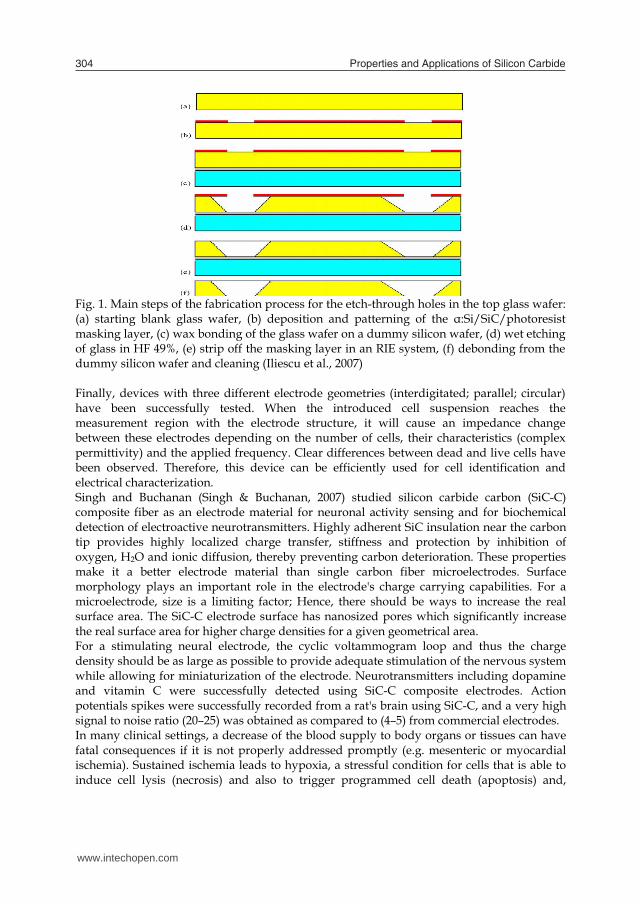
Fig. 1. Main steps of the fabrication process for the etch-through holes in the top glass wafer: (a) starting blank glass wafer, (b) deposition and patterning of the α:Si/SiC/photoresist masking layer, (c) wax bonding of the glass wafer on a dummy silicon wafer, (d) wet etching of glass in HF 49%, (e) strip off the masking layer in an RIE system, (f) debonding from the dummy silicon wafer and cleaning (Iliescu et al., 2007) Finally, devices with three different electrode geometries (interdigitated; parallel; circular) have been successfully tested. When the introduced cell suspension reaches the measurement region with the electrode structure, it will cause an impedance change between these electrodes depending on the number of cells, their characteristics (complex permittivity) and the applied frequency. Clear differences between dead and live cells have been observed. Therefore, this device can be efficiently used for cell identification and electrical characterization. Singh and Buchanan (Singh & Buchanan, 2007) studied silicon carbide carbon (SiC-C) composite fiber as an electrode material for neuronal activity sensing and for biochemical detection of electroactive neurotransmitters. Highly adherent SiC insulation near the carbon tip provides highly localized charge transfer, stiffness and protection by inhibition of oxygen, H2O and ionic diffusion, thereby preventing carbon deterioration. These properties make it a better electrode material than single carbon fiber microelectrodes. Surface morphology plays an important role in the electrode's charge carrying capabilities. For a microelectrode, size is a limiting factor; Hence, there should be ways to increase the real surface area. The SiC-C electrode surface has nanosized pores which significantly increase the real surface area for higher charge densities for a given geometrical area. For a stimulating neural electrode, the cyclic voltammogram loop and thus the charge density should be as large as possible to provide adequate stimulation of the nervous system while allowing for miniaturization of the electrode. Neurotransmitters including dopamine and vitamin C were successfully detected using SiC-C composite electrodes. Action potentials spikes were successfully recorded from a rat's brain using SiC-C, and a very high signal to noise ratio (20–25) was obtained as compared to (4–5) from commercial electrodes. In many clinical settings, a decrease of the blood supply to body organs or tissues can have fatal consequences if it is not properly addressed promptly (e.g. mesenteric or myocardial ischemia). Sustained ischemia leads to hypoxia, a stressful condition for cells that is able to induce cell lysis (necrosis) and also to trigger programmed cell death (apoptosis) and,
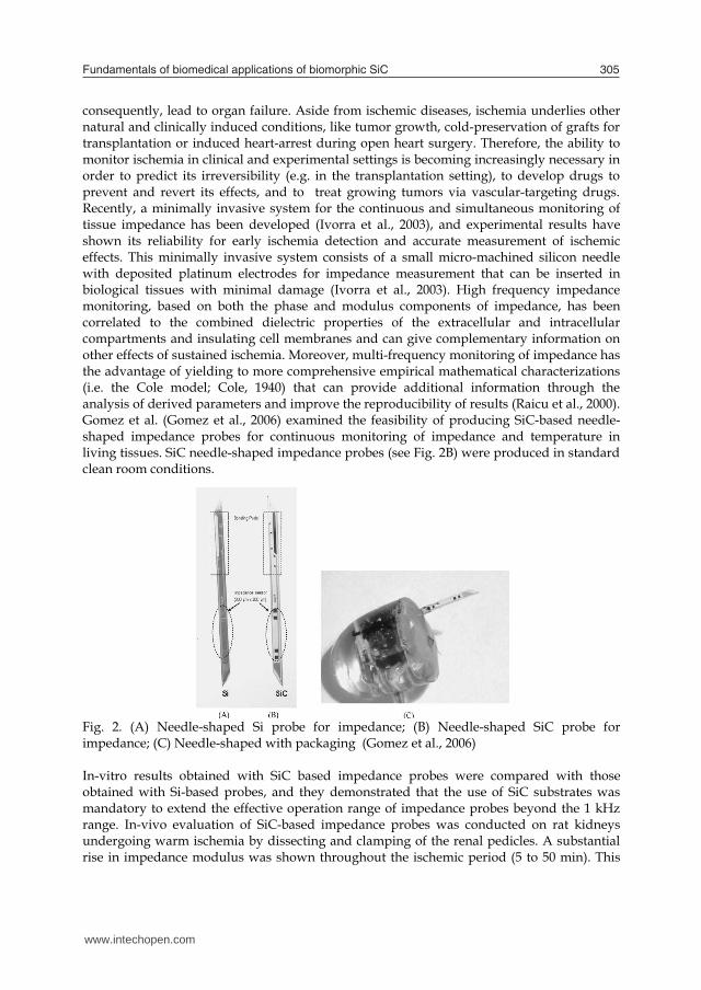
consequently, lead to organ failure. Aside from ischemic diseases, ischemia underlies other natural and clinically induced conditions, like tumor growth, cold-preservation of grafts for transplantation or induced heart-arrest during open heart surgery. Therefore, the ability to monitor ischemia in clinical and experimental settings is becoming increasingly necessary in order to predict its irreversibility (e.g. in the transplantation setting), to develop drugs to prevent and revert its effects, and to treat growing tumors via vascular-targeting drugs. Recently, a minimally invasive system for the continuous and simultaneous monitoring of tissue impedance has been developed (Ivorra et al., 2003), and experimental results have shown its reliability for early ischemia detection and accurate measurement of ischemic effects. This minimally invasive system consists of a small micro-machined silicon needle with deposited platinum electrodes for impedance measurement that can be inserted in biological tissues with minimal damage (Ivorra et al., 2003). High frequency impedance monitoring, based on both the phase and modulus components of impedance, has been correlated to the combined dielectric properties of the extracellular and intracellular compartments and insulating cell membranes and can give complementary information on other effects of sustained ischemia. Moreover, multi-frequency monitoring of impedance has the advantage of yielding to more comprehensive empirical mathematical characterizations (i.e. the Cole model; Cole, 1940) that can provide additional information through the analysis of derived parameters and improve the reproducibility of results (Raicu et al., 2000). Gomez et al. (Gomez et al., 2006) examined the feasibility of producing SiC-based needle-shaped impedance probes for continuous monitoring of impedance and temperature in living tissues. SiC needle-shaped impedance probes (see Fig. 2B) were produced in standard clean room conditions.
Fig. 2. (A) Needle-shaped Si probe for impedance; (B) Needle-shaped SiC probe for impedance; (C) Needle-shaped with packaging (Gomez et al., 2006) In-vitro results obtained with SiC based impedance probes were compared with those obtained with Si-based probes, and they demonstrated that the use of SiC substrates was mandatory to extend the effective operation range of impedance probes beyond the 1 kHz range. In-vivo evaluation of SiC-based impedance probes was conducted on rat kidneys undergoing warm ischemia by dissecting and clamping of the renal pedicles. A substantial rise in impedance modulus was shown throughout the ischemic period (5 to 50 min). This
www.intechopen.com
Fundamentals of biomedical applications of biomorphic SiC 305
Fig. 1. Main steps of the fabrication process for the etch-through holes in the top glass wafer: (a) starting blank glass wafer, (b) deposition and patterning of the α:Si/SiC/photoresist masking layer, (c) wax bonding of the glass wafer on a dummy silicon wafer, (d) wet etching of glass in HF 49%, (e) strip off the masking layer in an RIE system, (f) debonding from the dummy silicon wafer and cleaning (Iliescu et al., 2007) Finally, devices with three different electrode geometries (interdigitated; parallel; circular) have been successfully tested. When the introduced cell suspension reaches the measurement region with the electrode structure, it will cause an impedance change between these electrodes depending on the number of cells, their characteristics (complex permittivity) and the applied frequency. Clear differences between dead and live cells have been observed. Therefore, this device can be efficiently used for cell identification and electrical characterization. Singh and Buchanan (Singh & Buchanan, 2007) studied silicon carbide carbon (SiC-C) composite fiber as an electrode material for neuronal activity sensing and for biochemical detection of electroactive neurotransmitters. Highly adherent SiC insulation near the carbon tip provides highly localized charge transfer, stiffness and protection by inhibition of oxygen, H2O and ionic diffusion, thereby preventing carbon deterioration. These properties make it a better electrode material than single carbon fiber microelectrodes. Surface morphology plays an important role in the electrode's charge carrying capabilities. For a microelectrode, size is a limiting factor; Hence, there should be ways to increase the real surface area. The SiC-C electrode surface has nanosized pores which significantly increase the real surface area for higher charge densities for a given geometrical area. For a stimulating neural electrode, the cyclic voltammogram loop and thus the charge density should be as large as possible to provide adequate stimulation of the nervous system while allowing for miniaturization of the electrode. Neurotransmitters including dopamine and vitamin C were successfully detected using SiC-C composite electrodes. Action potentials spikes were successfully recorded from a rat's brain using SiC-C, and a very high signal to noise ratio (20–25) was obtained as compared to (4–5) from commercial electrodes. In many clinical settings, a decrease of the blood supply to body organs or tissues can have fatal consequences if it is not properly addressed promptly (e.g. mesenteric or myocardial ischemia). Sustained ischemia leads to hypoxia, a stressful condition for cells that is able to induce cell lysis (necrosis) and also to trigger programmed cell death (apoptosis) and,
consequently, lead to organ failure. Aside from ischemic diseases, ischemia underlies other natural and clinically induced conditions, like tumor growth, cold-preservation of grafts for transplantation or induced heart-arrest during open heart surgery. Therefore, the ability to monitor ischemia in clinical and experimental settings is becoming increasingly necessary in order to predict its irreversibility (e.g. in the transplantation setting), to develop drugs to prevent and revert its effects, and to treat growing tumors via vascular-targeting drugs. Recently, a minimally invasive system for the continuous and simultaneous monitoring of tissue impedance has been developed (Ivorra et al., 2003), and experimental results have shown its reliability for early ischemia detection and accurate measurement of ischemic effects. This minimally invasive system consists of a small micro-machined silicon needle with deposited platinum electrodes for impedance measurement that can be inserted in biological tissues with minimal damage (Ivorra et al., 2003). High frequency impedance monitoring, based on both the phase and modulus components of impedance, has been correlated to the combined dielectric properties of the extracellular and intracellular compartments and insulating cell membranes and can give complementary information on other effects of sustained ischemia. Moreover, multi-frequency monitoring of impedance has the advantage of yielding to more comprehensive empirical mathematical characterizations (i.e. the Cole model; Cole, 1940) that can provide additional information through the analysis of derived parameters and improve the reproducibility of results (Raicu et al., 2000). Gomez et al. (Gomez et al., 2006) examined the feasibility of producing SiC-based needle-shaped impedance probes for continuous monitoring of impedance and temperature in living tissues. SiC needle-shaped impedance probes (see Fig. 2B) were produced in standard clean room conditions.
Fig. 2. (A) Needle-shaped Si probe for impedance; (B) Needle-shaped SiC probe for impedance; (C) Needle-shaped with packaging (Gomez et al., 2006) In-vitro results obtained with SiC based impedance probes were compared with those obtained with Si-based probes, and they demonstrated that the use of SiC substrates was mandatory to extend the effective operation range of impedance probes beyond the 1 kHz range. In-vivo evaluation of SiC-based impedance probes was conducted on rat kidneys undergoing warm ischemia by dissecting and clamping of the renal pedicles. A substantial rise in impedance modulus was shown throughout the ischemic period (5 to 50 min). This
www.intechopen.com
Properties and Applications of Silicon Carbide306
increase can be attributed to the occurrence of hypoxic edema as the result of cell swelling, which leads to a reduction of extracellular space, an increase in extracellular resistance, and cell-to-cell uncoupling (Gersing, 1998). Upon unclamping of the renal artery (50 min), impedance modulus can be seen to return to its basal value, a fact that can be attributed in this experimental setting to a reversion from a short period of ischemia without substantial structural damage to the tissue. A fall in impedance modulus at low frequencies, however, has also been reported as a consequence of membrane breakdown and cell lysis due to the sustained ischemia (Haemmerich et al., 2002). It is in this respect that the multifrequency analysis of the phase component of impedance made possible by the use of SiC-based probes conveys useful complementary information. Researchers (Godignon, 2005) fabricated impedance and temperature sensors on bulk SiC for a biomedical needle that can be used for open heart surgery monitoring or graft monitoring of organs during transportation and transplantation. According to Godignon (Godignon, 2005) other applications can be foreseen, such as DNA polymerase chain reaction (PCR), electrophoresis chips and cell culture micro-arrays. In DNA electrophoresis devices, the high critical electric field and high resistivity of semi-insulating SiC would be beneficial. In DNA PCR, it is the high thermal conductivity which could improve the device behaviour. In addition, in most of these cases, the transparency of semi-insulating SiC can be used for optical monitoring of the biological process, as for example for the DNA reaction or the cell culture activity. Caputo et al. (Caputo et al., 2008) reported on biomolecule detection based on a two-color amorphous silicon photosensor. The revealed biomolecules were DNA strands labeled with two fluorochromes (Alexa Fluor 350 or Cy5) with different spectral properties and the device is a p-i-n-i-p amorphous silicon/amorphous silicon carbide stacked structure, that was able to detect different spectral regions depending on the voltage applied to its electrodes. The device design has been optimized in order to maximize the spectral match between the sensor responses and the emission spectra of the fluorochromes. This optimization process has been carried out by means of a numerical device simulator, taking into account the optical and the electrical properties of the amorphous silicon materials. Therefore, according to these set of materials, one can conclude SiC could be considered as a good candidate for biosensing applications.
7. Stent coating
In recent years, coronary stenting has become a well established therapy of coronary artery disease. However, in up to 30% of all stent implantations, the process of restenosis leads to a re-narrowing of the vessel within several months. The optimization of the stent design with regard to mechanical properties only resulted in limited success in reduction of the restenosis rate, and a hybrid concept for stent design was proposed; on the one hand, the mechanical requirements for an optimized geometrical design are met by using 316L stainless steel as bulk material. On the other hand, unwanted interactions of the implant's metal surface with surrounding tissue and blood diminishing biocompatibility and inducing the process of restenosis are reduced by a suitable coating working as a "magic hat" (Harder, 1999; Rzany & Schaldach, 2001). The surface properties of a stent determine the interactions with the surrounding physiologic environment, while properties such as the mechanical performance are determined by the bulk material, the design of which is shown
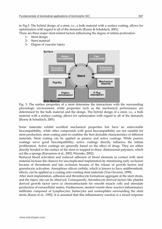
in Fig.3. The hybrid design of a stent, i.e., a bulk material with a surface coating, allows for optimization with regard to all of the demands (Rzany & Schaldach, 2001). There are three major stent-related factors influencing the degree of intima proferation:
1- Stent design 2- Stent material 3- Degree of vascular injury
Fig. 3. The surface properties of a stent determine the interactions with the surrounding physiologic environment, while properties such as the mechanical performance are determined by the bulk material and the design. The hybrid design of a stent, i.e., a bulk material with a surface coating, allows for optimization with regard to all of the demands (Rzany & Schaldach, 2001) Some materials exhibit excellent mechanical properties but have an unfavorable biocompatibility, while other compounds with good biocompatibility are not suitable for stent production, stent coating aims to combine the best desirable characteristics of different materials. Stent coating can be applied as passive and active coatings. While passive coatings serve good biocompatibility, active coatings directly influence the intima proliferation. Active coatings are generally based on the effect of drugs. They are either directly bonded to the surface of the stent or trapped in three- dimensional polymers, which act like a sponge (Karoussos et al., 2002; Wieneke, 2002). Reduced blood activation and reduced adhesion of blood elements in contact with stent material increase the chances for uncomplicated implantation by minimizing early occlusion became of thrombosis and late occlusion became of the release of growth factors and granulocyte activation. Amorphous silicon carbide, which is known to have antithrombotic effects, can be applied as a coating onto existing stent materials (Van Oeveren, 1999). After stent implantation, adhesion and thrombocyte formations aggregate at the stent struts, and the injury site can be observed. Consequently, thrombocyte derived factors like platelet derived growth factor serve as chemoattractants for smooth muscle cells and stimulate production of extracellular matrix. Furthermore, stented vessels show reactive inflammatory infiltrates composed of lymphocytes, histiocytes and eosinophiles surrounding the stent struts (Karas et al., 1992). It is assumed that this inflammatory reaction is a mixed response
www.intechopen.com
Fundamentals of biomedical applications of biomorphic SiC 307
increase can be attributed to the occurrence of hypoxic edema as the result of cell swelling, which leads to a reduction of extracellular space, an increase in extracellular resistance, and cell-to-cell uncoupling (Gersing, 1998). Upon unclamping of the renal artery (50 min), impedance modulus can be seen to return to its basal value, a fact that can be attributed in this experimental setting to a reversion from a short period of ischemia without substantial structural damage to the tissue. A fall in impedance modulus at low frequencies, however, has also been reported as a consequence of membrane breakdown and cell lysis due to the sustained ischemia (Haemmerich et al., 2002). It is in this respect that the multifrequency analysis of the phase component of impedance made possible by the use of SiC-based probes conveys useful complementary information. Researchers (Godignon, 2005) fabricated impedance and temperature sensors on bulk SiC for a biomedical needle that can be used for open heart surgery monitoring or graft monitoring of organs during transportation and transplantation. According to Godignon (Godignon, 2005) other applications can be foreseen, such as DNA polymerase chain reaction (PCR), electrophoresis chips and cell culture micro-arrays. In DNA electrophoresis devices, the high critical electric field and high resistivity of semi-insulating SiC would be beneficial. In DNA PCR, it is the high thermal conductivity which could improve the device behaviour. In addition, in most of these cases, the transparency of semi-insulating SiC can be used for optical monitoring of the biological process, as for example for the DNA reaction or the cell culture activity. Caputo et al. (Caputo et al., 2008) reported on biomolecule detection based on a two-color amorphous silicon photosensor. The revealed biomolecules were DNA strands labeled with two fluorochromes (Alexa Fluor 350 or Cy5) with different spectral properties and the device is a p-i-n-i-p amorphous silicon/amorphous silicon carbide stacked structure, that was able to detect different spectral regions depending on the voltage applied to its electrodes. The device design has been optimized in order to maximize the spectral match between the sensor responses and the emission spectra of the fluorochromes. This optimization process has been carried out by means of a numerical device simulator, taking into account the optical and the electrical properties of the amorphous silicon materials. Therefore, according to these set of materials, one can conclude SiC could be considered as a good candidate for biosensing applications.
7. Stent coating
In recent years, coronary stenting has become a well established therapy of coronary artery disease. However, in up to 30% of all stent implantations, the process of restenosis leads to a re-narrowing of the vessel within several months. The optimization of the stent design with regard to mechanical properties only resulted in limited success in reduction of the restenosis rate, and a hybrid concept for stent design was proposed; on the one hand, the mechanical requirements for an optimized geometrical design are met by using 316L stainless steel as bulk material. On the other hand, unwanted interactions of the implant's metal surface with surrounding tissue and blood diminishing biocompatibility and inducing the process of restenosis are reduced by a suitable coating working as a "magic hat" (Harder, 1999; Rzany & Schaldach, 2001). The surface properties of a stent determine the interactions with the surrounding physiologic environment, while properties such as the mechanical performance are determined by the bulk material, the design of which is shown
in Fig.3. The hybrid design of a stent, i.e., a bulk material with a surface coating, allows for optimization with regard to all of the demands (Rzany & Schaldach, 2001). There are three major stent-related factors influencing the degree of intima proferation:
1- Stent design 2- Stent material 3- Degree of vascular injury
Fig. 3. The surface properties of a stent determine the interactions with the surrounding physiologic environment, while properties such as the mechanical performance are determined by the bulk material and the design. The hybrid design of a stent, i.e., a bulk material with a surface coating, allows for optimization with regard to all of the demands (Rzany & Schaldach, 2001) Some materials exhibit excellent mechanical properties but have an unfavorable biocompatibility, while other compounds with good biocompatibility are not suitable for stent production, stent coating aims to combine the best desirable characteristics of different materials. Stent coating can be applied as passive and active coatings. While passive coatings serve good biocompatibility, active coatings directly influence the intima proliferation. Active coatings are generally based on the effect of drugs. They are either directly bonded to the surface of the stent or trapped in three- dimensional polymers, which act like a sponge (Karoussos et al., 2002; Wieneke, 2002). Reduced blood activation and reduced adhesion of blood elements in contact with stent material increase the chances for uncomplicated implantation by minimizing early occlusion became of thrombosis and late occlusion became of the release of growth factors and granulocyte activation. Amorphous silicon carbide, which is known to have antithrombotic effects, can be applied as a coating onto existing stent materials (Van Oeveren, 1999). After stent implantation, adhesion and thrombocyte formations aggregate at the stent struts, and the injury site can be observed. Consequently, thrombocyte derived factors like platelet derived growth factor serve as chemoattractants for smooth muscle cells and stimulate production of extracellular matrix. Furthermore, stented vessels show reactive inflammatory infiltrates composed of lymphocytes, histiocytes and eosinophiles surrounding the stent struts (Karas et al., 1992). It is assumed that this inflammatory reaction is a mixed response
www.intechopen.com
Properties and Applications of Silicon Carbide308
to vessel injury on the one hand, and non-specific activation mediated through metal ions released from the alloy of the stent on the other. Cytokines released by inflammatory cells not only serve as smooth muscle mitogens, but also regulate the production of extracellular matrix. Although the detailed mechanisms of inflammation are not completely understood, the correlation between the degree of inflammatory reaction and the extent of neointimal thickness suggest a central role for inflammation in the process of restenosis. It is well accepted that platelet activation and thrombus formation are one of the critical steps in the formation of restenosis. Since it has long been known that thrombus formation is based on electronic processes, semiconductor surfaces have been used for stent coatings (Wieneke, 2002). The prototype of this coating is a hypothrombogenic semi-conducting coating of amorphous hydrogenated silicon carbide (a-SiC:H). This material can suppress the electron transfer that is crucial in the transformation of fibrinogen to fibrin (Wieneke, 2002). Experimental studies using silicon carbide as a passive stent coating have shown a marked reduction in fibrin and thrombus deposits (Rzany et al., 2000). Based on this theoretical background, stents with silicon carbide coating have been used in patients with acute myocardial infarction with promising short- and long- term results (Scheller et al., 2001; Rzany & Schaldach, 2001). In one randomized study with the silicon carbide coating, the major adverse cardiac events rate after 6 months has reduced significantly as compared with a 316L stainless steel; however, the restenosis rate was similar (Unverdorben et al., 2000). The deposition of this particular modification of amorphous silicon carbide is performed by means of the PECVD. Since amorphous SiC is a ceramic material, its mechanical properties are significantly different from the metallic substrate. Especially during the dilatation of the stent, enormous mechanical stresses are created at the interface between coating and substrate, while deformations up to 30% are taking place. Therefore, the coating must have strong adhesion to the substrate. There are four steps in the coating process which have to be optimized in sequence to fit both the required electronic properties as well as strong adhesion: the cleaning process, surface activation, deposition of a thin intermediate film and finally coating the surface with a-SiC:H. The specific requirements for the electronic properties of the surface need a careful selection of process parameters. The electronic band gap is mainly influenced by two physical effects; on the one hand, the band gap of all semiconductors is a property of the material's chemical composition. On the other hand, the band gap of amorphous semiconductors is affected by the density of unsaturated bonds. To achieve a large band gap as well as a low density of states within the gap, the dangling bonds have to be saturated by hydrogen atoms. The most important benefit of the coating with regard to corrosion is that it acts as a diffusion barrier. The uncoated stents may cause cell reactions or reactions of the immune system. However, when coated, the ions must diffuse through the coating before they can get into the patient. Due to the internal structure of amorphous silicon carbide, this diffusion is so slow that the ion release is negligible (Harder, 1999). The amorphous silicon carbide has been reported to reduce fibrin deposition, which may result in reduced platelet and leukocyte adherence as well (Bolz, 1995; Van Oeveren, 1999). The a-SiC:H surface with multiple clean areas or a loose cell deposit without the fibrin network is shown in Fig. 4. Van Oeveren concluded that the acute response of stainless steel on blood activation can be quenched by a-SiC:H coating.
Fig. 4. (a) Scanning electron micrograph (1500x) of the stainless steel surface showing areas with a dense layer of blood proteins and formed elements, covered by fibrin strands; (b) Scanning electron micrograph (2000x) of the a-SiC:H coated surface showing areas with thrombi and erythrocytes, but not densely packed and not extensively covered with fibrin (Van Oeveren, 1999). Hanekamp and Koolen reported implantation of a silicon carbide coated stent in coronary arteries < 3.0 mm is a safe and effective treatment when compared to percutaneous transluminal coronary angioplasty alone (Hanekamp & Koolen, 2000). Monnink et al. (Monnink et al., 1999) reported a lower activation of leukocytes expressed by a significantly lower CD11b receptor-mediated adhesion at the SiC-coated stent. The coating of nitinol stents with a-SiC:H showed an improvement regarding thrombogenic properties as reflected in the platelet count, levels of β-TG and TAT III complex in a short-term in vitro setting. Although the currently used nitinol stents show good results in the iliac vasculature and have shown to be superior to percutaneous transluminal angioplasty alone in the superficial femoral artery, they still tend to restenose in a nonnegligible number (Schillinger, 2006).The coating with a-SiC:H might result in a further decrease of thrombogenicity especially for longer lesions and a conservation of the stent material by decreasing the removal of nickel ions from the alloy (Schmehl, 2008). Therefore, it can be concluded from these reports that the adhesion and activation of human platelets is significantly reduced at silicon carbide coated surfaces.
8. Tissue engineering
Tissue engineering was once categorized as a sub-field of bio materials, but having grown in scope and importance, it can be considered as a field in its own right. It is the use of a combination of cells, engineering and materials methods, and suitable biochemical and physio-chemical factors to improve or replace biological functions. While most definitions of tissue engineering cover a broad range of applications, the term is, in practice, closely associated with applications that repair or replace portions of or whole tissues (i.e., bone, cartilage, blood vessels, bladder, skin etc.). Often, the tissues involved require certain mechanical and structural properties for proper functioning. The term has also been applied to efforts to perform specific biochemical functions using cells within an artificially-created support system (e.g. an pancreas or a bio artificial liver). Cells are often implanted or 'seeded' into an artificial structure capable of supporting three-dimensional tissue formation. To achieve the goal of tissue reconstruction, scaffolds must meet some specific requirements. A high porosity and an adequate pore size are necessary
www.intechopen.com

Fundamentals of biomedical applications of biomorphic SiC 309
to vessel injury on the one hand, and non-specific activation mediated through metal ions released from the alloy of the stent on the other. Cytokines released by inflammatory cells not only serve as smooth muscle mitogens, but also regulate the production of extracellular matrix. Although the detailed mechanisms of inflammation are not completely understood, the correlation between the degree of inflammatory reaction and the extent of neointimal thickness suggest a central role for inflammation in the process of restenosis. It is well accepted that platelet activation and thrombus formation are one of the critical steps in the formation of restenosis. Since it has long been known that thrombus formation is based on electronic processes, semiconductor surfaces have been used for stent coatings (Wieneke, 2002). The prototype of this coating is a hypothrombogenic semi-conducting coating of amorphous hydrogenated silicon carbide (a-SiC:H). This material can suppress the electron transfer that is crucial in the transformation of fibrinogen to fibrin (Wieneke, 2002). Experimental studies using silicon carbide as a passive stent coating have shown a marked reduction in fibrin and thrombus deposits (Rzany et al., 2000). Based on this theoretical background, stents with silicon carbide coating have been used in patients with acute myocardial infarction with promising short- and long- term results (Scheller et al., 2001; Rzany & Schaldach, 2001). In one randomized study with the silicon carbide coating, the major adverse cardiac events rate after 6 months has reduced significantly as compared with a 316L stainless steel; however, the restenosis rate was similar (Unverdorben et al., 2000). The deposition of this particular modification of amorphous silicon carbide is performed by means of the PECVD. Since amorphous SiC is a ceramic material, its mechanical properties are significantly different from the metallic substrate. Especially during the dilatation of the stent, enormous mechanical stresses are created at the interface between coating and substrate, while deformations up to 30% are taking place. Therefore, the coating must have strong adhesion to the substrate. There are four steps in the coating process which have to be optimized in sequence to fit both the required electronic properties as well as strong adhesion: the cleaning process, surface activation, deposition of a thin intermediate film and finally coating the surface with a-SiC:H. The specific requirements for the electronic properties of the surface need a careful selection of process parameters. The electronic band gap is mainly influenced by two physical effects; on the one hand, the band gap of all semiconductors is a property of the material's chemical composition. On the other hand, the band gap of amorphous semiconductors is affected by the density of unsaturated bonds. To achieve a large band gap as well as a low density of states within the gap, the dangling bonds have to be saturated by hydrogen atoms. The most important benefit of the coating with regard to corrosion is that it acts as a diffusion barrier. The uncoated stents may cause cell reactions or reactions of the immune system. However, when coated, the ions must diffuse through the coating before they can get into the patient. Due to the internal structure of amorphous silicon carbide, this diffusion is so slow that the ion release is negligible (Harder, 1999). The amorphous silicon carbide has been reported to reduce fibrin deposition, which may result in reduced platelet and leukocyte adherence as well (Bolz, 1995; Van Oeveren, 1999). The a-SiC:H surface with multiple clean areas or a loose cell deposit without the fibrin network is shown in Fig. 4. Van Oeveren concluded that the acute response of stainless steel on blood activation can be quenched by a-SiC:H coating.
Fig. 4. (a) Scanning electron micrograph (1500x) of the stainless steel surface showing areas with a dense layer of blood proteins and formed elements, covered by fibrin strands; (b) Scanning electron micrograph (2000x) of the a-SiC:H coated surface showing areas with thrombi and erythrocytes, but not densely packed and not extensively covered with fibrin (Van Oeveren, 1999). Hanekamp and Koolen reported implantation of a silicon carbide coated stent in coronary arteries < 3.0 mm is a safe and effective treatment when compared to percutaneous transluminal coronary angioplasty alone (Hanekamp & Koolen, 2000). Monnink et al. (Monnink et al., 1999) reported a lower activation of leukocytes expressed by a significantly lower CD11b receptor-mediated adhesion at the SiC-coated stent. The coating of nitinol stents with a-SiC:H showed an improvement regarding thrombogenic properties as reflected in the platelet count, levels of β-TG and TAT III complex in a short-term in vitro setting. Although the currently used nitinol stents show good results in the iliac vasculature and have shown to be superior to percutaneous transluminal angioplasty alone in the superficial femoral artery, they still tend to restenose in a nonnegligible number (Schillinger, 2006).The coating with a-SiC:H might result in a further decrease of thrombogenicity especially for longer lesions and a conservation of the stent material by decreasing the removal of nickel ions from the alloy (Schmehl, 2008). Therefore, it can be concluded from these reports that the adhesion and activation of human platelets is significantly reduced at silicon carbide coated surfaces.
8. Tissue engineering
Tissue engineering was once categorized as a sub-field of bio materials, but having grown in scope and importance, it can be considered as a field in its own right. It is the use of a combination of cells, engineering and materials methods, and suitable biochemical and physio-chemical factors to improve or replace biological functions. While most definitions of tissue engineering cover a broad range of applications, the term is, in practice, closely associated with applications that repair or replace portions of or whole tissues (i.e., bone, cartilage, blood vessels, bladder, skin etc.). Often, the tissues involved require certain mechanical and structural properties for proper functioning. The term has also been applied to efforts to perform specific biochemical functions using cells within an artificially-created support system (e.g. an pancreas or a bio artificial liver). Cells are often implanted or 'seeded' into an artificial structure capable of supporting three-dimensional tissue formation. To achieve the goal of tissue reconstruction, scaffolds must meet some specific requirements. A high porosity and an adequate pore size are necessary
www.intechopen.com
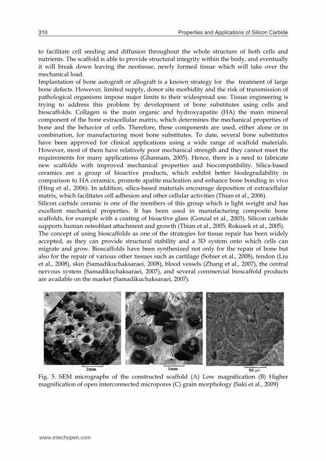
Properties and Applications of Silicon Carbide310
to facilitate cell seeding and diffusion throughout the whole structure of both cells and nutrients. The scaffold is able to provide structural integrity within the body, and eventually it will break down leaving the neotissue, newly formed tissue which will take over the mechanical load. Implantation of bone autograft or allograft is a known strategy for the treatment of large bone defects. However, limited supply, donor site morbidity and the risk of transmission of pathological organisms impose major limits to their widespread use. Tissue engineering is trying to address this problem by development of bone substitutes using cells and bioscaffolds. Collagen is the main organic and hydroxyapatite (HA) the main mineral component of the bone extracellular matrix, which determines the mechanical properties of bone and the behavior of cells. Therefore, these components are used, either alone or in combination, for manufacturing most bone substitutes. To date, several bone substitutes have been approved for clinical applications using a wide range of scaffold materials. However, most of them have relatively poor mechanical strength and they cannot meet the requirements for many applications (Ghannam, 2005). Hence, there is a need to fabricate new scaffolds with improved mechanical properties and biocompatibility. Silica-based ceramics are a group of bioactive products, which exhibit better biodegradability in comparison to HA ceramics, promote apatite nucleation and enhance bone bonding in vivo (Hing et al., 2006). In addition, silica-based materials encourage deposition of extracellular matrix, which facilitates cell adhesion and other cellular activities (Thian et al., 2006). Silicon carbide ceramic is one of the members of this group which is light weight and has excellent mechanical properties. It has been used in manufacturing composite bone scaffolds, for example with a coating of bioactive glass (Gonzal et al., 2003). Silicon carbide supports human osteoblast attachment and growth (Thian et al., 2005; Rokusek et al., 2005). The concept of using bioscaffolds as one of the strategies for tissue repair has been widely accepted, as they can provide structural stability and a 3D system onto which cells can migrate and grow. Bioscaffolds have been synthesized not only for the repair of bone but also for the repair of various other tissues such as cartilage (Sohier et al., 2008), tendon (Liu et al., 2008), skin (Samadikuchaksaraei, 2008), blood vessels (Zhang et al., 2007), the central nervous system (Samadikuchaksaraei, 2007), and several commercial bioscaffold products are available on the market (Samadikuchaksaraei, 2007).
Fig. 5. SEM micrographs of the constructed scaffold (A) Low magnification (B) Higher magnification of open interconnected micropores (C) grain morphology (Saki et al., 2009)
Saki et al. synthesized a hydroxyapatite-alumina and silicon carbide composite scaffold for bone tissue engineering (Saki et al., 2009). SEM captured images of scaffold morphology show fairly uniform pores, which are suitable for growth of bone tissue cells (Fig 5A, B). Fig. 5C shows the grain morphology of the constructed scaffold. The grains are of uniform morphology and their size ranges between 2.5-5 μm. Cell growth and viability studies show that the scaffold does not significantly change the behaviour of osteoblasts. They show many cells attached to the scaffold (Fig 6).
Fig.6. SEM micrographs of osteoblast-like cells attached to HA-alumina-SiC composite surfaces; the cell filaments attaching them to the surface are observed in Fig. 6B (Saki et al., 2009) Saki et al. used a recently developed method involving impregnation of an organic foam for synthesis of a three dimensional scaffold. One of the advantages of this method of construction is the synthesis of scaffolds with fairly uniform pore morphologies, which are interconnected and in the range suitable for penetration by osteoblasts and vascular tissue. Silicon-containing HA surfaces have also been shown to be suitable for osteoblast adhesion, migration and proliferation. Biological materials with complex composite forms and microstructures often display outstanding mechanical properties, which have inspired material scientists in the design of novel materials (Meyers et al., 2008). In the last decade, biotemplating has been widely used to fabricate biomorphic porous materials with various components, such as zeolite, metals, oxides, carbides, nitrides, and with different microstructures, such as fibrilla macroscopic structures, laminated ceramics, or complex micro/macro-pore structures, which might be suitable for technical applications, for example as filters, catalyst carriers or biomedical materials (Zampieri et al., 2006; Liu et al., 2007, Fan et al., 2006; Luo et al., 2007). Due to their hierarchical structure and uniaxial pore morphologies with anisotropic mechanical properties, woods have become the most commonly used biotemplates. Several investigations were carried out in the recent years to exploit the biomorphic ceramics as new scaffold for bone implants. Porous Si3N4/SiC composites have attracted increasing attention because of their excellent physical and chemical properties, such as their strength, resistance to oxidation and corrosion. The mechanical properties of porous ceramics not only depend on the component and porosity, but are also highly dependent on the size, shape, and orientation of the pores as well as grains (Ohji et al., 2008). Fibrous bioceramic scaffolds are favorable candidates since they offer a large specific surface area, giving rise to a high bioactivity for bone tissue engineering. Interconnected pores provide a
www.intechopen.com

Fundamentals of biomedical applications of biomorphic SiC 311
to facilitate cell seeding and diffusion throughout the whole structure of both cells and nutrients. The scaffold is able to provide structural integrity within the body, and eventually it will break down leaving the neotissue, newly formed tissue which will take over the mechanical load. Implantation of bone autograft or allograft is a known strategy for the treatment of large bone defects. However, limited supply, donor site morbidity and the risk of transmission of pathological organisms impose major limits to their widespread use. Tissue engineering is trying to address this problem by development of bone substitutes using cells and bioscaffolds. Collagen is the main organic and hydroxyapatite (HA) the main mineral component of the bone extracellular matrix, which determines the mechanical properties of bone and the behavior of cells. Therefore, these components are used, either alone or in combination, for manufacturing most bone substitutes. To date, several bone substitutes have been approved for clinical applications using a wide range of scaffold materials. However, most of them have relatively poor mechanical strength and they cannot meet the requirements for many applications (Ghannam, 2005). Hence, there is a need to fabricate new scaffolds with improved mechanical properties and biocompatibility. Silica-based ceramics are a group of bioactive products, which exhibit better biodegradability in comparison to HA ceramics, promote apatite nucleation and enhance bone bonding in vivo (Hing et al., 2006). In addition, silica-based materials encourage deposition of extracellular matrix, which facilitates cell adhesion and other cellular activities (Thian et al., 2006). Silicon carbide ceramic is one of the members of this group which is light weight and has excellent mechanical properties. It has been used in manufacturing composite bone scaffolds, for example with a coating of bioactive glass (Gonzal et al., 2003). Silicon carbide supports human osteoblast attachment and growth (Thian et al., 2005; Rokusek et al., 2005). The concept of using bioscaffolds as one of the strategies for tissue repair has been widely accepted, as they can provide structural stability and a 3D system onto which cells can migrate and grow. Bioscaffolds have been synthesized not only for the repair of bone but also for the repair of various other tissues such as cartilage (Sohier et al., 2008), tendon (Liu et al., 2008), skin (Samadikuchaksaraei, 2008), blood vessels (Zhang et al., 2007), the central nervous system (Samadikuchaksaraei, 2007), and several commercial bioscaffold products are available on the market (Samadikuchaksaraei, 2007).
Fig. 5. SEM micrographs of the constructed scaffold (A) Low magnification (B) Higher magnification of open interconnected micropores (C) grain morphology (Saki et al., 2009)
Saki et al. synthesized a hydroxyapatite-alumina and silicon carbide composite scaffold for bone tissue engineering (Saki et al., 2009). SEM captured images of scaffold morphology show fairly uniform pores, which are suitable for growth of bone tissue cells (Fig 5A, B). Fig. 5C shows the grain morphology of the constructed scaffold. The grains are of uniform morphology and their size ranges between 2.5-5 μm. Cell growth and viability studies show that the scaffold does not significantly change the behaviour of osteoblasts. They show many cells attached to the scaffold (Fig 6).
Fig.6. SEM micrographs of osteoblast-like cells attached to HA-alumina-SiC composite surfaces; the cell filaments attaching them to the surface are observed in Fig. 6B (Saki et al., 2009) Saki et al. used a recently developed method involving impregnation of an organic foam for synthesis of a three dimensional scaffold. One of the advantages of this method of construction is the synthesis of scaffolds with fairly uniform pore morphologies, which are interconnected and in the range suitable for penetration by osteoblasts and vascular tissue. Silicon-containing HA surfaces have also been shown to be suitable for osteoblast adhesion, migration and proliferation. Biological materials with complex composite forms and microstructures often display outstanding mechanical properties, which have inspired material scientists in the design of novel materials (Meyers et al., 2008). In the last decade, biotemplating has been widely used to fabricate biomorphic porous materials with various components, such as zeolite, metals, oxides, carbides, nitrides, and with different microstructures, such as fibrilla macroscopic structures, laminated ceramics, or complex micro/macro-pore structures, which might be suitable for technical applications, for example as filters, catalyst carriers or biomedical materials (Zampieri et al., 2006; Liu et al., 2007, Fan et al., 2006; Luo et al., 2007). Due to their hierarchical structure and uniaxial pore morphologies with anisotropic mechanical properties, woods have become the most commonly used biotemplates. Several investigations were carried out in the recent years to exploit the biomorphic ceramics as new scaffold for bone implants. Porous Si3N4/SiC composites have attracted increasing attention because of their excellent physical and chemical properties, such as their strength, resistance to oxidation and corrosion. The mechanical properties of porous ceramics not only depend on the component and porosity, but are also highly dependent on the size, shape, and orientation of the pores as well as grains (Ohji et al., 2008). Fibrous bioceramic scaffolds are favorable candidates since they offer a large specific surface area, giving rise to a high bioactivity for bone tissue engineering. Interconnected pores provide a
www.intechopen.com
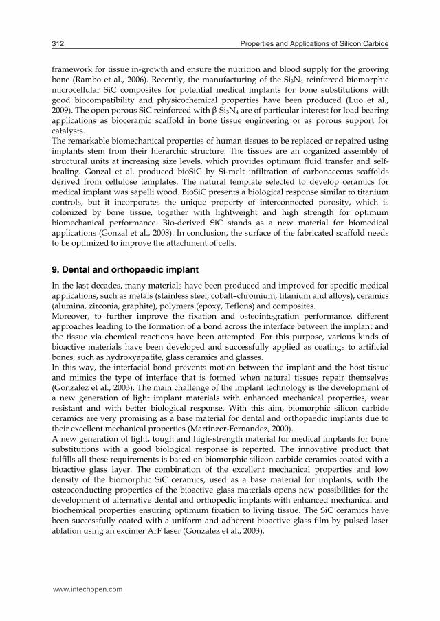
Properties and Applications of Silicon Carbide312
framework for tissue in-growth and ensure the nutrition and blood supply for the growing bone (Rambo et al., 2006). Recently, the manufacturing of the Si3N4 reinforced biomorphic microcellular SiC composites for potential medical implants for bone substitutions with good biocompatibility and physicochemical properties have been produced (Luo et al., 2009). The open porous SiC reinforced with β-Si3N4 are of particular interest for load bearing applications as bioceramic scaffold in bone tissue engineering or as porous support for catalysts. The remarkable biomechanical properties of human tissues to be replaced or repaired using implants stem from their hierarchic structure. The tissues are an organized assembly of structural units at increasing size levels, which provides optimum fluid transfer and self-healing. Gonzal et al. produced bioSiC by Si-melt infiltration of carbonaceous scaffolds derived from cellulose templates. The natural template selected to develop ceramics for medical implant was sapelli wood. BioSiC presents a biological response similar to titanium controls, but it incorporates the unique property of interconnected porosity, which is colonized by bone tissue, together with lightweight and high strength for optimum biomechanical performance. Bio-derived SiC stands as a new material for biomedical applications (Gonzal et al., 2008). In conclusion, the surface of the fabricated scaffold needs to be optimized to improve the attachment of cells.
9. Dental and orthopaedic implant
In the last decades, many materials have been produced and improved for specific medical applications, such as metals (stainless steel, cobalt–chromium, titanium and alloys), ceramics (alumina, zirconia, graphite), polymers (epoxy, Teflons) and composites. Moreover, to further improve the fixation and osteointegration performance, different approaches leading to the formation of a bond across the interface between the implant and the tissue via chemical reactions have been attempted. For this purpose, various kinds of bioactive materials have been developed and successfully applied as coatings to artificial bones, such as hydroxyapatite, glass ceramics and glasses. In this way, the interfacial bond prevents motion between the implant and the host tissue and mimics the type of interface that is formed when natural tissues repair themselves (Gonzalez et al., 2003). The main challenge of the implant technology is the development of a new generation of light implant materials with enhanced mechanical properties, wear resistant and with better biological response. With this aim, biomorphic silicon carbide ceramics are very promising as a base material for dental and orthopaedic implants due to their excellent mechanical properties (Martinzer-Fernandez, 2000). A new generation of light, tough and high-strength material for medical implants for bone substitutions with a good biological response is reported. The innovative product that fulfills all these requirements is based on biomorphic silicon carbide ceramics coated with a bioactive glass layer. The combination of the excellent mechanical properties and low density of the biomorphic SiC ceramics, used as a base material for implants, with the osteoconducting properties of the bioactive glass materials opens new possibilities for the development of alternative dental and orthopedic implants with enhanced mechanical and biochemical properties ensuring optimum fixation to living tissue. The SiC ceramics have been successfully coated with a uniform and adherent bioactive glass film by pulsed laser ablation using an excimer ArF laser (Gonzalez et al., 2003).
Titanium implants made of commercially pure titanium, medical grade, have a reference tensile yield strength of between 280–345 MPa (Mangonon, 1999). Higher strength implants made of Ti–6Al–4V (alpha-beta alloys) have a reference tensile yield strength of 830–924 MPa. It is then possible to conclude that beech-based SiC biomorphic implants appear as a quite interesting alternative to Ti implants, by showing higher strength and less than 40% of its density. Moreover, taking into account the biomechanical requirements (density, elastic modulus, strain to failure, etc.) of a particular type of bone in the body that should be repaired, biomorphic SiC ceramics can be tailored by an appropriate wood precursor selection. An alternative material for medical implants for bone substitutions based on high-strength and low density biomorphic SiC ceramics coated with bioactive glass is reported, combining the characteristics of both materials into a new product with enhanced mechanical and biochemical properties (Gonzalez et al., 2003). Several reports (Gonzalez, et al., 2003, 2004; Mayor et al., 1998; LeGeros et al., 1967) showed biomorphic silicon carbide coated with bioactive glass has been proposed as an alternative to titanium and titanium alloy devices due to its low density, bio-inertness, interconnected porosity and improved mechanical properties. Hydroxylapatite coatings was produced by pulsed laser deposition (PLD) on biomorphic silicon carbide ceramics by ablation of non-sintered HA discs with an ArF excimer laser (193 nm, 25 ns, 4.2 J cm_2) at different conditions of water vapour pressure and substrate temperature for dental and orthopaedic applications, reported by Barrajo et al. (Borrajo et al., 2005). Bioactive silica-based glasses are good candidates to be applied as coatings, thereby improving the physiological response of the ceramic substrate because they promote the intimate bonding of living tissues through the formation of a calcium phosphate layer similar to the apatite found in bone (Fujibayashi et al., 2003), thus preventing the formation of a fibrous capsule around the implant. Recently (Carlos et al., 2006), in vitro cytotoxicity of wood-based biomorphic silicon carbide ceramics coated with bioactive glass, using MG-63 human osteoblast-like cells, and their application in bone implantology have been reported. The MG-63 osteoblast-like cell monolayer time course formation. A, 1 hr; B, 6 hrs and C, 24 hrs after seeding on a representative beech-based SiC ceramic was coated with bioactive glass is shown in Fig. 7. One hour after seeding (A), rounded cells have involved in cellular division events and can be seen attached to the outer surface inside the pores. Cells begin to penetrate and colonise the inner surface of the existing pores. At 6 hrs after seeding (B), cells were attached and had spread out, displaying a flat configuration and a normal morphology. Neighbouring cells maintained physical contact with one another through extensions of the cytoplasm. At 24 hrs (C), the bioactive glass coated surface is almost completely covered by the MG-63 cells. No evidence of major deleterious or cytotoxic responses has been observed. The biomorphic beech-based SiC ceramics coated with bioactive glass supports the cellular monolayer formation and the colonisation of the surface of the material. The same results have been obtained for the eucalyptus and the sapelli-based coated ceramics. SiC ceramics coated with bioactive glass showed the same biological response as the reference materials Ti6Al4V and bulk bioactive glass. The biomorphic SiC ceramics coated with bioactive glass by PLD did not produce a cytotoxic response on the MG-63 osteoblast-like cells. The same behavior was observed for uncoated ceramics. The cellular activity on coated and uncoated SiC ceramics was similar to well known implant materials like Ti6Al4V and bulk bioactive glass (Carlos et al., 2006).
www.intechopen.com

Fundamentals of biomedical applications of biomorphic SiC 313
framework for tissue in-growth and ensure the nutrition and blood supply for the growing bone (Rambo et al., 2006). Recently, the manufacturing of the Si3N4 reinforced biomorphic microcellular SiC composites for potential medical implants for bone substitutions with good biocompatibility and physicochemical properties have been produced (Luo et al., 2009). The open porous SiC reinforced with β-Si3N4 are of particular interest for load bearing applications as bioceramic scaffold in bone tissue engineering or as porous support for catalysts. The remarkable biomechanical properties of human tissues to be replaced or repaired using implants stem from their hierarchic structure. The tissues are an organized assembly of structural units at increasing size levels, which provides optimum fluid transfer and self-healing. Gonzal et al. produced bioSiC by Si-melt infiltration of carbonaceous scaffolds derived from cellulose templates. The natural template selected to develop ceramics for medical implant was sapelli wood. BioSiC presents a biological response similar to titanium controls, but it incorporates the unique property of interconnected porosity, which is colonized by bone tissue, together with lightweight and high strength for optimum biomechanical performance. Bio-derived SiC stands as a new material for biomedical applications (Gonzal et al., 2008). In conclusion, the surface of the fabricated scaffold needs to be optimized to improve the attachment of cells.
9. Dental and orthopaedic implant
In the last decades, many materials have been produced and improved for specific medical applications, such as metals (stainless steel, cobalt–chromium, titanium and alloys), ceramics (alumina, zirconia, graphite), polymers (epoxy, Teflons) and composites. Moreover, to further improve the fixation and osteointegration performance, different approaches leading to the formation of a bond across the interface between the implant and the tissue via chemical reactions have been attempted. For this purpose, various kinds of bioactive materials have been developed and successfully applied as coatings to artificial bones, such as hydroxyapatite, glass ceramics and glasses. In this way, the interfacial bond prevents motion between the implant and the host tissue and mimics the type of interface that is formed when natural tissues repair themselves (Gonzalez et al., 2003). The main challenge of the implant technology is the development of a new generation of light implant materials with enhanced mechanical properties, wear resistant and with better biological response. With this aim, biomorphic silicon carbide ceramics are very promising as a base material for dental and orthopaedic implants due to their excellent mechanical properties (Martinzer-Fernandez, 2000). A new generation of light, tough and high-strength material for medical implants for bone substitutions with a good biological response is reported. The innovative product that fulfills all these requirements is based on biomorphic silicon carbide ceramics coated with a bioactive glass layer. The combination of the excellent mechanical properties and low density of the biomorphic SiC ceramics, used as a base material for implants, with the osteoconducting properties of the bioactive glass materials opens new possibilities for the development of alternative dental and orthopedic implants with enhanced mechanical and biochemical properties ensuring optimum fixation to living tissue. The SiC ceramics have been successfully coated with a uniform and adherent bioactive glass film by pulsed laser ablation using an excimer ArF laser (Gonzalez et al., 2003).
Titanium implants made of commercially pure titanium, medical grade, have a reference tensile yield strength of between 280–345 MPa (Mangonon, 1999). Higher strength implants made of Ti–6Al–4V (alpha-beta alloys) have a reference tensile yield strength of 830–924 MPa. It is then possible to conclude that beech-based SiC biomorphic implants appear as a quite interesting alternative to Ti implants, by showing higher strength and less than 40% of its density. Moreover, taking into account the biomechanical requirements (density, elastic modulus, strain to failure, etc.) of a particular type of bone in the body that should be repaired, biomorphic SiC ceramics can be tailored by an appropriate wood precursor selection. An alternative material for medical implants for bone substitutions based on high-strength and low density biomorphic SiC ceramics coated with bioactive glass is reported, combining the characteristics of both materials into a new product with enhanced mechanical and biochemical properties (Gonzalez et al., 2003). Several reports (Gonzalez, et al., 2003, 2004; Mayor et al., 1998; LeGeros et al., 1967) showed biomorphic silicon carbide coated with bioactive glass has been proposed as an alternative to titanium and titanium alloy devices due to its low density, bio-inertness, interconnected porosity and improved mechanical properties. Hydroxylapatite coatings was produced by pulsed laser deposition (PLD) on biomorphic silicon carbide ceramics by ablation of non-sintered HA discs with an ArF excimer laser (193 nm, 25 ns, 4.2 J cm_2) at different conditions of water vapour pressure and substrate temperature for dental and orthopaedic applications, reported by Barrajo et al. (Borrajo et al., 2005). Bioactive silica-based glasses are good candidates to be applied as coatings, thereby improving the physiological response of the ceramic substrate because they promote the intimate bonding of living tissues through the formation of a calcium phosphate layer similar to the apatite found in bone (Fujibayashi et al., 2003), thus preventing the formation of a fibrous capsule around the implant. Recently (Carlos et al., 2006), in vitro cytotoxicity of wood-based biomorphic silicon carbide ceramics coated with bioactive glass, using MG-63 human osteoblast-like cells, and their application in bone implantology have been reported. The MG-63 osteoblast-like cell monolayer time course formation. A, 1 hr; B, 6 hrs and C, 24 hrs after seeding on a representative beech-based SiC ceramic was coated with bioactive glass is shown in Fig. 7. One hour after seeding (A), rounded cells have involved in cellular division events and can be seen attached to the outer surface inside the pores. Cells begin to penetrate and colonise the inner surface of the existing pores. At 6 hrs after seeding (B), cells were attached and had spread out, displaying a flat configuration and a normal morphology. Neighbouring cells maintained physical contact with one another through extensions of the cytoplasm. At 24 hrs (C), the bioactive glass coated surface is almost completely covered by the MG-63 cells. No evidence of major deleterious or cytotoxic responses has been observed. The biomorphic beech-based SiC ceramics coated with bioactive glass supports the cellular monolayer formation and the colonisation of the surface of the material. The same results have been obtained for the eucalyptus and the sapelli-based coated ceramics. SiC ceramics coated with bioactive glass showed the same biological response as the reference materials Ti6Al4V and bulk bioactive glass. The biomorphic SiC ceramics coated with bioactive glass by PLD did not produce a cytotoxic response on the MG-63 osteoblast-like cells. The same behavior was observed for uncoated ceramics. The cellular activity on coated and uncoated SiC ceramics was similar to well known implant materials like Ti6Al4V and bulk bioactive glass (Carlos et al., 2006).
www.intechopen.com
Properties and Applications of Silicon Carbide314
Fig. 7. Scanning electron microscopy images showing the MG-63 osteoblast-like cell monolayer time course formation. A, 1 hour; B, 6 hrs and C, 24 hrs after seeding on a representative beech-based SiC ceramic coated with bioactive glass. All magnifications are 1000×(Carlos et al., 2006) When using the combination of 316L and gold in liquid, electrolytic media such as blood, the corrosion resistance of the system comes into question. The main reasons for this are contact and crevice corrosion. The corrosive attack in particular could lead to the destruction of the implant or to a release of metallic ions. Along with the potential problem of corrosion, there is the question of bio- and hemocompatibility of gold that is in direct contact with the biological environment. While some authors do not see any deleterious interactions between gold and the vessel wall (Tanigawa et al., 1991, 1995), a significant thrombogenic effect has been attributed to this material in other publications (Sawyer et al., 1965; Schomig et al., 1999). The corrosion behavior of gold-covered stents that have been coated with an additional, amorphous silicon carbide film that is known to be antithrombogenic (a-SiC:H), and which prevents direct contact between the gold coating and the biological environment (Wendler et al., 2000). For improved osseointegration, titanium-based total hip replacement (THR) components were coated by some manufacturers with hydroxyapatite, which also prevents titanium metallosis. However, hydroxyapatite has a tendency to dissolve from the implant surface, and thus a hydroxyapatite coating does not prevent periprosthetic metallosis permanently. Wear particles from THR prosthetic components cause a local host response directly and indirectly, which leads to component loosening (Santavirta et al., 1990). Silicon carbide as a ceramic coating material of titanium-based THR implants. The idea is to prevent wear debris formation from the soft titanium surface. SiC is a hard and tightly bonding ceramic surface material, and because of these physical properties it is not easily degradable, as is the case with hydroxyapatite. SiC can be deposited on a titanium implant surface, e.g., by sputtering techniques, and such coatings bond very well to the substrate. In human monocyte cultures phagocytized SiC particles cause a similar stimulation to hydroxyapatite (Santavirta et al., 1998). Recently titanium-based implant manufacturers have begun to consider modifying surfaces, so that bonds can form a mechanical interlock. The approach is the creation of meshwork through sintered beads or threads that have 350–500 µm pores to promote osteoconduction. The obtained mechanical properties, i.e. elastic modulus ≥335 GPa are favorable as compared with human cancellous bone and titanium. Ceramic on ceramic total hip prostheses are developed to apply to young patients because lifetime of polyethylene joint prostheses is limited by loosening due to biological response. As mating faces of all-ceramic joint must be
highly conformed to reduce stress concentration, wear properties of SiC surface were investigated by Ikeuchi et al. (Ikeuchi et al., 2000). The conclusions are as follows: (1) Among the four ceramics, alumina and silicon carbide can be applied to ceramic on ceramic joint prostheses because they keep low wear rate, smooth surface and high hardness during sliding in water environments. (2) Surface film formed on the ceramic surface may contribute to boundary lubrication in a ceramic on ceramic joint prostheses. Therefore, SiC is a candidate material that can be applied for dental and orthopaedic implants.
10. Surface functionalization
Surface functionalization introduces chemical functional groups to a surface. This way, materials with functional groups on their surfaces can be designed from substrates with standard bulk material properties. Prominent examples can be found in the semiconductor industry and biomaterial research. In the recent survey by Stutzmann et al. (Stutzmann et al., 2006) a particular emphasis on the direct covalent attachment of biomolecules on semiconductor surfaces and the resulting electronic properties was given. In that context SiC was suggested as a suitable material for biofunctionalization of H-terminated surfaces. It was also emphasized (Stutzmann et al., 2006) that the different polytypes of SiC were quite well matched to organic systems in terms of band gap and band alignment. Therefore, SiC should be a very interesting substrate material for semiconductor/organic heterostructures. Attachment of covalently bound organic monolayers onto SiC (vide infra) required a pre-treatment that provided the surface of this material with a reproducible reactivity. This pre-treatment involved cleaning of as-received SiC with organic solvents, subsequent oxidation by air plasma, and wet etching in 2.5% aqueous HF solution. Direct, covalent attachment of organic layers to a semiconductor interface provides for the incorporation of many new properties, including lubrication, optical response, chemical sensing, or biocompatibility. In combination with a biocompatible semiconductor material, the hybrid system could be the basis for implantable biosensors or other electrical components inside the human body. One of the major challenges in this area is the stable surface functionalization of mechanically and physicochemically robust materials. Compared to silicon, both diamond and SiC have the same advantages, like stability and biocompatibility, but SiC processing is easier. The development of methods to tune the surface properties of two robust high bandgap materials, silicon-rich silicon nitride (SixN4, 3.5 < x < 4.5) and SiC would significantly increase the possible use of these materials. SixN4 is widely used, for example, as waveguide material in refractometric (McDonagh et al., 2008) or fluorescence (Anderson et al., 2008) detection, and as coating material for sensors based on electrical impedance (Tlili et al., 2005) or vibrating microcantilevers (Goeders et al., 2008) SiC has a high potential for similar applications (Yakimova et al., 2007). For such sensing and biomedical applications, both materials would benefit from specific surface modification. For the realization, a strategy for the covalent immobilization of the active molecules, e.g. enzymes, on SiC has to be developed. The general method is shown in Fig. 8. Usually the functionalization is carried out in three or more steps.
www.intechopen.com
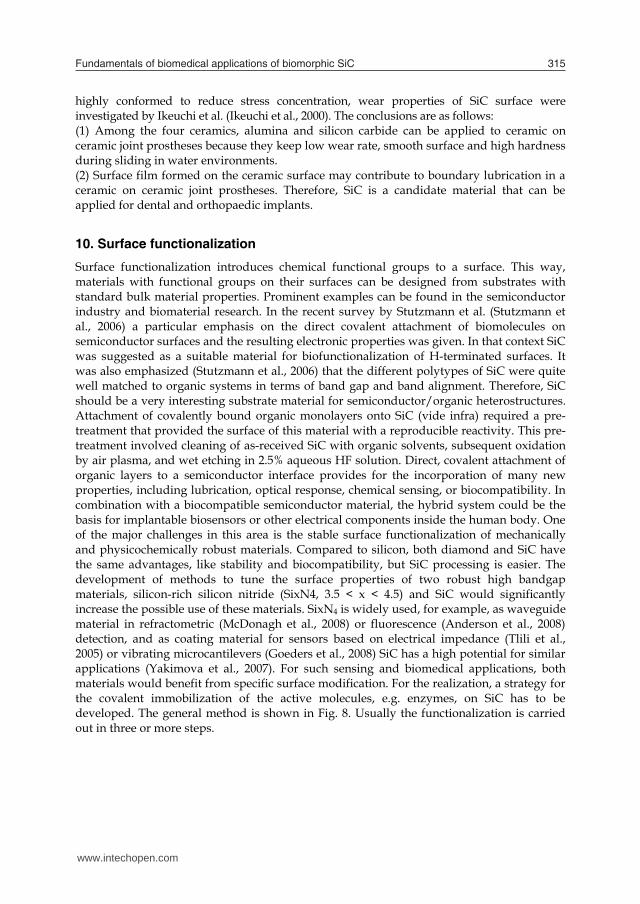
Fundamentals of biomedical applications of biomorphic SiC 315
Fig. 7. Scanning electron microscopy images showing the MG-63 osteoblast-like cell monolayer time course formation. A, 1 hour; B, 6 hrs and C, 24 hrs after seeding on a representative beech-based SiC ceramic coated with bioactive glass. All magnifications are 1000×(Carlos et al., 2006) When using the combination of 316L and gold in liquid, electrolytic media such as blood, the corrosion resistance of the system comes into question. The main reasons for this are contact and crevice corrosion. The corrosive attack in particular could lead to the destruction of the implant or to a release of metallic ions. Along with the potential problem of corrosion, there is the question of bio- and hemocompatibility of gold that is in direct contact with the biological environment. While some authors do not see any deleterious interactions between gold and the vessel wall (Tanigawa et al., 1991, 1995), a significant thrombogenic effect has been attributed to this material in other publications (Sawyer et al., 1965; Schomig et al., 1999). The corrosion behavior of gold-covered stents that have been coated with an additional, amorphous silicon carbide film that is known to be antithrombogenic (a-SiC:H), and which prevents direct contact between the gold coating and the biological environment (Wendler et al., 2000). For improved osseointegration, titanium-based total hip replacement (THR) components were coated by some manufacturers with hydroxyapatite, which also prevents titanium metallosis. However, hydroxyapatite has a tendency to dissolve from the implant surface, and thus a hydroxyapatite coating does not prevent periprosthetic metallosis permanently. Wear particles from THR prosthetic components cause a local host response directly and indirectly, which leads to component loosening (Santavirta et al., 1990). Silicon carbide as a ceramic coating material of titanium-based THR implants. The idea is to prevent wear debris formation from the soft titanium surface. SiC is a hard and tightly bonding ceramic surface material, and because of these physical properties it is not easily degradable, as is the case with hydroxyapatite. SiC can be deposited on a titanium implant surface, e.g., by sputtering techniques, and such coatings bond very well to the substrate. In human monocyte cultures phagocytized SiC particles cause a similar stimulation to hydroxyapatite (Santavirta et al., 1998). Recently titanium-based implant manufacturers have begun to consider modifying surfaces, so that bonds can form a mechanical interlock. The approach is the creation of meshwork through sintered beads or threads that have 350–500 µm pores to promote osteoconduction. The obtained mechanical properties, i.e. elastic modulus ≥335 GPa are favorable as compared with human cancellous bone and titanium. Ceramic on ceramic total hip prostheses are developed to apply to young patients because lifetime of polyethylene joint prostheses is limited by loosening due to biological response. As mating faces of all-ceramic joint must be
highly conformed to reduce stress concentration, wear properties of SiC surface were investigated by Ikeuchi et al. (Ikeuchi et al., 2000). The conclusions are as follows: (1) Among the four ceramics, alumina and silicon carbide can be applied to ceramic on ceramic joint prostheses because they keep low wear rate, smooth surface and high hardness during sliding in water environments. (2) Surface film formed on the ceramic surface may contribute to boundary lubrication in a ceramic on ceramic joint prostheses. Therefore, SiC is a candidate material that can be applied for dental and orthopaedic implants.
10. Surface functionalization
Surface functionalization introduces chemical functional groups to a surface. This way, materials with functional groups on their surfaces can be designed from substrates with standard bulk material properties. Prominent examples can be found in the semiconductor industry and biomaterial research. In the recent survey by Stutzmann et al. (Stutzmann et al., 2006) a particular emphasis on the direct covalent attachment of biomolecules on semiconductor surfaces and the resulting electronic properties was given. In that context SiC was suggested as a suitable material for biofunctionalization of H-terminated surfaces. It was also emphasized (Stutzmann et al., 2006) that the different polytypes of SiC were quite well matched to organic systems in terms of band gap and band alignment. Therefore, SiC should be a very interesting substrate material for semiconductor/organic heterostructures. Attachment of covalently bound organic monolayers onto SiC (vide infra) required a pre-treatment that provided the surface of this material with a reproducible reactivity. This pre-treatment involved cleaning of as-received SiC with organic solvents, subsequent oxidation by air plasma, and wet etching in 2.5% aqueous HF solution. Direct, covalent attachment of organic layers to a semiconductor interface provides for the incorporation of many new properties, including lubrication, optical response, chemical sensing, or biocompatibility. In combination with a biocompatible semiconductor material, the hybrid system could be the basis for implantable biosensors or other electrical components inside the human body. One of the major challenges in this area is the stable surface functionalization of mechanically and physicochemically robust materials. Compared to silicon, both diamond and SiC have the same advantages, like stability and biocompatibility, but SiC processing is easier. The development of methods to tune the surface properties of two robust high bandgap materials, silicon-rich silicon nitride (SixN4, 3.5 < x < 4.5) and SiC would significantly increase the possible use of these materials. SixN4 is widely used, for example, as waveguide material in refractometric (McDonagh et al., 2008) or fluorescence (Anderson et al., 2008) detection, and as coating material for sensors based on electrical impedance (Tlili et al., 2005) or vibrating microcantilevers (Goeders et al., 2008) SiC has a high potential for similar applications (Yakimova et al., 2007). For such sensing and biomedical applications, both materials would benefit from specific surface modification. For the realization, a strategy for the covalent immobilization of the active molecules, e.g. enzymes, on SiC has to be developed. The general method is shown in Fig. 8. Usually the functionalization is carried out in three or more steps.
www.intechopen.com
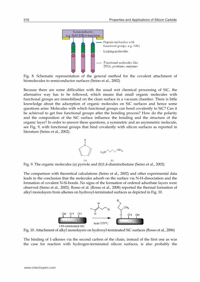
Properties and Applications of Silicon Carbide316
Fig. 8. Schematic representation of the general method for the covalent attachment of biomolecules to semiconductor surfaces (Seino et al., 2002) Because there are some difficulties with the usual wet chemical processing of SiC, the alternative way has to be followed, which means that small organic molecules with functional groups are immobilised on the clean surface in a vacuum chamber. There is little knowledge about the adsorption of organic molecules on SiC surfaces and hence some questions arise: Molecules with which functional groups can bond covalently to SiC? Can it be achieved to get free functional groups after the bonding process? How do the polarity and the composition of the SiC surface influence the bonding and the structure of the organic layer? In order to answer these questions, a symmetric and an asymmetric molecule, see Fig. 9, with functional groups that bind covalently with silicon surfaces as reported in literature (Seino et al., 2002).
Fig. 9. The organic molecules (a) pyrrole and (b)1,4-diaminobutane (Seino et al., 2002) The comparison with theoretical calculations (Seino et al., 2002) and other experimental data leads to the conclusion that the molecules adsorb on the surface via N-H-dissociation and the formation of covalent N-Si-bonds. No signs of the formation of ordered adsorbate layers were observed (Seino et al., 2002). Rosso et al. (Rosso et al., 2008) reported the thermal formation of alkyl monolayers from alkenes on hydroxyl-terminated surfaces as depicted in Fig. 10.
Fig. 10. Attachment of alkyl monolayers on hydroxyl-terminated SiC surfaces (Rosso et al., 2006) The binding of 1-alkenes via the second carbon of the chain, instead of the first one as was the case for reaction with hydrogen-terminated silicon surfaces, is also probably the
explanation for the formation of somewhat less ordered monolayers. Apart from the stability of methyl-terminated surfaces, the integration of organic functionalities onto inorganic materials opens an extended field of research. Rosso et al. (Rosso et al., 2009) investigated the UV-assisted formation of organic monolayers on 3C-SiC and SixN4, using only wet chemistry under ambient conditions (temperature and pressure), as this would significantly increase the range of monolayer functionalities that can be attached. After hydrolysis of ester monolayers and subsequent N-hydrosuccinimide activation of the obtained carboxylic acid-terminated surfaces, amide formation with m-(trifluoromethyl) benzylamine (TFBA) showed the possibility of easy surface (bio-) functionalization of SiC. Beside alkyl monolayers, functional coatings from covalently attached ω-functionalized 1-alkenes can be successfully formed onto both SiC surface. Methyl undec-10-enoate and 2,2,2-trifluoroethyl undec-10-enoate (TFE) were successfully attached onto this material using UV light. TFE monolayers were subsequently subjected to further reactions at room temperature, including hydrolysis with a 0.25 M solution of potassium tert-butoxide in DMSO, followed by a NHS activation of the obtained carboxylic acids and subsequent reaction with TFBA, as depicted in Fig. 11.
Fig. 11. Functionalization of TFE monolayers on SiC or SixN4 surfaces (Rosso et al., 2009) Robust functionalization of SiC surface with a wide variety of (bio-)functional moieties is possible using a protecting trifluoethyl ester. A rich chemistry including surface patterning can then be performed. Protein-repellent monolayers can also be formed with these methods, using oligoethylene glycol compounds. Cicero and Catellani (Cicero and Catellani, 2005) used first-principle predictions of the chemical processes that eventually led to adsorption of organic molecules on the Si-terminated SiC in the search of feasible ways to surface functionalization. Their results suggest a larger stability of the functionalization for the studied groups on SiC rather than on Si. Moreover, the preferential chemisorption of thiolates can lead to the realization of stable self-assembled monolayers (SAMs), with no need of preliminary metallic deposition. The interaction of water with polar SiC surfaces was first investigated. The researchers found that irrespective of coverage, water dissociates on the Si-terminated surface, substantially modifying the clean surface reconstruction. In contrast, the C-terminated surface is non-reactive and hydrophobic. This suggests that by growing SiC substrates with adjacent islands which are either Si- or C-terminated, an atomic control of hydrophilic and hydrophobic environments may be attained. Such surfaces are interesting for DNA and protein attachment. Kanai et al (Kanai et al., 2005) studied theoretically biotin chemisorption on clean and hydrohylated Si-terminated SiC (001) surfaces. These results gained preliminary insight into the understanding of experimental observations showing that biotin molecules directly adsorbed on the hydrohylated surfaces bind strongly to some proteins. Preuss et al (Preuss et al., 2006) made a combined theoretical and experimental study of pyrrole-functionalized Si- and C-terminated SiC surfaces. Recent experimental
www.intechopen.com
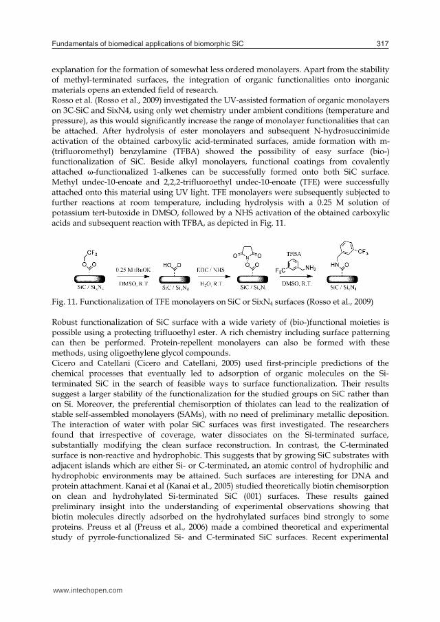
Fundamentals of biomedical applications of biomorphic SiC 317
Fig. 8. Schematic representation of the general method for the covalent attachment of biomolecules to semiconductor surfaces (Seino et al., 2002) Because there are some difficulties with the usual wet chemical processing of SiC, the alternative way has to be followed, which means that small organic molecules with functional groups are immobilised on the clean surface in a vacuum chamber. There is little knowledge about the adsorption of organic molecules on SiC surfaces and hence some questions arise: Molecules with which functional groups can bond covalently to SiC? Can it be achieved to get free functional groups after the bonding process? How do the polarity and the composition of the SiC surface influence the bonding and the structure of the organic layer? In order to answer these questions, a symmetric and an asymmetric molecule, see Fig. 9, with functional groups that bind covalently with silicon surfaces as reported in literature (Seino et al., 2002).
Fig. 9. The organic molecules (a) pyrrole and (b)1,4-diaminobutane (Seino et al., 2002) The comparison with theoretical calculations (Seino et al., 2002) and other experimental data leads to the conclusion that the molecules adsorb on the surface via N-H-dissociation and the formation of covalent N-Si-bonds. No signs of the formation of ordered adsorbate layers were observed (Seino et al., 2002). Rosso et al. (Rosso et al., 2008) reported the thermal formation of alkyl monolayers from alkenes on hydroxyl-terminated surfaces as depicted in Fig. 10.
Fig. 10. Attachment of alkyl monolayers on hydroxyl-terminated SiC surfaces (Rosso et al., 2006) The binding of 1-alkenes via the second carbon of the chain, instead of the first one as was the case for reaction with hydrogen-terminated silicon surfaces, is also probably the
explanation for the formation of somewhat less ordered monolayers. Apart from the stability of methyl-terminated surfaces, the integration of organic functionalities onto inorganic materials opens an extended field of research. Rosso et al. (Rosso et al., 2009) investigated the UV-assisted formation of organic monolayers on 3C-SiC and SixN4, using only wet chemistry under ambient conditions (temperature and pressure), as this would significantly increase the range of monolayer functionalities that can be attached. After hydrolysis of ester monolayers and subsequent N-hydrosuccinimide activation of the obtained carboxylic acid-terminated surfaces, amide formation with m-(trifluoromethyl) benzylamine (TFBA) showed the possibility of easy surface (bio-) functionalization of SiC. Beside alkyl monolayers, functional coatings from covalently attached ω-functionalized 1-alkenes can be successfully formed onto both SiC surface. Methyl undec-10-enoate and 2,2,2-trifluoroethyl undec-10-enoate (TFE) were successfully attached onto this material using UV light. TFE monolayers were subsequently subjected to further reactions at room temperature, including hydrolysis with a 0.25 M solution of potassium tert-butoxide in DMSO, followed by a NHS activation of the obtained carboxylic acids and subsequent reaction with TFBA, as depicted in Fig. 11.
Fig. 11. Functionalization of TFE monolayers on SiC or SixN4 surfaces (Rosso et al., 2009) Robust functionalization of SiC surface with a wide variety of (bio-)functional moieties is possible using a protecting trifluoethyl ester. A rich chemistry including surface patterning can then be performed. Protein-repellent monolayers can also be formed with these methods, using oligoethylene glycol compounds. Cicero and Catellani (Cicero and Catellani, 2005) used first-principle predictions of the chemical processes that eventually led to adsorption of organic molecules on the Si-terminated SiC in the search of feasible ways to surface functionalization. Their results suggest a larger stability of the functionalization for the studied groups on SiC rather than on Si. Moreover, the preferential chemisorption of thiolates can lead to the realization of stable self-assembled monolayers (SAMs), with no need of preliminary metallic deposition. The interaction of water with polar SiC surfaces was first investigated. The researchers found that irrespective of coverage, water dissociates on the Si-terminated surface, substantially modifying the clean surface reconstruction. In contrast, the C-terminated surface is non-reactive and hydrophobic. This suggests that by growing SiC substrates with adjacent islands which are either Si- or C-terminated, an atomic control of hydrophilic and hydrophobic environments may be attained. Such surfaces are interesting for DNA and protein attachment. Kanai et al (Kanai et al., 2005) studied theoretically biotin chemisorption on clean and hydrohylated Si-terminated SiC (001) surfaces. These results gained preliminary insight into the understanding of experimental observations showing that biotin molecules directly adsorbed on the hydrohylated surfaces bind strongly to some proteins. Preuss et al (Preuss et al., 2006) made a combined theoretical and experimental study of pyrrole-functionalized Si- and C-terminated SiC surfaces. Recent experimental
www.intechopen.com
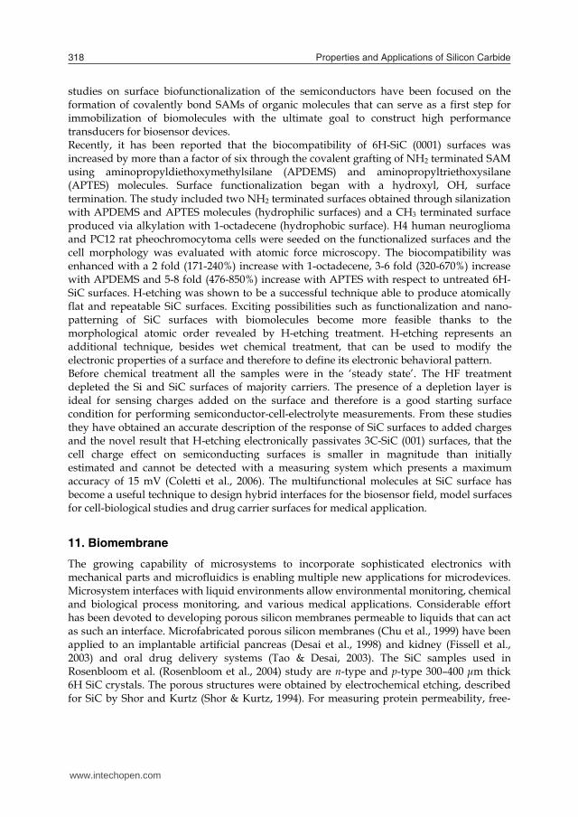
Properties and Applications of Silicon Carbide318
studies on surface biofunctionalization of the semiconductors have been focused on the formation of covalently bond SAMs of organic molecules that can serve as a first step for immobilization of biomolecules with the ultimate goal to construct high performance transducers for biosensor devices. Recently, it has been reported that the biocompatibility of 6H-SiC (0001) surfaces was increased by more than a factor of six through the covalent grafting of NH2 terminated SAM using aminopropyldiethoxymethylsilane (APDEMS) and aminopropyltriethoxysilane (APTES) molecules. Surface functionalization began with a hydroxyl, OH, surface termination. The study included two NH2 terminated surfaces obtained through silanization with APDEMS and APTES molecules (hydrophilic surfaces) and a CH3 terminated surface produced via alkylation with 1-octadecene (hydrophobic surface). H4 human neuroglioma and PC12 rat pheochromocytoma cells were seeded on the functionalized surfaces and the cell morphology was evaluated with atomic force microscopy. The biocompatibility was enhanced with a 2 fold (171-240%) increase with 1-octadecene, 3-6 fold (320-670%) increase with APDEMS and 5-8 fold (476-850%) increase with APTES with respect to untreated 6H-SiC surfaces. H-etching was shown to be a successful technique able to produce atomically flat and repeatable SiC surfaces. Exciting possibilities such as functionalization and nano-patterning of SiC surfaces with biomolecules become more feasible thanks to the morphological atomic order revealed by H-etching treatment. H-etching represents an additional technique, besides wet chemical treatment, that can be used to modify the electronic properties of a surface and therefore to define its electronic behavioral pattern. Before chemical treatment all the samples were in the ‘steady state’. The HF treatment depleted the Si and SiC surfaces of majority carriers. The presence of a depletion layer is ideal for sensing charges added on the surface and therefore is a good starting surface condition for performing semiconductor-cell-electrolyte measurements. From these studies they have obtained an accurate description of the response of SiC surfaces to added charges and the novel result that H-etching electronically passivates 3C-SiC (001) surfaces, that the cell charge effect on semiconducting surfaces is smaller in magnitude than initially estimated and cannot be detected with a measuring system which presents a maximum accuracy of 15 mV (Coletti et al., 2006). The multifunctional molecules at SiC surface has become a useful technique to design hybrid interfaces for the biosensor field, model surfaces for cell-biological studies and drug carrier surfaces for medical application.
11. Biomembrane
The growing capability of microsystems to incorporate sophisticated electronics with mechanical parts and microfluidics is enabling multiple new applications for microdevices. Microsystem interfaces with liquid environments allow environmental monitoring, chemical and biological process monitoring, and various medical applications. Considerable effort has been devoted to developing porous silicon membranes permeable to liquids that can act as such an interface. Microfabricated porous silicon membranes (Chu et al., 1999) have been applied to an implantable artificial pancreas (Desai et al., 1998) and kidney (Fissell et al., 2003) and oral drug delivery systems (Tao & Desai, 2003). The SiC samples used in Rosenbloom et al. (Rosenbloom et al., 2004) study are n-type and p-type 300–400 μm thick 6H SiC crystals. The porous structures were obtained by electrochemical etching, described for SiC by Shor and Kurtz (Shor & Kurtz, 1994). For measuring protein permeability, free-
standing nanoporous SiC membranes were glued onto circular polystyrene plastic supports and placed into a small fluidic chamber. Molecules within the source solution below the membrane diffused through the membrane into the receiving solution above. Biofouling is a significant problem with any nanoporous membrane exposed to high protein concentrations. The two porous SiC morphologies shown in Fig. 12 were used by Rosenbloom et al. (Rosenbloom et al., 2004). Despite considerably different pore structure, membranes of both n-type and p-type SiC exclude proteins in the same size range. The n-type material allowed much more protein to diffuse through, by a factor of as much as four times (for myoglobin). Each membrane type passed proteins of up to 29000 Da molecular weight but excluded larger proteins with a molecular weight of 45000 Da and higher. These molecular weights correspond to molecular diameters of less than 4.7 nm (permeable) and greater than 5.0 nm (excluded). The discrimination between the 29 kD and 44 kD proteins is likely complex, and not simply based on size (4.7 and 5.0 nm, respectively). For example, charge, hydrophilic or hydrophobic interactions, as well as dimerization in solution or differential adsorption to the SiC, to the glue, to the plastic tube or membrane backing, etc., could all play roles. This same behaviour was observed when they tested the same 6-protein mix with commercial membranes, i.e., ovalbumin is excluded to a much greater degree than carbonic anhydrase.
Fig. 12. The cross-sectional SEM images taken from pieces of porous membranes used for the tests: (a) n-type 6 H SiC and (b) p-type 6 H SiC. The p-type porous structure has a more feathery appearance, corresponding to more material removal. These membranes are more fragile than the n-type membranes (Rosenbloom et al., 2004) It has been shown (Bai et al., 2003), that nanoporous SiC membranes can be made with a wide variety of pore sizes, shapes and densities by varying the conditions of pore formation. The size range of proteins able to cross the membranes includes many important cell signaling molecules, which tend to be small. Nanoporous SiC has a low protein adsorption comparable to the best commercially available polymeric membranes specifically designed for low protein adsorption. This resistance to protein fouling is another desirable property of this material. Early membranes that were tested had nonuniform pores. Having demonstrated good resistance to protein adhesion for SiC, and the ability to make free-standing semi-permeable membranes, it was decided to attempt to improve the membranes by making more uniform and consistent pore structures. The goal was to improve the protein throughput. These membranes had areas of large, relatively straight pores with a high density in some areas. The protein permeability of these membranes was very good. Although these membranes are usable for probe interfaces, it is obvious that the pore structures are nonuniform. Most of the diffusion likely occurs in a few areas of high porosity
www.intechopen.com
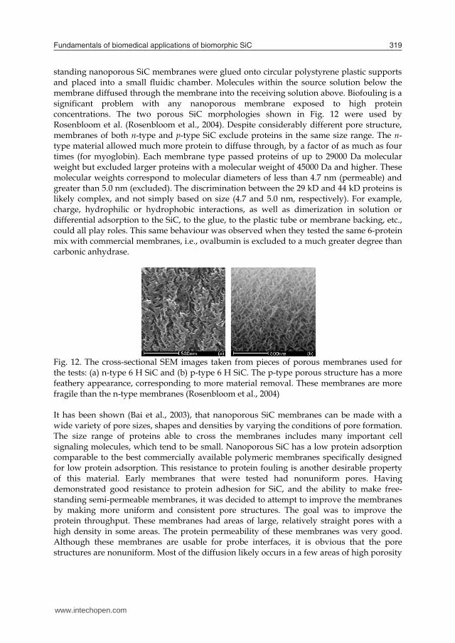
Fundamentals of biomedical applications of biomorphic SiC 319
studies on surface biofunctionalization of the semiconductors have been focused on the formation of covalently bond SAMs of organic molecules that can serve as a first step for immobilization of biomolecules with the ultimate goal to construct high performance transducers for biosensor devices. Recently, it has been reported that the biocompatibility of 6H-SiC (0001) surfaces was increased by more than a factor of six through the covalent grafting of NH2 terminated SAM using aminopropyldiethoxymethylsilane (APDEMS) and aminopropyltriethoxysilane (APTES) molecules. Surface functionalization began with a hydroxyl, OH, surface termination. The study included two NH2 terminated surfaces obtained through silanization with APDEMS and APTES molecules (hydrophilic surfaces) and a CH3 terminated surface produced via alkylation with 1-octadecene (hydrophobic surface). H4 human neuroglioma and PC12 rat pheochromocytoma cells were seeded on the functionalized surfaces and the cell morphology was evaluated with atomic force microscopy. The biocompatibility was enhanced with a 2 fold (171-240%) increase with 1-octadecene, 3-6 fold (320-670%) increase with APDEMS and 5-8 fold (476-850%) increase with APTES with respect to untreated 6H-SiC surfaces. H-etching was shown to be a successful technique able to produce atomically flat and repeatable SiC surfaces. Exciting possibilities such as functionalization and nano-patterning of SiC surfaces with biomolecules become more feasible thanks to the morphological atomic order revealed by H-etching treatment. H-etching represents an additional technique, besides wet chemical treatment, that can be used to modify the electronic properties of a surface and therefore to define its electronic behavioral pattern. Before chemical treatment all the samples were in the ‘steady state’. The HF treatment depleted the Si and SiC surfaces of majority carriers. The presence of a depletion layer is ideal for sensing charges added on the surface and therefore is a good starting surface condition for performing semiconductor-cell-electrolyte measurements. From these studies they have obtained an accurate description of the response of SiC surfaces to added charges and the novel result that H-etching electronically passivates 3C-SiC (001) surfaces, that the cell charge effect on semiconducting surfaces is smaller in magnitude than initially estimated and cannot be detected with a measuring system which presents a maximum accuracy of 15 mV (Coletti et al., 2006). The multifunctional molecules at SiC surface has become a useful technique to design hybrid interfaces for the biosensor field, model surfaces for cell-biological studies and drug carrier surfaces for medical application.
11. Biomembrane
The growing capability of microsystems to incorporate sophisticated electronics with mechanical parts and microfluidics is enabling multiple new applications for microdevices. Microsystem interfaces with liquid environments allow environmental monitoring, chemical and biological process monitoring, and various medical applications. Considerable effort has been devoted to developing porous silicon membranes permeable to liquids that can act as such an interface. Microfabricated porous silicon membranes (Chu et al., 1999) have been applied to an implantable artificial pancreas (Desai et al., 1998) and kidney (Fissell et al., 2003) and oral drug delivery systems (Tao & Desai, 2003). The SiC samples used in Rosenbloom et al. (Rosenbloom et al., 2004) study are n-type and p-type 300–400 μm thick 6H SiC crystals. The porous structures were obtained by electrochemical etching, described for SiC by Shor and Kurtz (Shor & Kurtz, 1994). For measuring protein permeability, free-
standing nanoporous SiC membranes were glued onto circular polystyrene plastic supports and placed into a small fluidic chamber. Molecules within the source solution below the membrane diffused through the membrane into the receiving solution above. Biofouling is a significant problem with any nanoporous membrane exposed to high protein concentrations. The two porous SiC morphologies shown in Fig. 12 were used by Rosenbloom et al. (Rosenbloom et al., 2004). Despite considerably different pore structure, membranes of both n-type and p-type SiC exclude proteins in the same size range. The n-type material allowed much more protein to diffuse through, by a factor of as much as four times (for myoglobin). Each membrane type passed proteins of up to 29000 Da molecular weight but excluded larger proteins with a molecular weight of 45000 Da and higher. These molecular weights correspond to molecular diameters of less than 4.7 nm (permeable) and greater than 5.0 nm (excluded). The discrimination between the 29 kD and 44 kD proteins is likely complex, and not simply based on size (4.7 and 5.0 nm, respectively). For example, charge, hydrophilic or hydrophobic interactions, as well as dimerization in solution or differential adsorption to the SiC, to the glue, to the plastic tube or membrane backing, etc., could all play roles. This same behaviour was observed when they tested the same 6-protein mix with commercial membranes, i.e., ovalbumin is excluded to a much greater degree than carbonic anhydrase.
Fig. 12. The cross-sectional SEM images taken from pieces of porous membranes used for the tests: (a) n-type 6 H SiC and (b) p-type 6 H SiC. The p-type porous structure has a more feathery appearance, corresponding to more material removal. These membranes are more fragile than the n-type membranes (Rosenbloom et al., 2004) It has been shown (Bai et al., 2003), that nanoporous SiC membranes can be made with a wide variety of pore sizes, shapes and densities by varying the conditions of pore formation. The size range of proteins able to cross the membranes includes many important cell signaling molecules, which tend to be small. Nanoporous SiC has a low protein adsorption comparable to the best commercially available polymeric membranes specifically designed for low protein adsorption. This resistance to protein fouling is another desirable property of this material. Early membranes that were tested had nonuniform pores. Having demonstrated good resistance to protein adhesion for SiC, and the ability to make free-standing semi-permeable membranes, it was decided to attempt to improve the membranes by making more uniform and consistent pore structures. The goal was to improve the protein throughput. These membranes had areas of large, relatively straight pores with a high density in some areas. The protein permeability of these membranes was very good. Although these membranes are usable for probe interfaces, it is obvious that the pore structures are nonuniform. Most of the diffusion likely occurs in a few areas of high porosity
www.intechopen.com
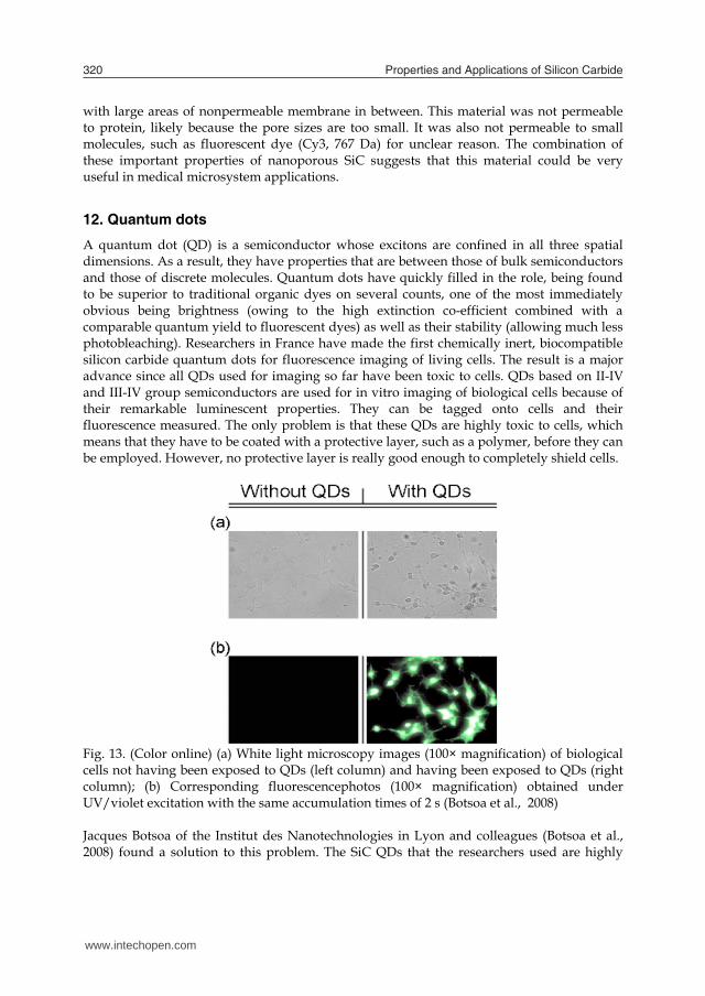
Properties and Applications of Silicon Carbide320
with large areas of nonpermeable membrane in between. This material was not permeable to protein, likely because the pore sizes are too small. It was also not permeable to small molecules, such as fluorescent dye (Cy3, 767 Da) for unclear reason. The combination of these important properties of nanoporous SiC suggests that this material could be very useful in medical microsystem applications.
12. Quantum dots
A quantum dot (QD) is a semiconductor whose excitons are confined in all three spatial dimensions. As a result, they have properties that are between those of bulk semiconductors and those of discrete molecules. Quantum dots have quickly filled in the role, being found to be superior to traditional organic dyes on several counts, one of the most immediately obvious being brightness (owing to the high extinction co-efficient combined with a comparable quantum yield to fluorescent dyes) as well as their stability (allowing much less photobleaching). Researchers in France have made the first chemically inert, biocompatible silicon carbide quantum dots for fluorescence imaging of living cells. The result is a major advance since all QDs used for imaging so far have been toxic to cells. QDs based on II-IV and III-IV group semiconductors are used for in vitro imaging of biological cells because of their remarkable luminescent properties. They can be tagged onto cells and their fluorescence measured. The only problem is that these QDs are highly toxic to cells, which means that they have to be coated with a protective layer, such as a polymer, before they can be employed. However, no protective layer is really good enough to completely shield cells.
Fig. 13. (Color online) (a) White light microscopy images (100× magnification) of biological cells not having been exposed to QDs (left column) and having been exposed to QDs (right column); (b) Corresponding fluorescencephotos (100× magnification) obtained under UV/violet excitation with the same accumulation times of 2 s (Botsoa et al., 2008) Jacques Botsoa of the Institut des Nanotechnologies in Lyon and colleagues (Botsoa et al., 2008) found a solution to this problem. The SiC QDs that the researchers used are highly
luminescent. They could be used to bio-image and label living cells, as well as study the mechanisms of quantum transport through cells and nuclear membranes. Botsoa and co-workers made their QDs by electrochemical anodization etching of low resistivity grade bulk 3C-SiC polycrystalline wafer. These QDs have dimensions smaller than the Bohr diameter of the exciton (around 5.4凹nm) and exhibit highly efficient "above-gap" luminescence thanks to quantum confinement. To test their QDs, the researchers added the suspension to cell cultures of 3T3-L1 fibroblasts (Fig. 13). Rossi et al (Rossi et al., 2008) prepared SiC nanocrystals by single crystal silicon carbide wafer electrochemical etching. Particles in the size range of 1-3 nm exhibit stable UV-VIS photoluminescence in aqueous media, acids, and organic solvents. Given that the SiC nanostructures below ~ 3 nm would acquire a bandgap above 3 eV that effectively extends their luminescence into the UV range. Moreover, due to the exceptional SiC thermal and chemical stability, such nanostructures could be considered for the UV emitter development and serve as an alternative to the Group-III nitride based semiconductors. SiC based QDs are water-soluble and they may be ideal as biological labels. The nanoparticles show high resistance against photobleaching with no significant cytotoxicity (Fig. 14). In conclusion, SiC-based QDs for biomedical application sounds to be a promising approach for future research.
Fig. 14. After the uptake of 3C-SiC nanocrystals, human fetal osteoblast (hFOB) cells exhibit brightgreen-yellow fluorescence (Fan et al., 2008)
13. Nanosilicon carbide
Silicon carbide nanomaterials are widely investigated due to high strength, good creep, oxidation resistance at elevated temperature, chemical inertness, thermal stability, and resistance to corrosion (Kamakaran et al., 2004). The size, shape, and surface composition of these nanostructured materials are some of the few factors that contribute to these unique and fascinating properties (Xia et al., 2003). Efforts have been made to develop nanosize hollow spheres of silicon carbide for technical applications in optical, electronic, acoustic, and sensing devices (Sun and Xia, 2002, Kidambi et al., 2004). Recently, the formation of silicon carbide nanowires from inorganic–organic hybrid of sol–gel-derived silica and the commercially available lignin that is naturally abundant amorphous biopolymer was reported by Mishra et al. (Mishra et al., 2009). The biocompatibility of the SiC nanomesh renders it a candidate template for biomolecular assembly, opening many unexplored and stimulating possibilities for fundamental and applied studies. As a consequence the sophisticated tools available to surface scientists can shed new light onto nanoscale aspects of biocompatibility (Oliveria & Nanci, 2004), which are related mainly to surface and interface properties, thus guiding the exploration of novel potential biomaterials. Another crucial point for a thorough understanding of the SiC
www.intechopen.com
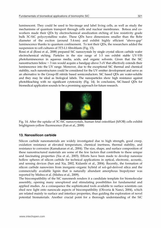
Fundamentals of biomedical applications of biomorphic SiC 321
with large areas of nonpermeable membrane in between. This material was not permeable to protein, likely because the pore sizes are too small. It was also not permeable to small molecules, such as fluorescent dye (Cy3, 767 Da) for unclear reason. The combination of these important properties of nanoporous SiC suggests that this material could be very useful in medical microsystem applications.
12. Quantum dots
A quantum dot (QD) is a semiconductor whose excitons are confined in all three spatial dimensions. As a result, they have properties that are between those of bulk semiconductors and those of discrete molecules. Quantum dots have quickly filled in the role, being found to be superior to traditional organic dyes on several counts, one of the most immediately obvious being brightness (owing to the high extinction co-efficient combined with a comparable quantum yield to fluorescent dyes) as well as their stability (allowing much less photobleaching). Researchers in France have made the first chemically inert, biocompatible silicon carbide quantum dots for fluorescence imaging of living cells. The result is a major advance since all QDs used for imaging so far have been toxic to cells. QDs based on II-IV and III-IV group semiconductors are used for in vitro imaging of biological cells because of their remarkable luminescent properties. They can be tagged onto cells and their fluorescence measured. The only problem is that these QDs are highly toxic to cells, which means that they have to be coated with a protective layer, such as a polymer, before they can be employed. However, no protective layer is really good enough to completely shield cells.
Fig. 13. (Color online) (a) White light microscopy images (100× magnification) of biological cells not having been exposed to QDs (left column) and having been exposed to QDs (right column); (b) Corresponding fluorescencephotos (100× magnification) obtained under UV/violet excitation with the same accumulation times of 2 s (Botsoa et al., 2008) Jacques Botsoa of the Institut des Nanotechnologies in Lyon and colleagues (Botsoa et al., 2008) found a solution to this problem. The SiC QDs that the researchers used are highly
luminescent. They could be used to bio-image and label living cells, as well as study the mechanisms of quantum transport through cells and nuclear membranes. Botsoa and co-workers made their QDs by electrochemical anodization etching of low resistivity grade bulk 3C-SiC polycrystalline wafer. These QDs have dimensions smaller than the Bohr diameter of the exciton (around 5.4凹nm) and exhibit highly efficient "above-gap" luminescence thanks to quantum confinement. To test their QDs, the researchers added the suspension to cell cultures of 3T3-L1 fibroblasts (Fig. 13). Rossi et al (Rossi et al., 2008) prepared SiC nanocrystals by single crystal silicon carbide wafer electrochemical etching. Particles in the size range of 1-3 nm exhibit stable UV-VIS photoluminescence in aqueous media, acids, and organic solvents. Given that the SiC nanostructures below ~ 3 nm would acquire a bandgap above 3 eV that effectively extends their luminescence into the UV range. Moreover, due to the exceptional SiC thermal and chemical stability, such nanostructures could be considered for the UV emitter development and serve as an alternative to the Group-III nitride based semiconductors. SiC based QDs are water-soluble and they may be ideal as biological labels. The nanoparticles show high resistance against photobleaching with no significant cytotoxicity (Fig. 14). In conclusion, SiC-based QDs for biomedical application sounds to be a promising approach for future research.
Fig. 14. After the uptake of 3C-SiC nanocrystals, human fetal osteoblast (hFOB) cells exhibit brightgreen-yellow fluorescence (Fan et al., 2008)
13. Nanosilicon carbide
Silicon carbide nanomaterials are widely investigated due to high strength, good creep, oxidation resistance at elevated temperature, chemical inertness, thermal stability, and resistance to corrosion (Kamakaran et al., 2004). The size, shape, and surface composition of these nanostructured materials are some of the few factors that contribute to these unique and fascinating properties (Xia et al., 2003). Efforts have been made to develop nanosize hollow spheres of silicon carbide for technical applications in optical, electronic, acoustic, and sensing devices (Sun and Xia, 2002, Kidambi et al., 2004). Recently, the formation of silicon carbide nanowires from inorganic–organic hybrid of sol–gel-derived silica and the commercially available lignin that is naturally abundant amorphous biopolymer was reported by Mishra et al. (Mishra et al., 2009). The biocompatibility of the SiC nanomesh renders it a candidate template for biomolecular assembly, opening many unexplored and stimulating possibilities for fundamental and applied studies. As a consequence the sophisticated tools available to surface scientists can shed new light onto nanoscale aspects of biocompatibility (Oliveria & Nanci, 2004), which are related mainly to surface and interface properties, thus guiding the exploration of novel potential biomaterials. Another crucial point for a thorough understanding of the SiC
www.intechopen.com
Properties and Applications of Silicon Carbide322
template is the determination of the chemical nature of the carbonaceous species at the surface (Cicoira & Rosei, 2006). Bioceramic nanocomposites were synthesized by sintering compacted bodies of hydroxyapatite mixed with 5 or 15 wt% nanosilicon carbide at 1100 or 1200 °C in a reducing atmosphere. The results indicate that the composite of 95 wt% hydroxyapatite and 5 wt% SiC exhibited better mechanical and biological properties than pure hydroxyapatite and further addition of SiC failed strength and toughness (Hesaraki et al., 2010). The preparation of nano-sized silicon carbide has received considerable attention, because it allows the preparation of bulk materials with increased plasticity (Stobierski & Gubernat, 2003) or nanocomposites with enhanced mechanical and tribological properties. In conclusion, it opens up exciting possibilities in the area of template-assisted growth at the nanoscale.
14. Drug delivery
Drug delivery systems (DDS) are an area of study in which researchers from almost every scientific discipline can make a significant contribution. Understanding the fate of drugs inside the human body is a high standard classical endeavor, where basic and mathematical analysis can be used to achieve an important practical end. No doubt the effectiveness of drug therapy is closely related to biophysics and physiology of drug movement through tissue. Therefore, DDS requires an understanding of the characteristics of the system, the molecular mechanisms of drug transport and elimination, particularly at the site of delivery. In the last decade DDS have received much attention since they can significantly improve the therapeutic effects of the drug while minimizing its side effects.In recent years, Poly (D,L-Lactide) (PLA) and Poly (D,L-Lactide-co-Glycolide) (PLGA) have been extensively investigated for use as implantable biodegradable carriers for controlled release of drugs. Silicon carbide coated stents have been coated with a layer of PLA or PLGA containing the drug by dip coating or spray coating techniques. Several drugs have been considered as candidates for stent coatings preventing instent restenosis. SiC is used as a basis for drug delivery systems or bioactive coatings in order to modulate vascular cell growth. For a sufficient polymer-drug coating of a silicon carbide stent and a long-term release of the desired agent, PLA and PLGA are biocompatible materials useful for a variety of applications, including the design and properties of the controlled-release systems for pharmaceutical agents. Despite the phenomenal pace of stent design technology and the improvements in biocompatibility that have been achieved with the SiC coating, the incidence of in-stent restenosis remains unacceptably high. To address this problem, intense research is being conducted in order to find new stent coatings. Coatings with specific polymer-drug composites or with specific glycosaminoglycans showed promising results in modulating the proliferation of vascular smooth muscle cells and endothelial cells (Bayer, 2001). Using an existing technology for dip coating, glycosaminoglycans can be covalently bonded to the silicon carbide surface via a spacer molecule (Hildebrandt, 2001). Crosslinking the network of coated glycosaminoglycans should result in a stable bioactive layer with long-term anti-proliferative effects (Bayer, 2001). Finally, it is suggested that more detailed experiments are required and would be useful to distinguish and clarify SiC-based materials application in drug delivery.
15. Surface modification of Ti-6Al-4V alloy by SiC paper for orthopaedic applications
It is possible to change localized areas of metals in order to obtain both compositions and microstructures with improved properties. Titanium and titanium alloys are the most frequently used material for load-bearing orthopaedic implants, due to their specific properties such as high corrosion resistance, surface oxidation layer, high strength and high-temperature resistance (Feng et al., 2003). Titanium and its alloys’ application like any other biomaterials involve the creation of at least one interface between the material and biological tissues. Biocompatibility and bioactivity of biomaterials rely on the interactions that take place between the interface of the biomaterials and the biological system (Wang & Zheng, 2009). It is generally believed that proteins adsorbed on implant surface can play an important role in cell-surface response. Different proteins such as collagen, fibronectin and vitronectin which are acting as ligands are particularly important in osteoblast interaction with surface. Ligands are the junctions which facilitate adhesion of bone cells to implant surface. In another word, more ligand formation implies a better cell-surface interaction (Tirrell et al., 2002). In vitro studies can be used to study the influence of surface properties on processes such as cell attachment, cell proliferation and cell differentiation. However, in vivo studies must be performed to achieve a complete understanding of the healing process around implants. Previous studies have shown that surface characteristics named above have a significant influence on adhesion, morphology and maturation of cultured osteoblasts (Masuda et al., 1998). Also, it has been demonstrated that for primary bovine osteoblasts, the wettability is one of the key factors. In our studies (Khosroshahi et al., 2007; Khosroshahi), 2007; Khosroshahi et al., 2008; Khosroshahi et al., 2008; Khosroshahi et al., 2009), it is shown that the wettability of the surface can provide a better spreading condition for osteoblast cells due to reduced contact angle. Bearing in mind that the adhesion of bone cells to implant surface consists of two stages. In primary stage the cells must get close enough to surface at an appropriate distance known as focal distance over which the cells can easily be spread over it. In this respect, the wettability can be effective in providing a preferred accessability to surface and thus reaching the focal distance. The secondary stage includes cell-cell attachment which obeys the regular biological facts. Interface reactions between metallic implants and the surrounding tissues play a crucial role in the success of osseointegration. The titanium and its alloys like some other medical grade metals are the materials of choice for long-term implants. The effect of implant surface characteristics on bone reactions has thus attracted much attention and is still considered to be an important issue (Buchter et al., 2006). So far as the surface characteristics of the implants are concerned, two main features that can influence the establishment of the osseointegration are the physico-chemical properties and the surface morphology. Cell adhesion is involved in various phenomena such as embryogenesis, wound healing, immune response and metastasis as well as tissue integration of biomaterial. Thus, attachment, adhesion and spreading will depend on the cell-material interaction and the cell’s capacity to proliferate and to differentiate itself on contact with the implant (Bigerelle et al., 2005). Cell behavior, such as adhesion, morphologic change and functional alteration are greatly influenced by surface properties including texture, roughness, hydrophilicity and morphology. In extensive investigations of tissue response to implant surfaces, it has been shown that surface treatment of implant materials significantly influences the attachment of
www.intechopen.com
Fundamentals of biomedical applications of biomorphic SiC 323
template is the determination of the chemical nature of the carbonaceous species at the surface (Cicoira & Rosei, 2006). Bioceramic nanocomposites were synthesized by sintering compacted bodies of hydroxyapatite mixed with 5 or 15 wt% nanosilicon carbide at 1100 or 1200 °C in a reducing atmosphere. The results indicate that the composite of 95 wt% hydroxyapatite and 5 wt% SiC exhibited better mechanical and biological properties than pure hydroxyapatite and further addition of SiC failed strength and toughness (Hesaraki et al., 2010). The preparation of nano-sized silicon carbide has received considerable attention, because it allows the preparation of bulk materials with increased plasticity (Stobierski & Gubernat, 2003) or nanocomposites with enhanced mechanical and tribological properties. In conclusion, it opens up exciting possibilities in the area of template-assisted growth at the nanoscale.
14. Drug delivery
Drug delivery systems (DDS) are an area of study in which researchers from almost every scientific discipline can make a significant contribution. Understanding the fate of drugs inside the human body is a high standard classical endeavor, where basic and mathematical analysis can be used to achieve an important practical end. No doubt the effectiveness of drug therapy is closely related to biophysics and physiology of drug movement through tissue. Therefore, DDS requires an understanding of the characteristics of the system, the molecular mechanisms of drug transport and elimination, particularly at the site of delivery. In the last decade DDS have received much attention since they can significantly improve the therapeutic effects of the drug while minimizing its side effects.In recent years, Poly (D,L-Lactide) (PLA) and Poly (D,L-Lactide-co-Glycolide) (PLGA) have been extensively investigated for use as implantable biodegradable carriers for controlled release of drugs. Silicon carbide coated stents have been coated with a layer of PLA or PLGA containing the drug by dip coating or spray coating techniques. Several drugs have been considered as candidates for stent coatings preventing instent restenosis. SiC is used as a basis for drug delivery systems or bioactive coatings in order to modulate vascular cell growth. For a sufficient polymer-drug coating of a silicon carbide stent and a long-term release of the desired agent, PLA and PLGA are biocompatible materials useful for a variety of applications, including the design and properties of the controlled-release systems for pharmaceutical agents. Despite the phenomenal pace of stent design technology and the improvements in biocompatibility that have been achieved with the SiC coating, the incidence of in-stent restenosis remains unacceptably high. To address this problem, intense research is being conducted in order to find new stent coatings. Coatings with specific polymer-drug composites or with specific glycosaminoglycans showed promising results in modulating the proliferation of vascular smooth muscle cells and endothelial cells (Bayer, 2001). Using an existing technology for dip coating, glycosaminoglycans can be covalently bonded to the silicon carbide surface via a spacer molecule (Hildebrandt, 2001). Crosslinking the network of coated glycosaminoglycans should result in a stable bioactive layer with long-term anti-proliferative effects (Bayer, 2001). Finally, it is suggested that more detailed experiments are required and would be useful to distinguish and clarify SiC-based materials application in drug delivery.
15. Surface modification of Ti-6Al-4V alloy by SiC paper for orthopaedic applications
It is possible to change localized areas of metals in order to obtain both compositions and microstructures with improved properties. Titanium and titanium alloys are the most frequently used material for load-bearing orthopaedic implants, due to their specific properties such as high corrosion resistance, surface oxidation layer, high strength and high-temperature resistance (Feng et al., 2003). Titanium and its alloys’ application like any other biomaterials involve the creation of at least one interface between the material and biological tissues. Biocompatibility and bioactivity of biomaterials rely on the interactions that take place between the interface of the biomaterials and the biological system (Wang & Zheng, 2009). It is generally believed that proteins adsorbed on implant surface can play an important role in cell-surface response. Different proteins such as collagen, fibronectin and vitronectin which are acting as ligands are particularly important in osteoblast interaction with surface. Ligands are the junctions which facilitate adhesion of bone cells to implant surface. In another word, more ligand formation implies a better cell-surface interaction (Tirrell et al., 2002). In vitro studies can be used to study the influence of surface properties on processes such as cell attachment, cell proliferation and cell differentiation. However, in vivo studies must be performed to achieve a complete understanding of the healing process around implants. Previous studies have shown that surface characteristics named above have a significant influence on adhesion, morphology and maturation of cultured osteoblasts (Masuda et al., 1998). Also, it has been demonstrated that for primary bovine osteoblasts, the wettability is one of the key factors. In our studies (Khosroshahi et al., 2007; Khosroshahi), 2007; Khosroshahi et al., 2008; Khosroshahi et al., 2008; Khosroshahi et al., 2009), it is shown that the wettability of the surface can provide a better spreading condition for osteoblast cells due to reduced contact angle. Bearing in mind that the adhesion of bone cells to implant surface consists of two stages. In primary stage the cells must get close enough to surface at an appropriate distance known as focal distance over which the cells can easily be spread over it. In this respect, the wettability can be effective in providing a preferred accessability to surface and thus reaching the focal distance. The secondary stage includes cell-cell attachment which obeys the regular biological facts. Interface reactions between metallic implants and the surrounding tissues play a crucial role in the success of osseointegration. The titanium and its alloys like some other medical grade metals are the materials of choice for long-term implants. The effect of implant surface characteristics on bone reactions has thus attracted much attention and is still considered to be an important issue (Buchter et al., 2006). So far as the surface characteristics of the implants are concerned, two main features that can influence the establishment of the osseointegration are the physico-chemical properties and the surface morphology. Cell adhesion is involved in various phenomena such as embryogenesis, wound healing, immune response and metastasis as well as tissue integration of biomaterial. Thus, attachment, adhesion and spreading will depend on the cell-material interaction and the cell’s capacity to proliferate and to differentiate itself on contact with the implant (Bigerelle et al., 2005). Cell behavior, such as adhesion, morphologic change and functional alteration are greatly influenced by surface properties including texture, roughness, hydrophilicity and morphology. In extensive investigations of tissue response to implant surfaces, it has been shown that surface treatment of implant materials significantly influences the attachment of
www.intechopen.com
Properties and Applications of Silicon Carbide324
cells (Heinrich et al., 2008). Additionally, these modified surfaces must resist both the mechanical wear and the corrosion (Sighvi et al., 1998). It is therefore important to evaluate systematically the role of different surface properties and to assess the biological performance of different implant materials. The surface morphology, as well as manipulation with the physical state and chemical composition of implant surfaces may be significant for bone-implant integration. Surfaces are treated to facilitate an intimate contact between bone and implant. So, the tissue response to an implant involves physical factors, depending on implant design, surface topography, surface charge density, surface free energy and chemical factors associated with the composition of the materials. These substrate characteristics may directly influence cell adhesion, spreading and signaling, events that regulate a wide variety of biological functions (Ronold et al., 2003). Numerous surface treatments including Ion implantation, coating, shot blast, machining, plasma spray, plasma nitrid, nitrogen diffusion hardening are some of the relatively older techniques in the field of material processing which can be used to change implant’s surface topography. Thus, the main intention of this work is to extend the earlier research by carrying out some detailed In vitro and In vivo experiments using a 300 and 800 grit SiC papers on surface physico-chemical changes, surface wettability, corrosion resistance, microhardness and osteoblast cells adhesivity of Ti6Al4V with respect to possible orthopaedic applications.
16. Materials and methods
Rectangular–shaped specimens with 20×10 mm dimensions and the thickness of 2 mm, were made from a medical grade Ti6Al4V (ASTM F136, Friadent, Mannheim- Germany- GmbH) with chemical formulation Ti(91.63%)Al(5.12% V(3.25%). The samples were divided into three groups of untreated, 300 and 800 grit SiC paper. Prior to treatment, all samples were cleaned with 97% ethanol and were subsequently washed twice by distilled water in an ultrasonic bath (Mattachanna, Barcelona-Spain). A final rinse was done by de-ionized water at a neutral pH to ensure a clean surface was obtained. They were polished using 300 and 800 grit SiC paper. Finally, an optical microscope with magnification of ×20 was used to ensure that no particles were left on the sample surface. Surface roughness
The surface micro roughness (Ra) measurements were carried out using a non-contact laser profilemeter (NCLP) (Messtechnik, Germany) equipped with a micro focus sensor based on an auto focusing system. Ra is the arithmetical mean of the absolute values of the profile deviations from the mean line. Five two-dimensional NCLP profiles were obtained for each surface over a distance of 3.094 mm with a lateral resolution of 1µm using a Gaussian filter and an attenuation factor of 60% at a cut-off wavelength of 0.59 mm . The roughness parameters were calculated with the NCLP software similar to that described by Wieland et al. (Wieland et al., 2001). Surface hardness
Surface microhardness test was carried out with 50 gram load in 10 seconds by a diamond squared pyramid tip (Celemx CMT, Automatic). Each related test was considered at 5 points
and reported as an average. The Vickers diamond pyramid hardness number is the applied load divided by the surface area of the indentation (mm2) which could be calculated from equation bellow:
VHN = {2FSin (136°/2)}/d2 (1) This equation could be re-written approximately as:
VHN = 1.854(F/d2) (2) Corrosion tests
The standard Tafel photodynamic polarization tests (EG&G, PARC 273) were carried out to study the corrosion behavior of specimens in Hank’s salt balanced physiological solution at 37ºC. The metal corrosion behavior was studied by measuring the current and plotting the E-logI (Voltage – Current) diagram. The corrosion rate (milli per year (mpy)) was determined using equation:
C.R. = 0.129 ( M/n ) ( Icorr /ρ ) (3)
Where M is the molecular weight, n is the charge, Icorr is the corrosion current and ρ is the density. Surface tension
The surface energy of the samples were determined by measuring the contact angle (θ) of test liquids (diiodo-Methane and water; Busscher) on the titanium plates using Kruss-G40-instrument (Germany).The geometric mean equation divides the surface energy in to two components of dispersive and polar and when combined with Young’s equation it yields: γlv (1+cosθ)=2(γld . γsd)0.5 +2(γlp. γsp)0.5 (4) Equation (4) can be rearranged as by Ownes-Wendt-Kaeble’s equation: γlv (1+cosθ)/ (γld )0.5 = (γsp)0.5 ((γlp)0.5 / (γld )0.5)+ ( γsd)0.5 (5) Where s and l represent solid and liquid surfaces respectively, γd stands for the dispersion component of the total surface energy (γ) and γp is the polar component. In vitro test
Mice connective tissue fibroblasts (L-929) with 4×10⁵ ml were provided and maintained in culture medium (RPMI-1640) consisting of 100U/ml Penicillin, 100U/ml Streptomicine, and 10% fetal calf serum (FCS) .The untreated sample, and SiC treated samples along with a negative control (ie. fibroblast cells only in the cell culture medium) were then placed inside the culture medium in a polystyrene dish. All the samples were incubated at 37°C in 5% CO2 atmosphere and 90% humidity for 24h. Then the samples were washed with the de-ionized water and sterilized by water steam for 20 min at 120 °C. Subsequently, the samples were then fixed by using 50%, 65%, 75%, 85%, 96% ethanol and stained by Gimsa. Finally, they were evaluated, without extracting the samples from cell culture dish, with an optical microscope
www.intechopen.com
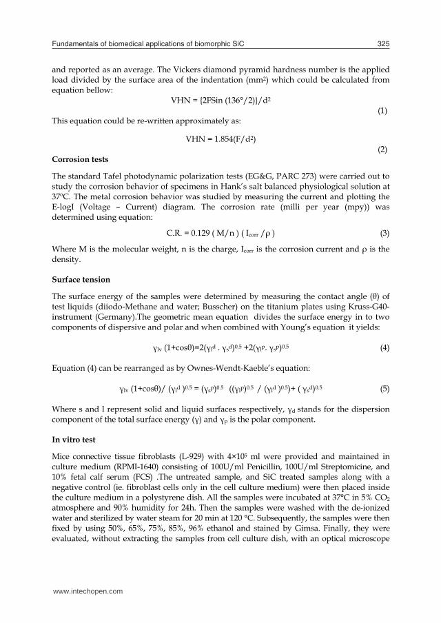
Fundamentals of biomedical applications of biomorphic SiC 325
cells (Heinrich et al., 2008). Additionally, these modified surfaces must resist both the mechanical wear and the corrosion (Sighvi et al., 1998). It is therefore important to evaluate systematically the role of different surface properties and to assess the biological performance of different implant materials. The surface morphology, as well as manipulation with the physical state and chemical composition of implant surfaces may be significant for bone-implant integration. Surfaces are treated to facilitate an intimate contact between bone and implant. So, the tissue response to an implant involves physical factors, depending on implant design, surface topography, surface charge density, surface free energy and chemical factors associated with the composition of the materials. These substrate characteristics may directly influence cell adhesion, spreading and signaling, events that regulate a wide variety of biological functions (Ronold et al., 2003). Numerous surface treatments including Ion implantation, coating, shot blast, machining, plasma spray, plasma nitrid, nitrogen diffusion hardening are some of the relatively older techniques in the field of material processing which can be used to change implant’s surface topography. Thus, the main intention of this work is to extend the earlier research by carrying out some detailed In vitro and In vivo experiments using a 300 and 800 grit SiC papers on surface physico-chemical changes, surface wettability, corrosion resistance, microhardness and osteoblast cells adhesivity of Ti6Al4V with respect to possible orthopaedic applications.
16. Materials and methods
Rectangular–shaped specimens with 20×10 mm dimensions and the thickness of 2 mm, were made from a medical grade Ti6Al4V (ASTM F136, Friadent, Mannheim- Germany- GmbH) with chemical formulation Ti(91.63%)Al(5.12% V(3.25%). The samples were divided into three groups of untreated, 300 and 800 grit SiC paper. Prior to treatment, all samples were cleaned with 97% ethanol and were subsequently washed twice by distilled water in an ultrasonic bath (Mattachanna, Barcelona-Spain). A final rinse was done by de-ionized water at a neutral pH to ensure a clean surface was obtained. They were polished using 300 and 800 grit SiC paper. Finally, an optical microscope with magnification of ×20 was used to ensure that no particles were left on the sample surface. Surface roughness
The surface micro roughness (Ra) measurements were carried out using a non-contact laser profilemeter (NCLP) (Messtechnik, Germany) equipped with a micro focus sensor based on an auto focusing system. Ra is the arithmetical mean of the absolute values of the profile deviations from the mean line. Five two-dimensional NCLP profiles were obtained for each surface over a distance of 3.094 mm with a lateral resolution of 1µm using a Gaussian filter and an attenuation factor of 60% at a cut-off wavelength of 0.59 mm . The roughness parameters were calculated with the NCLP software similar to that described by Wieland et al. (Wieland et al., 2001). Surface hardness
Surface microhardness test was carried out with 50 gram load in 10 seconds by a diamond squared pyramid tip (Celemx CMT, Automatic). Each related test was considered at 5 points
and reported as an average. The Vickers diamond pyramid hardness number is the applied load divided by the surface area of the indentation (mm2) which could be calculated from equation bellow:
VHN = {2FSin (136°/2)}/d2 (1) This equation could be re-written approximately as:
VHN = 1.854(F/d2) (2) Corrosion tests
The standard Tafel photodynamic polarization tests (EG&G, PARC 273) were carried out to study the corrosion behavior of specimens in Hank’s salt balanced physiological solution at 37ºC. The metal corrosion behavior was studied by measuring the current and plotting the E-logI (Voltage – Current) diagram. The corrosion rate (milli per year (mpy)) was determined using equation:
C.R. = 0.129 ( M/n ) ( Icorr /ρ ) (3)
Where M is the molecular weight, n is the charge, Icorr is the corrosion current and ρ is the density. Surface tension
The surface energy of the samples were determined by measuring the contact angle (θ) of test liquids (diiodo-Methane and water; Busscher) on the titanium plates using Kruss-G40-instrument (Germany).The geometric mean equation divides the surface energy in to two components of dispersive and polar and when combined with Young’s equation it yields: γlv (1+cosθ)=2(γld . γsd)0.5 +2(γlp. γsp)0.5 (4) Equation (4) can be rearranged as by Ownes-Wendt-Kaeble’s equation: γlv (1+cosθ)/ (γld )0.5 = (γsp)0.5 ((γlp)0.5 / (γld )0.5)+ ( γsd)0.5 (5) Where s and l represent solid and liquid surfaces respectively, γd stands for the dispersion component of the total surface energy (γ) and γp is the polar component. In vitro test
Mice connective tissue fibroblasts (L-929) with 4×10⁵ ml were provided and maintained in culture medium (RPMI-1640) consisting of 100U/ml Penicillin, 100U/ml Streptomicine, and 10% fetal calf serum (FCS) .The untreated sample, and SiC treated samples along with a negative control (ie. fibroblast cells only in the cell culture medium) were then placed inside the culture medium in a polystyrene dish. All the samples were incubated at 37°C in 5% CO2 atmosphere and 90% humidity for 24h. Then the samples were washed with the de-ionized water and sterilized by water steam for 20 min at 120 °C. Subsequently, the samples were then fixed by using 50%, 65%, 75%, 85%, 96% ethanol and stained by Gimsa. Finally, they were evaluated, without extracting the samples from cell culture dish, with an optical microscope
www.intechopen.com
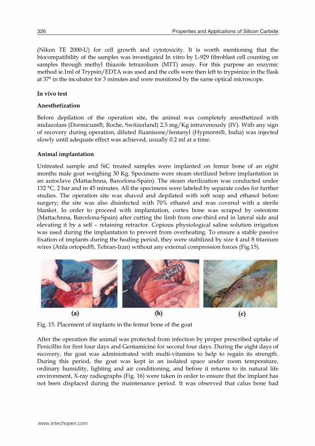
Properties and Applications of Silicon Carbide326
(Nikon TE 2000-U) for cell growth and cytotoxicity. It is worth mentioning that the biocompatibility of the samples was investigated In vitro by L-929 fibroblast cell counting on samples through methyl thiazole tetrazolium (MTT) assay. For this purpose an enzymic method ie.1ml of Trypsin/EDTA was used and the cells were then left to trypsinize in the flask at 37° in the incubator for 3 minutes and were monitored by the same optical microscope. In vivo test Anesthetization
Before depilation of the operation site, the animal was completely anesthetized with midazolam (Dormicum®, Roche, Switzerland) 2.5 mg/Kg intravenously (IV). With any sign of recovery during operation, diluted fluanisone/fentanyl (Hypnorm®, India) was injected slowly until adequate effect was achieved, usually 0.2 ml at a time. Animal implantation
Untreated sample and SiC treated samples were implanted on femur bone of an eight months male goat weighing 30 Kg. Specimens were steam sterilized before implantation in an autoclave (Mattachnna, Barcelona-Spain). The steam sterilization was conducted under 132 °C, 2 bar and in 45 minutes. All the specimens were labeled by separate codes for further studies. The operation site was shaved and depilated with soft soap and ethanol before surgery; the site was also disinfected with 70% ethanol and was covered with a sterile blanket. In order to proceed with implantation, cortex bone was scraped by osteotom (Mattachnna, Barcelona-Spain) after cutting the limb from one-third end in lateral side and elevating it by a self – retaining retractor. Copious physiological saline solution irrigation was used during the implantation to prevent from overheating. To ensure a stable passive fixation of implants during the healing period, they were stabilized by size 4 and 8 titanium wires (Atila ortoped®, Tehran-Iran) without any external compression forces (Fig.15).
Fig. 15. Placement of implants in the femur bone of the goat After the operation the animal was protected from infection by proper prescribed uptake of Penicillin for first four days and Gentamicine for second four days. During the eight days of recovery, the goat was administrated with multi-vitamins to help to regain its strength. During this period, the goat was kept in an isolated space under room temperature, ordinary humidity, lighting and air conditioning, and before it returns to its natural life environment, X-ray radiographs (Fig. 16) were taken in order to ensure that the implant has not been displaced during the maintenance period. It was observed that calus bone had
grown in the vicinity of the implant. After five months the animal was sacrificed and the specimens were removed (Fig. 17).
Fig. 16. The X-ray of implants wired to the bone
Fig. 17. Implant removal from the femur bone of the goat: (a) before detachment of the wires, (b) after detachmented (c, d) the foot-print of the implants on the bone The experiments had been approved by the Yazd School of Veterinary Science (Iran) and its animal research authority and conducted in accordance with the Animal Welfare Act of December 20th 1974 and the Regulation on Animal Experimentation of January 15th 1996. The explantation procedure was performed by first cutting the upper and lower section of femur bone using an electric saw and then the implant together with its surrounding tissues was placed in 4% formalin solution for pathological assessment and SEM. Cell analysis
www.intechopen.com
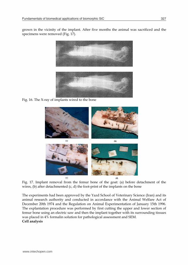
Fundamentals of biomedical applications of biomorphic SiC 327
(Nikon TE 2000-U) for cell growth and cytotoxicity. It is worth mentioning that the biocompatibility of the samples was investigated In vitro by L-929 fibroblast cell counting on samples through methyl thiazole tetrazolium (MTT) assay. For this purpose an enzymic method ie.1ml of Trypsin/EDTA was used and the cells were then left to trypsinize in the flask at 37° in the incubator for 3 minutes and were monitored by the same optical microscope. In vivo test Anesthetization
Before depilation of the operation site, the animal was completely anesthetized with midazolam (Dormicum®, Roche, Switzerland) 2.5 mg/Kg intravenously (IV). With any sign of recovery during operation, diluted fluanisone/fentanyl (Hypnorm®, India) was injected slowly until adequate effect was achieved, usually 0.2 ml at a time. Animal implantation
Untreated sample and SiC treated samples were implanted on femur bone of an eight months male goat weighing 30 Kg. Specimens were steam sterilized before implantation in an autoclave (Mattachnna, Barcelona-Spain). The steam sterilization was conducted under 132 °C, 2 bar and in 45 minutes. All the specimens were labeled by separate codes for further studies. The operation site was shaved and depilated with soft soap and ethanol before surgery; the site was also disinfected with 70% ethanol and was covered with a sterile blanket. In order to proceed with implantation, cortex bone was scraped by osteotom (Mattachnna, Barcelona-Spain) after cutting the limb from one-third end in lateral side and elevating it by a self – retaining retractor. Copious physiological saline solution irrigation was used during the implantation to prevent from overheating. To ensure a stable passive fixation of implants during the healing period, they were stabilized by size 4 and 8 titanium wires (Atila ortoped®, Tehran-Iran) without any external compression forces (Fig.15).
Fig. 15. Placement of implants in the femur bone of the goat After the operation the animal was protected from infection by proper prescribed uptake of Penicillin for first four days and Gentamicine for second four days. During the eight days of recovery, the goat was administrated with multi-vitamins to help to regain its strength. During this period, the goat was kept in an isolated space under room temperature, ordinary humidity, lighting and air conditioning, and before it returns to its natural life environment, X-ray radiographs (Fig. 16) were taken in order to ensure that the implant has not been displaced during the maintenance period. It was observed that calus bone had
grown in the vicinity of the implant. After five months the animal was sacrificed and the specimens were removed (Fig. 17).
Fig. 16. The X-ray of implants wired to the bone
Fig. 17. Implant removal from the femur bone of the goat: (a) before detachment of the wires, (b) after detachmented (c, d) the foot-print of the implants on the bone The experiments had been approved by the Yazd School of Veterinary Science (Iran) and its animal research authority and conducted in accordance with the Animal Welfare Act of December 20th 1974 and the Regulation on Animal Experimentation of January 15th 1996. The explantation procedure was performed by first cutting the upper and lower section of femur bone using an electric saw and then the implant together with its surrounding tissues was placed in 4% formalin solution for pathological assessment and SEM. Cell analysis
www.intechopen.com
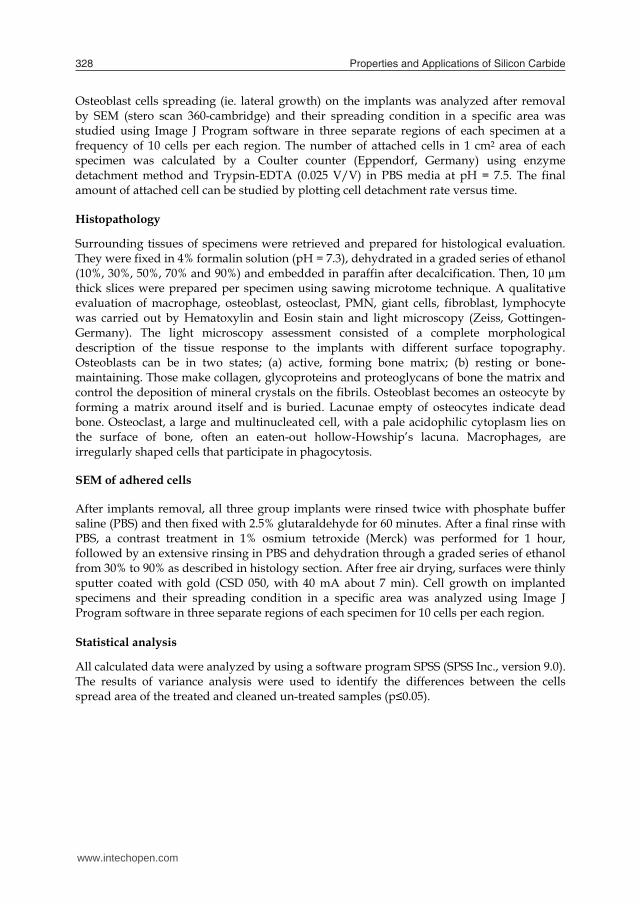
Properties and Applications of Silicon Carbide328
Osteoblast cells spreading (ie. lateral growth) on the implants was analyzed after removal by SEM (stero scan 360-cambridge) and their spreading condition in a specific area was studied using Image J Program software in three separate regions of each specimen at a frequency of 10 cells per each region. The number of attached cells in 1 cm2 area of each specimen was calculated by a Coulter counter (Eppendorf, Germany) using enzyme detachment method and Trypsin-EDTA (0.025 V/V) in PBS media at pH = 7.5. The final amount of attached cell can be studied by plotting cell detachment rate versus time. Histopathology
Surrounding tissues of specimens were retrieved and prepared for histological evaluation. They were fixed in 4% formalin solution (pH = 7.3), dehydrated in a graded series of ethanol (10%, 30%, 50%, 70% and 90%) and embedded in paraffin after decalcification. Then, 10 µm thick slices were prepared per specimen using sawing microtome technique. A qualitative evaluation of macrophage, osteoblast, osteoclast, PMN, giant cells, fibroblast, lymphocyte was carried out by Hematoxylin and Eosin stain and light microscopy (Zeiss, Gottingen-Germany). The light microscopy assessment consisted of a complete morphological description of the tissue response to the implants with different surface topography. Osteoblasts can be in two states; (a) active, forming bone matrix; (b) resting or bone-maintaining. Those make collagen, glycoproteins and proteoglycans of bone the matrix and control the deposition of mineral crystals on the fibrils. Osteoblast becomes an osteocyte by forming a matrix around itself and is buried. Lacunae empty of osteocytes indicate dead bone. Osteoclast, a large and multinucleated cell, with a pale acidophilic cytoplasm lies on the surface of bone, often an eaten-out hollow-Howship’s lacuna. Macrophages, are irregularly shaped cells that participate in phagocytosis. SEM of adhered cells After implants removal, all three group implants were rinsed twice with phosphate buffer saline (PBS) and then fixed with 2.5% glutaraldehyde for 60 minutes. After a final rinse with PBS, a contrast treatment in 1% osmium tetroxide (Merck) was performed for 1 hour, followed by an extensive rinsing in PBS and dehydration through a graded series of ethanol from 30% to 90% as described in histology section. After free air drying, surfaces were thinly sputter coated with gold (CSD 050, with 40 mA about 7 min). Cell growth on implanted specimens and their spreading condition in a specific area was analyzed using Image J Program software in three separate regions of each specimen for 10 cells per each region. Statistical analysis
All calculated data were analyzed by using a software program SPSS (SPSS Inc., version 9.0). The results of variance analysis were used to identify the differences between the cells spread area of the treated and cleaned un-treated samples (p≤0.05).
17. Results and discussion
Characterization of surface topography SiC paper effect
Figure 18 indicates that SiC treated surfaces have some unevenly distributed microgrooves with occasional scratch and pitting made on it by SiC paper. More directionally defined track lines were produced by 800 than 300.
Fig. 18. SEM of SiC paper treated surface by: (a) 300 grit, (b) 800grit Surface roughness
In order to obtain a quantitative comparison between the original and treated surface, the arithmetic average of the absolute values of all points of profile (Ra) was calculated for all samples. The Ra values for untreated, 800, and 300 SiC paper were 12.3±0.03, 16.6±0.15, and 21.8 ±0.05 respectively. All the calculations were performed for n=5 and reported as a mean value of standard deviation (SD). Surface hardness The surface hardness measurements presented in table 1 clearly indicate that micro hardness of the metal decreases with SiC paper. The surface hardness was found to vary from 377 VHN for SiC treated to 394 VHN for untreated. Table 1. Surface hardness tests before and after treatment EDX analysis
The experimental results of EDX spectroscopy of the untreated and SiC treated samples in the ambient condition is given in table 2. The analysis exhibited K-α lines for aluminium and titanium for both samples, though it was expected that carbon would be detected too.
Sample Microhardness (HVN) Untreated 394
SiC paper ( 300 grit) 377 SiC paper ( 800 grit) 378
www.intechopen.com
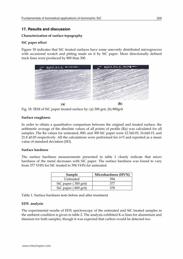
Fundamentals of biomedical applications of biomorphic SiC 329
Osteoblast cells spreading (ie. lateral growth) on the implants was analyzed after removal by SEM (stero scan 360-cambridge) and their spreading condition in a specific area was studied using Image J Program software in three separate regions of each specimen at a frequency of 10 cells per each region. The number of attached cells in 1 cm2 area of each specimen was calculated by a Coulter counter (Eppendorf, Germany) using enzyme detachment method and Trypsin-EDTA (0.025 V/V) in PBS media at pH = 7.5. The final amount of attached cell can be studied by plotting cell detachment rate versus time. Histopathology
Surrounding tissues of specimens were retrieved and prepared for histological evaluation. They were fixed in 4% formalin solution (pH = 7.3), dehydrated in a graded series of ethanol (10%, 30%, 50%, 70% and 90%) and embedded in paraffin after decalcification. Then, 10 µm thick slices were prepared per specimen using sawing microtome technique. A qualitative evaluation of macrophage, osteoblast, osteoclast, PMN, giant cells, fibroblast, lymphocyte was carried out by Hematoxylin and Eosin stain and light microscopy (Zeiss, Gottingen-Germany). The light microscopy assessment consisted of a complete morphological description of the tissue response to the implants with different surface topography. Osteoblasts can be in two states; (a) active, forming bone matrix; (b) resting or bone-maintaining. Those make collagen, glycoproteins and proteoglycans of bone the matrix and control the deposition of mineral crystals on the fibrils. Osteoblast becomes an osteocyte by forming a matrix around itself and is buried. Lacunae empty of osteocytes indicate dead bone. Osteoclast, a large and multinucleated cell, with a pale acidophilic cytoplasm lies on the surface of bone, often an eaten-out hollow-Howship’s lacuna. Macrophages, are irregularly shaped cells that participate in phagocytosis. SEM of adhered cells After implants removal, all three group implants were rinsed twice with phosphate buffer saline (PBS) and then fixed with 2.5% glutaraldehyde for 60 minutes. After a final rinse with PBS, a contrast treatment in 1% osmium tetroxide (Merck) was performed for 1 hour, followed by an extensive rinsing in PBS and dehydration through a graded series of ethanol from 30% to 90% as described in histology section. After free air drying, surfaces were thinly sputter coated with gold (CSD 050, with 40 mA about 7 min). Cell growth on implanted specimens and their spreading condition in a specific area was analyzed using Image J Program software in three separate regions of each specimen for 10 cells per each region. Statistical analysis
All calculated data were analyzed by using a software program SPSS (SPSS Inc., version 9.0). The results of variance analysis were used to identify the differences between the cells spread area of the treated and cleaned un-treated samples (p≤0.05).
17. Results and discussion
Characterization of surface topography SiC paper effect
Figure 18 indicates that SiC treated surfaces have some unevenly distributed microgrooves with occasional scratch and pitting made on it by SiC paper. More directionally defined track lines were produced by 800 than 300.
Fig. 18. SEM of SiC paper treated surface by: (a) 300 grit, (b) 800grit Surface roughness
In order to obtain a quantitative comparison between the original and treated surface, the arithmetic average of the absolute values of all points of profile (Ra) was calculated for all samples. The Ra values for untreated, 800, and 300 SiC paper were 12.3±0.03, 16.6±0.15, and 21.8 ±0.05 respectively. All the calculations were performed for n=5 and reported as a mean value of standard deviation (SD). Surface hardness The surface hardness measurements presented in table 1 clearly indicate that micro hardness of the metal decreases with SiC paper. The surface hardness was found to vary from 377 VHN for SiC treated to 394 VHN for untreated. Table 1. Surface hardness tests before and after treatment EDX analysis
The experimental results of EDX spectroscopy of the untreated and SiC treated samples in the ambient condition is given in table 2. The analysis exhibited K-α lines for aluminium and titanium for both samples, though it was expected that carbon would be detected too.
Sample Microhardness (HVN) Untreated 394
SiC paper ( 300 grit) 377 SiC paper ( 800 grit) 378
www.intechopen.com
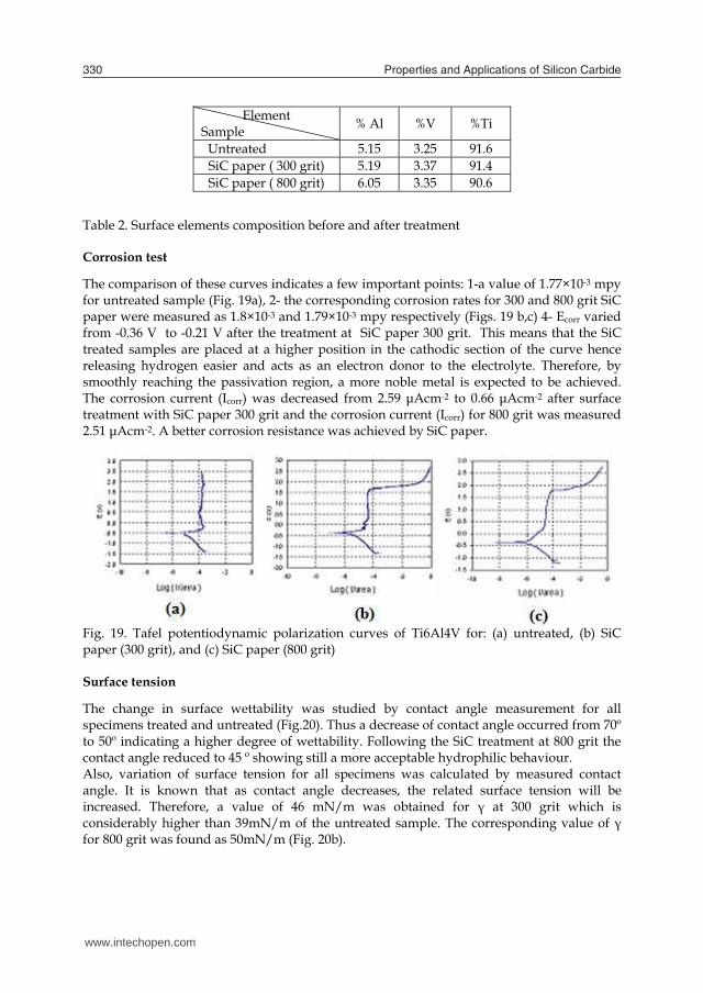
Properties and Applications of Silicon Carbide330
Table 2. Surface elements composition before and after treatment Corrosion test The comparison of these curves indicates a few important points: 1-a value of 1.77×10⁻³ mpy for untreated sample (Fig. 19a), 2- the corresponding corrosion rates for 300 and 800 grit SiC paper were measured as 1.8×10⁻³ and 1.79×10⁻³ mpy respectively (Figs. 19 b,c) 4- Ecorr varied from -0.36 V to -0.21 V after the treatment at SiC paper 300 grit. This means that the SiC treated samples are placed at a higher position in the cathodic section of the curve hence releasing hydrogen easier and acts as an electron donor to the electrolyte. Therefore, by smoothly reaching the passivation region, a more noble metal is expected to be achieved. The corrosion current (Icorr) was decreased from 2.59 μAcm-2 to 0.66 μAcm-2 after surface treatment with SiC paper 300 grit and the corrosion current (Icorr) for 800 grit was measured 2.51 μAcm-2. A better corrosion resistance was achieved by SiC paper.
Fig. 19. Tafel potentiodynamic polarization curves of Ti6Al4V for: (a) untreated, (b) SiC paper (300 grit), and (c) SiC paper (800 grit) Surface tension
The change in surface wettability was studied by contact angle measurement for all specimens treated and untreated (Fig.20). Thus a decrease of contact angle occurred from 70º to 50º indicating a higher degree of wettability. Following the SiC treatment at 800 grit the contact angle reduced to 45 º showing still a more acceptable hydrophilic behaviour. Also, variation of surface tension for all specimens was calculated by measured contact angle. It is known that as contact angle decreases, the related surface tension will be increased. Therefore, a value of 46 mN/m was obtained for γ at 300 grit which is considerably higher than 39mN/m of the untreated sample. The corresponding value of γ for 800 grit was found as 50mN/m (Fig. 20b).
Element Sample % Al %V %Ti
Untreated 5.15 3.25 91.6 SiC paper ( 300 grit) 5.19 3.37 91.4 SiC paper ( 800 grit) 6.05 3.35 90.6
Fig. 20. Variation of contact angle: (a) and surface tension, (b) with sample surface texture In vitro
Figures 21 a-c illustrate the morphology and the spreading of cells on the negative control, the untreated and SiC treatment respectively. As it is observed in all cases, some of the attached cells spread radially from the centre and developed a filopodia type shape. The surface of cells which are not spread, were convoluted in to micro ridges and the neighboring cells maintain a physical contact with one another through multiple extensions. Cell spreading is an essential function of cell adhesivity to any surface and it proceeds the proliferation until the surface is fully covered by the cellular network. The number of cells attached to the surface was evaluated by SiC treated samples assay. More cells are attached to the surface for 300 and 800 grits of SiC paper, 9× 10⁵ and 10 × 105 respectively, which are higher than 8×10⁵ for untreated sample.
Fig. 21. Light microscopy of cell culture evaluation (a) negative control, (b) untreated sample, (c) SiC paper ( 800 grit). In vivo Cell spreading analysis
The experimental results of bone cell growth are given in table 3. As it can be seen, cells spreading over the specimen surface are related to surface texture which was measured by Image J program software (IJP). The highest spreading area (383 µm2) belongs to SiC treated sample (800 grit).
www.intechopen.com
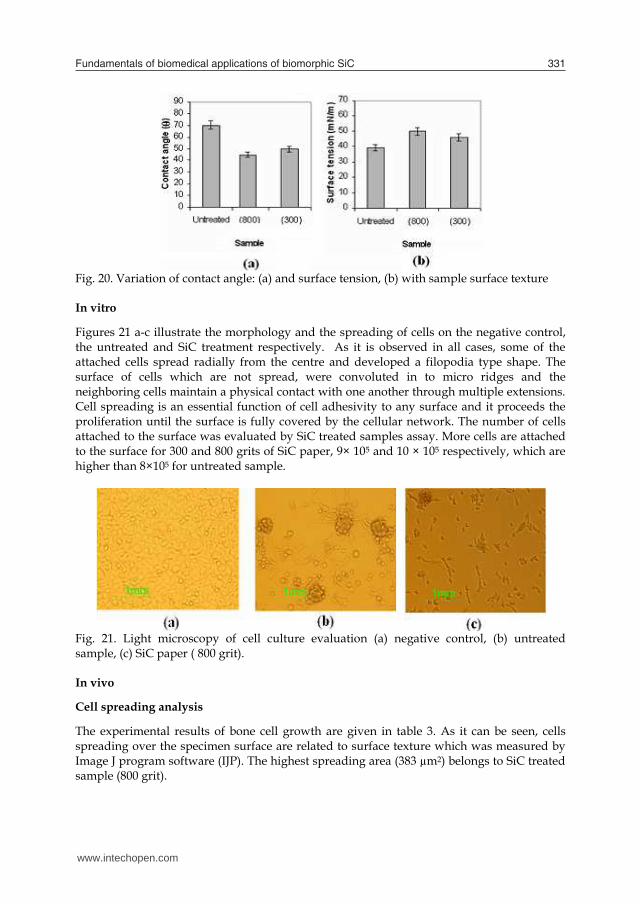
Fundamentals of biomedical applications of biomorphic SiC 331
Table 2. Surface elements composition before and after treatment Corrosion test The comparison of these curves indicates a few important points: 1-a value of 1.77×10⁻³ mpy for untreated sample (Fig. 19a), 2- the corresponding corrosion rates for 300 and 800 grit SiC paper were measured as 1.8×10⁻³ and 1.79×10⁻³ mpy respectively (Figs. 19 b,c) 4- Ecorr varied from -0.36 V to -0.21 V after the treatment at SiC paper 300 grit. This means that the SiC treated samples are placed at a higher position in the cathodic section of the curve hence releasing hydrogen easier and acts as an electron donor to the electrolyte. Therefore, by smoothly reaching the passivation region, a more noble metal is expected to be achieved. The corrosion current (Icorr) was decreased from 2.59 μAcm-2 to 0.66 μAcm-2 after surface treatment with SiC paper 300 grit and the corrosion current (Icorr) for 800 grit was measured 2.51 μAcm-2. A better corrosion resistance was achieved by SiC paper.
Fig. 19. Tafel potentiodynamic polarization curves of Ti6Al4V for: (a) untreated, (b) SiC paper (300 grit), and (c) SiC paper (800 grit) Surface tension
The change in surface wettability was studied by contact angle measurement for all specimens treated and untreated (Fig.20). Thus a decrease of contact angle occurred from 70º to 50º indicating a higher degree of wettability. Following the SiC treatment at 800 grit the contact angle reduced to 45 º showing still a more acceptable hydrophilic behaviour. Also, variation of surface tension for all specimens was calculated by measured contact angle. It is known that as contact angle decreases, the related surface tension will be increased. Therefore, a value of 46 mN/m was obtained for γ at 300 grit which is considerably higher than 39mN/m of the untreated sample. The corresponding value of γ for 800 grit was found as 50mN/m (Fig. 20b).
Element Sample % Al %V %Ti
Untreated 5.15 3.25 91.6 SiC paper ( 300 grit) 5.19 3.37 91.4 SiC paper ( 800 grit) 6.05 3.35 90.6
Fig. 20. Variation of contact angle: (a) and surface tension, (b) with sample surface texture In vitro
Figures 21 a-c illustrate the morphology and the spreading of cells on the negative control, the untreated and SiC treatment respectively. As it is observed in all cases, some of the attached cells spread radially from the centre and developed a filopodia type shape. The surface of cells which are not spread, were convoluted in to micro ridges and the neighboring cells maintain a physical contact with one another through multiple extensions. Cell spreading is an essential function of cell adhesivity to any surface and it proceeds the proliferation until the surface is fully covered by the cellular network. The number of cells attached to the surface was evaluated by SiC treated samples assay. More cells are attached to the surface for 300 and 800 grits of SiC paper, 9× 10⁵ and 10 × 105 respectively, which are higher than 8×10⁵ for untreated sample.
Fig. 21. Light microscopy of cell culture evaluation (a) negative control, (b) untreated sample, (c) SiC paper ( 800 grit). In vivo Cell spreading analysis
The experimental results of bone cell growth are given in table 3. As it can be seen, cells spreading over the specimen surface are related to surface texture which was measured by Image J program software (IJP). The highest spreading area (383 µm2) belongs to SiC treated sample (800 grit).
www.intechopen.com
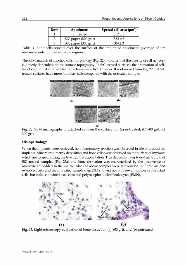
Properties and Applications of Silicon Carbide332
Row Specimens Spread cell area (μm2) 1 untreated 352 ± 6 2 SiC paper (800 grit) 383 ± 5 3 SiC paper (300 grit) 367± 3
Table 3. Bone cells spread over the surface of the implanted specimens (average of ten measurements in three separate regions) The SEM analysis of attached cells morphology (Fig. 22) indicates that the density of cell network is directly dependent on the surface topography. In SiC treated surfaces, the orientation of cells was longitudinal and parallel to the lines made by SiC paper. It is observed from Fig. 22 that SiC treated surfaces have more fibroblast cells compared with the untreated sample.
Fig. 22. SEM micrographs of attached cells on the surface for: (a) untreated, (b) 800 grit, (c) 300 grit Histopathology
When the implants were retrieved, no inflammatory reaction was observed inside or around the implants. Mineralized matrix deposition and bone cells were observed on the surface of implants which are formed during the five months implantation. This deposition was found all around of SiC treated samples (Fig. 23a) and bone formation was characterized by the occurrence of osteocyte embedded in the matrix. Also the above samples were surrounded by fibroblast and osteoblast cells and the untreated sample (Fig. 23b) showed not only fewer number of fibroblast cells, but it also contained osteoclast and polymorpho nuclear leukocytes (PMN).
Fig. 23. Light microscopy evaluation of bone tissue for: (a) 800 grit, and (b) untreated
In table 4, the symbols indicate the presence of 2-3 cells (+), 3-5 cells (++), more than 5 cells (+++) and lack of cells (-) respectively. No PMN, giant cells and osteoclast were seen in SiC treated samples. Also tissue healing was better conducted near mentioned implant. Fibroblast and osteoblast cells were seen in samples. The successful incorporation of bone implants strongly depends on a firm longstanding adhesion of the tissue surrounding the implants. The cellular reaction is influenced by the properties of the bulk materials as well as the specifications of the surface, that is, the chemical composition and the topography (Birte et al., 2003, Siikavitsas et al., 2003). When one is considering materials for application of orthopaedic implants, it is important to consider a number of factors, such as biocompatibility and surface wettability. The interaction of living cells with foreign materials is complicated matter, but fundamental for biology medicine and is a key for understanding the biocompatibility. The initial cellular events which take place at the biomaterials interface mimic to a certain extent the natural adhesive interaction of cells with the extra cellular matrix (ECM).
Sample Cell SiC paper (800 grit) SiC paper (300 grit) untreated Fibroblast ++ ++ ++ Osteoblast + + + Giant cell - - - Osteoclast - - + PMN - - + Lymphocyte ++ ++ ++ Macrophage +++ +++ ++ Healing + + +
Table 4. Qualitative evaluation of histology results of bone tissue around the implants The osteoblasts, which play a principal role in bone formation, readily attach to the material surfaces via adsorbed protein layer consisting or RGD containing ligands like fibronectin, vitronectin or fibrinogen. Family of cell surface receptors that provide trans- membrane links between the ECM and the cytoskeleton. Our study showed that surface micro grooves can affect the orientation guidance of bone cells i.e. the deeper grooves were more effective in guiding the cells as it was evaluated by SEM. However, we did not conduct or evaluate systematically the exact effects of grooves depth and size on cell orientation, but our preliminary results were similar to those reported by Xiong et.al (Xiong et al., 2003). This study was focused on the topographic effects of Ti-6Al-4V produced by SiC paper on goat bone cell adhesion. The results showed a common feature reported in the previous studies on a variety of cell types and substrates ie, topographic features strongly affects the cell guidance. Micro grooved surfaces increase of surface tension and reduction of contact angle. The test confirmed that the highest number of cells is attached to SiC paper modified surface. It is also concluded from the SEM, contact angle measurements and preliminary in vitro and in vivo tests that SiC paper can induce a desirable surface modification on Ti-6Al-4V alloy for cell adhesivity and that a noble and biocompatible. Finally, it is suggested that more detailed experiments are required and would be useful to distinguish and clarify the relation between the grooves size and their orientation must be studied more carefully with respect to cell attachment and their reliability as well as endurance.
www.intechopen.com
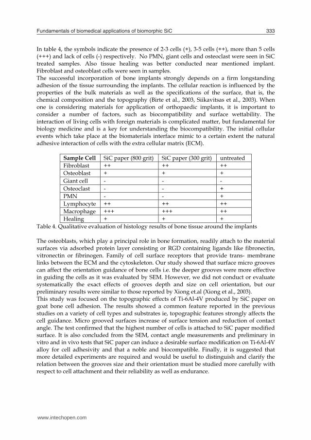
Fundamentals of biomedical applications of biomorphic SiC 333
Row Specimens Spread cell area (μm2) 1 untreated 352 ± 6 2 SiC paper (800 grit) 383 ± 5 3 SiC paper (300 grit) 367± 3
Table 3. Bone cells spread over the surface of the implanted specimens (average of ten measurements in three separate regions) The SEM analysis of attached cells morphology (Fig. 22) indicates that the density of cell network is directly dependent on the surface topography. In SiC treated surfaces, the orientation of cells was longitudinal and parallel to the lines made by SiC paper. It is observed from Fig. 22 that SiC treated surfaces have more fibroblast cells compared with the untreated sample.
Fig. 22. SEM micrographs of attached cells on the surface for: (a) untreated, (b) 800 grit, (c) 300 grit Histopathology
When the implants were retrieved, no inflammatory reaction was observed inside or around the implants. Mineralized matrix deposition and bone cells were observed on the surface of implants which are formed during the five months implantation. This deposition was found all around of SiC treated samples (Fig. 23a) and bone formation was characterized by the occurrence of osteocyte embedded in the matrix. Also the above samples were surrounded by fibroblast and osteoblast cells and the untreated sample (Fig. 23b) showed not only fewer number of fibroblast cells, but it also contained osteoclast and polymorpho nuclear leukocytes (PMN).
Fig. 23. Light microscopy evaluation of bone tissue for: (a) 800 grit, and (b) untreated
In table 4, the symbols indicate the presence of 2-3 cells (+), 3-5 cells (++), more than 5 cells (+++) and lack of cells (-) respectively. No PMN, giant cells and osteoclast were seen in SiC treated samples. Also tissue healing was better conducted near mentioned implant. Fibroblast and osteoblast cells were seen in samples. The successful incorporation of bone implants strongly depends on a firm longstanding adhesion of the tissue surrounding the implants. The cellular reaction is influenced by the properties of the bulk materials as well as the specifications of the surface, that is, the chemical composition and the topography (Birte et al., 2003, Siikavitsas et al., 2003). When one is considering materials for application of orthopaedic implants, it is important to consider a number of factors, such as biocompatibility and surface wettability. The interaction of living cells with foreign materials is complicated matter, but fundamental for biology medicine and is a key for understanding the biocompatibility. The initial cellular events which take place at the biomaterials interface mimic to a certain extent the natural adhesive interaction of cells with the extra cellular matrix (ECM).
Sample Cell SiC paper (800 grit) SiC paper (300 grit) untreated Fibroblast ++ ++ ++ Osteoblast + + + Giant cell - - - Osteoclast - - + PMN - - + Lymphocyte ++ ++ ++ Macrophage +++ +++ ++ Healing + + +
Table 4. Qualitative evaluation of histology results of bone tissue around the implants The osteoblasts, which play a principal role in bone formation, readily attach to the material surfaces via adsorbed protein layer consisting or RGD containing ligands like fibronectin, vitronectin or fibrinogen. Family of cell surface receptors that provide trans- membrane links between the ECM and the cytoskeleton. Our study showed that surface micro grooves can affect the orientation guidance of bone cells i.e. the deeper grooves were more effective in guiding the cells as it was evaluated by SEM. However, we did not conduct or evaluate systematically the exact effects of grooves depth and size on cell orientation, but our preliminary results were similar to those reported by Xiong et.al (Xiong et al., 2003). This study was focused on the topographic effects of Ti-6Al-4V produced by SiC paper on goat bone cell adhesion. The results showed a common feature reported in the previous studies on a variety of cell types and substrates ie, topographic features strongly affects the cell guidance. Micro grooved surfaces increase of surface tension and reduction of contact angle. The test confirmed that the highest number of cells is attached to SiC paper modified surface. It is also concluded from the SEM, contact angle measurements and preliminary in vitro and in vivo tests that SiC paper can induce a desirable surface modification on Ti-6Al-4V alloy for cell adhesivity and that a noble and biocompatible. Finally, it is suggested that more detailed experiments are required and would be useful to distinguish and clarify the relation between the grooves size and their orientation must be studied more carefully with respect to cell attachment and their reliability as well as endurance.
www.intechopen.com
Properties and Applications of Silicon Carbide334
18. Future considerations in biomedical applications of SiC
The next decade will see a great increase in scientific research into the biomedical applications of SiC. Many analysis techniques may be used to analyze SiC biocompatibility. In particular, primary cell lines could be cultured on SiC surfaces in the future since their behavior would be a closer description of the in vivo performance of the material. While proof-of concept studies in research laboratories have demonstrated great promise in the use of SiC for scaffold of tissue engineering, several issues will need to be addressed before SiC find way to large-scale clinical application. In particular, researches will need to study toxic and pharmacokinetic effects of SiC in vivo. In addition, research will focuse on the synthesis SiC nanopartiicles that may facilitate the development of multifunctional nanostructures for use in drug delivery and tissue engineering applications. More experiments are required to clarify the relation between SiC and cell attachment in scaffold of tissue engineering. The different polytypes of SiC were quite well matched to organic systems in terms of band gap and band alignment. Therefore, SiC should be a very interesting substrate material for future semiconductor/organic heterostructures. Finally, the feasibility of surface functionalization of SiC leaving free functional groups has been shown while deeper understanding of the chemisorptions of various organic molecules is still needed in order to optimize surface functionalization processes. The preparation and complete characterization of atomically ordered SiC surfaces may lead to the successful implementation of a large variety of biotechnological applications. It is suggested that more investigations are required and would be useful to distinguish SiC biomedical applications.
19. References
Amon, M.; Bolz, A. & Schaldach, M. (1996). Improvement of stenting therapy with a silicon carbide coated tantalum stent. J Mater Sci: Mater Med, 7, 5, 273–8
Amon, M.; Winkler, S.; Dekker, A.; Bolz, A.; Mittermayer, C. & Schaldach, M. (1995). Introduction of a new coronary stent with enhanced radiopacity and hemocompatibility. pp. 107–8 Proceedings of the Annual International Conference of the IEEE Engineers in Medicine and Biology Society 95CB35746, vol. 17, 1. New York: IEEE Press.
Anderson, A.S.; Dattelbaum, A.M.; Montano, G.A.; Price, D.N.; Schmidt, J.G.; Martinez, J.S.; Grace, W.K.; Grace, K.M.; Swanson, B.I. (2008). Functioal PEG-modified thin films for biological detection. Langmuir, 24, 2240-2247
Bai, S.; Ke, Y.; Shishkin, Y.; Shigiltchoff, O.; Devaty, R.P.; Choyke, W.J.; Strauch, D.; Stojetz, B.; Dorner, B.; Hobgood, D.; Serrano, J.; Cardona, M.; Nagasawa, H.; Kimoto, T. & Porter L.M. (2003). Four Current Examples of Characterization of Silicon Carbide, Mat. Res. Soc. Symp. Proc. 742, K3.1.1
Bayer, G.; Hartwig, S.; Nagel, M.; Tilttelbach, M.; Rzany, A. & Schaldach, M. (2001). Future Strategies for Antiproliferative Stent Coatings. Progress in Biomedical Research, 222-225
Berthold, A.; Laugere F.; Schellevis, H.; De Boer, Ch. R.; Laros, M.; Guijt, R. M.; Sarro, P. M. & Vellekoop, M. J. (2002). Fabrication of a glass-implemented microcapillary electrophoresis device with integrated contactless conductivity detection. Electrophoresis, 23, 3511–3519
Bigerelle, M. & Anselme, K. (2005). Bootstrap analysis of the relation between initial adhesive events and long-term cellular functions of human osteoblasts cultured on biocompatible metallic substrates. Acta biomaterialia, 1, 499-510
Birte, G.S.; Neubert, A.; Hopp, M.; Griepentrog, M. & Lange, K.P. (2003). Fibroblast growth on surface modified dental implants: An in vitro study. J. Biomed. Mater. Res., 64A, 591-599
Bolz, A. & Schaldach, M., (1993). Heamocompatibility optimization of implants by hybrid structuring, S123-S130, World Congress Supplement, Biomaterials, Koyoto
Bolz, A. (1995). Applications of Thin-Film Technology in Biomedical Engineering. Bolz, A. & Schaldach, M. (1990). Artificial heart valves: improved blood compatibility by PECVD a-SiC:H coating. Artificial organs, 14(4), 260-9
Bolz, A. & Schaldach, M. (1993). Biomaterials haemocompatibility optimization of implants by hybrid structuring. Med. & Biol. Eng. & Comput., 31, 123-130
Bolz, A; Amon, M; Ozbek, C; Heublein, B; Schaldach, M. (1996). Coating of cardiovascular stents with a semiconductor to improve their hemocompatibility. Texas Heart Institute J. 23, 2, 162-6
D.L. , Trantolo, D.J. & et al. Encyclopedic Handbook of Biomaterials and Bioengineering. Part A: Materials, In: Wise, 2, 1287-1330
Borrajo, J.P.; Serra, J.; Liste, S.; Gonza´lez, P.; Chiussi, S.; Leo´n, B. & Pe´rez-Amor, M. (2005). Pulsed laser deposition of hydroxylapatite thin films on biomorphic silicon carbide ceramics. Applied Surface Science, 248, 2005, 355–359
Botsoa, J., Lysenko, V., G_eloen, A., Marty, O., Bluet, J. M. & Guillot, G. (2008). Application of 3C-SiC quantum dots for living cell imaging. Appl. Phys. Lett., 92, 173902
Buchter, A.; Joos, U.; Wiessman, H.P.; Seper, L. & Meyer, U. ( 2006). Biological and biomechanical evaluation of interface reaction at conical screw-type implant. Head and Face Med., 2, 5-18
Carlos, A. D.; Borrajo, J.P.; Serra, J.; Gonz´alez, P. & Le´on, B. (2006). Behaviour of MG-63 osteoblast-like cells on wood-based biomorphic SiC ceramics coated with bioactive glass. J Mater Sci: Mater Med , 17, 523–529
Carrie, K.; Khalife, M.; Hamon, B.; Citron, J.P.; Monassier, R.; Sabatier, J.; Lipiecky, S.; Mourali, L.; Sarfaty,M.; Elbaz, J.; Fourcade & Puel, J. (2001). Initial and Follow-Up Results of the Tenax Coronary Stent. J. Interventional Cardiology 14(1), 1-5
Calderon, N.R.; Martinez-Escandell, M.; Narciso, J. & Rodríguez-Reinoso, F. (2009). The role of carbon biotemplate density in mechanical properties of biomorphic SiC. J. of the European Ceramic Society, 29, 465–472
Caputo, D.; de Cesare, G.; Nascetti, A.; Scipinotti, R., (2008). Two-Color Sensor for Biomolecule Detection. Sensor Letters, 6, 4, 542-547
Chakrabarti, O.P.; Maiti, H.S., & Majumdar, R. (2004). Biomimetic synthesis of cellular SiC based ceramics from plant precursor. Bull. Mater. Sci., 27, 5, 467–470
Chu, W.H.; Chin ,R.; Huen, T. & Ferrari, M. (1999). Silicon Membrane Nanofilters from Sacrificial Oxide Removal. J. Microelectromech. Syst. 8, 34-42
Cicero, G. & Catellani, A. (2005). Towards SiC surface functionalization: An ab initio study. J. Chem. Phys., 122, 214716, 1-5
Cicoira, f. & Rosei, F. (2006). Playing Tetris at the nanoscale. Surface Science, 600, 1–5
www.intechopen.com
Fundamentals of biomedical applications of biomorphic SiC 335
18. Future considerations in biomedical applications of SiC
The next decade will see a great increase in scientific research into the biomedical applications of SiC. Many analysis techniques may be used to analyze SiC biocompatibility. In particular, primary cell lines could be cultured on SiC surfaces in the future since their behavior would be a closer description of the in vivo performance of the material. While proof-of concept studies in research laboratories have demonstrated great promise in the use of SiC for scaffold of tissue engineering, several issues will need to be addressed before SiC find way to large-scale clinical application. In particular, researches will need to study toxic and pharmacokinetic effects of SiC in vivo. In addition, research will focuse on the synthesis SiC nanopartiicles that may facilitate the development of multifunctional nanostructures for use in drug delivery and tissue engineering applications. More experiments are required to clarify the relation between SiC and cell attachment in scaffold of tissue engineering. The different polytypes of SiC were quite well matched to organic systems in terms of band gap and band alignment. Therefore, SiC should be a very interesting substrate material for future semiconductor/organic heterostructures. Finally, the feasibility of surface functionalization of SiC leaving free functional groups has been shown while deeper understanding of the chemisorptions of various organic molecules is still needed in order to optimize surface functionalization processes. The preparation and complete characterization of atomically ordered SiC surfaces may lead to the successful implementation of a large variety of biotechnological applications. It is suggested that more investigations are required and would be useful to distinguish SiC biomedical applications.
19. References
Amon, M.; Bolz, A. & Schaldach, M. (1996). Improvement of stenting therapy with a silicon carbide coated tantalum stent. J Mater Sci: Mater Med, 7, 5, 273–8
Amon, M.; Winkler, S.; Dekker, A.; Bolz, A.; Mittermayer, C. & Schaldach, M. (1995). Introduction of a new coronary stent with enhanced radiopacity and hemocompatibility. pp. 107–8 Proceedings of the Annual International Conference of the IEEE Engineers in Medicine and Biology Society 95CB35746, vol. 17, 1. New York: IEEE Press.
Anderson, A.S.; Dattelbaum, A.M.; Montano, G.A.; Price, D.N.; Schmidt, J.G.; Martinez, J.S.; Grace, W.K.; Grace, K.M.; Swanson, B.I. (2008). Functioal PEG-modified thin films for biological detection. Langmuir, 24, 2240-2247
Bai, S.; Ke, Y.; Shishkin, Y.; Shigiltchoff, O.; Devaty, R.P.; Choyke, W.J.; Strauch, D.; Stojetz, B.; Dorner, B.; Hobgood, D.; Serrano, J.; Cardona, M.; Nagasawa, H.; Kimoto, T. & Porter L.M. (2003). Four Current Examples of Characterization of Silicon Carbide, Mat. Res. Soc. Symp. Proc. 742, K3.1.1
Bayer, G.; Hartwig, S.; Nagel, M.; Tilttelbach, M.; Rzany, A. & Schaldach, M. (2001). Future Strategies for Antiproliferative Stent Coatings. Progress in Biomedical Research, 222-225
Berthold, A.; Laugere F.; Schellevis, H.; De Boer, Ch. R.; Laros, M.; Guijt, R. M.; Sarro, P. M. & Vellekoop, M. J. (2002). Fabrication of a glass-implemented microcapillary electrophoresis device with integrated contactless conductivity detection. Electrophoresis, 23, 3511–3519
Bigerelle, M. & Anselme, K. (2005). Bootstrap analysis of the relation between initial adhesive events and long-term cellular functions of human osteoblasts cultured on biocompatible metallic substrates. Acta biomaterialia, 1, 499-510
Birte, G.S.; Neubert, A.; Hopp, M.; Griepentrog, M. & Lange, K.P. (2003). Fibroblast growth on surface modified dental implants: An in vitro study. J. Biomed. Mater. Res., 64A, 591-599
Bolz, A. & Schaldach, M., (1993). Heamocompatibility optimization of implants by hybrid structuring, S123-S130, World Congress Supplement, Biomaterials, Koyoto
Bolz, A. (1995). Applications of Thin-Film Technology in Biomedical Engineering. Bolz, A. & Schaldach, M. (1990). Artificial heart valves: improved blood compatibility by PECVD a-SiC:H coating. Artificial organs, 14(4), 260-9
Bolz, A. & Schaldach, M. (1993). Biomaterials haemocompatibility optimization of implants by hybrid structuring. Med. & Biol. Eng. & Comput., 31, 123-130
Bolz, A; Amon, M; Ozbek, C; Heublein, B; Schaldach, M. (1996). Coating of cardiovascular stents with a semiconductor to improve their hemocompatibility. Texas Heart Institute J. 23, 2, 162-6
D.L. , Trantolo, D.J. & et al. Encyclopedic Handbook of Biomaterials and Bioengineering. Part A: Materials, In: Wise, 2, 1287-1330
Borrajo, J.P.; Serra, J.; Liste, S.; Gonza´lez, P.; Chiussi, S.; Leo´n, B. & Pe´rez-Amor, M. (2005). Pulsed laser deposition of hydroxylapatite thin films on biomorphic silicon carbide ceramics. Applied Surface Science, 248, 2005, 355–359
Botsoa, J., Lysenko, V., G_eloen, A., Marty, O., Bluet, J. M. & Guillot, G. (2008). Application of 3C-SiC quantum dots for living cell imaging. Appl. Phys. Lett., 92, 173902
Buchter, A.; Joos, U.; Wiessman, H.P.; Seper, L. & Meyer, U. ( 2006). Biological and biomechanical evaluation of interface reaction at conical screw-type implant. Head and Face Med., 2, 5-18
Carlos, A. D.; Borrajo, J.P.; Serra, J.; Gonz´alez, P. & Le´on, B. (2006). Behaviour of MG-63 osteoblast-like cells on wood-based biomorphic SiC ceramics coated with bioactive glass. J Mater Sci: Mater Med , 17, 523–529
Carrie, K.; Khalife, M.; Hamon, B.; Citron, J.P.; Monassier, R.; Sabatier, J.; Lipiecky, S.; Mourali, L.; Sarfaty,M.; Elbaz, J.; Fourcade & Puel, J. (2001). Initial and Follow-Up Results of the Tenax Coronary Stent. J. Interventional Cardiology 14(1), 1-5
Calderon, N.R.; Martinez-Escandell, M.; Narciso, J. & Rodríguez-Reinoso, F. (2009). The role of carbon biotemplate density in mechanical properties of biomorphic SiC. J. of the European Ceramic Society, 29, 465–472
Caputo, D.; de Cesare, G.; Nascetti, A.; Scipinotti, R., (2008). Two-Color Sensor for Biomolecule Detection. Sensor Letters, 6, 4, 542-547
Chakrabarti, O.P.; Maiti, H.S., & Majumdar, R. (2004). Biomimetic synthesis of cellular SiC based ceramics from plant precursor. Bull. Mater. Sci., 27, 5, 467–470
Chu, W.H.; Chin ,R.; Huen, T. & Ferrari, M. (1999). Silicon Membrane Nanofilters from Sacrificial Oxide Removal. J. Microelectromech. Syst. 8, 34-42
Cicero, G. & Catellani, A. (2005). Towards SiC surface functionalization: An ab initio study. J. Chem. Phys., 122, 214716, 1-5
Cicoira, f. & Rosei, F. (2006). Playing Tetris at the nanoscale. Surface Science, 600, 1–5
www.intechopen.com
Properties and Applications of Silicon Carbide336
Cogan, S.F.; Edell, D.J.; Guzellan, A.A.; Ying, L.P. & Edell, R. (2003). Plasma-enhanced chemical vapor deposited silicon carbide as an implantable dielectric coating. J. Biomed. Mater. Res. A, 67, 3, 856-67
Cole, K.S. (1940). Permiability and impermiability of cell membranes for ions. Sympos Quant.Biol., 8, 110-122
Coletti, C.; Jaroszeski, M.; Hoff, A.M. & Saddow, S.E. (2006). Culture of mammalian cells on single crystal SiC substrates, Mater. Res. Soc. Symp. Proc. 950
Coletti, C.; Jaroszeski, M.J.; Pallaoro, A.; Hoff, A.M.; Iannotta, S. & Saddow, S.E. (2007). Biocompatibility and wettability of crystalline SiC and Si surfaces. IEEE EMBS Proceedings, 5849-5852
Desai, T.A.; Chu, W.H.; Tu, J.K.; Beattie, G.M.; Hayek, A. & Ferrari, M. (1997). Microfabricated Immunoisolating Biocapsules. Biotech. and Bioeng., 57(1), 118-120
Elbaz, M.; El Mokhtar, E.; Fourcade, J.; Mourali, S.; Hobeika, R.; Carrie, D. & Puel, J. (2002). Dose stent design affect the long-term outcome after coronary stenting? Catheterization & Cardiovascular Interventions, 56, 305-311
El Ghannam, A. (2005). Bone reconstruction: from bioceramics to tissue engineering. Expert Rev Med Devices. 2, 1, 87-101
Fan, T.X.; Li, X.F.; Liu, Z.T.; Gu, J.J.; Zhang, D.; Guo, Q.X. & Am, J. (2006). Microstructure and infrared absorption of biomorphic chromium oxides templated by wood tissues. Ceram. Soc. 89, 3511-3515
Fan, J.; Li, H.; Jiang, J., So L. K. Y.; Lam Y. W. & Chu, P. K. (2008). 3C-SiC Nanocrystals as Fluorescent Biological Labels. Small, 4, 8, 1058–1062
Feng, B.; Weng, J.; Yang, B.C.; Qu, S.X. & Zhang, X.D. (2003). Characterization of surface oxide films on titanium and adhesion of osteoblast. Biomaterials, 24, 4663-4670
Feng, X.; Yang, Q. & De Jonghe, L.C. (2003). Microstructure development of hot-pressed silicon carbide: effects of aluminum, boron, and carbon additives. Acta Mater, 51, 3849-60
Fissell, W.H. (2003). Nanoporous Hemofiltration Membranes for Bioartificial Kidneys. oral presentation at 4th Annual BioMEMS and Nanotech World,Washington, DC
Frewin, C.L.; Jaroszeski, M.; Weeber, E.; Muffly, K.E.; Kumar, A.; Peters, M.; Oliveros, A.& Saddow, S.E.J. (2009). Atomic force microscopy analysis of central nervous system cell morphology on silicon carbide and diamond substrates. Mol. Recognit., 22, 5, 380
Fujibayashi, S.; Neo, M.; Kim, H.; Kokubo, T. & Nakamura, T. (2003). A comparative study between in vivo bone in growth and in vitro apatite formation on Na2O-CaO-SiO2 glasses. Biomaterials, 24, 1349-1356
Gabriel, G.; Erill, I.; Caro, J.; Gomez, R.; Riera, D.; Villa, R. & Godignon, Ph. (2007). Manufacturing and full characterization of silicon carbide-based multi-sensor micro-probes for biomedical applications. Microelec. J., 38, 406–415
Gersing, E. (1998). Impedance spectroscopy on living tissue for determination of the state of organs. Bioelectrochem. Bioenerg. 45, 145–149
Goeders, K. M.; Colton, J. S.; Bottomley, L. A. (2008). Microcantilevers: Sensing Chemical Interactions via Mechanical Motion. Chem. Rev., 108, 522-542
Godignon, P. (2005). New Generation of SiC Based Biodevices Implemented on 4” Wafers. Mater. Sci. Forum 483–485. 1097-1100
Gomez, R.; Ivorra, A.; Villa, R.; Godignon, Ph.; Mill´an, J.; Erill, I.; Sol`a, A.; Hotter, G. & Palacios, L. (2006). A SiC microdevice for the minimally invasive monitoring of ischemia in living tissues. Biomed. Microdevices, 8, 43–49
Gonzalez, P.; Serra, J.; Liste, S.; Chiussi, S.; Leon, B.; Perez-Amor, M.; Martinez-Fernandez, J.; de Arellano-Lopez, A.R. & Varela-Feria, F.M. (2003).New biomorphic SiC ceramics coated with bioactive glass for biomedical applications. Biomaterials, 24, 4827–4832
Gonzalez, P.; Borrajo, J.P.; Serra, J.; Liste, S.; Chiussi, S.; Leo´n, B.; Semmelmann, K.; De Carlos, A.; Varela-Feria, F.M.; Mart´nez-Ferna´ndez, J. & De Arellano-Lo´pez, A.R. (2004). Key Eng. Mater., 1029, 254–256
Gonzalez, P.; Serra, J.; Liste, S.; Chiussi, S.; Leon, B.; Perez-Amor, M. & et al. (2003). New biomorphic SiC ceramics coated with bioactive glass for biomedical applications. Biomaterials, 24, 26, 4827-432
Gonzalez, P.; Borrajo, J.P.; Serra, J.; Chiussi, J.; Leo, B.; Mart nez-Ferna ndez, J.; Varela-Feria, F.M.; de Arellano-Lopez, A.R.; de Carlos, A.; Mun oz, F.M.; Lo pez, M. & Singh, M. (2008). A new generation of bio-derived ceramic materials for medical applications. 2008 Wiley Periodicals, Inc.
Greil, P. (2002). Advanced engineering ceramics. Adv. Eng. Mater, 4, 5, 247–254 Gutierrez-Mora, F.; Goretta, K.C.; Varela-Feria, F.M.; Arellano Lo pez, A.R. & Martnez
Ferna ndez, J. (2005). Indentation hardness of biomorphic SiC. Inter J. of Refractory Metals & Hard Materials, 23, 369–374
Haemmerich, D.; Ozkan, R.; Tungjitkusolmun, S.; Tsai, J.Z.; Mahvi, D.M.; Staelin, S.T.; Webster, J.G.; (2002). Changes in electrical resistivity of swine liver after occlusion and postmortem, Medical and Biological Engineering and Computing, 40, 1, 29-33
Hamm, C.W.; Hugenholtz, P.G. & Trust, I. (2003). Silicon carbide-coated stents in patients with acute coronary syndrome. Catheterization & Cardiovascular Interventions, 60, 3, 375-381
Hanekamp, C.E.E. & Koolen, J.J. (2000). Coated Stents in Small Coronary Vessels – A Successful Strategy? Progress in Biomedical Research, 221-223
Harder, C.; Rzany,A. & Schaldach, M. (1999). Coating of Vascular Stents with Antithrombogenic Amorphous Silicon Carbide, Progress in Biomedical Research, 71-77
Heinrich, A.; Dengler, K.; Koerner, T.; Haczek, C.; Deppe, H. & Stritzker, B. (2008). Laser-modified titanium implants for improved cell adhesion. Laser in Medical science, 23, 55-58
Hesaraki, S.; Ebadzadeh, T. & Ahmadzadeh-Asl, S. (2010). Nanosilicon carbide/hydroxyapatite nanocomposites: structural, mechanical and in vitro cellular properties. J Mater Sci: Mater Med, DOI 10.1007/s10856-010-4068-7
Hildebrandt, P.; Sayyad, M.; Rzany, A. & et al. (2001). Prevention of surface encrustation of urological implants by coating with inhibitors. Biomaterials, 22, 503-507
Hing, K.A.; Revell, P.A.; Smith, N. & Buckland, T. (2006). Effect of silicon level on rate, quality and progression of bone healing within silicate-substituted porous hydroxyapatite scaffolds. Biomaterials, 27, 29, 5014-5026
Ikeuchi, K.; Kusaka J. & Yoshida, H. (2000). Tribology for all-ceramic joint prostheses. Journal of Ceramic Processing Research, 1, 1, 53-56
www.intechopen.com
Fundamentals of biomedical applications of biomorphic SiC 337
Cogan, S.F.; Edell, D.J.; Guzellan, A.A.; Ying, L.P. & Edell, R. (2003). Plasma-enhanced chemical vapor deposited silicon carbide as an implantable dielectric coating. J. Biomed. Mater. Res. A, 67, 3, 856-67
Cole, K.S. (1940). Permiability and impermiability of cell membranes for ions. Sympos Quant.Biol., 8, 110-122
Coletti, C.; Jaroszeski, M.; Hoff, A.M. & Saddow, S.E. (2006). Culture of mammalian cells on single crystal SiC substrates, Mater. Res. Soc. Symp. Proc. 950
Coletti, C.; Jaroszeski, M.J.; Pallaoro, A.; Hoff, A.M.; Iannotta, S. & Saddow, S.E. (2007). Biocompatibility and wettability of crystalline SiC and Si surfaces. IEEE EMBS Proceedings, 5849-5852
Desai, T.A.; Chu, W.H.; Tu, J.K.; Beattie, G.M.; Hayek, A. & Ferrari, M. (1997). Microfabricated Immunoisolating Biocapsules. Biotech. and Bioeng., 57(1), 118-120
Elbaz, M.; El Mokhtar, E.; Fourcade, J.; Mourali, S.; Hobeika, R.; Carrie, D. & Puel, J. (2002). Dose stent design affect the long-term outcome after coronary stenting? Catheterization & Cardiovascular Interventions, 56, 305-311
El Ghannam, A. (2005). Bone reconstruction: from bioceramics to tissue engineering. Expert Rev Med Devices. 2, 1, 87-101
Fan, T.X.; Li, X.F.; Liu, Z.T.; Gu, J.J.; Zhang, D.; Guo, Q.X. & Am, J. (2006). Microstructure and infrared absorption of biomorphic chromium oxides templated by wood tissues. Ceram. Soc. 89, 3511-3515
Fan, J.; Li, H.; Jiang, J., So L. K. Y.; Lam Y. W. & Chu, P. K. (2008). 3C-SiC Nanocrystals as Fluorescent Biological Labels. Small, 4, 8, 1058–1062
Feng, B.; Weng, J.; Yang, B.C.; Qu, S.X. & Zhang, X.D. (2003). Characterization of surface oxide films on titanium and adhesion of osteoblast. Biomaterials, 24, 4663-4670
Feng, X.; Yang, Q. & De Jonghe, L.C. (2003). Microstructure development of hot-pressed silicon carbide: effects of aluminum, boron, and carbon additives. Acta Mater, 51, 3849-60
Fissell, W.H. (2003). Nanoporous Hemofiltration Membranes for Bioartificial Kidneys. oral presentation at 4th Annual BioMEMS and Nanotech World,Washington, DC
Frewin, C.L.; Jaroszeski, M.; Weeber, E.; Muffly, K.E.; Kumar, A.; Peters, M.; Oliveros, A.& Saddow, S.E.J. (2009). Atomic force microscopy analysis of central nervous system cell morphology on silicon carbide and diamond substrates. Mol. Recognit., 22, 5, 380
Fujibayashi, S.; Neo, M.; Kim, H.; Kokubo, T. & Nakamura, T. (2003). A comparative study between in vivo bone in growth and in vitro apatite formation on Na2O-CaO-SiO2 glasses. Biomaterials, 24, 1349-1356
Gabriel, G.; Erill, I.; Caro, J.; Gomez, R.; Riera, D.; Villa, R. & Godignon, Ph. (2007). Manufacturing and full characterization of silicon carbide-based multi-sensor micro-probes for biomedical applications. Microelec. J., 38, 406–415
Gersing, E. (1998). Impedance spectroscopy on living tissue for determination of the state of organs. Bioelectrochem. Bioenerg. 45, 145–149
Goeders, K. M.; Colton, J. S.; Bottomley, L. A. (2008). Microcantilevers: Sensing Chemical Interactions via Mechanical Motion. Chem. Rev., 108, 522-542
Godignon, P. (2005). New Generation of SiC Based Biodevices Implemented on 4” Wafers. Mater. Sci. Forum 483–485. 1097-1100
Gomez, R.; Ivorra, A.; Villa, R.; Godignon, Ph.; Mill´an, J.; Erill, I.; Sol`a, A.; Hotter, G. & Palacios, L. (2006). A SiC microdevice for the minimally invasive monitoring of ischemia in living tissues. Biomed. Microdevices, 8, 43–49
Gonzalez, P.; Serra, J.; Liste, S.; Chiussi, S.; Leon, B.; Perez-Amor, M.; Martinez-Fernandez, J.; de Arellano-Lopez, A.R. & Varela-Feria, F.M. (2003).New biomorphic SiC ceramics coated with bioactive glass for biomedical applications. Biomaterials, 24, 4827–4832
Gonzalez, P.; Borrajo, J.P.; Serra, J.; Liste, S.; Chiussi, S.; Leo´n, B.; Semmelmann, K.; De Carlos, A.; Varela-Feria, F.M.; Mart´nez-Ferna´ndez, J. & De Arellano-Lo´pez, A.R. (2004). Key Eng. Mater., 1029, 254–256
Gonzalez, P.; Serra, J.; Liste, S.; Chiussi, S.; Leon, B.; Perez-Amor, M. & et al. (2003). New biomorphic SiC ceramics coated with bioactive glass for biomedical applications. Biomaterials, 24, 26, 4827-432
Gonzalez, P.; Borrajo, J.P.; Serra, J.; Chiussi, J.; Leo, B.; Mart nez-Ferna ndez, J.; Varela-Feria, F.M.; de Arellano-Lopez, A.R.; de Carlos, A.; Mun oz, F.M.; Lo pez, M. & Singh, M. (2008). A new generation of bio-derived ceramic materials for medical applications. 2008 Wiley Periodicals, Inc.
Greil, P. (2002). Advanced engineering ceramics. Adv. Eng. Mater, 4, 5, 247–254 Gutierrez-Mora, F.; Goretta, K.C.; Varela-Feria, F.M.; Arellano Lo pez, A.R. & Martnez
Ferna ndez, J. (2005). Indentation hardness of biomorphic SiC. Inter J. of Refractory Metals & Hard Materials, 23, 369–374
Haemmerich, D.; Ozkan, R.; Tungjitkusolmun, S.; Tsai, J.Z.; Mahvi, D.M.; Staelin, S.T.; Webster, J.G.; (2002). Changes in electrical resistivity of swine liver after occlusion and postmortem, Medical and Biological Engineering and Computing, 40, 1, 29-33
Hamm, C.W.; Hugenholtz, P.G. & Trust, I. (2003). Silicon carbide-coated stents in patients with acute coronary syndrome. Catheterization & Cardiovascular Interventions, 60, 3, 375-381
Hanekamp, C.E.E. & Koolen, J.J. (2000). Coated Stents in Small Coronary Vessels – A Successful Strategy? Progress in Biomedical Research, 221-223
Harder, C.; Rzany,A. & Schaldach, M. (1999). Coating of Vascular Stents with Antithrombogenic Amorphous Silicon Carbide, Progress in Biomedical Research, 71-77
Heinrich, A.; Dengler, K.; Koerner, T.; Haczek, C.; Deppe, H. & Stritzker, B. (2008). Laser-modified titanium implants for improved cell adhesion. Laser in Medical science, 23, 55-58
Hesaraki, S.; Ebadzadeh, T. & Ahmadzadeh-Asl, S. (2010). Nanosilicon carbide/hydroxyapatite nanocomposites: structural, mechanical and in vitro cellular properties. J Mater Sci: Mater Med, DOI 10.1007/s10856-010-4068-7
Hildebrandt, P.; Sayyad, M.; Rzany, A. & et al. (2001). Prevention of surface encrustation of urological implants by coating with inhibitors. Biomaterials, 22, 503-507
Hing, K.A.; Revell, P.A.; Smith, N. & Buckland, T. (2006). Effect of silicon level on rate, quality and progression of bone healing within silicate-substituted porous hydroxyapatite scaffolds. Biomaterials, 27, 29, 5014-5026
Ikeuchi, K.; Kusaka J. & Yoshida, H. (2000). Tribology for all-ceramic joint prostheses. Journal of Ceramic Processing Research, 1, 1, 53-56
www.intechopen.com
Properties and Applications of Silicon Carbide338
Iliescu, C.; Poenar, D. P.; Carp, M.; Loe, F. C. (2007). A microfluidic device for impedance spectroscopy analysis of biological samples. Sensors and Actuators B, 123, 168–176
Ivorra, A.; Gómez, R.; Noguera, N.; Villa, R.; Sola, A.; Palacios, L.; Hotter, G. & Aguiló, J. (2003). Minimally invasive silicon probe for electrical impedance measurements in small animals. Biosensors & Biolectronics, 19, 4, 391-399
Kamakaran, R.; Lupo, F.F.; Grobert, N.; Scheu, T.; Phillipp, N.Y.G. & Ruhle, M. (2004). Microstructural Characterization of C–SiC–Carbon Nanotube Composite Flakes. Carbon, 42, 1–4
Kanai, Y.; Cicero, G.; Selloni, A.; Car, R. & Galli, G. (2005). A Theoretical Study of Biotin Chemisorption on Si-SiC(001) Surfaces. J. Phys. Chem. B 109, 13656
Kalnins, U.; Erglis, A.; Dinne, I.; Kumsars, I.; & Jegere, S. (2002). Medical Science Monitor 8, I16
Karas, S.P.; Gravanis, M.B.; Santoian, E.C.; Robinson, K.A.; Andernerg, K.A. & King, S.B. (1992). Coronary intimal proliferation after balloon injury and stenting in swine: an animal model of restenosis. J. Am. Coll .Cardiol., 20, 467–74
Karoussos, A.; Wieneke, H.; Sawitowski, T.; Wnendt, S.; Fischer, O.; Dirsch, O.; Dahmen, U. & Erbel, R. (2002). Inorganic materials as drug delivery systems in coronary artery stenting. Mat. Wiss.U. Werkstofftech., 33, 738-746
Khosroshahi, M.E.; Mahmoodi, M. & Tavakoli, J. (2007). Characterization of Ti-6Al-4V implant surface treated by Nd:YAG laser and emery paper for orthopaedic applications. Applied surface science, 253, 8772-8781
Khosroshahi, M.E.; Tavakoli, J. & Mahmoodi, M. (2007). Analysis of bioadhesivity of osteoblast on titanium by Nd:YAG laser. J. of Adhesion, 83, 151-172
Khosroshahi, M.E.; Mahmoodi, M.; Saedinasab, H. & Tahriri, M. (2008). Evaluation of mechanical and electrochemical properties of laser surface modified Ti-6AL-4V for biomedical application: in vitro study. Surface Engineering, 24, 209-218
Khosroshahi, M.E.; Mahmoodi, M.; Tavakoli, J. & Tahriri, M. (2008). Effect of Nd: YAG laser radiation on Ti6AL4V alloy properties for biomedical application. J. of Laser Applications, 20, 209-217
Khosroshahi, M.E.; Mahmoodi, M. & Saedinasab, H. (2009). In vitro and in vivo studies of osteoblast cell response to a Ti6A14V surface modified by Nd:YAG laser and silicon carbide paper. Laser Med. Sci, 24, 925-939
Kidambi, S.; Dai, J.H. & Bruening, M.L. (2004). Selective Hydrogenation of Pd Nanoparticles Embedded in Polyelectrolyte Multilayers. J. Am. Chem. Soc., 126, 2658–9
Kotzara, G.; Freasa, M.; Abelb, Ph.; Fleischmanc, A.; Royc, Sh.; Zormand, Ch.; Morane, J. M. & Melzak, J. (2002). Evaluation of MEMS materials of construction for implantable medical devices. Biomaterials, 23, 2737–2750
LeGeros, R.Z.; LeGeros, J.P.; Klein, E. & Shirra, W.P. (1967). Apatite crystallites. Effect of carbonate on mor- phology. Science, 155, 1409-1411
Liu, Z.; Fan, T.; Gu, J.; Zhang, D.; Gong, X.; Gu, Q. & Xu, J. (2007). Preparation of Porous Fe from Biomorphic Fe2O3 Precursors with Wood Templates. Mater. Trans., 48, 878-881
Liu, Y.; Ramanath, H.S. & Wang, D.A. (2008). Tendon tissue engineering using scaffold enhancing strategies. TrendsBiotechnol., 26, 4, 201-209
Luo, M.; Gao, J.Q.; Yang, J.F. & Am, J. (2007). Biomorphic silicon nitride ceramics with and reduction–nitridation. Ceram. Soc. 90, 4036-4039
Luo, M.; Hou, G.Y.; Yang, J.F.; Fang, J.Z.; Gao, J.Q.; Zhao, L. & Li, X. (2009). Manufacture of fibrous β-Si3N4-reinforced biomorphic SiC matrix composites for bioceramic scaffold applications. Materials Science and Engineering C, 29, 1422–1427
Maitz, M.F.; Pham, M. & Wieser, E. (2003). Blood compatibility of titanium oxides with various crystal structure and element doping. J. biomaterials applications, 17, 4, 303-319
Mangonon, P.L. (1999). The principles of materials selection for engineering design, 1st Ed.Upper Saddle River, NJ: Prentice-Hall
Mart nez-Fern!andez, J.; Varela-Feria, F.M. & Singh, M. (2000). Microstructure and thermomechanical characterizacion of biomorphic silicon carbide-based ceramics. Scr Mater, 43, 813–8
Mart nez Ferna ndez, J.; de Arellano-Lo pez, A.R.; Varela-Feria, F.M. & Singh, M. (2001). Procedimiento para la Fabricacio´n de Carburo de Silicio a Partir de Precursores Vegetales. Spanish Patent, P200102278, submitted by Universidad de Sevilla
Masuda, T.; Yliheikkila, P.K.; Felton, D.A. & Cooper, L.F. (1998). Generolization regarding the process and phenomena of osseointegration: in vivo studies, part I. Int. J. Oral Maxillofac. Implants, 13, 17-29
Mayor, B.; Arias, J.; Chiussi, S.; Garcia, F.; Pou, J.; Leo´n, B. & Pe´rez-Amor, M. (1998). Calcium phosphate coatings grown at different substrate temperatures by pulsed ArF-laser deposition. Thin Solid Films, 317, 363-366
McDonagh, C.; Burke, C.S. & MacCraith, B.D. (2008). Optical chemical sensors. Chem. Rev., 108, 400-422
Meyers, M.A.; Chen, P.Y.; Lin, A.Y.M. & Seki, Y. (2008). Biological materials: structure and mechanical properties. Prog. Mater. Sci., 53, 1-206
Monnink, S.H.; van Boven, A.J.; Peels, H.O.; Tigchelaar, I.; de Kam, P.J.; Crijns, H.J. & van Oeveren,W. (1999). Silicon-carbide coated coronary stents have low platelet and leukocyte adhesion during platelet activation. J. Investig Med, 47, 304–10
Naji, A. & Harmand, M.F. (1991). Cyto compatibilio Qf two coating materials, amorphous alumina and silicon carbide, using human differentiated cell cultures. Biomaterials, 12, 690-694
Nordsletten, L.; Hogasen, A.K.; Konttinen, Y.T.; Santavirta, S.; Aspenberg, P. & Aasen, A.O. (1996). Human monocytes stimulation by particles of hydroxyapatite, silicon carbide and diamond: in vitro studies of new prosthesis coatings. Biomaterials, 17, 15, 1521–7
Nurdin, N.; François, P.; Mugnier, Y.; Moret, M.; Aronsson, B.O.; Krumeich, J. & Descouts, P. (2003). Haemocompatibility evaluation of DLC and SiC-coated surfaces. European Cells and Materials, 5, 17-28
Presas, M.; Pastor, J.Y.; Liorca, J.; Arellano Lo pez, A.R.; Mart nez Ferna ndez, J. & Sepu lveda, R. (2006). Microstructure and fracture properties of biomorphic SiC. Inter J. of Refractory Metals & Hard Materials, 24, 49–54
Preuss, M.; Bechstedt, F.; Schmidt, W.G.; Sochos, J.; Schroter Band Richter W (2006). Clean and pyrrole-functionalized Si- and C-terminated SiC surfaces: First-principles calculations of geometry and energetics compared with LEED and XPS. Phys. Rev. B, 74, 235406
Ohji, T. (2008). Microstructural design and mechanical properties of porous silicon nitride Ceramics. Mater. Sci. Eng. A, 498, 5-11
www.intechopen.com
Fundamentals of biomedical applications of biomorphic SiC 339
Iliescu, C.; Poenar, D. P.; Carp, M.; Loe, F. C. (2007). A microfluidic device for impedance spectroscopy analysis of biological samples. Sensors and Actuators B, 123, 168–176
Ivorra, A.; Gómez, R.; Noguera, N.; Villa, R.; Sola, A.; Palacios, L.; Hotter, G. & Aguiló, J. (2003). Minimally invasive silicon probe for electrical impedance measurements in small animals. Biosensors & Biolectronics, 19, 4, 391-399
Kamakaran, R.; Lupo, F.F.; Grobert, N.; Scheu, T.; Phillipp, N.Y.G. & Ruhle, M. (2004). Microstructural Characterization of C–SiC–Carbon Nanotube Composite Flakes. Carbon, 42, 1–4
Kanai, Y.; Cicero, G.; Selloni, A.; Car, R. & Galli, G. (2005). A Theoretical Study of Biotin Chemisorption on Si-SiC(001) Surfaces. J. Phys. Chem. B 109, 13656
Kalnins, U.; Erglis, A.; Dinne, I.; Kumsars, I.; & Jegere, S. (2002). Medical Science Monitor 8, I16
Karas, S.P.; Gravanis, M.B.; Santoian, E.C.; Robinson, K.A.; Andernerg, K.A. & King, S.B. (1992). Coronary intimal proliferation after balloon injury and stenting in swine: an animal model of restenosis. J. Am. Coll .Cardiol., 20, 467–74
Karoussos, A.; Wieneke, H.; Sawitowski, T.; Wnendt, S.; Fischer, O.; Dirsch, O.; Dahmen, U. & Erbel, R. (2002). Inorganic materials as drug delivery systems in coronary artery stenting. Mat. Wiss.U. Werkstofftech., 33, 738-746
Khosroshahi, M.E.; Mahmoodi, M. & Tavakoli, J. (2007). Characterization of Ti-6Al-4V implant surface treated by Nd:YAG laser and emery paper for orthopaedic applications. Applied surface science, 253, 8772-8781
Khosroshahi, M.E.; Tavakoli, J. & Mahmoodi, M. (2007). Analysis of bioadhesivity of osteoblast on titanium by Nd:YAG laser. J. of Adhesion, 83, 151-172
Khosroshahi, M.E.; Mahmoodi, M.; Saedinasab, H. & Tahriri, M. (2008). Evaluation of mechanical and electrochemical properties of laser surface modified Ti-6AL-4V for biomedical application: in vitro study. Surface Engineering, 24, 209-218
Khosroshahi, M.E.; Mahmoodi, M.; Tavakoli, J. & Tahriri, M. (2008). Effect of Nd: YAG laser radiation on Ti6AL4V alloy properties for biomedical application. J. of Laser Applications, 20, 209-217
Khosroshahi, M.E.; Mahmoodi, M. & Saedinasab, H. (2009). In vitro and in vivo studies of osteoblast cell response to a Ti6A14V surface modified by Nd:YAG laser and silicon carbide paper. Laser Med. Sci, 24, 925-939
Kidambi, S.; Dai, J.H. & Bruening, M.L. (2004). Selective Hydrogenation of Pd Nanoparticles Embedded in Polyelectrolyte Multilayers. J. Am. Chem. Soc., 126, 2658–9
Kotzara, G.; Freasa, M.; Abelb, Ph.; Fleischmanc, A.; Royc, Sh.; Zormand, Ch.; Morane, J. M. & Melzak, J. (2002). Evaluation of MEMS materials of construction for implantable medical devices. Biomaterials, 23, 2737–2750
LeGeros, R.Z.; LeGeros, J.P.; Klein, E. & Shirra, W.P. (1967). Apatite crystallites. Effect of carbonate on mor- phology. Science, 155, 1409-1411
Liu, Z.; Fan, T.; Gu, J.; Zhang, D.; Gong, X.; Gu, Q. & Xu, J. (2007). Preparation of Porous Fe from Biomorphic Fe2O3 Precursors with Wood Templates. Mater. Trans., 48, 878-881
Liu, Y.; Ramanath, H.S. & Wang, D.A. (2008). Tendon tissue engineering using scaffold enhancing strategies. TrendsBiotechnol., 26, 4, 201-209
Luo, M.; Gao, J.Q.; Yang, J.F. & Am, J. (2007). Biomorphic silicon nitride ceramics with and reduction–nitridation. Ceram. Soc. 90, 4036-4039
Luo, M.; Hou, G.Y.; Yang, J.F.; Fang, J.Z.; Gao, J.Q.; Zhao, L. & Li, X. (2009). Manufacture of fibrous β-Si3N4-reinforced biomorphic SiC matrix composites for bioceramic scaffold applications. Materials Science and Engineering C, 29, 1422–1427
Maitz, M.F.; Pham, M. & Wieser, E. (2003). Blood compatibility of titanium oxides with various crystal structure and element doping. J. biomaterials applications, 17, 4, 303-319
Mangonon, P.L. (1999). The principles of materials selection for engineering design, 1st Ed.Upper Saddle River, NJ: Prentice-Hall
Mart nez-Fern!andez, J.; Varela-Feria, F.M. & Singh, M. (2000). Microstructure and thermomechanical characterizacion of biomorphic silicon carbide-based ceramics. Scr Mater, 43, 813–8
Mart nez Ferna ndez, J.; de Arellano-Lo pez, A.R.; Varela-Feria, F.M. & Singh, M. (2001). Procedimiento para la Fabricacio´n de Carburo de Silicio a Partir de Precursores Vegetales. Spanish Patent, P200102278, submitted by Universidad de Sevilla
Masuda, T.; Yliheikkila, P.K.; Felton, D.A. & Cooper, L.F. (1998). Generolization regarding the process and phenomena of osseointegration: in vivo studies, part I. Int. J. Oral Maxillofac. Implants, 13, 17-29
Mayor, B.; Arias, J.; Chiussi, S.; Garcia, F.; Pou, J.; Leo´n, B. & Pe´rez-Amor, M. (1998). Calcium phosphate coatings grown at different substrate temperatures by pulsed ArF-laser deposition. Thin Solid Films, 317, 363-366
McDonagh, C.; Burke, C.S. & MacCraith, B.D. (2008). Optical chemical sensors. Chem. Rev., 108, 400-422
Meyers, M.A.; Chen, P.Y.; Lin, A.Y.M. & Seki, Y. (2008). Biological materials: structure and mechanical properties. Prog. Mater. Sci., 53, 1-206
Monnink, S.H.; van Boven, A.J.; Peels, H.O.; Tigchelaar, I.; de Kam, P.J.; Crijns, H.J. & van Oeveren,W. (1999). Silicon-carbide coated coronary stents have low platelet and leukocyte adhesion during platelet activation. J. Investig Med, 47, 304–10
Naji, A. & Harmand, M.F. (1991). Cyto compatibilio Qf two coating materials, amorphous alumina and silicon carbide, using human differentiated cell cultures. Biomaterials, 12, 690-694
Nordsletten, L.; Hogasen, A.K.; Konttinen, Y.T.; Santavirta, S.; Aspenberg, P. & Aasen, A.O. (1996). Human monocytes stimulation by particles of hydroxyapatite, silicon carbide and diamond: in vitro studies of new prosthesis coatings. Biomaterials, 17, 15, 1521–7
Nurdin, N.; François, P.; Mugnier, Y.; Moret, M.; Aronsson, B.O.; Krumeich, J. & Descouts, P. (2003). Haemocompatibility evaluation of DLC and SiC-coated surfaces. European Cells and Materials, 5, 17-28
Presas, M.; Pastor, J.Y.; Liorca, J.; Arellano Lo pez, A.R.; Mart nez Ferna ndez, J. & Sepu lveda, R. (2006). Microstructure and fracture properties of biomorphic SiC. Inter J. of Refractory Metals & Hard Materials, 24, 49–54
Preuss, M.; Bechstedt, F.; Schmidt, W.G.; Sochos, J.; Schroter Band Richter W (2006). Clean and pyrrole-functionalized Si- and C-terminated SiC surfaces: First-principles calculations of geometry and energetics compared with LEED and XPS. Phys. Rev. B, 74, 235406
Ohji, T. (2008). Microstructural design and mechanical properties of porous silicon nitride Ceramics. Mater. Sci. Eng. A, 498, 5-11
www.intechopen.com
Properties and Applications of Silicon Carbide340
Oliveira, T.D. & Nanci, A. (2004). Nanotexturing of titanium-based surfaces upregulates expression of bone sialoprotein and osteopontin by cultured osteogenic cells. Biomaterials, 25, 403-413
Raicu, V.; Saibara, T. & Irimajiri, A. (2000). Phys. Multifrequency method for dielectric monitoring of cold-preserved organs. Phys. Med. Biol., 45, 1397-1407
Rambo, C.R.; Muller F.A.; Muller, L.; Sieber, H.; Hofmann, I. & Greil, P. (2006). Biomimetic apatite coating on biomorphous alumina scaffolds.Mater. Sci. Eng. C, 26, 92-99
Rokusek, D.; Davitt, C.; Bandyopadhyay, A.; Bose, S. & Hosick, H.L. (2005) Interaction of human osteoblasts with bioinert and bioactive ceramic substrates. J Biomed Mater Res Part A, 75, 3, 588-594
Ronold, H.J.; Lyngstadaas, S.P. & Ellingsen, J.E. (2003). A study on the effect of dual blasting with TiO2 on titanium implant surfaces on functional attachment in bone. J. Biomed. Mater. Res., 67A, 524-530
Rosenbloom, A.J.; Sipe, D.M.; Shishkin, Y.; Ke, Y.; Devaty, R.P. & Choyke, W.J. (2004). Nanoporous SiC: A Candidate Semi-Permeable Material for Biomedical Applications. Biomed.l Microdevices, 6, 4, 261–267
Rossi, A.M.; Reipa, V. & Murphy, T.E. (2008). Luminescence emission from Silicon Carbide Quantum Dot. Nanotech Conference Program Abstract NIST, US
Rosso, M.; Arafat, A.; Schroën, K.; Giesbers, M.; Roper, C.S.; Maboudian, R.; Zuilhof, H. (2008). Covalent attachment of organic monolayers to silicon carbide surfaces. Langmuir, 24, 4007-4012
Rosso, M.; Giesbers, M.; Arafat, A.; Schroën, K. & Zuilhof, H. (2009). Covalently Attached Organic Monolayers on SiC and SixN4 Surfaces: Formation Using UV Light at Room Temperature. Langmuir, 25, 2172-2180
Rzany, A.; Harder, C. & Schaldach, M. (2000). Silicon carbide as an anti-thrombogenic stent coating; an example of a science-based development strategy. Prog Biomed Res, 5, 168-178
Rzany, A. & Schaldach, M. (2001). Smart Material Silicon Carbide: Reduced Activation of Cells and Proteins on a-SiC:H-coated Stainless Steel. Progress in Biomedical Research, May 182-194
Saki, M.; Kazemzadeh, M.N.M.; Samadikuchaksaraei, A.; Basir, H.G. & Gorjipour, F. (2009). Yakhteh Medical Journal, 11, 1, 55-60
Samadikuchaksaraei, A. (2007). An overview of tissue engineering approaches for management of spinal cord injuries. J Neuroeng Rehabil. 4, 15-30
Samadikuchaksaraei, A. (2007). Scientific and industrial status of tissue engineering. Afr J Biotechnol., 6, 25, 897-2909
Samadikuchaksaraei, A. (2008). Engineering of skin substitutes: current methods and products. Tissue Engineering Research Trends. In: Greco, G.N. editor. 1st ed, NY: Nova Science Publishers Inc., 251-266, Hauppauge
Santavirta, S.; Konttinen, Y.T.; Bergroth, V.; Eskola, A.; Tallroth, K. & Lindholm, T.S. (1990). Aggressive granulomatous lesions associated with hip arthroplasty. Immunopathological studies. J Bone Joint Surg (Am), 72, 252–258
Santavirta, S.; Takagi, M.; Nordsletten, L.; Anttila, A.; Lappalainen, R. & Konttinen, Y.T. (1998). Biocompatibility of silicon carbide in colony formation test in vitro A promising new ceramic THR implant coating material. Arch Orthop Trauma Surg, 118, 89–91
Sawyer, P.N.; Brattain, W.H. & Boddy, P.J. (1965).Electrochemical criteria in the choice of materials used in vascular prostheses, In: Biophysical mechanism in vascular hemostasis and intravascular thrombosis, Sawyer, P.N. (Ed.), 337-348, Appleton-Century-Crofts, New York
Seino, K.; Schmidt, W.G.; Furthmüller, J. & Bechstedt, F. (2002). Chemisorption of pyrrole and polypyrrole on Si(001), Phys. Rev. B, 66, 235323
Scheller, B.; Hennen, B.; Severin-Kneib, S.; zbek, C.; Schieffer, H. & Markwirth, T. (2001). Long-term follow-up of a randomized study of primary stenting versus angioplasty in acute myocardial infarction. Am J Med, 110, 1–6
Schillinger, M.; Sabeti, S.; Loewe, C.; Dick, P.; Amighi, J.; Mlekusch, W.; Schlager, O.; Cejna, M.; Lammer, J. & Minar, E. (2006). Balloon angioplasty versus implantation of nitinol stents in the superficial femoral artery. N Engl J Med, 354, 1879–88
Schmehla, J.M.; Harderb, C.; Wendelc, H.P.; Claussena, C.D. & Tepea, G. (2008). Silicon carbide coating of nitinol stents to increase antithrombogenic properties and reduce nickel release. Cardiovascular Revascularization Medicine, 9, 255–262
Schömig, A.; Kastrati, A.; Dirschinger; J. & et al. (1999). Randomized comparison of gold-plated steel stent with conventional steel stent: Results of the angiographic follow-up. JACC. ACCIS, 1217-54 (abstr.)
Shivani, B.; Mishra, w.; Ajay, K. & et al. (2009). Synthesis of Silicon Carbide Nanowires from a Hybrid of Amorphous Biopolymer and Sol–Gel-Derived Silica. J. Am. Ceram. Soc., 92, 12, 3052–3058
Shor, J.S. & Kurtz, A.D. (1994). Photoelectrochemical Etching of 6H-SiC. J. Electrochemical Society 141(3), 778-781
Sieber, H.; Vogli, E. & Greil P. (2001). Biomorphic SiC-ceramic manufactured by gas-phase infiltration of pine wood. Ceram Eng Sci Proc, 22, 4, 109–16
Sighvi, R. & Wang, D.I. (1998). Review: Effects of substratum morphology on cell physiology. Biotech. Bioeng., 43, 764-771
Sikavitsas, V.I.; Dolder, J.; Bancroft, G. & Jansen, J. (2003). Influence of the in vitro culture period on the in vivo performance of cell/titanium bone tissue-engineered constructs using a rat cranial size defect model. J. Biomed. Mater. Res., 67A, 944-951.
Singh, Sh. & Buchanan R.C. (2007). SiC-C fiber electrode for biological sensing. Mater. Sci. and Eng. C, 27, 551–557
Sohier, J.; Moroni, L.; van Blitterswijk, C.; de Groot, K. & Bezemer, J.M. (2008). Critical factors in the design of growth factor releasing scaffolds for cartilage tissueengineering. Expert Opin Drug Deliv., 5, 5, 543-566
Starke, U.; Lee, W.Y.; Coletti, C.; Saddow, S.E.; Devaty, R.P. & Choyke, W.J. (2006). SiC Pore Surfaces: Surface Studies of 4H-SiC (-1102) and 4H-SiC (-110 -2). Applied Physics Letters, 88, (3), 031915
Steenackers,M.; Sharp, I.D.; Larsson, K.; Hutter, N.A.; Stutzmann, M. & Jordan, R. (2010). Structured Polymer Brushes on Silicon Carbide. Chem. Mater., 22, 272–278
Stobierski, L. & Gubernat, A. (2003) Sintering of silicon carbide Effect of carbon. Ceram Int., 29, 287–92
Stutzmann, M.; Garrido, J.A.; Eickhoff, M. & Bandt, M.S. (2006) Direct biofunctionalization of semiconductors: A survey. Phys. Status Solidi, 203(14), 3424-3437
Sun, Y. & Xia, Y. (2002). Shape-Controlled Synthesis of Gold and Silver Nanoparticles. Science, 298, 2176–9
www.intechopen.com
Fundamentals of biomedical applications of biomorphic SiC 341
Oliveira, T.D. & Nanci, A. (2004). Nanotexturing of titanium-based surfaces upregulates expression of bone sialoprotein and osteopontin by cultured osteogenic cells. Biomaterials, 25, 403-413
Raicu, V.; Saibara, T. & Irimajiri, A. (2000). Phys. Multifrequency method for dielectric monitoring of cold-preserved organs. Phys. Med. Biol., 45, 1397-1407
Rambo, C.R.; Muller F.A.; Muller, L.; Sieber, H.; Hofmann, I. & Greil, P. (2006). Biomimetic apatite coating on biomorphous alumina scaffolds.Mater. Sci. Eng. C, 26, 92-99
Rokusek, D.; Davitt, C.; Bandyopadhyay, A.; Bose, S. & Hosick, H.L. (2005) Interaction of human osteoblasts with bioinert and bioactive ceramic substrates. J Biomed Mater Res Part A, 75, 3, 588-594
Ronold, H.J.; Lyngstadaas, S.P. & Ellingsen, J.E. (2003). A study on the effect of dual blasting with TiO2 on titanium implant surfaces on functional attachment in bone. J. Biomed. Mater. Res., 67A, 524-530
Rosenbloom, A.J.; Sipe, D.M.; Shishkin, Y.; Ke, Y.; Devaty, R.P. & Choyke, W.J. (2004). Nanoporous SiC: A Candidate Semi-Permeable Material for Biomedical Applications. Biomed.l Microdevices, 6, 4, 261–267
Rossi, A.M.; Reipa, V. & Murphy, T.E. (2008). Luminescence emission from Silicon Carbide Quantum Dot. Nanotech Conference Program Abstract NIST, US
Rosso, M.; Arafat, A.; Schroën, K.; Giesbers, M.; Roper, C.S.; Maboudian, R.; Zuilhof, H. (2008). Covalent attachment of organic monolayers to silicon carbide surfaces. Langmuir, 24, 4007-4012
Rosso, M.; Giesbers, M.; Arafat, A.; Schroën, K. & Zuilhof, H. (2009). Covalently Attached Organic Monolayers on SiC and SixN4 Surfaces: Formation Using UV Light at Room Temperature. Langmuir, 25, 2172-2180
Rzany, A.; Harder, C. & Schaldach, M. (2000). Silicon carbide as an anti-thrombogenic stent coating; an example of a science-based development strategy. Prog Biomed Res, 5, 168-178
Rzany, A. & Schaldach, M. (2001). Smart Material Silicon Carbide: Reduced Activation of Cells and Proteins on a-SiC:H-coated Stainless Steel. Progress in Biomedical Research, May 182-194
Saki, M.; Kazemzadeh, M.N.M.; Samadikuchaksaraei, A.; Basir, H.G. & Gorjipour, F. (2009). Yakhteh Medical Journal, 11, 1, 55-60
Samadikuchaksaraei, A. (2007). An overview of tissue engineering approaches for management of spinal cord injuries. J Neuroeng Rehabil. 4, 15-30
Samadikuchaksaraei, A. (2007). Scientific and industrial status of tissue engineering. Afr J Biotechnol., 6, 25, 897-2909
Samadikuchaksaraei, A. (2008). Engineering of skin substitutes: current methods and products. Tissue Engineering Research Trends. In: Greco, G.N. editor. 1st ed, NY: Nova Science Publishers Inc., 251-266, Hauppauge
Santavirta, S.; Konttinen, Y.T.; Bergroth, V.; Eskola, A.; Tallroth, K. & Lindholm, T.S. (1990). Aggressive granulomatous lesions associated with hip arthroplasty. Immunopathological studies. J Bone Joint Surg (Am), 72, 252–258
Santavirta, S.; Takagi, M.; Nordsletten, L.; Anttila, A.; Lappalainen, R. & Konttinen, Y.T. (1998). Biocompatibility of silicon carbide in colony formation test in vitro A promising new ceramic THR implant coating material. Arch Orthop Trauma Surg, 118, 89–91
Sawyer, P.N.; Brattain, W.H. & Boddy, P.J. (1965).Electrochemical criteria in the choice of materials used in vascular prostheses, In: Biophysical mechanism in vascular hemostasis and intravascular thrombosis, Sawyer, P.N. (Ed.), 337-348, Appleton-Century-Crofts, New York
Seino, K.; Schmidt, W.G.; Furthmüller, J. & Bechstedt, F. (2002). Chemisorption of pyrrole and polypyrrole on Si(001), Phys. Rev. B, 66, 235323
Scheller, B.; Hennen, B.; Severin-Kneib, S.; zbek, C.; Schieffer, H. & Markwirth, T. (2001). Long-term follow-up of a randomized study of primary stenting versus angioplasty in acute myocardial infarction. Am J Med, 110, 1–6
Schillinger, M.; Sabeti, S.; Loewe, C.; Dick, P.; Amighi, J.; Mlekusch, W.; Schlager, O.; Cejna, M.; Lammer, J. & Minar, E. (2006). Balloon angioplasty versus implantation of nitinol stents in the superficial femoral artery. N Engl J Med, 354, 1879–88
Schmehla, J.M.; Harderb, C.; Wendelc, H.P.; Claussena, C.D. & Tepea, G. (2008). Silicon carbide coating of nitinol stents to increase antithrombogenic properties and reduce nickel release. Cardiovascular Revascularization Medicine, 9, 255–262
Schömig, A.; Kastrati, A.; Dirschinger; J. & et al. (1999). Randomized comparison of gold-plated steel stent with conventional steel stent: Results of the angiographic follow-up. JACC. ACCIS, 1217-54 (abstr.)
Shivani, B.; Mishra, w.; Ajay, K. & et al. (2009). Synthesis of Silicon Carbide Nanowires from a Hybrid of Amorphous Biopolymer and Sol–Gel-Derived Silica. J. Am. Ceram. Soc., 92, 12, 3052–3058
Shor, J.S. & Kurtz, A.D. (1994). Photoelectrochemical Etching of 6H-SiC. J. Electrochemical Society 141(3), 778-781
Sieber, H.; Vogli, E. & Greil P. (2001). Biomorphic SiC-ceramic manufactured by gas-phase infiltration of pine wood. Ceram Eng Sci Proc, 22, 4, 109–16
Sighvi, R. & Wang, D.I. (1998). Review: Effects of substratum morphology on cell physiology. Biotech. Bioeng., 43, 764-771
Sikavitsas, V.I.; Dolder, J.; Bancroft, G. & Jansen, J. (2003). Influence of the in vitro culture period on the in vivo performance of cell/titanium bone tissue-engineered constructs using a rat cranial size defect model. J. Biomed. Mater. Res., 67A, 944-951.
Singh, Sh. & Buchanan R.C. (2007). SiC-C fiber electrode for biological sensing. Mater. Sci. and Eng. C, 27, 551–557
Sohier, J.; Moroni, L.; van Blitterswijk, C.; de Groot, K. & Bezemer, J.M. (2008). Critical factors in the design of growth factor releasing scaffolds for cartilage tissueengineering. Expert Opin Drug Deliv., 5, 5, 543-566
Starke, U.; Lee, W.Y.; Coletti, C.; Saddow, S.E.; Devaty, R.P. & Choyke, W.J. (2006). SiC Pore Surfaces: Surface Studies of 4H-SiC (-1102) and 4H-SiC (-110 -2). Applied Physics Letters, 88, (3), 031915
Steenackers,M.; Sharp, I.D.; Larsson, K.; Hutter, N.A.; Stutzmann, M. & Jordan, R. (2010). Structured Polymer Brushes on Silicon Carbide. Chem. Mater., 22, 272–278
Stobierski, L. & Gubernat, A. (2003) Sintering of silicon carbide Effect of carbon. Ceram Int., 29, 287–92
Stutzmann, M.; Garrido, J.A.; Eickhoff, M. & Bandt, M.S. (2006) Direct biofunctionalization of semiconductors: A survey. Phys. Status Solidi, 203(14), 3424-3437
Sun, Y. & Xia, Y. (2002). Shape-Controlled Synthesis of Gold and Silver Nanoparticles. Science, 298, 2176–9
www.intechopen.com
Properties and Applications of Silicon Carbide342
Tanigawa, N.; Sawada, S.; Koyama, T. & et al. (1991). An animal experiment on arterial wall reaction to stents coated with gold, silver and copper. Nippon Igaku Hoshasen Gakkai Zasshi, 51, 10, 1195-1200
Takami, Y.; Yamane, S.; Makinouchi, K.; Otsuka, G.; Glueck, J.; Benkowski, R. & Nose Y. (1998). Protein adsorption onto ceramic surfaces. J. Biomed. Mater. Res., 40(1), 24-30
Tanigawa, N.; Sawada, S. & Kobayashi, M. (1995). Reaction of the aortic wall to six metallic stent materials. Acad Radiol., 2, 5, 379-384
Thian, E.S.; Huang, J.; Best, S.M.; Barber, Z.H. & Bonfield, W. (2005). A new way ofincorporating silicon in hydroxyapatite (Si-HA) as thin films. J Mater Sci Mater Med., 16, 5, 411-415
Thian, E.S.; Huang, J.; Best, S.M.; Barber, Z.H.; Brooks, R.A.; Rushton, N. & et al. (2006). The response of osteoblasts to nanocrystalline silicon-substituted hydroxyapatite thin films. Biomaterials, 27, 13, 2692-2698
Tlili, C.; Korri-Youssoufi, H.; Ponsonnet, L.; Martelet, C.; Jaffrezic-Renault, N. J. (2005). Electrochemical impedance probing of DNA hybridization on oligonucleotide-functionalised polypyrrole. Talanta, 68, 131-137
Tirrell, M.; Kokkoli, E. & Biesalski, M. (2002). The role of surface science in bioengineered materials. Surf. Sci., 500, 61-63 Unverdorben, M.; Sippe, B.; Degenhardt, R.; Sattler, K.; Fries, R.; Abt, B.; Wagner, E.; Scholz, M.; Koehler, H.; Ibrahim, H.; Tews, K.H.; Hennen, B.; Schieffer, H.; Berthold, H.K. & Vallbracht, C. (2000). Langzeitvergleich des Siliziumkarbid-beschichteten Stents mit einem Stahlstent: Die Tenax‘- vs. NIR‘-Stent Studienresultate (RENISS-L). Z Kardiol ,90, 18
Van Oeveren, W. (1999). Reduced Deposition of Blood Formed Elements and Fibrin onto Amorphous Silicon Carbide Coated Stainless Steel. Progress in Biomedical Research, February: 78-83
Varadan, V.K. (2003). Nanotechnology: MEMS and NEMS and their applications to smart systems and devices. Conference Title: Smart Materials, Structures, and Systems, Proc. SPIE, Vol. 5062, 20
Varela-Feria, F.M.; Lo pez-Pombero, S.; de Arellano-Lo pez, A.R. & Mart nez-Ferna ndez, J. (2002). Maderas Cera micas: Fabricacio´n y Propiedades del Carburo de Silicio Biomo´rfico. Bol Soc Esp Ceram Vidrio, 41, 4, 377–384
Wang, Y.B. & Zheng, Y.F. (2009). Corrosion behaviour and biocompatibility evalution of low modulus Ti-16Nb shape memory alloy as potential biomaterial. Material Letters, 63, 1293-1295
Wendler-Kalsch, E.; Mueller, H. & Bonner, S. (2000). Corrosion Behavior of Stents Coated with Gold and a-SiC:H. Progress in Biomedical Research. 179-183
Wieneke, H.; Sawitowski, T.; Wnendt, S.; Fischer, A.; Dirsch, O.; Karoussos, I.A. & Erbel, R. (2002). Stent Coating: A New Approach in Interventional Cardiology. Herz, 27, 518–26
Wieland, M.; Textor, M.; Spencer, N.D. & Brunette, D.M. (2001). Wavelength-roughness: a quantitative approach to characterizing the topography of rough titanium surfaces. Int J Oral Maxilloface Impl., 16, 2, 163-181
Xia, Y.; Yang, P.; Sun, Y.; Wu, Y.; Mayers, B.; Gates, B.; Yin, Y.; Kim, F. & Yan, H. (2003). One-Dimensional Nanostructures: Synthesis, Characterization, and Applications. Adv. Mater., 15, 353–89
Xiong, L. & Yang, L. (2003). Quantitative analysis of osteoblast behavior on microgrooved hydroxyapatite and titanium substrata. J. Biomed. Mater. Res., 66A, 677-687
Yakimova, R.; Petoral, R. M.; Yazdi, G.R.; Vahlberg, C.; Spetz, L. & Uvdal, K. (2007). Surface functionalization and biomedical applications based on SiC. J. Phys. D: Appl. Phys., 40, 6435-6442
Zampieri, A.; Mabande, G.T.P.; Selvam, T.; Schwieger, W.; Rudolph, A.; Hermann, R.; Sieber, H. & Greil, P. (2006). Biotemplating of Luffa Cylindrica Sponges to ... Catalytic Reactors. Mater. Sci. Eng. C, 26 , 130-135
Zhang, W.J.; Liu, W.; Cui, L. & Cao, Y. (2007). Tissue engineering of blood vessel. J Cell Mol Med. 11, 5, 945-957
Zawrah, M.F. & El-Gazery, M. (2007). Mechanical properties of SiC ceramics by ultrasonic nondestructive technique and its bioactivity. Materials Chemistry and Physics, 106, 330–337
www.intechopen.com

Fundamentals of biomedical applications of biomorphic SiC 343
Tanigawa, N.; Sawada, S.; Koyama, T. & et al. (1991). An animal experiment on arterial wall reaction to stents coated with gold, silver and copper. Nippon Igaku Hoshasen Gakkai Zasshi, 51, 10, 1195-1200
Takami, Y.; Yamane, S.; Makinouchi, K.; Otsuka, G.; Glueck, J.; Benkowski, R. & Nose Y. (1998). Protein adsorption onto ceramic surfaces. J. Biomed. Mater. Res., 40(1), 24-30
Tanigawa, N.; Sawada, S. & Kobayashi, M. (1995). Reaction of the aortic wall to six metallic stent materials. Acad Radiol., 2, 5, 379-384
Thian, E.S.; Huang, J.; Best, S.M.; Barber, Z.H. & Bonfield, W. (2005). A new way ofincorporating silicon in hydroxyapatite (Si-HA) as thin films. J Mater Sci Mater Med., 16, 5, 411-415
Thian, E.S.; Huang, J.; Best, S.M.; Barber, Z.H.; Brooks, R.A.; Rushton, N. & et al. (2006). The response of osteoblasts to nanocrystalline silicon-substituted hydroxyapatite thin films. Biomaterials, 27, 13, 2692-2698
Tlili, C.; Korri-Youssoufi, H.; Ponsonnet, L.; Martelet, C.; Jaffrezic-Renault, N. J. (2005). Electrochemical impedance probing of DNA hybridization on oligonucleotide-functionalised polypyrrole. Talanta, 68, 131-137
Tirrell, M.; Kokkoli, E. & Biesalski, M. (2002). The role of surface science in bioengineered materials. Surf. Sci., 500, 61-63 Unverdorben, M.; Sippe, B.; Degenhardt, R.; Sattler, K.; Fries, R.; Abt, B.; Wagner, E.; Scholz, M.; Koehler, H.; Ibrahim, H.; Tews, K.H.; Hennen, B.; Schieffer, H.; Berthold, H.K. & Vallbracht, C. (2000). Langzeitvergleich des Siliziumkarbid-beschichteten Stents mit einem Stahlstent: Die Tenax‘- vs. NIR‘-Stent Studienresultate (RENISS-L). Z Kardiol ,90, 18
Van Oeveren, W. (1999). Reduced Deposition of Blood Formed Elements and Fibrin onto Amorphous Silicon Carbide Coated Stainless Steel. Progress in Biomedical Research, February: 78-83
Varadan, V.K. (2003). Nanotechnology: MEMS and NEMS and their applications to smart systems and devices. Conference Title: Smart Materials, Structures, and Systems, Proc. SPIE, Vol. 5062, 20
Varela-Feria, F.M.; Lo pez-Pombero, S.; de Arellano-Lo pez, A.R. & Mart nez-Ferna ndez, J. (2002). Maderas Cera micas: Fabricacio´n y Propiedades del Carburo de Silicio Biomo´rfico. Bol Soc Esp Ceram Vidrio, 41, 4, 377–384
Wang, Y.B. & Zheng, Y.F. (2009). Corrosion behaviour and biocompatibility evalution of low modulus Ti-16Nb shape memory alloy as potential biomaterial. Material Letters, 63, 1293-1295
Wendler-Kalsch, E.; Mueller, H. & Bonner, S. (2000). Corrosion Behavior of Stents Coated with Gold and a-SiC:H. Progress in Biomedical Research. 179-183
Wieneke, H.; Sawitowski, T.; Wnendt, S.; Fischer, A.; Dirsch, O.; Karoussos, I.A. & Erbel, R. (2002). Stent Coating: A New Approach in Interventional Cardiology. Herz, 27, 518–26
Wieland, M.; Textor, M.; Spencer, N.D. & Brunette, D.M. (2001). Wavelength-roughness: a quantitative approach to characterizing the topography of rough titanium surfaces. Int J Oral Maxilloface Impl., 16, 2, 163-181
Xia, Y.; Yang, P.; Sun, Y.; Wu, Y.; Mayers, B.; Gates, B.; Yin, Y.; Kim, F. & Yan, H. (2003). One-Dimensional Nanostructures: Synthesis, Characterization, and Applications. Adv. Mater., 15, 353–89
Xiong, L. & Yang, L. (2003). Quantitative analysis of osteoblast behavior on microgrooved hydroxyapatite and titanium substrata. J. Biomed. Mater. Res., 66A, 677-687
Yakimova, R.; Petoral, R. M.; Yazdi, G.R.; Vahlberg, C.; Spetz, L. & Uvdal, K. (2007). Surface functionalization and biomedical applications based on SiC. J. Phys. D: Appl. Phys., 40, 6435-6442
Zampieri, A.; Mabande, G.T.P.; Selvam, T.; Schwieger, W.; Rudolph, A.; Hermann, R.; Sieber, H. & Greil, P. (2006). Biotemplating of Luffa Cylindrica Sponges to ... Catalytic Reactors. Mater. Sci. Eng. C, 26 , 130-135
Zhang, W.J.; Liu, W.; Cui, L. & Cao, Y. (2007). Tissue engineering of blood vessel. J Cell Mol Med. 11, 5, 945-957
Zawrah, M.F. & El-Gazery, M. (2007). Mechanical properties of SiC ceramics by ultrasonic nondestructive technique and its bioactivity. Materials Chemistry and Physics, 106, 330–337
www.intechopen.com

Properties and Applications of Silicon Carbide344
www.intechopen.com
Properties and Applications of Silicon CarbideEdited by Prof. Rosario Gerhardt
ISBN 978-953-307-201-2Hard cover, 536 pagesPublisher InTechPublished online 04, April, 2011Published in print edition April, 2011
InTech EuropeUniversity Campus STeP Ri Slavka Krautzeka 83/A 51000 Rijeka, Croatia Phone: +385 (51) 770 447 Fax: +385 (51) 686 166www.intechopen.com
InTech ChinaUnit 405, Office Block, Hotel Equatorial Shanghai No.65, Yan An Road (West), Shanghai, 200040, China
Phone: +86-21-62489820 Fax: +86-21-62489821
In this book, we explore an eclectic mix of articles that highlight some new potential applications of SiC anddifferent ways to achieve specific properties. Some articles describe well-established processing methods,while others highlight phase equilibria or machining methods. A resurgence of interest in the structural arena isevident, while new ways to utilize the interesting electromagnetic properties of SiC continue to increase.
How to referenceIn order to correctly reference this scholarly work, feel free to copy and paste the following:
Mahboobeh Mahmoodi and Lida Ghazanfari (2011). Fundamentals of Biomedical Applications of BiomorphicSiC, Properties and Applications of Silicon Carbide, Prof. Rosario Gerhardt (Ed.), ISBN: 978-953-307-201-2,InTech, Available from: http://www.intechopen.com/books/properties-and-applications-of-silicon-carbide/fundamentals-of-biomedical-applications-of-biomorphic-sic

© 2011 The Author(s). Licensee IntechOpen. This chapter is distributedunder the terms of the Creative Commons Attribution-NonCommercial-ShareAlike-3.0 License, which permits use, distribution and reproduction fornon-commercial purposes, provided the original is properly cited andderivative works building on this content are distributed under the samelicense.


![Chapter 2 SiC Materials and Processing Technology€¦ · 34 2 SiC Materials and Processing Technology Table 2.1 Key electrical parameters of SiC [1] Property 4H-SiC 6H-SiC 3C-SiC](https://static.fdocuments.in/doc/165x107/5f4fd11797ddad63bf719816/chapter-2-sic-materials-and-processing-technology-34-2-sic-materials-and-processing.jpg)