
Functionally distinct ERAP1 allotype combinations …Functionally distinct ERAP1 allotype...
6
Functionally distinct ERAP1 allotype combinations distinguish individuals with Ankylosing Spondylitis Emma Reeves a , Alexandra Colebatch-Bourn b , Tim Elliott a,c , Christopher J. Edwards b,c,1 , and Edward James a,c,1 a Cancer Sciences Unit, Faculty of Medicine and b National Institute for Health Research Wellcome Trust Clinical Research Facility, University Hospital Southampton National Health Service Foundation Trust, Southampton General Hospital, Southampton SO16 6YD, United Kingdom; and c Institute for Life Sciences, University of Southampton, Southampton SO17 1BJ, United Kingdom Edited by Peter Cresswell, Yale University School of Medicine, New Haven, CT, and approved November 5, 2014 (received for review May 15, 2014) For more than 40 y, expression of HLA-B27 has been strongly associated with the chronic inflammatory disease Ankylosing Spondylitis (AS); however, the mechanisms underlying this asso- ciation are still unknown. Single nucleotide polymorphisms with- in the aminopeptidase endoplasmic reticulum aminopeptidase 1 (ERAP1), which is essential for trimming peptides before they are presented to T cells by major histocompatibility complex (MHC) class I molecules, have been linked with disease. We show that ERAP1 is a highly polymorphic molecule comprising allotypes of single nucleotide polymorphisms. The prevalence of specific ERAP1 allotypes is different between AS cases and controls. Both chromo- somal copies of ERAP1 are codominantly expressed, and analysis of allotype pairs provided clear stratification of individuals with AS versus controls. Functional analyses demonstrated that ERAP1 allo- type pairs seen in AS cases were poor at generating optimal peptide ligands for binding to murine H-2K b and -D b and the AS-associated HLA-B*2705. We therefore provide strong evidence that polymor- phic ERAP1 alters protein function predisposing an individual to AS via its influence on the antigen processing pathway. ERAP1 | Ankylosing Spondylitis | HLA-B27 | antigen processing | antigen presentation A nkylosing Spondylitis (AS) is a chronic inflammatory rheu- matic disease that causes peripheral joint inflammation, enthesitis, and typical lesions in the spine that may lead to fusion. Many factors are thought to contribute to disease susceptibility, with the primary genetic risk factor being the expression of hu- man leukocyte antigen (HLA)-B27, found in 95% of AS-affected individuals. This strong association between HLA-B27 and AS (P > 10 −200 ) has been known for 40 y; however, the precise mechanisms underlying this association are still not known. Re- cent genome-wide association studies (GWAS) have revealed single nucleotide polymorphisms (SNPs) in two genes associated with AS, endoplasmic reticulum aminopeptidase 1 (ERAP1) (ARTS1), and IL-23R (1). Subsequent studies have confirmed this association to the extent that ERAP1 has the second stron- gest association after HLA-B27 (P > 10 −27 ) (2, 3). The ER resident aminopeptidase, ERAP1, performs a key final step within the major histocompatibility complex class I (MHC I) antigen processing pathway, trimming N-terminal residues to generate peptides that are an optimal length for loading onto MHC I (4, 5). This trimming is critical for the gen- eration of many antigenic epitopes in vivo and influences the generation of the antigenic peptide repertoire (6–8). Because the functions of ERAP1 and HLA-B27 intersect in the ER at the final stages of the antigen processing pathway, it is reasonable to pos- tulate that the disease mechanism or pathogenicity arise from defects/variations in the function of molecules that lie on this pathway. Interestingly, ERAP1 is only associated with AS in individuals expressing HLA-B27 further supporting this theory (2). The contribution of individual SNPs to AS risk has been ex- amined in many studies (reviewed in ref. 9). However, the ability to apportion disease risk to each SNP is often difficult because of strong linkage disequilibrium between polymorphisms of the same gene. Despite this, studies have identified the combination of the major allele of rs30187 (coding for R528) and the minor allele at rs10050860 (coding for N575) being protective against AS (a fourfold reduction in AS risk) (2). In addition, haplotypes K528/D575/R725, K528/D575/E730, R528/D575/R725/E730, and K528/D575/R725/Q730 have been shown to be associated with AS (10–12). The in vitro assessment of single amino acid ERAP1 variants corresponding to the AS-associated SNPs re- vealed reduced trimming activity for R528, Q725, and E730 but not N575 (2, 13, 14). Additional ERAP1 polymorphisms may contribute to function and/or disease association because fine mapping SNP analysis of ERAP1 identified the presence of ad- ditional SNPs in AS patients (3). These types of analyses, however, do not allow for the effects of SNPs that occur in combinations/haplotypes to be determined and are of particular importance because, in a recent study, we showed that naturally occurring ERAP1 molecules are polymorphic, existing as hap- lotypes, hereafter referred to as allotypes, which consist of multiple combinations of AS-associated SNPs, and that they have func- tional differences with different amino acid specificities (15). In addition, natural polymorphic ERAP1 molecules have been shown to alter the repertoire of peptides presented by HLA-B27 (16). Indeed, the combined effects of two AS-associated SNPs, K528R and D575N, reveals a hierarchy of activity; K528/N575 have the greatest and R528/D575 the lowest with the relative activity of K528 and R528 dependent on residue 575 (17). These findings show that the SNPs are cis acting and suggest the role of ERAP1 SNPs in AS association works at a level more complex Significance The immune system performs surveillance to identify infected or cancerous cells through recognition of small protein frag- ments called antigenic peptides on their surface. To do this, the peptides must be cut to a specific length by an enzyme called endoplasmic reticulum aminopeptidase 1 (ERAP1). Variation in this enzyme has recently been linked to the inflammatory rheumatic disease Ankylosing Spondylitis (AS). We have found that ERAP1 is highly polymorphic in humans and that specific combinations of ERAP1 are found in people with AS. These disease-associated combinations have a reduced ability to gen- erate peptides for presentation at the cell surface by MHC class I molecules, including HLA-B27. Understanding this finding may allow easier identification of individuals with AS and allow stratification into prognostic groups. Author contributions: E.R., T.E., C.J.E., and E.J. designed research; E.R., A.C.-B., and E.J. performed research; E.R., A.C.-B., T.E., C.J.E., and E.J. analyzed data; and E.R., T.E., C.J.E., and E.J. wrote the paper. Conflict of interest statement: A patent relating to the work has been filed. This article is a PNAS Direct Submission. Freely available online through the PNAS open access option. Data deposition: The sequences reported in this paper have been deposited in the Gen- Bank database (accession nos. KM357870–KM357891). 1 To whom correspondence may be addressed. Email: [email protected] or [email protected]. This article contains supporting information online at www.pnas.org/lookup/suppl/doi:10. 1073/pnas.1408882111/-/DCSupplemental. 17594–17599 | PNAS | December 9, 2014 | vol. 111 | no. 49 www.pnas.org/cgi/doi/10.1073/pnas.1408882111 Downloaded by guest on February 4, 2020
Transcript of Functionally distinct ERAP1 allotype combinations …Functionally distinct ERAP1 allotype...
Functionally distinct ERAP1 allotype combinationsdistinguish individuals with Ankylosing SpondylitisEmma Reevesa, Alexandra Colebatch-Bournb, Tim Elliotta,c, Christopher J. Edwardsb,c,1, and Edward Jamesa,c,1
aCancer Sciences Unit, Faculty of Medicine and bNational Institute for Health Research Wellcome Trust Clinical Research Facility, University HospitalSouthampton National Health Service Foundation Trust, Southampton General Hospital, Southampton SO16 6YD, United Kingdom; and cInstitute for LifeSciences, University of Southampton, Southampton SO17 1BJ, United Kingdom
Edited by Peter Cresswell, Yale University School of Medicine, New Haven, CT, and approved November 5, 2014 (received for review May 15, 2014)
For more than 40 y, expression of HLA-B27 has been stronglyassociated with the chronic inflammatory disease AnkylosingSpondylitis (AS); however, the mechanisms underlying this asso-ciation are still unknown. Single nucleotide polymorphisms with-in the aminopeptidase endoplasmic reticulum aminopeptidase 1(ERAP1), which is essential for trimming peptides before they arepresented to T cells by major histocompatibility complex (MHC)class I molecules, have been linked with disease. We show thatERAP1 is a highly polymorphic molecule comprising allotypes ofsingle nucleotide polymorphisms. The prevalence of specific ERAP1allotypes is different between AS cases and controls. Both chromo-somal copies of ERAP1 are codominantly expressed, and analysis ofallotype pairs provided clear stratification of individuals with ASversus controls. Functional analyses demonstrated that ERAP1 allo-type pairs seen in AS cases were poor at generating optimal peptideligands for binding to murine H-2Kb and -Db and the AS-associatedHLA-B*2705. We therefore provide strong evidence that polymor-phic ERAP1 alters protein function predisposing an individual to ASvia its influence on the antigen processing pathway.
ERAP1 | Ankylosing Spondylitis | HLA-B27 | antigen processing |antigen presentation
Ankylosing Spondylitis (AS) is a chronic inflammatory rheu-matic disease that causes peripheral joint inflammation,
enthesitis, and typical lesions in the spine that may lead to fusion.Many factors are thought to contribute to disease susceptibility,with the primary genetic risk factor being the expression of hu-man leukocyte antigen (HLA)-B27, found in 95% of AS-affectedindividuals. This strong association between HLA-B27 and AS(P > 10−200) has been known for 40 y; however, the precisemechanisms underlying this association are still not known. Re-cent genome-wide association studies (GWAS) have revealedsingle nucleotide polymorphisms (SNPs) in two genes associatedwith AS, endoplasmic reticulum aminopeptidase 1 (ERAP1)(ARTS1), and IL-23R (1). Subsequent studies have confirmedthis association to the extent that ERAP1 has the second stron-gest association after HLA-B27 (P > 10−27) (2, 3).The ER resident aminopeptidase, ERAP1, performs a key
final step within the major histocompatibility complex classI (MHC I) antigen processing pathway, trimming N-terminalresidues to generate peptides that are an optimal length forloading onto MHC I (4, 5). This trimming is critical for the gen-eration of many antigenic epitopes in vivo and influences thegeneration of the antigenic peptide repertoire (6–8). Because thefunctions of ERAP1 and HLA-B27 intersect in the ER at the finalstages of the antigen processing pathway, it is reasonable to pos-tulate that the disease mechanism or pathogenicity arise fromdefects/variations in the function of molecules that lie on thispathway. Interestingly, ERAP1 is only associated with AS inindividuals expressing HLA-B27 further supporting this theory (2).The contribution of individual SNPs to AS risk has been ex-
amined in many studies (reviewed in ref. 9). However, the abilityto apportion disease risk to each SNP is often difficult because ofstrong linkage disequilibrium between polymorphisms of thesame gene. Despite this, studies have identified the combination
of the major allele of rs30187 (coding for R528) and the minorallele at rs10050860 (coding for N575) being protective againstAS (a fourfold reduction in AS risk) (2). In addition, haplotypesK528/D575/R725, K528/D575/E730, R528/D575/R725/E730,and K528/D575/R725/Q730 have been shown to be associatedwith AS (10–12). The in vitro assessment of single amino acidERAP1 variants corresponding to the AS-associated SNPs re-vealed reduced trimming activity for R528, Q725, and E730but not N575 (2, 13, 14). Additional ERAP1 polymorphisms maycontribute to function and/or disease association because finemapping SNP analysis of ERAP1 identified the presence of ad-ditional SNPs in AS patients (3). These types of analyses,however, do not allow for the effects of SNPs that occur incombinations/haplotypes to be determined and are of particularimportance because, in a recent study, we showed that naturallyoccurring ERAP1 molecules are polymorphic, existing as hap-lotypes, hereafter referred to as allotypes, which consist of multiplecombinations of AS-associated SNPs, and that they have func-tional differences with different amino acid specificities (15). Inaddition, natural polymorphic ERAP1 molecules have beenshown to alter the repertoire of peptides presented by HLA-B27(16). Indeed, the combined effects of two AS-associated SNPs,K528R and D575N, reveals a hierarchy of activity; K528/N575have the greatest and R528/D575 the lowest with the relativeactivity of K528 and R528 dependent on residue 575 (17). Thesefindings show that the SNPs are cis acting and suggest the role ofERAP1 SNPs in AS association works at a level more complex
Significance
The immune system performs surveillance to identify infectedor cancerous cells through recognition of small protein frag-ments called antigenic peptides on their surface. To do this, thepeptides must be cut to a specific length by an enzyme calledendoplasmic reticulum aminopeptidase 1 (ERAP1). Variation inthis enzyme has recently been linked to the inflammatoryrheumatic disease Ankylosing Spondylitis (AS). We have foundthat ERAP1 is highly polymorphic in humans and that specificcombinations of ERAP1 are found in people with AS. Thesedisease-associated combinations have a reduced ability to gen-erate peptides for presentation at the cell surface by MHC classI molecules, including HLA-B27. Understanding this finding mayallow easier identification of individuals with AS and allowstratification into prognostic groups.
Author contributions: E.R., T.E., C.J.E., and E.J. designed research; E.R., A.C.-B., and E.J.performed research; E.R., A.C.-B., T.E., C.J.E., and E.J. analyzed data; and E.R., T.E., C.J.E.,and E.J. wrote the paper.
Conflict of interest statement: A patent relating to the work has been filed.
This article is a PNAS Direct Submission.
Freely available online through the PNAS open access option.
Data deposition: The sequences reported in this paper have been deposited in the Gen-Bank database (accession nos. KM357870–KM357891).1To whom correspondence may be addressed. Email: [email protected] [email protected].
This article contains supporting information online at www.pnas.org/lookup/suppl/doi:10.1073/pnas.1408882111/-/DCSupplemental.
17594–17599 | PNAS | December 9, 2014 | vol. 111 | no. 49 www.pnas.org/cgi/doi/10.1073/pnas.1408882111
Dow
nloa
ded
by g
uest
on
Feb
ruar
y 4,
202
0
than individual SNPs. Therefore, it is important to identify thedegree of polymorphism and allotype heterogeneity withinERAP1, whether particular allotypes associate with AS, and therole of ERAP1 molecules in AS pathogenesis.Here, we characterize the full-length coding sequence of in-
dividual ERAP1 allotypes from a cohort of AS cases and non-AScontrols, revealing 13 different allotypes, and propose a stan-dardized nomenclature reflecting the highly polymorphic natureof ERAP1. These ERAP1 allotypes revealed a level of diseasedistinction with the frequency of those identified in non-AS con-trols, significantly different from those observed in AS cases. Fur-thermore, characterization of allotype pairs revealed stratificationof AS cases and control groups because no combination wasshared between groups. Functional analysis of these pairs alsosegregated case and control groups, with those found in AS casesbeing predominantly dysfunctional. Importantly, ERAP1 allotypepairs from AS cases are unable to restore MHC I levels to thoseobserved from non-AS controls, indicating an inability to generateoptimal HLA-B*2705 ligands and, therefore, significantly differ-ent peptide repertoires were presented at the cell surface.
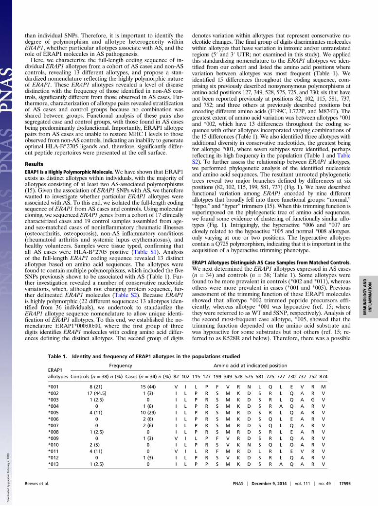
ResultsERAP1 Is a Highly PolymorphicMolecule.We have shown that ERAP1exists as distinct allotypes within individuals, with the majority ofallotypes consisting of at least two AS-associated polymorphisms(15). Given the association of ERAP1 SNPs with AS, we thereforewanted to investigate whether particular ERAP1 allotypes wereassociated with AS. To this end, we isolated the full-length codingsequence of ERAP1 from AS cases and controls. Using molecularcloning, we sequenced ERAP1 genes from a cohort of 17 clinicallycharacterized cases and 19 control samples assembled from age-and sex-matched cases of noninflammatory rheumatic illnesses(osteoarthritis, osteoporosis), non-AS inflammatory conditions(rheumatoid arthritis and systemic lupus erythematosus), andhealthy volunteers. Samples were tissue typed, confirming thatall AS cases were HLA-B*2705 positive (Table S1). Analysisof the full-length ERAP1 coding sequence revealed 13 distinctallotypes based on amino acid sequences. The allotypes werefound to contain multiple polymorphisms, which included the fiveSNPs previously shown to be associated with AS (Table 1). Fur-ther investigation revealed a number of conservative nucleotidevariations, which, although not changing protein sequence, fur-ther delineated ERAP1 molecules (Table S2). Because ERAP1is highly polymorphic (22 different sequences: 13 allotypes iden-tified from 36 individuals), we undertook to standardize theERAP1 allotype sequence nomenclature to allow unique identi-fication of ERAP1 allotypes. To this end, we established the no-menclature ERAP1*000:00:00, where the first group of threedigits identifies ERAP1 molecules with coding amino acid differ-ences defining the distinct allotypes. The second group of digits
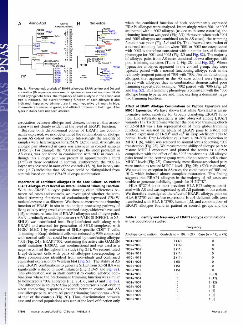
denotes variation within allotypes that represent conservative nu-cleotide changes. The final group of digits discriminates moleculeswithin allotypes that have variation in intronic and/or untranslatedregions (5′ and 3′ UTR; not examined in this study). We appliedthis standardizing nomenclature to the ERAP1 allotypes we iden-tified from our cohort and listed the amino acid positions wherevariation between allotypes was most frequent (Table 1). Weidentified 15 differences throughout the coding sequence, com-prising six previously described nonsynonymous polymorphisms atamino acid positions 127, 349, 528, 575, 725, and 730; six that havenot been reported previously at positions 82, 102, 115, 581, 737,and 752; and three others at previously described positions butencoding different amino acids (F199C, L727P, and M874T). Thegreatest extent of amino acid variation was between allotypes *001and *002, which have 13 differences throughout the coding se-quence with other allotypes incorporated varying combinations ofthe 15 differences (Table 1). We also identified three allotypes withadditional diversity in conservative nucleotides, the greatest beingfor allotype *001, where seven subtypes were identified, perhapsreflecting its high frequency in the population (Table 1 and TableS2). To further assess the relationship between ERAP1 allotypes,we performed phylogenetic analysis of the identified nucleotideand amino acid sequences. The resultant unrooted phylogenetictrees reveal two major branches defined by differences at sixpositions (82, 102, 115, 199, 581, 737) (Fig. 1). We have describedfunctional variation among ERAP1 encoded by nine differentallotypes that broadly fell into three functional groups: “normal,”“hypo,” and “hyper” trimmers (15). When this trimming function issuperimposed on the phylogenetic tree of amino acid sequences,we found some evidence of clustering of functionally similar allo-types (Fig. 1). Intriguingly, the hyperactive *006 and *007 areclosely related to the hypoactive *005 and normal *008 allotypes,only varying at one or two positions. The hyperactive allotypescontain a Q725 polymorphism, indicating that it is important in theacquisition of a hyperactive trimming phenotype.
ERAP1 Allotypes Distinguish AS Case Samples from Matched Controls.We next determined the ERAP1 allotypes expressed in AS cases(n = 34) and controls (n = 38; Table 1). Some allotypes werefound to be more prevalent in controls (*002 and *011), whereasothers were more prevalent in cases (*001 and *005). Previousassessment of the trimming function of these ERAP1 moleculesshowed that allotype *002 trimmed peptide precursors effi-ciently, whereas allotype *001 was hypoactive (ref. 15; wherethey were referred to as WT and 5SNP, respectively). Analysis ofthe second most-frequent case allotype, *005, showed that thetrimming function depended on the amino acid substrate andwas hypoactive for some substrates but not others (ref. 15; re-ferred to as K528R and below). Therefore, there was a possible
Table 1. Identity and frequency of ERAP1 allotypes in the populations studied
ERAP1allotypes
Frequency Amino acid at indicated position
Controls (n = 38) n (%) Cases (n = 34) n (%) 82 102 115 127 199 349 528 575 581 725 727 730 737 752 874
*001 8 (21) 15 (44) V I L P F V R N L Q L E V R M*002 17 (44.5) 1 (3) I L P R S M K D S R L Q A R V*003 1 (2.5) 0 I L P R S M K D S R L Q A G V*004 0 1 (6) I L P R S M K D S R A Q A R V*005 4 (11) 10 (29) I L P R S M R D S R L Q A R V*006 0 2 (6) I L P R S M K D S Q L E A R V*007 0 2 (6) I L P R S M R D S Q L Q A R V*008 1 (2.5) 0 I L P R S M R D S R L E A R V*009 0 1 (3) V I L P F V R D S R L Q A R V*010 2 (5) 0 I L P R S V K N S Q L Q A R V*011 4 (11) 0 V I L R F M R D L R L E V R V*012 0 1 (3) I L P R S V K D S R L Q A R V*013 1 (2.5) 0 I L P P S M K D S R A Q A R V
Reeves et al. PNAS | December 9, 2014 | vol. 111 | no. 49 | 17595
IMMUNOLO
GYAND
INFLAMMATION
Dow
nloa
ded
by g
uest
on
Feb
ruar
y 4,
202
0
association between allotype and disease; however, this associ-ation was not clearly evident at the level of ERAP1 function.Because both chromosomal copies of ERAP1 are codomi-
nantly expressed, we next determined the combinations of allotypein our AS cohort and control group. Interestingly, the majority ofsamples were heterozygous for ERAP1 (32/36) and, strikingly, noallotype pair observed in cases was also seen in control samples(Table 2). For example, the *001 allotype, the most prevalent inAS cases, was not found in combination with *002 in cases, al-though this allotype pair was present in approximately a third(37%) of those identified in controls. Furthermore, the *002 al-lotype was observed in most of the controls (15/19), but in only onecase (1/17) indicating that AS cases could be distinguished fromcontrols based on their ERAP1 allotype combination.
Importance of Combined Allotypes in the Case Cohort: AS PatientERAP1 Allotype Pairs Reveal an Overall Reduced Trimming Function.With the ERAP1 allotype pairs showing clear differences be-tween AS cases and controls, we investigated whether the com-bined trimming functions of codominantly expressed ERAP1molecules were also different. We chose to measure the trimmingfunction of ERAP1 in situ in the antigen processing pathway ofliving cells by using a well-characterized assay, which we have used(15), to measure function of ERAP1 allotypes and allotype pairs.An N-terminally extended precursor (AIVMK-SIINFEHL or X5-SHL8) was transfected into Erap1-deficient cells along withERAP1 and assessed for generation of SHL8 complexed withH-2Kb MHC I by activation of SHL8-specific CD8+ T cells.Trimming in Erap1-deficient cells was reduced by 90% comparedwith normal cells but could be restored by transfecting allotype*002 (Fig. 2A). ERAP1*002, containing the active site GAMENmotif mutation (E320A), was nonfunctional and was used as anegative control throughout the study (Fig. 2A). We reconstitutedErap1-deficient cells with pairs of allotypes corresponding tothose combinations identified from individuals and confirmedequivalent expression by Western blot (Fig. S1). The ability of AScase ERAP1 combinations to generate SHL8 from X5-SHL8 wassignificantly reduced in most instances (Fig. 2 B–D and Fig. S2).This observation was in stark contrast to control allotype com-binations where the predominant trimming function was similarto homozygous *002 allotypes (Fig. 2 A, C, and D and Fig. S2).The difference in ability to trim peptide precursor is most evidentwhen comparing responses observed between control and AScase allotype pairs, where AS group trimming function was ∼50%of that of the controls (Fig. 2C). Thus, discrimination betweencase and control populations was seen at the level of function only
when the combined function of both codominantly expressedERAP1 allotypes were analyzed. Interestingly, when *001 or *005are paired with a *002 allotype (as occurs in some controls), thetrimming function was good (Fig. 2D). However, when both *001and *005 allotypes are combined (as in AS cases), the trimmingfunction was poor (Fig. 2 A and D). The observed restoration ofa normal trimming function when *001 or *005 are coexpressedwith *002 is therefore consistent with a simple loss-of-functionphenotype for *001 and *005 (Fig. 2D and Fig. S2). The majorityof allotype pairs from AS cases consisted of two allotypes withpoor trimming activities (Table 2, Fig. 2D, and Fig. S2). Wherehypoactive allotypes appeared in the control group, they weretypically paired with a normal functioning allotype such as therelatively frequent pairing of *001 with *002. Normal functioningallotypes that appeared in the AS case cohort were typicallypaired with allotypes that in combination demonstrated poortrimming capacity; for example, *002 paired with *006 (Fig. 2Dand Fig. S2). This trimming phenotype is consistent with the *006allotype being hyperactive and, thus, exerting a dominant nega-tive trimming function.
Affect of ERAP1 Allotype Combinations on Peptide Repertoire andMHC I Expression. We have shown that while X5-SHL8 is an in-formative index substrate for broadly classifying ERAP1 func-tion, fine substrate specificity is also observed among ERAP1variants (15). To determine whether the observed trimming effectsof X5-SHL8 was a fair representation of more global trimmingfunction, we assessed the ability of ERAP1 pairs to restore cellsurface expression of H-2Db and -Kb in Erap1-deficient cells tonormal levels; Erap1-deficient cells have a 20–30% reduction inMHC I (6), which was restored to normal levels following *002transfection (Fig. 2E). We measured the ability of allotype pairs torestore MHC I expression and plotted the results as a directcomparison with the effect of the *002 transfectants. All allotypepairs found in the control group were able to restore cell surfaceMHC I levels (Fig. 2E). Conversely, most disease-associated pairswere unable to restore MHC I levels (Fig. 2E; >50% reduction).We noted one exception in AS cases, the combination of *003 and*012, which induced almost complete restoration. This findingsuggests that ERAP1 allotypes in the majority of AS cases areunable to generate stabilizing ligands for H-2Db/Kb.HLA-B*2705 is the most prevalent HLA-B27 subtype associ-
ated with AS and was expressed by all AS patients in our cohort.We therefore investigated the effect of ERAP1 allotype pairs onHLA-B*2705 cell surface expression. Erap1-deficient cells weretransfected with HLA-B*2705, human β2M, and combinations ofERAP1 allotypes found in patient or control groups and the
Fig. 1. Phylogenetic analysis of ERAP1 allotypes. ERAP1 amino acid (A) andnucleotide (B) sequences were used to generate unrooted maximum likeli-hood phylogenetic trees. The frequency of each allotype in the amino acidtree is indicated. The overall trimming function of each allotype is alsoindicated; hyperactive trimmers are in red, hypoactive trimmers in blue,intermediate trimmers in green, and efficient trimmers in bold type. Allo-types in italics have not been assessed.
Table 2. Identity and frequency of ERAP1 allotype combinationsin the populations studied
Allotype combination
Frequency
Controls (n = 19), n (%) Case (n = 17), n (%)
*001+*002 7 (37) 0*002+*005 3 (16) 0*002+*002 2 (11) 0*002+*011 2 (11) 0*010+*011 2 (11) 0*001+*008 1 (5) 0*002+*003 1 (5) 0*005+*013 1 (5) 0*001+*005 0 9 (53)*001+*001 0 2 (12)*001+*007 0 2 (12)*002+*006 0 1 (6)*004+*006 0 1 (6)*005+*009 0 1 (6)*003+*012 0 1 (6)
17596 | www.pnas.org/cgi/doi/10.1073/pnas.1408882111 Reeves et al.
Dow
nloa
ded
by g
uest
on
Feb
ruar
y 4,
202
0
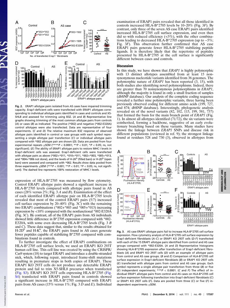
expression of HLA-B*2705 was measured by flow cytometry.Control ERAP1 allotype pairs showed a significant increase inHLA-B*2705 levels compared with allotype pairs found in AScases (28% versus 2%; Fig. 3 A and B). Examination of the effectof each identified ERAP1 allotype pair on HLA-B*2705 levelsrevealed that most of the control ERAP1 pairs (5/7) increasedcell surface expression by 20–40% (Fig. 3C) with the remainingtwo ERAP1 combinations (*002+*003 and *005+*013) increasingexpression by <10% compared with the nonfunctional *002-E320A(Fig. 3C). By contrast, all of the ERAP1 pairs from AS individualsshowed little difference in B*2705 expression compared with *002-E320A, with some even decreasing HLA-B*2705 levels (Fig. 3 Band C). These data suggest that, similar to the results obtained forH-2Db and H-Kb, the ERAP1 pairs found in AS cases generatefewer peptides capable of stabilizing B*2705 compared with com-binations found in controls.To further investigate the effect of ERAP1 combinations on
HLA-B*2705 cell surface levels, we used an ERAP1 KO 293Thuman cell line. This cell line was created by using the CRISPR/Cas9 system (18) to target ERAP1 and introduce a double-strandednick, which, following repair, introduced frame-shift mutationsresulting in premature stops in both copies of ERAP1. TheseERAP1 KO 293T cells do not produce any detectable ERAP1protein and fail to trim X5-SHL8 precursor when transfected(Fig. S3). ERAP1 KO 293T cells expressing HLA-B*2705 (Fig.S4) transfected with ERAP1 pairs found in controls showeda significant increase in HLA-B*2705 compared with ERAP1pairs from AS cases (15% versus 1%; Fig. 3 D and E). Individual
examination of ERAP1 pairs revealed that all those identified incontrols increased HLA-B*2705 levels by 10–20% (Fig. 3F). Bycontrast, only three of the seven AS case ERAP1 pairs identifiedincreased HLA-B*2705 cell surface expression, and even thendid so with reduced efficiency (<5%), with the other combina-tions leading to decreased HLA-B*2705 expression (up to −5%;Fig. 3F). This observation further confirmed that AS caseERAP1 pairs generate fewer HLA-B*2705 stabilizing peptideligands. It is therefore likely that the repertoire of peptidespresented by HLA-B*2705 at the cell surface is significantlydifferent between cases and controls.
DiscussionIn this study, we have shown that ERAP1 is highly polymorphicwith 13 distinct allotypes assembled from at least 15 non-synonymous nucleotide variants identified from 36 genomes. Thepolymorphic nature of ERAP1 has been reported (3, 15), withboth studies also identifying novel polymorphisms. Indeed, thereare greater than 70 nonsynonymous polymorphisms in ERAP1,although the majority is found in only a small fraction of samples(dbSNP database). Our analysis of the complete coding sequencerevealed a further nine polymorphic variants, three having beenpreviously observed coding for different amino acids (199, 727,and 874; dbSNP database). Interestingly, phylogenetic analysisrevealed six of the novel variants (82, 102, 115, 199, 581, 737)that formed the basis for the main branch point of ERAP1 (Fig.1). In almost all allotypes identified (71/72), the six variants werecoinherited, forming a backbone, suggestive of an early evolu-tionary branching based on these variants. Many studies haveshown the linkage between ERAP1 SNPs and disease risk indifferent populations (reviewed in ref. 9); the strongest linkagefound at residues 528 and 730 (3), observed in allotypes from
Fig. 2. ERAP1 allotype pairs isolated from AS cases have impaired trimmingcapacity. Erap1-deficient cells were transfected with ERAP1 allotypes corre-sponding to individual allotype pairs identified in cases and controls and X5-SHL8 and assessed for trimming using B3Z. (A and B) Representative linegraphs showing trimming of the most common allotype pairs from controls(A) or cases (B) as indicated. The positive (*002) and negative (*002-E320A)control allotypes were also transfected. Data are representative of fourexperiments. (C and D) The relative maximum B3Z response of observedallotype pairs identified in control or case groups with each symbol repre-senting a single allotype pair transfection (C) or individual allotype pairscompared with *002 allotype pair are shown (D). Data are pooled from fourexperimental repeats ±SEM (****P < 0.0001; **P < 0.01; *P < 0.05; ns, notsignificant). (E) The ability of ERAP1 allotype pairs to restore MHC I levels inErap1-deficient cells was assessed. Erap1-deficient cells were transfectedwith allotype pairs as above (*002+*011, *010+*011, *002+*003, *005+*013,and *004+*006 not done), and the levels of H-2Kb (filled bars) or H-2Db (openbars) were assessed and compared with *002. Results show data pooled fromthree experiments ±SEM (***P < 0.001; **P < 0.01; *P < 0.05; ns, not signifi-cant). The dashed line represents 100% restoration of MHC I levels.
Fig. 3. AS case ERAP1 allotype pairs fail to increase HLA-B*2705 cell surfaceexpression. Flow cytometry analysis of HLA-B*2705 cell surface expression byErap1-deficient fibroblasts (A–C) or ERAP1 KO 293T cells (D–F) transfectedwith each of the 15 ERAP1 allotype pairs identified from control and AS casegroups compared with *002-E320A. (A and D) Representative histogramsshowing HLA-B*2705 expression after transfection of Erap1-deficient fibro-blasts (A) and ERAP1 KO 293T cells (D) with an example of allotype pairsfrom control and AS case groups. (B and E) Comparison of HLA-B*2705 cellsurface expression in Erap1-deficient fibroblasts (B) or ERAP1 KO 293T cells(E) transfected with allotype pairs from control and AS case groups. Eachsymbol represents a single allotype pair transfection from three (B) or five(E) independent experiments; ***P < 0.0001. (C and F) The effect of in-dividual ERAP1 allotype pairs from control and AS cases on HLA-B*2705 cellsurface expression following transfection into Erap1-deficient fibroblasts (C)or ERAP1 KO 293T cells (F). Data are pooled from three (C) or five (F) in-dependent experiments ±SEM.
Reeves et al. PNAS | December 9, 2014 | vol. 111 | no. 49 | 17597
IMMUNOLO
GYAND
INFLAMMATION
Dow
nloa
ded
by g
uest
on
Feb
ruar
y 4,
202
0
both case and control groups. Previous studies have indicatedan association between K528 and AS susceptibility, whereaswe found an association with R528 (controls = 17/21 (R/K),cases = 28/6; P = 0.002); the reason for this difference is unclear.However, previous studies have noted an association with singleSNP, whereas our study unequivocally identifies individualERAP1 allotypes showing that the context in which SNPs appearwithin an allotype is important in determining disease associ-ation and ERAP1 function. Homozygosity at 528 was also as-sociated with AS susceptibility (controls = 4/19, cases = 17/17;P = 8 × 10⁻⁶), consistent with a poor trimming phenotype beingassociated with disease (R528 shows a 30% reduction in trim-ming compared with K528) (15), and our observation that poortrimming R528-containing allotypes are rescued in heterozygoteswhen the accompanying allotype has normal function. Haplotypeslinked with AS containing these polymorphisms have also beenimputed from SNP analysis (refs. 10–12; K528/D575/R725, K528/D575/E730, R528/D575/R725/E730, and K528/D575/R725/Q730).We identified one of these (K528/D575/E730), or ERAP1*006unambiguously in our full-length sequencing approach. This allo-type was only present in AS cases, albeit at a low frequency(Tables 1 and 2). Despite the association of single SNPs with AS,the demonstration that ERAP1 SNPs assemble into allotypes andthat SNPs affect ERAP1 function in cis (15–17), in the context ofthese allotypes, shows the limited expediency of simple SNPanalysis as a disease marker. Even allotype analysis was of limitedvalue, with the highest precision being achieved only when bothallotypes present in a single individual’s genome were identifiedunambiguously. Moreover, this stratification was rationalized atthe functional level because AS-associated allotype pairs encodedERAP1 with dysfunctional trimming activity arising from thepairing of two hypofunctional allotypes or a normal function anda hyperfunctional allotype indicating that, in general, the overalltrimming phenotype of two allotypes is summative which may beachieved through independent activity either in competition or insequence, or via modulation resulting from a physical interactionbetween the two ERAPs, similar to the alteration of functionobserved when ERAP1 heterodimerizes with ERAP2 (19). In-terestingly, one allotype combination (*003+*012) showed resto-ration of H-2Kb/Db, but not B27 expression (Figs. 2 and 3) whichmay reflect a combination where the substrate specificity of eachallotype is such that the peptidome thus generated contains abun-dant epitopes for H-2 but not HLA-B27. In support of this idea, thetrimming phenotype of *012 is substrate specific and similar to thatobserved for X-SHL8 substrates (*012 = MV in ref. 15), indicatingan overtrimming phenotype for B27.The mechanism by which differences in ERAP1 primary
structure contribute to the differences in function we observe isnot clear. Four of the six (82, 102, 115, and 199) together withresidue 127 are located in domain I away from the active site.Interestingly, the end of the S1 specificity pocket in domain IIborders residues 181 and 183 in domain I and, therefore, theobserved polymorphisms may affect the formation of the cata-lytic site (14, 20). In addition, residue 127 may affect confor-mational transition from open to closed states as proposed (21).Similarly, the AS-associated residue 528 is likely to affect theability to adopt a correct catalytic conformation because it lies ina region of the molecule that has been proposed to articulate theconformational change. Residue 581 is situated in a β-strand indomain III and, similarly to residue 575 (closely located as partof a loop), may affect flexibility of domain III (17). Residue 349is close to the active site and, therefore, may affect trimming. Bycontrast, residues 727 and 737 form part of an α-helix alsocontaining the AS-associated residues 725 and 730 in domain IV.These residues are located within the substrate binding cavity,which may interact with the C terminus of peptide substrates aspart of the “regulatory” domain and alter the binding and/ortrimming specificity of ERAP1.Although it is not known why ERAP1 is so polymorphic, the
identification of an ERAP1 trimming-resistant HIV gag epitope(22) and targeting of ERAP1 by human cytomegalovirus (23)
indicates selective pressure from infectious agents/pathogenssimilar to, but to a lesser extent than, that observed for HLA(MHC). It will be interesting to observe whether particularERAP1 allotypes are associated with better protection to somepathogens. One consequence of increased genetic diversity inERAP1 could be that the evolution of allotypes that confer betterprotection to a particular pathogen may, when expressed in indi-viduals of particular HLA types such as B*2705 and B*5701,predispose these individuals to autoimmune disease (1, 24).How allotypic variation of ERAP1 function could contribute
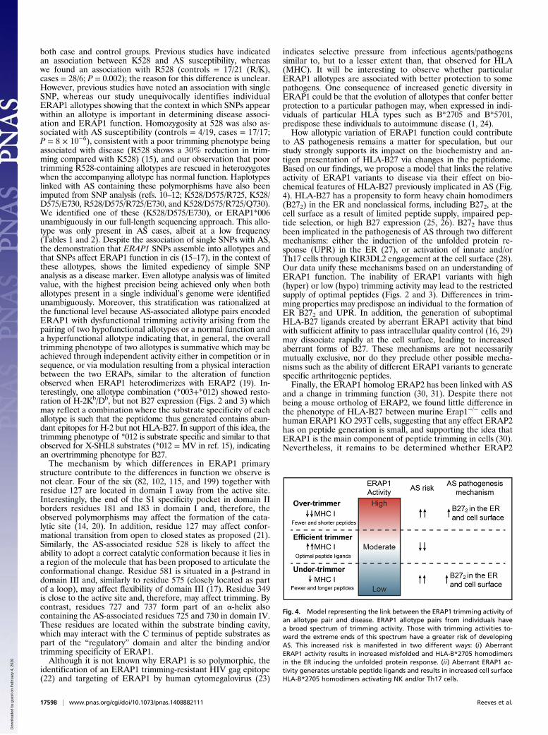
to AS pathogenesis remains a matter for speculation, but ourstudy strongly supports its impact on the biochemistry and an-tigen presentation of HLA-B27 via changes in the peptidome.Based on our findings, we propose a model that links the relativeactivity of ERAP1 variants to disease via their effect on bio-chemical features of HLA-B27 previously implicated in AS (Fig.4). HLA-B27 has a propensity to form heavy chain homodimers(B272) in the ER and nonclassical forms, including B272, at thecell surface as a result of limited peptide supply, impaired pep-tide selection, or high B27 expression (25, 26). B272 have thusbeen implicated in the pathogenesis of AS through two differentmechanisms: either the induction of the unfolded protein re-sponse (UPR) in the ER (27), or activation of innate and/orTh17 cells through KIR3DL2 engagement at the cell surface (28).Our data unify these mechanisms based on an understanding ofERAP1 function. The inability of ERAP1 variants with high(hyper) or low (hypo) trimming activity may lead to the restrictedsupply of optimal peptides (Figs. 2 and 3). Differences in trim-ming properties may predispose an individual to the formation ofER B272 and UPR. In addition, the generation of suboptimalHLA-B27 ligands created by aberrant ERAP1 activity that bindwith sufficient affinity to pass intracellular quality control (16, 29)may dissociate rapidly at the cell surface, leading to increasedaberrant forms of B27. These mechanisms are not necessarilymutually exclusive, nor do they preclude other possible mecha-nisms such as the ability of different ERAP1 variants to generatespecific arthritogenic peptides.Finally, the ERAP1 homolog ERAP2 has been linked with AS
and a change in trimming function (30, 31). Despite there notbeing a mouse ortholog of ERAP2, we found little difference inthe phenotype of HLA-B27 between murine Erap1−/− cells andhuman ERAP1 KO 293T cells, suggesting that any effect ERAP2has on peptide generation is small, and supporting the idea thatERAP1 is the main component of peptide trimming in cells (30).Nevertheless, it remains to be determined whether ERAP2
Fig. 4. Model representing the link between the ERAP1 trimming activity ofan allotype pair and disease. ERAP1 allotype pairs from individuals havea broad spectrum of trimming activity. Those with trimming activities to-ward the extreme ends of this spectrum have a greater risk of developingAS. This increased risk is manifested in two different ways: (i) AberrantERAP1 activity results in increased misfolded and HLA-B*2705 homodimersin the ER inducing the unfolded protein response. (ii) Aberrant ERAP1 ac-tivity generates unstable peptide ligands and results in increased cell surfaceHLA-B*2705 homodimers activating NK and/or Th17 cells.
17598 | www.pnas.org/cgi/doi/10.1073/pnas.1408882111 Reeves et al.
Dow
nloa
ded
by g
uest
on
Feb
ruar
y 4,
202
0
molecules in 293T cells are a low activity variant, which can beverified by using the ERAP1 KO 293T cells.In conclusion, this study provides a framework for under-
standing how ERAP1 function could impact on disease patho-genesis and how the distinct allotype combinations in AS casesmay serve as biomarkers for disease stratification and a targetfor treatment.
Materials and MethodsAS Case and Control Samples. All samples were obtained from patients andcontrols recruited with informed consent according to the protocol ap-proved by the National Research Ethics Services and Southampton ResearchEthics Committee. Diagnosis of AS was confirmed by using the Assessment ofSpondyloArthritis international Society (ASAS) classification criteria for axialspondyloarthritis and the modified New York criteria (Table S1).
ERAP1 Isolation. ERAP1 was amplified from cDNA as described (15). The PCRamplicon was cloned into vectors pcDNA3.1, pcDNA3.1V5/His (Life Tech-nologies), or a modified pcDNA3.1HA and sequenced to identify individualallotypes. ERAP1 allotype sequences were aligned, and phylogenetic analysiswere performed by using MEGA 5 software.
Cell Lines, T-Cell Activation, and MHC I Recovery Assays. Erap1-deficient cellswere transfected with both ERAP1 allotypes, recreating the combinationspresent within individuals and ES-AIVMK-SHL8 (X5-SHL8) minigene con-struct (15) by using FuGENE 6 (Promega). Presentation of trimmed SHL8and activation of B3Z T-cell hybridoma was assessed as described (15). Formurine MHC I recovery, 48 h after transfection Erap1-deficient cells werestained for H-2Kb (Y3) and H-2Db (B22.249). For HLA-B*2705 expression,
either Erap1-deficient or ERAP1 KO 293T-B27 (generated by using theCRISPR/Cas9 system; ref. 18) expressing cells were transfected with ERAP1allotype pairs as above and stained for HLA-B*2705 expression (ME1). Cellswere analyzed by flow cytometry with a BD FACS Canto II. The percentageof MHC class I recovery was calculated thus: [mean fluorescence intensity(MFI) of ERAP1 combination – MFI *002-E320A]/(MFI *002 – MFI *002-E320A) × 100. Percent change in HLA-B*2705 expression by ERAP1 allotypeswas calculated relative to cells transfected with the nonfunctional *002-E320A (SI Materials and Methods and Fig. S4).
Immunoblots. Expression of ERAP1 was determined by immunoblot as de-scribed (15). Immunoblots were probed with anti-human ARTS1 (R&D Sys-tems), anti-V5 (Life technologies), anti-HA (Abcam), or anti-glyceraldehyde3-phosphate dehydrogenase, -GAPDH (Abcam) antibodies followed by HRP-conjugated secondary antibody and SuperSignal West Pico or Femtochemiluminescent substrate (Thermo Scientific). ERAP1 protein expressionwas quantified from immunoblots by using ImageJ software and comparedwith GAPDH.
Statistical Analysis. One-way ANOVA with Dunnett’s post hoc test was per-formed for analysis of differences between multiple groups and control(GraphPad prism).
ACKNOWLEDGMENTS. We acknowledge the technical expertise of NasiaKontouli, clinical support from Helen Platten, and ethics submission supportfrom Pamela Freeman. This study was supported by the Southampton NationalInstitute for Health Research Wellcome Trust Clinical Research Facility. Thiswork was supported by Cancer Research UK Project Grant C25722/A8410 (toE.J. and T.E.), Cancer Research UK Programme Grant C7056/A11946 (to T.E.),and a Medical Research Council Studentship (to E.R.).
1. Burton PR, et al.; Wellcome Trust Case Control Consortium; Australo-Anglo-AmericanSpondylitis Consortium (TASC); Biologics in RA Genetics and Genomics Study Syndi-cate (BRAGGS) Steering Committee; Breast Cancer Susceptibility Collaboration (UK)(2007) Association scan of 14,500 nonsynonymous SNPs in four diseases identifiesautoimmunity variants. Nat Genet 39(11):1329–1337.
2. Evans DM, et al.; Spondyloarthritis Research Consortium of Canada (SPARCC); Aus-tralo-Anglo-American Spondyloarthritis Consortium (TASC); Wellcome Trust CaseControl Consortium 2 (WTCCC2) (2011) Interaction between ERAP1 and HLA-B27 inankylosing spondylitis implicates peptide handling in the mechanism for HLA-B27 indisease susceptibility. Nat Genet 43(8):761–767.
3. Harvey D, et al. (2009) Investigating the genetic association between ERAP1 andankylosing spondylitis. Hum Mol Genet 18(21):4204–4212.
4. Serwold T, Gonzalez F, Kim J, Jacob R, Shastri N (2002) ERAAP customizes peptides forMHC class I molecules in the endoplasmic reticulum. Nature 419(6906):480–483.
5. York IA, et al. (2002) The ER aminopeptidase ERAP1 enhances or limits antigen pre-sentation by trimming epitopes to 8-9 residues. Nat Immunol 3(12):1177–1184.
6. Hammer GE, Gonzalez F, Champsaur M, Cado D, Shastri N (2006) The aminopeptidaseERAAP shapes the peptide repertoire displayed by major histocompatibility complexclass I molecules. Nat Immunol 7(1):103–112.
7. Yan J, et al. (2006) In vivo role of ER-associated peptidase activity in tailoring peptidesfor presentation by MHC class Ia and class Ib molecules. J Exp Med 203(3):647–659.
8. York IA, Brehm MA, Zendzian S, Towne CF, Rock KL (2006) Endoplasmic reticulumaminopeptidase 1 (ERAP1) trims MHC class I-presented peptides in vivo and plays animportant role in immunodominance. Proc Natl Acad Sci USA 103(24):9202–9207.
9. Keidel S, Chen L, Pointon J, Wordsworth P (2013) ERAP1 and ankylosing spondylitis.Curr Opin Immunol 25(1):97–102.
10. Choi CB, et al. (2010) ARTS1 polymorphisms are associated with ankylosing spondylitisin Koreans. Ann Rheum Dis 69(3):582–584.
11. Kadi A, et al. (2013) Investigating the genetic association between ERAP1 andspondyloarthritis. Ann Rheum Dis 72(4):608–613.
12. MaksymowychWP, et al. (2009) Association of a specific ERAP1/ARTS1 haplotype withdisease susceptibility in ankylosing spondylitis. Arthritis Rheum 60(5):1317–1323.
13. Evnouchidou I, et al. (2011) Cutting Edge: Coding single nucleotide polymorphisms ofendoplasmic reticulum aminopeptidase 1 can affect antigenic peptide generation invitro by influencing basic enzymatic properties of the enzyme. J Immunol 186(4):1909–1913.
14. Kochan G, et al. (2011) Crystal structures of the endoplasmic reticulum aminopepti-dase-1 (ERAP1) reveal the molecular basis for N-terminal peptide trimming. Proc NatlAcad Sci USA 108(19):7745–7750.
15. Reeves E, Edwards CJ, Elliott T, James E (2013) Naturally occurring ERAP1 haplotypesencode functionally distinct alleles with fine substrate specificity. J Immunol 191(1):35–43.
16. García-Medel N, et al. (2012) Functional interaction of the ankylosing spondylitis-
associated endoplasmic reticulum aminopeptidase 1 polymorphism and HLA-B27 in
vivo. Mol Cell Proteomics 11(11):1416–1429.17. Martín-Esteban A, Gómez-Molina P, Sanz-Bravo A, López de Castro JA (2014) Com-
bined effects of ankylosing spondylitis-associated ERAP1 polymorphisms outside the
catalytic and peptide-binding sites on the processing of natural HLA-B27 ligands.
J Biol Chem 289(7):3978–3990.18. Cong L, et al. (2013) Multiplex genome engineering using CRISPR/Cas systems. Science
339(6121):819–823.19. Saveanu L, et al. (2005) Concerted peptide trimming by human ERAP1 and ERAP2
aminopeptidase complexes in the endoplasmic reticulum. Nat Immunol 6(7):689–697.20. Nguyen TT, et al. (2011) Structural basis for antigenic peptide precursor processing by
the endoplasmic reticulum aminopeptidase ERAP1. Nat Struct Mol Biol 18(5):604–613.21. Alvarez-Navarro C, López de Castro JA (2014) ERAP1 structure, function and patho-
genetic role in ankylosing spondylitis and other MHC-associated diseases. Mol Im-
munol 57(1):12–21.22. Draenert R, et al. (2004) Immune selection for altered antigen processing leads to
cytotoxic T lymphocyte escape in chronic HIV-1 infection. J Exp Med 199(7):905–915.23. Kim S, et al. (2011) Human cytomegalovirus microRNAmiR-US4-1 inhibits CD8(+) T cell
responses by targeting the aminopeptidase ERAP1. Nat Immunol 12(10):984–991.24. Kirino Y, et al. (2013) Genome-wide association analysis identifies new susceptibility
loci for Behçet’s disease and epistasis between HLA-B*51 and ERAP1. Nat Genet 45(2):
202–207.25. Bird LA, et al. (2003) Lymphoblastoid cells express HLA-B27 homodimers both in-
tracellularly and at the cell surface following endosomal recycling. Eur J Immunol
33(3):748–759.26. McHugh K, et al. (2014) Expression of aberrant HLA-B27 molecules is dependent on
B27 dosage and peptide supply. Ann Rheum Dis 73(4):763–770.27. Turner MJ, et al. (2005) HLA-B27 misfolding in transgenic rats is associated with ac-
tivation of the unfolded protein response. J Immunol 175(4):2438–2448.28. Bowness P, et al. (2011) Th17 cells expressing KIR3DL2+ and responsive to HLA-B27
homodimers are increased in ankylosing spondylitis. J Immunol 186(4):2672–2680.29. Chen L, et al. (2014) Critical role of endoplasmic reticulum aminopeptidase 1 in de-
termining the length and sequence of peptides bound and presented by HLA-B27.
Arthritis Rheum (Munch) 66(2):284–294.30. Evnouchidou I, et al. (2012) A common single nucleotide polymorphism in endo-
plasmic reticulum aminopeptidase 2 induces a specificity switch that leads to altered
antigen processing. J Immunol 189(5):2383–2392.31. Tsui FW, et al. (2010) Association of an ERAP1 ERAP2 haplotype with familial anky-
losing spondylitis. Ann Rheum Dis 69(4):733–736.
Reeves et al. PNAS | December 9, 2014 | vol. 111 | no. 49 | 17599
IMMUNOLO
GYAND
INFLAMMATION
Dow
nloa
ded
by g
uest
on
Feb
ruar
y 4,
202
0