Functional Synergies Underlying Control of Upright · PDF fileFunctional Synergies Underlying...
12
Functional Synergies Underlying Control of Upright Posture during Changes in Head Orientation Eunse Park 1 , Gregor Scho ¨ ner 2 , John P. Scholz 1,3 * 1 Biomechanics and Movement Science Program, University of Delaware, Newark, Delaware, United States of America, 2 Institute fu ¨ r Neuroinformatik, Ruhr University, Bochum, Germany, 3 Physical Therapy Department, University of Delaware, Newark, Delaware, United States of America Abstract Background: Studies of human upright posture typically have stressed the need to control ankle and hip joints to achieve postural stability. Recent studies, however, suggest that postural stability involves multi degree-of-freedom (DOF) coordination, especially when performing supra-postural tasks. This study investigated kinematic synergies related to control of the body’s position in space (two, four and six DOF models) and changes in the head’s orientation (six DOF model). Methodology/Principal Findings: Subjects either tracked a vertically moving target with a head-mounted laser pointer or fixated a stationary point during 4-min trials. Uncontrolled manifold (UCM) analysis was performed across tracking cycles at each point in time to determine the structure of joint configuration variance related to postural stability or tracking consistency. The effect of simulated removal of covariance among joints on that structure was investigated to further determine the role of multijoint coordination. Results indicated that cervical joint motion was poorly coordinated with other joints to stabilize the position of the body center of mass (CM). However, cervical joints were coordinated in a flexible manner with more caudal joints to achieve consistent changes in head orientation. Conclusions/Significance: An understanding of multijoint coordination requires reference to the stability/control of important performance variables. The nature of that coordination differs depending on the reference variable. Stability of upright posture primarily involved multijoint coordination of lower extremity and lower trunk joints. Consistent changes in the orientation of the head, however, required flexible coordination of those joints with motion of the cervical spine. A two- segment model of postural control was unable to account for the observed stability of the CM position during the tracking task, further supporting the need to consider multijoint coordination to understand postural stability. Citation: Park E, Scho ¨ ner G, Scholz JP (2012) Functional Synergies Underlying Control of Upright Posture during Changes in Head Orientation. PLoS ONE 7(8): e41583. doi:10.1371/journal.pone.0041583 Editor: Ramesh Balasubramaniam, McMaster University, Canada Received March 4, 2012; Accepted June 25, 2012; Published August 1, 2012 Copyright: ß 2012 Park et al. This is an open-access article distributed under the terms of the Creative Commons Attribution License, which permits unrestricted use, distribution, and reproduction in any medium, provided the original author and source are credited. Funding: This work was supported by a National Science Foundation Grant 0957920 to John Scholz (http://www.nsf.gov/funding/pgm_summ.jsp?pims_ id = 5686) and Deutsche Forschungsgemeinschaft grant to Gregor Scho ¨ ner (http://www.dfg.de/en/index.jsp) The funders had no role in study design, data collection and analysis, decision to publish, or preparation of the manuscript. Competing Interests: The authors have declared that no competing interests exist. * E-mail: [email protected] Introduction Except perhaps for the Queen’s Guard at Buckingham Palace or similar sentry-like occupations, humans rarely stand upright without performing other tasks. Even if the arms and body are generally quiet, individuals often track objects with their head, for example, a ball in flight before reaching up to catch it, or simply watching the flight of a shooting star. For an individual weighing 80-kg, the approximately 6.3-kg weight of the head is not insubstantial [1]. Thus, directional changes in the head’s orientation, especially if relatively rapid, have the potential to disturb posture and may require anticipatory postural adjustments elsewhere. This will depend, of course, on how closely head-neck motion is linked to that of the rest of the body. On the other hand, if head-neck DOFs were largely decoupled from body movement, then postural adjustments to head movement might be un- necessary. Assuming that postural adjustments are required when tracking objects with the head, it is of interest to know whether they involve all joints along the kinematic chain or only ankle and hip joint movements. Generally, standing quietly is presumed to require primarily active control of ankle joint motion [2–4], with additional coordination with hip motion when stronger environ- mental and/or task constraints are imposed [5–8]. There have been a limited number of studies of how head motion influences muscle and/or joint coordination related to postural control [9– 11]. Those studies did not distinguish between coordination related to maintaining upright posture versus that required for changing the head’s orientation. Instead, the focus was on the time course of changes in postural coordination of only the ankle and hip joints, indexed by the relative phase of their motions, as task and environmental constraints were varied. Whether a single or double inverted pendulum model of postural control is adequate to account for many aspects of upright stability is, nonetheless, an open question, particularly when additional tasks are performed in standing. Indeed, other joints along the body axis have been shown to be just as active as the ankle and hip joints even during quiet standing [12]. PLoS ONE | www.plosone.org 1 August 2012 | Volume 7 | Issue 8 | e41583
-
Upload
truongnhan -
Category
Documents
-
view
230 -
download
1
Transcript of Functional Synergies Underlying Control of Upright · PDF fileFunctional Synergies Underlying...

Functional Synergies Underlying Control of UprightPosture during Changes in Head OrientationEunse Park1, Gregor Schoner2, John P. Scholz1,3*
1 Biomechanics and Movement Science Program, University of Delaware, Newark, Delaware, United States of America, 2 Institute fur Neuroinformatik, Ruhr University,
Bochum, Germany, 3 Physical Therapy Department, University of Delaware, Newark, Delaware, United States of America
Abstract
Background: Studies of human upright posture typically have stressed the need to control ankle and hip joints to achievepostural stability. Recent studies, however, suggest that postural stability involves multi degree-of-freedom (DOF)coordination, especially when performing supra-postural tasks. This study investigated kinematic synergies related tocontrol of the body’s position in space (two, four and six DOF models) and changes in the head’s orientation (six DOFmodel).
Methodology/Principal Findings: Subjects either tracked a vertically moving target with a head-mounted laser pointer orfixated a stationary point during 4-min trials. Uncontrolled manifold (UCM) analysis was performed across tracking cycles ateach point in time to determine the structure of joint configuration variance related to postural stability or trackingconsistency. The effect of simulated removal of covariance among joints on that structure was investigated to furtherdetermine the role of multijoint coordination. Results indicated that cervical joint motion was poorly coordinated with otherjoints to stabilize the position of the body center of mass (CM). However, cervical joints were coordinated in a flexiblemanner with more caudal joints to achieve consistent changes in head orientation.
Conclusions/Significance: An understanding of multijoint coordination requires reference to the stability/control ofimportant performance variables. The nature of that coordination differs depending on the reference variable. Stability ofupright posture primarily involved multijoint coordination of lower extremity and lower trunk joints. Consistent changes inthe orientation of the head, however, required flexible coordination of those joints with motion of the cervical spine. A two-segment model of postural control was unable to account for the observed stability of the CM position during the trackingtask, further supporting the need to consider multijoint coordination to understand postural stability.
Citation: Park E, Schoner G, Scholz JP (2012) Functional Synergies Underlying Control of Upright Posture during Changes in Head Orientation. PLoS ONE 7(8):e41583. doi:10.1371/journal.pone.0041583
Editor: Ramesh Balasubramaniam, McMaster University, Canada
Received March 4, 2012; Accepted June 25, 2012; Published August 1, 2012
Copyright: � 2012 Park et al. This is an open-access article distributed under the terms of the Creative Commons Attribution License, which permits unrestricteduse, distribution, and reproduction in any medium, provided the original author and source are credited.
Funding: This work was supported by a National Science Foundation Grant 0957920 to John Scholz (http://www.nsf.gov/funding/pgm_summ.jsp?pims_id = 5686) and Deutsche Forschungsgemeinschaft grant to Gregor Schoner (http://www.dfg.de/en/index.jsp) The funders had no role in study design, datacollection and analysis, decision to publish, or preparation of the manuscript.
Competing Interests: The authors have declared that no competing interests exist.
* E-mail: [email protected]
Introduction
Except perhaps for the Queen’s Guard at Buckingham Palace
or similar sentry-like occupations, humans rarely stand upright
without performing other tasks. Even if the arms and body are
generally quiet, individuals often track objects with their head, for
example, a ball in flight before reaching up to catch it, or simply
watching the flight of a shooting star. For an individual weighing
80-kg, the approximately 6.3-kg weight of the head is not
insubstantial [1]. Thus, directional changes in the head’s
orientation, especially if relatively rapid, have the potential to
disturb posture and may require anticipatory postural adjustments
elsewhere. This will depend, of course, on how closely head-neck
motion is linked to that of the rest of the body. On the other hand,
if head-neck DOFs were largely decoupled from body movement,
then postural adjustments to head movement might be un-
necessary.
Assuming that postural adjustments are required when tracking
objects with the head, it is of interest to know whether they involve
all joints along the kinematic chain or only ankle and hip joint
movements. Generally, standing quietly is presumed to require
primarily active control of ankle joint motion [2–4], with
additional coordination with hip motion when stronger environ-
mental and/or task constraints are imposed [5–8]. There have
been a limited number of studies of how head motion influences
muscle and/or joint coordination related to postural control [9–
11]. Those studies did not distinguish between coordination
related to maintaining upright posture versus that required for
changing the head’s orientation. Instead, the focus was on the time
course of changes in postural coordination of only the ankle and
hip joints, indexed by the relative phase of their motions, as task
and environmental constraints were varied. Whether a single or
double inverted pendulum model of postural control is adequate to
account for many aspects of upright stability is, nonetheless, an
open question, particularly when additional tasks are performed in
standing. Indeed, other joints along the body axis have been
shown to be just as active as the ankle and hip joints even during
quiet standing [12].
PLoS ONE | www.plosone.org 1 August 2012 | Volume 7 | Issue 8 | e41583

Movement of the head when performing tracking or orienting
movements leads to transformations of sensory information from
visual and vestibular receptors as well as their integration with
changes in neck proprioceptor signals [13,14]. Such sensory
transformations are unlikely to have a strong effect on posture
unless sensory conflict arises because the nervous system is
believed to interpret such information via efference copy related
to the motor commands generated to produce the head movement
(Although alternative explanations have been offered [15]).
Nevertheless, this process is not perfect [16,17] so that the
transformations of visual, vestibular and neck proprioceptive
information during the tracking task could have subtle effects on
posture beyond any mechanical coupling of neck DOFs with
caudal body segments.
Accurate tracking of an external object when eye movement
alone is inadequate would require, at the very least, adequate
control of cervical joint motion. In principle, the task could be
accomplished by independent control of cervical movements,
including the atlanto-occipital (AO) joint. This would require that
the excursion of more caudal joints of the body be of limited
extent. Ankle joint movement, for example, has the potential to
affect the head’s spatial orientation given its long lever arm with
respect to the head’s position. Whether movement of other joints
significantly affects the tracking task will depend, of course, on the
required precision of that task. Thus, the need to coordinate
caudal joints that are more related to postural control with those
primarily involved in producing the desired changes in head
orientation would arise only when the typically small postural sway
exceeds the required precision of the task. For example, a 1.8-m
shooter aiming at a 10-cm diameter target at a distance of 100-m
who sways more than 0.05 degrees about the ankle risks missing
the target unless she coordinates posture with the act of aiming
and shooting at the target. Fortunately, normal postural sway is of
relatively small amplitude, so the effect of postural sway on head
orientation might be inconsequential if the precision requirements
of tracking are not too great.
The purposes of this study, then, were to investigate multijoint
coordination related to both stabilization of upright posture and
the control of head orientation when having to track a visual
target. Specifically, we asked whether the coordination of cervical
joint motion with other joints of the body was necessary to stabilize
upright posture during performance of the tracking task or was
primarily related to producing consistent changes in the head’s
orientation required for accurate tracking. To be clear, if cervical
DOFs have little effect on the anterior-posterior (AP) motion of the
whole body, then changes in the head’s orientation required to
track a moving target could be achieved without related postural
adjustments to stabilize upright posture. If so, movements of
cervical joints should have weak indices of coordination with more
caudal joints that are most related to postural control. However, to
the extent that changes in head orientation affect the body posture,
cervical joint motions should be more strongly coordinated with
those of other joints. Moreover, if natural postural sway were
within the precision requirements of the tracking task, then
coordination of cervical motion with other joints should be
unnecessary to accomplish tracking. However, the amount of sway
about the ankle observed in the current study, combined with the
target distance and size, indicated that it would affect targeting
accuracy if postural changes were not coordinated with cervical
movement. Thus, we expected to find strong indices of co-
ordination between cervical and more caudal joint motion related
to control of head orientation but weaker indices of coordination
related to the stability of the body’s position in space. Most indices
of coordination, e.g., cross-correlation analysis, relative phase
analysis, non-negative matrix factorization or principal compo-
nents analysis, cannot easily differentiate between these possibil-
ities, however. For example, if such measures were to show a high
correlation between the motions of cervical joints and joints more
directly related to the control of body posture, it would be
impossible to tell directly what was the goal of that coordination,
i.e., to stabilize posture, change the orientation of the head in
space, or a combination of both. Analysis based on the Un-
controlled Manifold (UCM) approach, however, provides a frame-
work for addressing such questions because it is possible to
determine how the variance of joint motions relates to changes in
the value of variables more directly related to a task, such as
consistency of head orientation or postural stability. This method
was applied to experimentally obtained data in the current study,
combined with UCM analysis of data with covariation among the
actual joint motions artificially removed to simulate incoordination
[18,19]. That analysis helped to determine the extent to which
UCM effects were primarily due to geometric constraints (i.e.,
variance of a variable that lies parallel to a dimension of the UCM
and, therefore, cannot affect the task variable of interest) versus
interjoint coordination.
Methods
Ethics StatementThe University of Delaware IRB approved the research
described in this submission. Approval was based on an
appropriate risk/benefit ratio and a study design wherein the
risks were minimized. All research was conducted in accordance
with the approved protocol. This protocol received expedited
review based on the applicable federal regulation. Informed
consent is a process beginning with a description of the study and
insurance of participant understanding followed by a signed
consent form. Informed consent continued throughout the study
via a dialogue between the researcher and research participants. In
accordance with Federal regulations, each participant received
a copy of the signed consent document.
SubjectsTwelve subjects (24.465.05 years old; 5 females and 7 males)
volunteered and were paid to participate in this study in response
to an online solicitation. Subjects had no balance disorders or
dizziness, and no musculoskeletal injuries, neurological disorders
or uncorrected visual acuity deficits by self-report. All subjects
provided written informed consent according to the procedures
approved by the Institutional Review Board of the University of
Delaware consistent with the Declaration of Helsinki.
Experimental SetupTwo stationary, 10-cm diameter circular targets were projected
on a wall 2-m in front of the subject. The upper circular target was
placed at eye level and the lower circular target was placed at 70%
of eye level, measured from the floor. One 5-cm diameter, yellow
circular cursor also was projected and oscillated sinusoidally
between the two stationary circular targets at a frequency of 0.258-
Hz. This frequency was chosen based on pilot experiments with
three subjects that attempted to identify a frequency of tracking
with which subjects could be successful while still being challenged.
Experimental data were collected at a sampling rate of 120-Hz
with a ViconTM MX-13 (Oxford Metrics) motion-measurement
system composed of eight infrared cameras. All analyses were
performed in the sagittal plane. Reflective markers were placed at
approximate joint centers on the right side of the body and on the
Functional Synergies Underlying Upright Posture
PLoS ONE | www.plosone.org 2 August 2012 | Volume 7 | Issue 8 | e41583
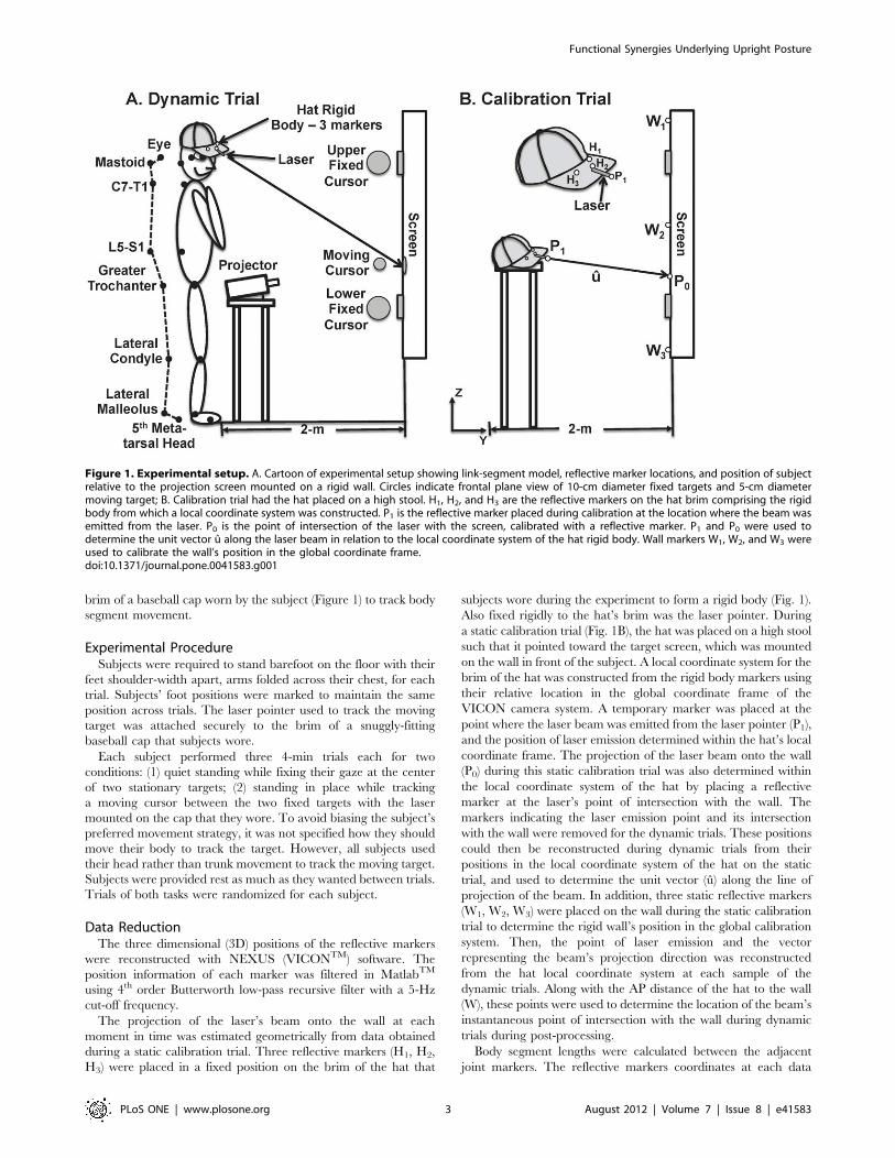
brim of a baseball cap worn by the subject (Figure 1) to track body
segment movement.
Experimental ProcedureSubjects were required to stand barefoot on the floor with their
feet shoulder-width apart, arms folded across their chest, for each
trial. Subjects’ foot positions were marked to maintain the same
position across trials. The laser pointer used to track the moving
target was attached securely to the brim of a snuggly-fitting
baseball cap that subjects wore.
Each subject performed three 4-min trials each for two
conditions: (1) quiet standing while fixing their gaze at the center
of two stationary targets; (2) standing in place while tracking
a moving cursor between the two fixed targets with the laser
mounted on the cap that they wore. To avoid biasing the subject’s
preferred movement strategy, it was not specified how they should
move their body to track the target. However, all subjects used
their head rather than trunk movement to track the moving target.
Subjects were provided rest as much as they wanted between trials.
Trials of both tasks were randomized for each subject.
Data ReductionThe three dimensional (3D) positions of the reflective markers
were reconstructed with NEXUS (VICONTM) software. The
position information of each marker was filtered in MatlabTM
using 4th order Butterworth low-pass recursive filter with a 5-Hz
cut-off frequency.
The projection of the laser’s beam onto the wall at each
moment in time was estimated geometrically from data obtained
during a static calibration trial. Three reflective markers (H1, H2,
H3) were placed in a fixed position on the brim of the hat that
subjects wore during the experiment to form a rigid body (Fig. 1).
Also fixed rigidly to the hat’s brim was the laser pointer. During
a static calibration trial (Fig. 1B), the hat was placed on a high stool
such that it pointed toward the target screen, which was mounted
on the wall in front of the subject. A local coordinate system for the
brim of the hat was constructed from the rigid body markers using
their relative location in the global coordinate frame of the
VICON camera system. A temporary marker was placed at the
point where the laser beam was emitted from the laser pointer (P1),
and the position of laser emission determined within the hat’s local
coordinate frame. The projection of the laser beam onto the wall
(P0) during this static calibration trial was also determined within
the local coordinate system of the hat by placing a reflective
marker at the laser’s point of intersection with the wall. The
markers indicating the laser emission point and its intersection
with the wall were removed for the dynamic trials. These positions
could then be reconstructed during dynamic trials from their
positions in the local coordinate system of the hat on the static
trial, and used to determine the unit vector (u) along the line of
projection of the beam. In addition, three static reflective markers
(W1, W2, W3) were placed on the wall during the static calibration
trial to determine the rigid wall’s position in the global calibration
system. Then, the point of laser emission and the vector
representing the beam’s projection direction was reconstructed
from the hat local coordinate system at each sample of the
dynamic trials. Along with the AP distance of the hat to the wall
(W), these points were used to determine the location of the beam’s
instantaneous point of intersection with the wall during dynamic
trials during post-processing.
Body segment lengths were calculated between the adjacent
joint markers. The reflective markers coordinates at each data
Figure 1. Experimental setup. A. Cartoon of experimental setup showing link-segment model, reflective marker locations, and position of subjectrelative to the projection screen mounted on a rigid wall. Circles indicate frontal plane view of 10-cm diameter fixed targets and 5-cm diametermoving target; B. Calibration trial had the hat placed on a high stool. H1, H2, and H3 are the reflective markers on the hat brim comprising the rigidbody from which a local coordinate system was constructed. P1 is the reflective marker placed during calibration at the location where the beam wasemitted from the laser. P0 is the point of intersection of the laser with the screen, calibrated with a reflective marker. P1 and P0 were used todetermine the unit vector u along the laser beam in relation to the local coordinate system of the hat rigid body. Wall markers W1, W2, and W3 wereused to calibrate the wall’s position in the global coordinate frame.doi:10.1371/journal.pone.0041583.g001
Functional Synergies Underlying Upright Posture
PLoS ONE | www.plosone.org 3 August 2012 | Volume 7 | Issue 8 | e41583

sample were used to calculate the following sagittal plane joint
angles (hi): ankle, knee, hip, between lumbar 5th and sacral 1st
vertebrae (L5-S1), between cervical 7th and thoracic 1st vertebrae
(C7), and between the atlas and occipital condyles (AO) using
a link-segment model [20]. The positive angle was defined as the
upper (or cranial) segment moving anterior, based on the formula:
hi~ cos{1 (V1.V2)
where V1 and V2 are unit vectors for the proximal and distal
segments, respectively.
The location of the body center of mass (CMPOS) was calculated
by the sum of the product of each segment’s estimated mass and
the location of center of mass of each segment, divided by total
body mass. The estimated location of each segment’s center of
mass was calculated from marker data, and the contribution of
their masses to total body mass was estimated from Winter [20].
dCM~1
M
Xni~1
midi
CMPOS was estimated using both a six segment (i.e., foot plus
shank, thigh, pelvis, trunk plus arms, neck, head) and a four-
segment (i.e., foot plus shank, thigh, pelvis, head-arms-trunk
[HAT]) model.
Experimental MeasuresTargeting Error. Constant error and variable error with
respect to the moving cursor were computed during the tracking
movement to evaluate the accuracy and consistency of performing
the tracking task.
Constant error estimated performance accuracy as follows:
PNi~1
(xi{T)=N
where T is the target position, N is the number of frames and xi is
the position of the laser’s projection on the screen on the ith cycle.
Variable error estimated the consistency of targeting as follows:
ffiffiffiffiffiffiffiffiffiffiffiffiffiffiffiffiffiffiffiffiffiffiffiffiffiffiffiffiffiffiffiffiffiffiXNi~1
(xi{M)2=N
vuut
where M is the average position of the laser’s projection on the
screen across frames [21].
Components of Joint Configuration Variance. UCM
analysis was performed at each point in normalized time, across
all tracking cycles, to determine the extent to which the variance of
six sagittal plane joint motions led to variability of the presumed
performance variables, head orientation to the target (HEADORI)
and center of mass position (CMPOS), or reflected equivalent joint
configurations that stabilized the values of those performance
variables [22].
UCM analysis can be illustrated conceptually as follows.
Consider placing your index fingertip on a spot on your cheek
to stop the bleeding after cutting yourself while shaving. This
requires the control of the fingertip in three dimensions, X, Y and
Z because orientation of the finger is not crucial to place the
fingertip on the correct spot. Thus, the task requires three DOFs of
fingertip control. However, the combined scapula, arm and finger
have a total of at least thirteen joint angles that can be combined in
different ways to achieve the same fingertip position, and more if
we assume trunk motion. The system of joints is highly redundant
with respect to the task’s requirements. This allows for flexibility in
combining the joints across repetitions to place the fingertip on the
spot, or across time to keep the fingertip on the spot. For example,
if a fly lands on your elbow while maintaining pressure on the spot,
there are enough redundant DOFs (1323= 10) to flick the elbow
and chase the fly away. This has led to its characterization as
motor abundance [23]. Across repetitions or across time,
variations of the joint angles could lead to the finger leaving the
designated spot on the cheek or could have no effect on the
fingertip position. That is, if we consider the full space of the joints
angles (N= 13), a subspace exists within which changes in the joint
configuration have no effect on the selected fingertip position (13–
3= 10) and variations within this subspace allow for flexibility in
performance. This is referred to as the UCM subspace and its
linear estimate is the null space of the geometric model relating
small changes in joint angles to changes in the performance
variable of interest, here the fingertip position. However, the
complementary subspace that lies orthogonal to the UCM
subspace, mathematically, is the range space. Variations of the
configuration of joint angles within this subspace leads to a range
of fingertip positions that differ from the desired position. UCM
analysis provides a formal way to determine how much of joint
variance lies within the UCM (VUCM) versus the range space
(VORT). If you try to maintain a fixed position of your fingertip
across time, or achieve the same position across repetitions of
reaching to that spot, by using an identical joint configuration each
time, then to the extent you are successful, we can expect that
VUCM < VORT. To the extent that you are not very good at
replicating the spot, we can expect VORT . VUCM. There is no
a priori reason to expect VUCM . VORT. Such a result would
reflect a system that tends to make use of the available redundancy
where possible to allow for flexibility, e.g., so that multiple tasks
(keeping the finger on the spot while flicking that fly) can be
performed without disturbing each other. As discussed below,
however, some of VUCM can derive from a source other than
coordination among the joint motions. We introduce a method
here to distinguish between these two sources.
The analysis of CMPOS focused on stability in the anterior-
posterior (AP) direction because the amplitude of sway in the AP
direction on a normal surface is substantially higher than in the
medial-lateral (ML) direction during standing [3], as was the case
for the tasks studied here. HEADORI was defined as the angle
between 1) a vector defined between individual markers placed on
the right mastoid process and immediately lateral to the right eye
and 2) a vector from the right mastoid to the center of the moving
cursor for the tracking task and to the midpoint between the two
stationary targets for the quiet standing task.
Each cycle of tracking was identified based on the peak-to-peak
of head position and divided into two half-cycles (upward and
downward tracking). Each half-cycle was then extracted and time-
normalized to 100 data points. To make the analysis of the two
tasks more comparable, quiet standing trials also were divided into
pseudo-cycles by choosing a duration equal to the average
duration of cycles for each subject’s tracking task and then time-
normalizing that period to 100 data points. UCM analysis then
was performed across cycles of tracking or across pseudo-cycles of
quiet standing separately at each normalized time sample,
according to the following steps described with respect to CMPOS.
The method for testing hypotheses related to the head orientation
is similar, except that the geometric model does not include
Functional Synergies Underlying Upright Posture
PLoS ONE | www.plosone.org 4 August 2012 | Volume 7 | Issue 8 | e41583
segment masses. Details of this analysis can be found in Scholz and
Schoner [22] or Krishnamoorthy et al. [24].
1) A geometric model relating the CMPOS to the joint angles
(hi), limb segment lengths (li), relative segment masses (mi),
and the distance of the individual segment masses from
distal joint (di) [20] was developed (Appendix S1) [12].
2) The Jacobian matrix (J), composed of partial derivatives of
this geometric model, was computed. The Jacobian
describes how small changes in the joint angles (hi) affectthe CMPOS.
3) The null space of this Jacobian was obtained using
MatlabTM, providing a linear approximation to the UCM,
or the subspace in joint angle space within which changes in
the joint configuration have no effect on the mean CMPOS.
The complementary subspace (subspace orthogonal to the
UCM) or range space was also computed, representing
a subspace in joint space in which changes in the joint
configuration lead to changes in the CMPOS. Both subspaces
were estimated at each time-normalized sample based on
the mean joint configuration (hmean) across cycles at that
sample (N=67 per trial).
4) At each time-normalized sample, the difference between the
current joint configuration hi and hmean was projected into
the null-space (UCM) and the range space of J, producing
scalar estimates of the extent to which the joint configuration
was aligned with those two subspaces.
5) Variances of the projection lengths across cycles at each
point in normalized time were computed within both the
UCM and range spaces. The two variances then were
normalized to the appropriate subspace dimension, dORT= 1
for the range space related to control of the AP CMPOS and
dUCM (n2dORT) = 5 for the UCM subspace based on the 6-
DOF geometric model and dUCM (n2dORT) = 3 for the 4-
DOF geometric model of the CMPOS, yielding measures of
variance per DOF, VORT and VUCM, respectively. Because
sagittal plane head orientation is also a 1-dimensional
performance variable, the variance components were
normalized to the same number of dimensions as for
CMPOS-6DOF.
6) Because the variance components were relatively stable
across the tracking cycle, we averaged each variance
component across the cycle, yielding one measure of VUCM
and VORT for each performance variable of each condition
for each subject.
What has been referred to as a UCM effect (i.e., VUCM .
VORT) can result because a given performance variable is
stabilized by coordinating joint motions within the UCM
whenever there is variation of one or more of the joint angles to
keep the performance variable constant. In addition, however, if
the axis of a given joint’s motion lies nearly parallel to a dimension
of the UCM subspace, then most of its variance will lie within the
UCM geometrically and its effect on the performance variable will
be minimal (i.e., minimal contribution to VORT) even if its motion
is not coordinated with the motions of other joints (e.g., consider
the effect of forearm pronation and supination on the hand’s
spatial position when the wrist is in a neutral position). To
determine the extent to which identified UCM effects result from
joint coordination, UCM analysis was repeated on the experi-
mental data after removing covariation among the joints by
eliminating the off-diagonal terms of their covariance matrix,
simulating a lack of interjoint coordination [25]. The extent to
which the UCM effect was diminished by removing joint motion
covariation provided an indication of the extent to which interjoint
coordination was responsible for identified UCM effects. To
reduce the number of factors in this analysis, the relative difference
between VUCM and VORT was computed for all performance
variables:
DVAR~VUCM{VORTVUCMzVORT
If DVAR is close to ‘+19, most joint variance reflects the use of
motor abundance (i.e., many equivalent joint configurations) to
stabilize the performance variable [22,26]. To the extent that
coordination among the joints accounted for UCM effects found
in UCM analysis of the original data, removing joint covariation
should result in DVAR decreasing to near zero.
In addition, because of predicted differences in the coordination
of cervical DOFs related to stabilizing the body in space (CMPOS)
versus producing a consistent head orientation, the individual
contributions of each joint to DVAR was determined for the
CMPOS based on the 6-DOF model and for HEADORI. This can
be accomplished by adding an additional step between steps #4
and #5 above. Because the UCM space and range space are
subspaces of the original joint configuration space, the length of
projections of each mean-free joint configuration into those two
subspaces can be projected back into the full joint space to yield
the proportion of the UCM and range space vectors contributed
by each joint. Thus, for each joint there will be two contributions,
one to the UCM subspace and one to the range space. The
variances of each joint’s contribution to each subspace are then
computed. DVAR was then calculated based for each angle’s
contribution to VUCM and VORT.
Finally, because many postural studies assume that control of
only the ankle and hip joints are crucial for understanding upright
posture [4,6,7,27], including the performance of tracking tasks
with the head [9–11], a 2-DOF geometric model of CMPOS also
was evaluated. Moreover, several recent accounts of postural
control have assumed, at least implicitly, that the nervous system
cares more about the orientation of the leg and trunk segments in
space than positional control of the ankle or hip joints [8,28–31].
Therefore, UCM effects were computed based on a two-segment
model of the CM position, using the angles formed by the leg
(vector from the lateral malleolus to the hip joint) and the trunk
(vector from the hip joint to the 7th cervical marker), respectively,
with the horizontal. Masses and mass locations of the shank plus
thigh and the combined pelvis, head, arms and trunk were used to
estimate the overall CM of the body in the model.
StatisticsA repeated-measure analysis of variance (RM-ANOVA) first
was performed to determine if differences existed in the variance
components between the upward and downward phases of
tracking, as well as between the half-cycle times for these two
phases.
To investigate differences in the range of joint excursion
between the tasks, quiet standing and tracking the moving target,
a multivariate repeated-measure ANOVA was performed with
task as the repeated factor. Which joints accounted for a significant
multivariate effect were determined by the univariate ANOVAs.
A two-way repeated-measure ANOVA was performed for each
performance variable (i.e., CMPOS-6DOF, CMPOS-4DOF and
HEADORI) to test for effects of the task (quiet standing vs.
tracking) and differences between the variance components (VUCM
vs. VORT). Significant interactions or planed contrasts were
Functional Synergies Underlying Upright Posture
PLoS ONE | www.plosone.org 5 August 2012 | Volume 7 | Issue 8 | e41583

investigated using the m-matrix structure in SPSSTM version 18.0,
with acceptable p-value of 0.05 for all analysis.
To compare the effect of actual and simulated UCM results
(obtained after removing joint covariation), a RM-ANOVA was
performed on DVAR for all performance variables. In addition,
a multivariate RM-ANOVA was performed to evaluate the
contributions of each individual joint to DVAR related to
HEADORI and CMPOS-6DOF before and after removing co-
variation among the joints.
Results
Cycle TimeThe cycle time for the tracking task, based on the head’s
movement, was 3.85360.212 seconds. The half-cycle times did
not differ significantly (p = 0.343) between the upward
(1.92460.19 sec) and downward (1.9360.187 sec) phases of
tracking.
Range of Joint MotionAmultivariate repeated measures ANOVA revealed a significant
difference in the average within-cycle range of joint excursions
between the quiet standing and head tracking tasks (F6,6 = 80.1,
p,0.001). The univariate ANOVAs revealed that the range of
motion of the AO (F1,11 = 260.8, p,0.001) and C7-T1 joints
(F1,11 = 26.4, p,0.001) were higher during tracking than during
quiet standing. In contrast, excursions of L5-S1 (p = 0.65), hip
(p = 0.17), knee (p= 0.08) and ankle (p = 0.76) joints did not differ
between tasks (Figure 2). Moreover, differences between the tasks
in the magnitude of across-cycle joint variability (F6,6 = 38.1,
p,0.001) was found only for the AO (F1,11 = 38.1, p,0.001) and
C7-T1 joints (F1,11 = 10.6, p,0.01), but not the other joints (all
joints p.0.27).
Given the measured range of ankle joint excursion (Figure 2),
the target distance (2-m) and its size (5-cm), this amount of ankle
sway alone would lead to constant errors of targeting of between
2.8-cm and 9.0-cm for the range of subject heights (1.58-m to
1.85-m) without compensation by motion of other joints of the
body.
Performance AccuracyAccuracy and consistency of tracking performance was evalu-
ated by computing constant and variable errors of targeting [21].
These errors were computed for each half-cycle of tracking (i.e.,
upward and downward). The number of cycles was consistent
across subjects (65-cycles for each trial). Figure 3 shows the
average across-subjects constant error and variable error in the
medial-lateral (ML) and vertical directions. Constant error was
substantially higher in the vertical direction during both upward
(11.1160.48-cm) and downward (26.3560.47-cm) tracking com-
pared to the ML direction (upward = 20.1260.008-cm;
downward = 20.2560.1-cm). The same was true for variable
error (upward: Vertical = 13.6760.31-cm; ML=1.0760.04-cm;
downward: Vertical = 10.6260.2 cm; ML=1.160.05-cm).
Components of Joint Configuration VarianceNo significant differences were found when evaluating the effect
of upward versus downward tracking movements on the variance
components (F1,11 = 0.398, p = 0.533), nor was there a significant
interaction between direction and variance component
(F1,11 = 0.804, p = 0.377). Therefore, we confine our presentation
of the head tracking results to the upward phase of head tracking.
As noted, the UCM analyses were performed across multiple
cycles at each percentage of normalized time. Figure 4 illustrates
the results for each variance component at each percentage of the
cycle for the CMPOS-4DOF of one representative subject. The
results were similarly consistent for all performance variables.
Therefore, the remaining figures show the results after averaging
across the normalized time points.
In Figure 5, the average across-subjects components of joint
configuration variance for quiet standing and the tracking task is
presented for each performance variable. Strong UCM effects (i.e.,
VUCM .. VORT) were present for each performance variable for
both the quiet standing and tracking tasks: CMPOS-6DOF (quiet
standing: F1,11 = 153.1, p,0.001; tracking: F1,11 = 345.8,
p,0.001; upper left panel, Fig. 4) and HEADORI (quiet standing:
F1,11 = 133.9, p,0.001; tracking: F1,11 = 78.3, p,0.001; lower left
panel, Fig. 4). The results for center of mass position with the 4-
DOF model (i.e., ankle, knee, hip and L5-S1 joints) also revealed
large differences between VUCM and VORT (Fig. 4, upper right
panel; quiet standing: F1,11 = 91.0, p,0.001; tracking: F1,11 = 64.3,
p,0.001). The UCM results differed significantly between the 6-
DOF and 4-DOF models, reflected by a significant interaction of
task variable and variance component for both conditions (quiet
standing: F1,11 = 6.55, p,0.05; tracking: F1,11 = 10.4, p,0.01) in
a separate RM-ANOVA. The interaction revealed in both cases
that VUCM was significantly greater when the model included the
cervical joints, whereas VORT did not differ between the 4-DOF
and 6-DOF models of CMPOS.
There were no main effects of task (quiet standing vs. tracking;
HEADORI: p.0.86; CMPOS-6DOF: p.0.82; CMPOS-4DOF:
p.0.80), nor were there any significant interactions between the
tasks and the variance components (HEADORI: p.0.35; CMPOS-
4DOF: p.0.42). However, the interaction approached significance
for CMPOS-6DOF (F1,11 = 4.3, p= 0.06) as a result of slightly higher
VUCM and slightly lower VORT for tracking compared to quiet
standing (Figure 4, lower left panel).
Results of the UCM analysis for the CMPOS based on a two-
segment model are presented in the lower right panel of Figure 4.
There were no significant differences between VUCM and VORT
Figure 2. Range of joint excursion. Mean across subjects (6SEM) ofthe maximum joint excursions for all measured joint motions for quietstanding while visually fixating a point between two stationary targetsand across cycles of tracking the moving target with the head; L5-S1 =joint between 5th lumbar and 1st sacral vertebrae, C7-T1 = jointbetween 7th cervical and 1st thoracic vertebrae. Cycles were defined aspseudo-cycles for quiet standing with the same average cycle time aswith target tracking.doi:10.1371/journal.pone.0041583.g002
Functional Synergies Underlying Upright Posture
PLoS ONE | www.plosone.org 6 August 2012 | Volume 7 | Issue 8 | e41583

(p.0.51); there was also no effect of task (p.0.09) or interaction
between task and variance component (p.0.78).
Components of Joint Configuration Variance afterRemoving Joint CovariationFigure 6 presents the relative variance difference (DVAR) based
on UCM analysis of the actual data and after simulated removal of
covariation among the joints. Values of DVAR above zero indicate
that more of the joint variance was VUCM, i.e., variance that
would not change the value of the performance variable. A value
of DVAR approaching unity indicates that most of the joint
variance was VUCM. In addition, the more that DVAR decreased
after removing covariation among the joints, the stronger the
evidence that VUCM resulted from coordination among the joint
motions as compared to variation of individual joints whose axes
happened to lie close to a dimension of the UCM.
RM-ANOVAs revealed a significant effect of removing co-
variation (CMPOS-6DOF: F1,11 = 22.3, p,0.01; CMPOS-4DOF:
F1,11 = 129.0, p,0.001; HEADORI: F1,11 = 142.3, p,0.001). Only
for CMPOS-6DOF (upper left panel, Fig. 5) was there a significant
effect of task (F1,11 = 13.2, p,0.01) or an interaction of task with
method (i.e., normal or simulated analysis; F1,11 = 8.0, p,0.05).
This was due to a greater reduction in DVAR when covariation was
removed in the quiet standing task compared to the tracking task.
The pattern of change between the results on actual versus
Figure 3. Constant and variable error of targeting. Upper Panels: Average across trials projection of the laser pointer (black dots) onto thescreen at every 10 frames of tracking for one representative subject, after each cycle was normalized to 100 frames. Units are in meters (m). LowerPanels: Average across-subjects constant error (6SEM) and variable error (6SEM) of targeting with respect to the moving cursor are shown.doi:10.1371/journal.pone.0041583.g003
Functional Synergies Underlying Upright Posture
PLoS ONE | www.plosone.org 7 August 2012 | Volume 7 | Issue 8 | e41583

simulated data were identical for CMPOS-4DOF and HEADORI
(Wilcoxon signed rank test, p = 0.78), both performance variables
showing a large decrease in DVAR after removing joint covariation
(Fig. 6, upper right and lower left panels). Moreover, this decrease
was substantially greater than for CMPOS-6DOF (Wilcoxon signed
rank, p,0.01 and p,0.05 for comparison with CMPOS-4DOF and
HEADORI, respectively). For all performance variables, the
decrease in DVAR resulted both from a decrease in VUCM
(CMPOS-6DOF: F1,11 = 6.0, p,0.05; CMPOS-4DOF: F1,11 = 5.7,
p,0.05; HEADORI: F1,11 = 15.1, p,0.01) and an increase in
VORT (CMPOS-6DOF: F1,11 = 6.0, p,0.05; CMPOS-4DOF:
F1,11 = 11.2, p,0.01; HEADORI: F1,11 = 15.6, p,0.01).
The lower right panel of Figure 6 shows the contribution to
DVAR of individual joints before (light patterned bars) and after
(dark filled bars) removing joint covariation for the HEADORI and
CMPOS-6DOF performance variables. For the ankle, knee, hip and
lumbar spine, DVAR decreased substantially after removing
covariation. A multivariate RM-ANOVA revealed no differences
between the tasks (i.e., tracking vs. quiet standing, p.0.63) or
interaction (p.0.79) between the task and method (i.e., normal vs.
simulated data). Therefore, further analyses of differences between
individual contributions to HEADORI vs. CMPOS-6DOF by method
were examined for the tracking task only. For the ankle (p.0.70),
knee (p.0.28) and hip (p.0.63) joints, the decrease in DVAR after
removing covariation did not differ between CMPOS-6DOF and
HEADORI. In all cases, DVAR became negative, indicating VORT
. VUCM without joint covariation. A similar decrease in DVAR
after removing covariation occurred for the L5-S1 joint, although
there was a significant difference in the values related to stability of
HEADORI versus CMPOS-6DOF (Fig. 5, lower right panel;
F1,11 = 14.2, p,0.01). DVAR related to CMPOS-6DOF stability
decreased significantly when removing covariation but it remained
positive. In contrast, DVAR related to the consistency of the head’s
orientation was negative. On the other hand, differences in DVAR
before and after covariation removal between HEADORI and
CMPOS-6DOF for the cervical joints (C7-T1: F1,11 = 119.3,
p,0.001 and AO: F1,11 = 156.2, p,0.001) were substantial.
Although the cervical contributions to DVAR did decrease slightly
after removing joint covariation related to stability of CMPOS-
6DOF, this decrease was miniscule compared to the decrease
related to HEADORI.
Discussion
Results of the current study provide insights about the
coordination of multiple joints along the body axis in providing
for postural stability as well as consistency of the additional
tracking task. Both single and double inverted pendulum models of
posture have influenced strongly studies of postural control
[3,7,8,28,32–36]. Recent work indicates, however, that both
inverted pendulum models may be oversimplified [12,24,27,37].
For example, if the body were controlled as a single inverted
pendulum, all joint variance could be expected to be VORT
because movement of the ankle would produce corresponding
motion of the CM since that model presumes that all joints
proximal to the ankle are stiffened and move minimally during
quiet standing. If the body acted like a double inverted pendulum,
involving motion about the ankle and hip, VUCM could be higher
than VORT only if the joint motions were perfectly out of phase. In
such a case, the primary contribution of both ankle and hip joint
variance could be expected to be to VUCM. However, out of phase
motion about the ankle and hip appears to be limited to
frequencies of sway above 2.0-Hz [8]. Moreover, results of the
current study as well as previous analyses [38] indicate that,
depending on the context, ankle joint variance contributes
relatively equally to VUCM and VORT, with a tendency for a larger
contribution to VORT. Thus, a multi-DOF postural model based
on the UCM hypothesis provides a viable alternative framework
for understanding postural control [39]. The current results
support that contention while revealing the role of coordination
among the active DOFs in producing the experimentally identified
variance structure related to stability of the CMPOS and
consistency of head orientation. In addition, the results suggest
that different synergies using some shared DOFs are used to
stabilize the different performance variables.
Is CMPOS Stability Dependent on Coordination of CervicalJoint Motions with those of more Caudal Joints?The difference between UCM analyses performed with 4-DOF
vs. 6-DOF geometric models of the CMPOS was the inclusion of
cervical joint movements, including the AO joint. Indeed, the only
significant increase in joint excursion when tracking an object in
the current study, compared to the quiet standing condition, was
due to cervical joint motion (Fig. 2). Studies of anticipatory
postural adjustments (APAs) that have investigated muscle or joint
synergies underlying postural perturbations or tasks with increased
difficulty have shown, in general, similar differences in variance
components (i.e., VUCM . VORT) related to stabilizing upright
posture as those reported here [40–43]. Danna-Dos-Santos et al.
[40] suggested that synergies of leg and trunk muscles underlying
APAs help to stabilize the head, which also is important to ensure
stable visual and vestibular information about the body’s position
in space. Thus, it is somewhat surprising that adding the tracking
task to quiet standing in the current study resulted in no substantial
differences in either variance component across tasks. It may be
Figure 4. Example of components of joint configurationvariance across the tracking cycle. Components of joint configu-ration variance per dimension of joint subspace (deg2) across thetracking cycle, related to stability of the 4-DOF CM position ofa representative subject. Note the consistency of VUCM and VORT acrossthe cycle, leading to using the average across the cycle for furtherstatistical analyses.doi:10.1371/journal.pone.0041583.g004
Functional Synergies Underlying Upright Posture
PLoS ONE | www.plosone.org 8 August 2012 | Volume 7 | Issue 8 | e41583

that having subjects fixate on a stationary target during quiet
standing, which required controlled head orientation to the
fixation point, minimized differences compared to orienting to
a moving target, although one would think that the added cervical
and head motion in the latter case would affect postural stability.
The magnitude of the UCM effect (i.e., VUCM . VORT) was
larger for the 6-DOF model of CM positional stability than when
investigating the 4-DOF geometric model, due primarily to
a greater magnitude of VUCM. VORT did not differ significantly
between the two models (Fig. 4). This difference in VUCM between
the two models could be due largely to the fact that the axes of
cervical joint motions were closely aligned with dimensions of the
UCM. To distinguish between contributions to VUCM due to
inter-joint coordination versus body geometry, covariation among
joints was artificially removed from the actual data [18,19,25] and
the UCM analysis repeated. If joint covariation plays a dominant
role, then most if not all of UCM effects found in the actual data
set should disappear after removing it and repeating the UCM
analysis. Verrell et al. [44] applied this randomization method to
investigate the control of the CM position relative to the foot
position at the time of heel strike during walking.
In the current study, when removing joint covariation from the
6-DOF geometric model there was still a significant UCM effect
(VUCM . VORT). In contrast, the UCM effect largely disappeared
when removing joint covariation from the 4-DOF model. This was
because the values of the partial derivatives of the C7-T1 and AO
joints with respect to the AP CMPOS in the Jacobian were an order
of magnitude smaller than the values for more caudal joints (e.g.,
Ankle: 0.271; Knee: 0.265; Hip: 0.226; L5-T1:0.178; C7-
T1:0.039; AO: 0.023 m/rad). Therefore, motion of the cervical
joints could affect only minimally the CMPOS. This fact is further
emphasized when examining the contributions that individual
joint’s motions made to the differences between variance
components (Fig. 5, bottom right panel). For cervical joints,
removing their covariation with other joints led to a miniscule
reduction in DVAR when considered with respect to the CMPOS-
6DOF.
These results lead to the conclusion that cervical joint motion
was largely decoupled from the synergy that stabilized the AP
CMPOS in the current task. It is conceivable that more vigorous
head movements, perhaps associated with tracking at a higher
frequency or larger amplitude, or tracking with the head in the
Figure 5. Average components of joint configuration variance. Mean (6SEM) components of joint configuration variance per dimension ofjoint subspace, related to stability of the center of mass position based on a 6-joint (CMPOS-6DOF) and a 4-joint (CMPOS-4DOF) geometric model and a 2segment model, and for head orientation with the target (HEADORI), all computed during quiet standing while visually fixating a point between twostationary targets and across cycles of tracking the moving target with the head. Significant differences between VUCM and VORT:
*p,0.05; #p,0.005;##p,0.001.doi:10.1371/journal.pone.0041583.g005
Functional Synergies Underlying Upright Posture
PLoS ONE | www.plosone.org 9 August 2012 | Volume 7 | Issue 8 | e41583

horizontal plane, could lead to the need to coordinate cervical
joint motions with those of more caudal joints to stabilize upright
posture. The effect of these factors on the synergies identified here
might be interesting to investigate in future work, but were beyond
the scope of the current study.
Is Orienting the Head to a Moving Object Accomplishedby Independent Control of Cervical Joint Motions?For the tallest subject of this study (1.85-m), tracking a 5-cm
diameter target at a distance of 2-m, considering postural sway
about the ankle of 2–3u, could lead to approximately a 10-cm
vertical deviation of the laser pointer. Thus, in principal,
coordinating the motions of joints most related to the body’s
position in space with cervical joint motion should be important
for accurate tracking performance. Nevertheless, the constant
error of aiming at the target was, on average, greater than 10-cm
in the upward direction. This amount of error could have resulted
because subjects did not coordinate their body sway with cervical
joint motion.
Alternatively, this degree of error could have resulted despite
attempts to coordinate cervical and more caudal joint motions
because of the inherent difficulty of tracking a target with a head-
mounted laser. That is, larger errors in the vertical direction may
have resulted because individuals typically track moving objects
primarily with eye motion, predicting the object’s future location
[45–47], unless the object’s excursion is too great. Because the
laser was mounted on the brim of a baseball cap, above eye level,
attempting to track the moving cursor with the eyes likely would
result in a tendency for the laser’s projection to lead the moving
target during its vertical ascent, although subjects were instructed
Figure 6. Relative difference between components of joint configuration variance, overall and for individual joints. Mean (6SEM)normalized variance difference between VUCM and VORT for quiet standing and target tracking tasks before (patterned bars) and after (solid bars)removal of covariance among the joints (COVAR REM) by randomization, displayed for each performance variable. Values close to 1.0 indicate thatmost of the joint variance was VUCM. The amount of decrease after removing joint covariance reflects the extent to which VUCM was due to multijointcoordination. Possible values range between 21.0 and 1.0. Only the positive range is shown for the upper two panels and lower left panel becauseno values fell below zero, unlike individual joint contributions. Lower right panel shows individual joint contributions based on both the 6-DOF CMmodel and HEADORI variables for the target tracking condition. Results for quiet standing differed minimally from those for target tracking.doi:10.1371/journal.pone.0041583.g006
Functional Synergies Underlying Upright Posture
PLoS ONE | www.plosone.org 10 August 2012 | Volume 7 | Issue 8 | e41583

to keep the laser on target throughout the movement. However, if
this explanation were true, then one could expect a phase lag of
the laser projection behind the moving cursor during the
downward movement, which was not the case (upper right panel
of Fig. 3). Instead, the laser’s projection tended to lead the target
during the downward movements as well, albeit to a lesser extent.
Thus, the consistent phase lead of the laser’s projection with
respect to the target may have resulted from subjects’ inexperience
precisely performing such tracking tasks primarily with head
movement, as well as difficulty dealing with the inertia of the head,
particularly at the turn around points, where the phase lead was
found to be largest.
Evidence that cervical joint motion was coordinated with the
motion of joints more directly linked to postural control comes
from the strong UCM effect measured with respect to consistency
of the head’s orientation to the target (Fig. 4). This was true both
when subjects were tracking the moving target and when fixating
a stationary point in space during quiet standing. More important
is the fact that this effect was due largely to covariation among the
joint motions as revealed by reduction of DVAR to close to zero
after removing joint covariation (Fig. 5). Although the decrease in
DVAR related to HEADORI after removing covariation was smaller
for cervical joints, and particularly for the AO joint, than for other
more caudal joints, it was nonetheless substantial (Fig. 5, bottom
right panel).
Results of this study suggest two synergies working in parallel
while sharing some of the same resources. That is, motions of the
four caudal joints are coordinated to stabilize the CMPOS during
both quiet standing and the head-tracking task. This synergy
apparently does not need to involve cervical joint motions because
of their small effect on the CMPOS. At the same time, consistent
changes in the head’s orientation needed to track the moving
target were not limited to independent control of cervical joint
motions because distal joint motions can affect that orientation.
Instead, given the motion of the caudal joints related to CMPOS
control, another synergy appears to operate to coordinate cervical
joint motions with those caudal joint motions to achieve a stable
path of head orientation. Thus, the UCM method allowed
a distinction to be made between coordination of the same joints
related to two different performance variables, stability of the CM
position and consistency of head orientation to the target.
Is the Study of Multijoint Coordination Along theKinematic Chain Necessary to Understand PosturalControl?The vast majority of studies focus on understanding the control
of only the hip and/or ankle joints to achieve an understanding of
upright postural control [4,27,34,48–50]. Other recent work has
focused on control of the orientation of two body segments in
space, the leg (movement around the ankle) and trunk (movement
around the hip), in studying the sensory information used to guide
upright posture [8,28–31,51]. In contrast, recent research has
suggested that coordination of multiple joints along the kinematic
chain of the body is required for postural stability [12,24,52–55]. A
consideration of how multiple joints and muscles contribute to
posture is believed to be critical for an understanding of how
supra-postural tasks can be integrated with the control of posture
[41,53,56–58]. The current study revisited this question by
investigating UCM effects based on a 2-body segment geometric
model that estimated the effect of changes in the leg’s or trunk’s
spatial orientation on the AP CM position.
Results of the 2-body segment analysis revealed no differences
between the variance component that leads to CM variability
(VORT) and the variance component that reflects equivalent joint
postures (VUCM) that produce an identical CM position. In other
words, based on such a model, one would have to conclude that
CM stability is not a goal of the postural control system despite
suggestions to the contrary [59]. Moreover, this conclusion is at
odds with the results obtained in this study when using 4-DOF and
6-DOF joint models.
The greater effect of removing joint covariance on VORT in the
current analyses can be explained by a recently proposed model of
postural control [39]. The model proposes a control law that
stabilizes a performance variable such as the CM position by
transforming sensory information that specifies unwanted changes
in that variable into coordinated control signals to all joints that act
to minimize deviations of the value of that variable from a desired
value. Perturbations due to inherent noise, or self-generated by
volitional movement, affect all joints. Therefore, their effect will be
evidenced within both the UCM (the null space being a linear
estimate of that subspace) and the range space of joint space.
However, the proposed control law leads to strong resistance of the
perturbation effects only in the range space as a result of these
coordinated control signals. Thus, removing natural inter-joint
covariation would be expected to result in a larger increase in
VORT if multijoint coordination plays an important role in the
control of upright posture.
Supporting Information
Appendix S1 Geometric model relating the joint config-uration to the center of mass position. The geometric
model relating the joint configuration to the center of mass
position (dCMPOS) in the sagittal plane (AP) was formulated in terms
of ankle, knee, hip and L5-S1 joint angles, with the addition of C7-
T1 and atlanto-occipital (AO) joint angles, or leg and trunk
segment angles with the horizontal. The model with 6 joint angles
(hi) is provided here, including 6 limb segment lengths (lj), the
proportion of total body mass for each of these segments (mj), and
the distance of the individual segment masses from the disital end
where the mass of that segment is concentrated (dj), where
i={ankle, knee, hip, L5-S1, C7-T1 and AO} joint angles, and j =
{shank, thigh, pelvis, trunk, neck, head} segments [20].
(DOCX)
Acknowledgments
The authors would like to thank Geetanjali Gera, Daniela Mattos and
Joshua Kuhl for their assistance with the data collections involved in this
study.
Author Contributions
Conceived and designed the experiments: JPS GS. Performed the
experiments: ES JPS. Analyzed the data: ES JPS. Contributed reagents/
materials/analysis tools: JPS GS. Wrote the paper: ES JPS.
References
1. Dempster WT (1955) Space requirements of the seated operator. Wright-
Patterson AFB, OH: Wright-Patterson Air Force Base.
2. Loram ID, Lakie M (2002) Human balancing of an inverted pendulum: position
control by small, ballistic-like, throw and catch movements. J Physiol 540: 1111–
1124.
3. Winter DA, Patla AE, Prince F, Ishac M, Gielo-Perczak K (1998) Stiffness
control of balance in quiet standing. J Neurophysiol 80: 1211–1221.
4. Peterka RJ (2002) Sensorimotor integration in human postural control.
J Neurophysiol 88: 1097–1118.
Functional Synergies Underlying Upright Posture
PLoS ONE | www.plosone.org 11 August 2012 | Volume 7 | Issue 8 | e41583

5. Kuo AD, Speers RA, Peterka RJ, Horak FB (1998) Effect of altered sensory
conditions on multivariate descriptors of human postural sway. Experimentalbrain research Experimentelle Hirnforschung Experimentation cerebrale 122:
185–195.
6. Nashner LM, McCollum G (1985) The organization of human posturalmovements: A formal basis and experimental synthesis. The Behavioral and
Brain Sciences 8: 135–172.
7. Nashner LM, Shupert CL, Horak FB, Black FO (1989) Organization of posturecontrols: an analysis of sensory and mechanical constraints. Prog Brain Res 80:
411–418; discussion 395–417.
8. Creath R, Kiemel T, Horak F, Peterka R, Jeka J (2005) A unified view of quietand perturbed stance: simultaneous co-existing excitable modes. Neurosci Lett
377: 75–80.
9. Bardy BG, Stoffregen TA, Bootsma RJ (1999) Postural coordination modesconsidered as emergent phenomena. Journal of Experimental Psychology:
Human Perception & Performance 25: 1284–1301.
10. Oullier O, Bardy BG, Stoffregen TA, Bootsma RJ (2002) Postural coordinationin looking and tracking tasks. Human Movement Science 21: 147–167.
11. Oullier O, Bardy BG, Stoffregen TA, Bootsma RJ (2004) Task-specific
stabilization of postural coordination during stance on a beam. Motor Control7: 174–187.
12. Hsu WL, Scholz JP, Schoner G, Jeka JJ, Kiemel T (2007) Control and
estimation of posture during quiet stance depends on multijoint coordination.Journal of Neurophysiology 97: 3024–3035.
13. Vuillerme N, Pinsault N, Vaillant J (2005) Postural control during quiet standingfollowing cervical muscular fatigue: effects of changes in sensory inputs.
Neuroscience Letters 378: 135–139.
14. Bove M, Fenoggio C, Tacchino A, Pelosin E, Schieppati M (2009) Interactionbetween vision and neck proprioception in the control of stance. Neuroscience
164: 1601–1608.
15. Feldman AG (2009) New insights into action-perception coupling. ExperimentalBrain Research 194: 39–58.
16. Dyde RT, Harris LR (2008) The influence of retinal and extra-retinal motion
cues on perceived object motion during self-motion. Journal of Vision 8: 5, 1–10.
17. Wexler M (2003) Voluntary head movement and allocentric perception of space.Psychological Science 14: 340–346.
18. Muller H, Sternad D (2004) Decomposition of variability in the execution ofgoal-oriented tasks: three components of skill improvement. Journal of
experimental psychology Human perception and performance 30: 212–233.
19. Martin TA, Norris SA, Greger BE, Thach WT (2002) Dynamic coordination ofbody parts during prism adaptation. Journal of Neurphysiology 88: 1685–1694.
20. Winter DA (2009) Biomechanics and motor control of human movement; John
Wiley & Sons I, editor. Hoboken, New Jersy: John Wiley & Sons, Inc.
21. Schmidt RA, Lee TD (2005) Motor control and learning: A behavioralemphasis. Champaign, IL: Human Kinetics. 544 p.
22. Scholz JP, Schoner G (1999) The uncontrolled manifold concept: identifying
control variables for a functional task. Experimental Brain Research 126: 289–306.
23. Latash ML (2012) The bliss (not the problem) of motor abundance (not
redundancy). Experimental Brain Research 217: 1–5.
24. Krishnamoorthy V, Yang JF, Scholz JP (2005) Joint coordination during quiet
stance: effects of vision. Experimental Brain Research 164: 1–17.
25. Yen JT, Chang Y-H (2009) Rate-dependent control of synergies stabilize limbforces during human locomotion. Journal of the Royal Society Interface 7: 801–
810.
26. Gera G, Freitas S, Latash M, Monahan K, Schoner G, et al. (2011) Motorabundance contributes to resolving multiple kinematic task constraints. Motor
Control 14: 83–115.
27. Park S, Horak FB, Kuo AD (2004) Postural feedback responses scale withbiomechanical constraints in human standing. Exp Brain Res 154: 417–427.
28. Jeka J, Oie K, Schoner G, Dijkstra T, Henson E (1998) Position and velocity
coupling of postural sway to somatosensory drive. J Neurophysiol 79: 1661–1674.
29. Jeka JJ, Kiemel T, Creath R, Horak F, Peterka RJ (2004) Controlling humanupright posture: Velocity information is more accurate than position or
acceleration. Journal of Neurophysiology 92: 2368–2379.
30. Kiemel T, Oie KS, Jeka JJ (2006) Slow dynamics of postural sway are in thefeedback loop. Journal of Neurophysiology 95: 1410–1418.
31. Kiemel T, Zhang Y, Jeka JJ (2011) Identification of neural feedback for upright
stance in humans: stabilization rather than sway minimization. The journal ofNeuroscience 31: 15144–15153.
32. Winter DA, Patla AE, Ishac M, Gage WH (2003) Motor mechanisms of balance
during quiet standing. J Electromyogr Kinesiol 13: 49–56.
33. Winter DA, Patla AE, Rietdyk S, Ishac MG (2001) Ankle muscle stiffness in the
control of balance during quiet standing. J Neurophysiol 85: 2630–2633.34. McCollum G, Leen TK (1989) Form and exploration of mechanical stability
limits in erect stance. J Mot Behav 21: 225–244.
35. Loram ID, Golee H, Lakie M, Gawthrop PJ (2011) Human control of aninverted pendulum: is continuous control necessary? Is imtermittent control
effective? Is intermittent control physiological? Journal of Physiology 589: 302–324.
36. Suzuki Y, Nomura T, Morasso P (2011) Stability of a double inverted pendulum
model during human quiet stance with continuous delay feedback control.Boston, MA. 7450–7453.
37. Alexandrov AV, Frolov AA, Horak FB, Carlson-Kuhta P, Park S (2005)Feedback equilibrium control during human standing. Biol Cybern 93: 309–322.
38. Scholz JP, Schoner G, Hsu WL, Jeka JJ, Horak F, et al. (2007) Motor equivalentcontrol of the center of mass in response to support surface perturbations. Exp
Brain Res 180: 163–179.
39. Reimann H, Schoner G, Scholz JP (2011) Visual information is sufficient formaintaining upright stance – a multi-joint model of human posture. 41st Annual
Meeting of the Society for Neuroscience. Washington Convention Center,Washington, DC.
40. Danna-Dos-Santos A, Degani AM, Latash ML (2008) Flexible muscle modes
and synergies in challenging whole-body tasks. Experimental Brain Research189: 171–187.
41. Krishnamoorthy V, Latash ML (2005) Reversals of anticipatory posturaladjustments during voluntary sway in humans. J Physiol 565: 675–684.
42. Shiratori T, Aruin A (2007) Modulation of anticipatory postural adjustmentsassociated with unloading perturbation: effect of characteristics of a motor
action. Exp Brain Res 178: 206–215.
43. Vedula S, Stapley PJ, Kearney RE (2008) Reflex changes associated withanticipatory postural adjustments preceding voluntary arm movements in
standing humans. Conf Proc IEEE Eng Med Biol Soc 2008: 4523–4526.44. Verrel J, Lovden M, Lindenberger U (2010) Motor-equivalent covariation
stabilizes step parameters and center of mass position during treadmill walking.
Exp Brain Res 207: 13–26.45. Ariff G, Donchin O, Nanayakkara T, Shadmehr R (2002) A real-time state
predictor in motor control: study of saccadic eye movements during unseenreaching movements. The Journal of Neuroscience 22: 7721–7729.
46. Gielen CCAM, Dijkstra TMH, Roozen IJ, Welten J (2009) Coordination of gazeand hand movements for tracking and tracing in 3D. Cortex 45: 340–355.
47. Reina GA, Schwartz AB (2003) Eye–hand coupling during closed-loop drawing:
Evidence of shared motor planning? Human Movement Science 22: 137–152.48. Fitzpatrick R, McCloskey DI (1994) Proprioceptive, visual and vestibular
thresholds for the perception of sway during standing in human. Journal ofPhysiology 478.1: 173–186.
49. Horak FB, Nashner LM, Diener HC (1990) Postural strategies associated with
somatosensory and vestibular loss. Exp Brain Res 82: 167–177.50. McCollum G, Shupert CL, Nashner LM (1996) Organizing sensory information
for postural control in altered sensory environments. Journal of theoreticalbiology 180: 257–270.
51. Jeka J, Oie KS, Kiemel T (2000) Multisensory information for human posturalcontrol: integrating touch and vision. Experimental Brain Research 134: 107–
125.
52. Freitas SM, Duarte M, Latash ML (2006) Two kinematic synergies in voluntarywhole-body movements during standing. J Neurophysiol 95: 636–645.
53. Krishnamoorthy V, Goodman S, Zatsiorsky V, Latash ML (2003) Musclesynergies during shifts of the center of pressure by standing persons:
identification of muscle modes. Biological cybernetics 89: 152–161.
54. Krishnamoorthy V, Latash ML, Scholz JP, Zatsiorsky VM (2003) Musclesynergies during shifts of the center of pressure by standing persons.
Experimental brain research Experimentelle Hirnforschung Experimentationcerebrale 152: 281–292.
55. Scholz JP, Schoner G, Hsu WL, Jeka JJ, Horak F, et al. (2007) Motor equivalent
control of the center of mass in response to support surface perturbations.Experimental Brain Research 180: 163–179.
56. Hsu WL, Scholz J (2011) Motor abundance supports multitasking whilestanding. Human Movement Science: Elsevier.
57. Latash ML, Krishnamoorthy V, Scholz JP, Zatsiorsky VM (2005) Posturalsynergies and their development. Neural plasticity 12: 119–130; discussion 263–
172.
58. Latash ML, Scholz JP, Schoner G (2007) Toward a new theory of motorsynergies. Motor Control 11: 276–308.
59. Corriveau H, Hebert R, Raiche M, Dubois MF, Prince F (2004) Posturalstability in the elderly: empirical confirmation of a theoretical model. Arch
Gerontol Geriatr 39: 163–177.
Functional Synergies Underlying Upright Posture
PLoS ONE | www.plosone.org 12 August 2012 | Volume 7 | Issue 8 | e41583