Functional Neuroanatomy and Applications IGERT Bootcamp September 2006.
50
Functional Neuroanatomy and Applications IGERT Bootcamp September 2006
-
date post
20-Dec-2015 -
Category
Documents
-
view
216 -
download
0
Transcript of Functional Neuroanatomy and Applications IGERT Bootcamp September 2006.
Functional Neuroanatomy and Applications
IGERT BootcampSeptember 2006

Outline
1. Introduction
2. Visual processing
3. Memory and plasticity
4. Motor systems

QuickTime™ and aTIFF (Uncompressed) decompressor
are needed to see this picture.
Edwin Smith Surgical Papyrus (circa 3000 BC)
First writings of the brain.
QuickTime™ and aTIFF (Uncompressed) decompressor
are needed to see this picture.
Neuron DoctrineQuickTime™ and a
TIFF (Uncompressed) decompressorare needed to see this picture.
Ramon y Cajal

A Course Map of the Brain.
central sulcus
definitions: sulcus vs. gyrus

QuickTime™ and aTIFF (Uncompressed) decompressor
are needed to see this picture.
A majority of the cortex is devoted to vision.
Adapted from Felleman & Van Essen (1991)

QuickTime™ and aTIFF (Uncompressed) decompressor
are needed to see this picture.
webvision.med.utah.edu
Cortex is divided into 6 layers.
2 - 6 mmthick
“gray matter”

Outline
1. Introduction
2. Visual processing
3. Memory and plasticity
4. Motor systems

QuickTime™ and aTIFF (Uncompressed) decompressor
are needed to see this picture.
Carlson N (2004) Foundations of Physiological Psychology
The early visual pathway “flips” sides.

QuickTime™ and aTIFF (Uncompressed) decompressor
are needed to see this picture.
Kolb (2003)
The retina is the beginning of the visual processing.

QuickTime™ and aTIFF (Uncompressed) decompressor
are needed to see this picture.
Kolb (2003)
A cross-section of the canonical retinal circuit.

QuickTime™ and aTIFF (Uncompressed) decompressor
are needed to see this picture.
Carlson N (2004) Foundations of Physiological Psychology
Spectral sensitivity of each photoreceptor type.

Carlson N (2004) Foundations of Physiological Psychology
The basis of a receptive field.
QuickTime™ and aTIFF (Uncompressed) decompressor
are needed to see this picture.
light stimulus

QuickTime™ and aTIFF (Uncompressed) decompressor
are needed to see this picture.
Kolb (2003)
But, it’s more complicated …
QuickTime™ and aTIFF (Uncompressed) decompressor
are needed to see this picture.
Carlson N (2004) Foundations of Physiological Psychology
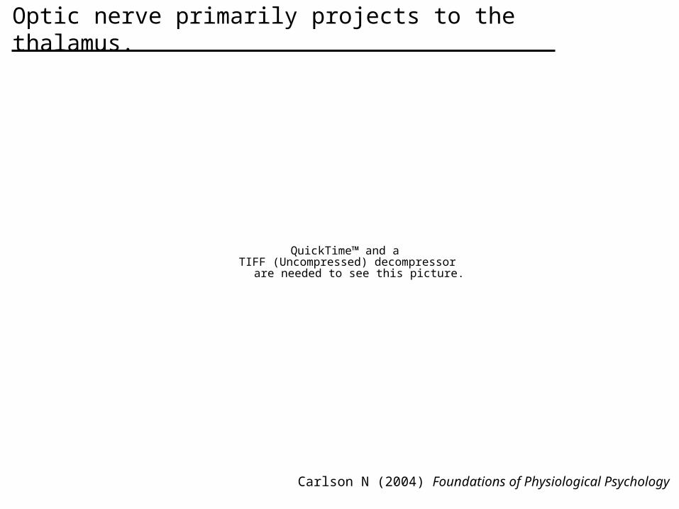
Optic nerve primarily projects to the thalamus.

QuickTime™ and aTIFF (Uncompressed) decompressor
are needed to see this picture.
Carlson N (2004) Foundations of Physiological Psychology
Primary visual cortex is the target of the thalamus.

QuickTime™ and aTIFF (Uncompressed) decompressor
are needed to see this picture.
webvision.med.utah.edu
Eye-specific layers project to segregated regions.
ocular dominance columns
QuickTime™ and aTIFF (Uncompressed) decompressor
are needed to see this picture.
QuickTime™ and aTIFF (Uncompressed) decompressor
are needed to see this picture.
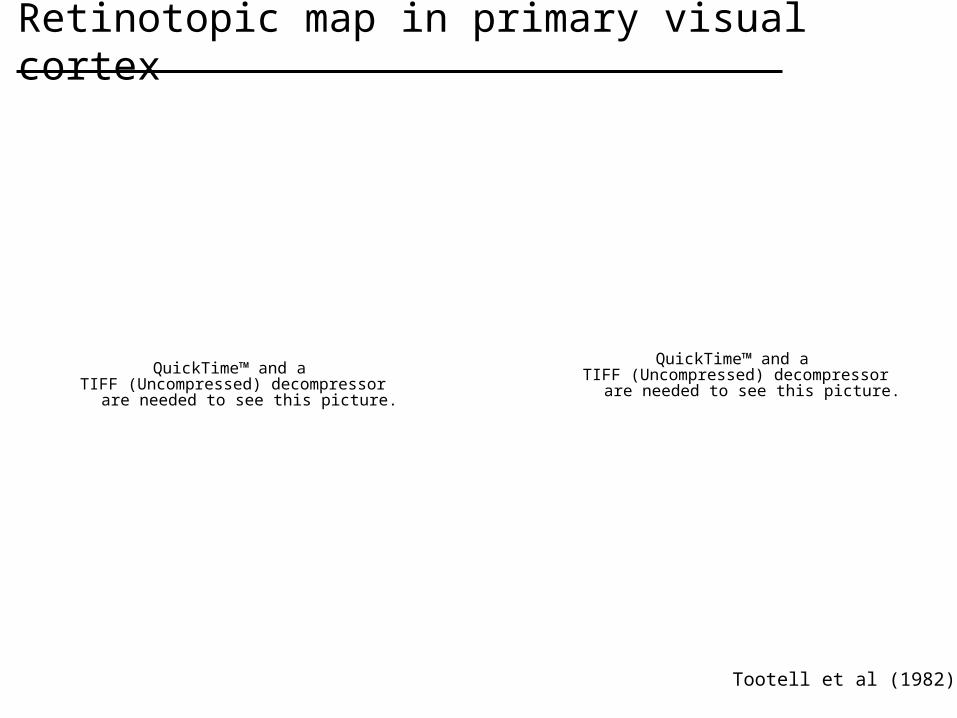
Tootell et al (1982)
Retinotopic map in primary visual cortex

QuickTime™ and aTIFF (Uncompressed) decompressor
are needed to see this picture.
Cortical magnification distorts representation.
webvision.med.utah.edu

QuickTime™ and aTIFF (Uncompressed) decompressor
are needed to see this picture.
Adapted from Hubel & Wiesel (19xx)
Neurons in primary visual cortex are tuned to orientation.
orientation of bar
tuning curve
stimulusstimulus
ONstimulus
OFFstimulus
OFF
response
Firing rate (Hz)

Ohki et al (2006)
The orientation preference of neurons form a topology on visual cortex.
QuickTime™ and aTIFF (Uncompressed) decompressor
are needed to see this picture.
• color represents orientation of tuning curve peak

QuickTime™ and aTIFF (Uncompressed) decompressor
are needed to see this picture.
Carlson N (2004) Foundations of Physiological Psychology
Radial columns are the basic sub-units of the cortex.
QuickTime™ and aTIFF (Uncompressed) decompressor
are needed to see this picture.
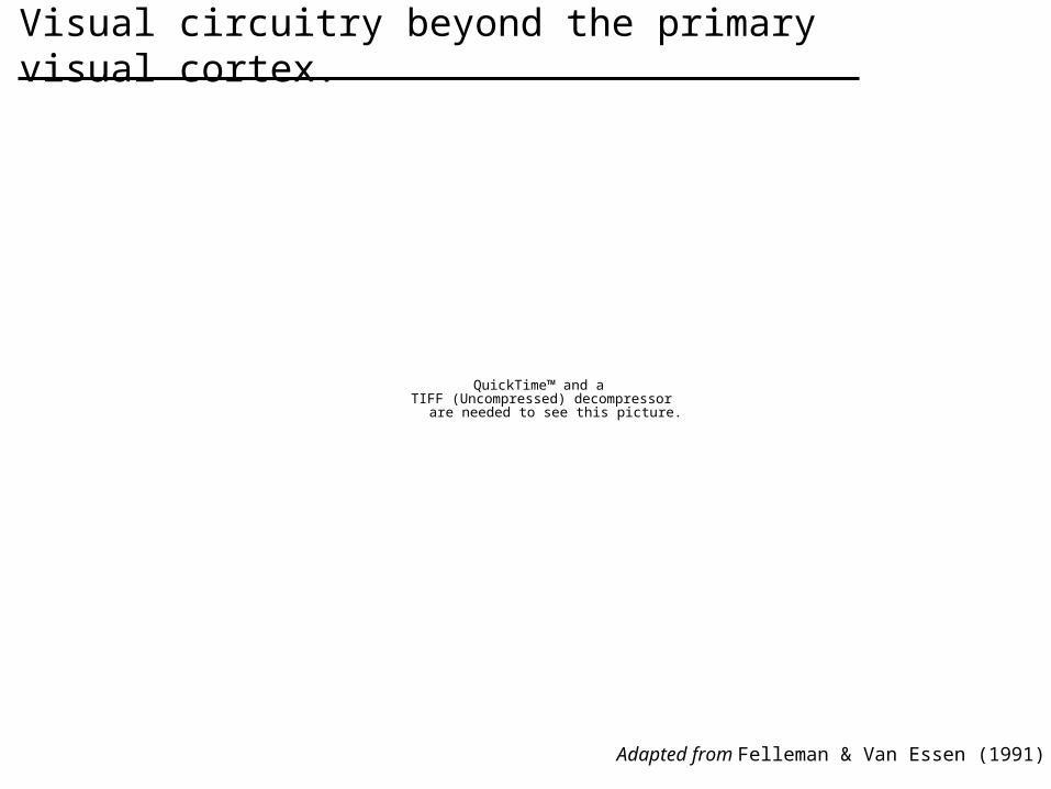
Adapted from Felleman & Van Essen (1991)
Visual circuitry beyond the primary visual cortex.
QuickTime™ and aTIFF (Uncompressed) decompressor
are needed to see this picture.
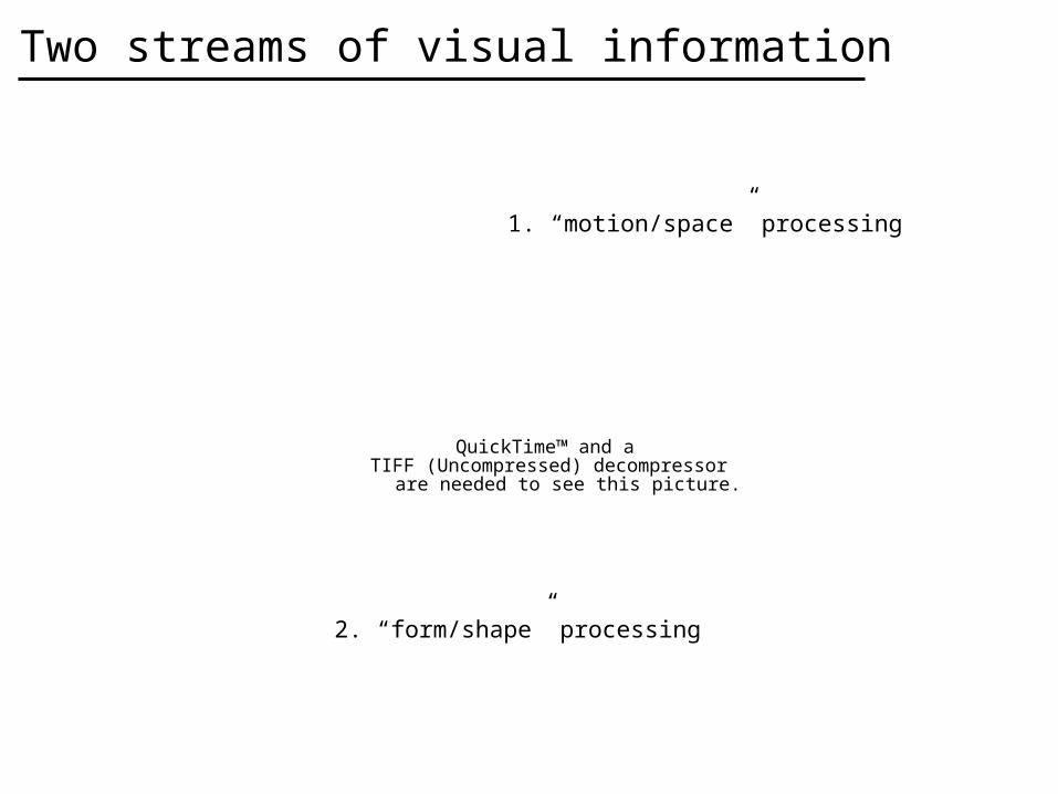
Two streams of visual information
1. “motion/space” processing
2. “form/shape” processing

Albright (1984)
Neurons in the medial temporal (MT) are tuned to motion.
QuickTime™ and aTIFF (Uncompressed) decompressor
are needed to see this picture.
medial temporal (MT) cortex

Albright et al (1984)
Motion direction is topologically organized in MT cortex.
QuickTime™ and aTIFF (Uncompressed) decompressor
are needed to see this picture.

QuickTime™ and aTIFF (Uncompressed) decompressor
are needed to see this picture.
Two streams of visual information
1. “motion/space” processing
2. “form/shape” processing

Desimone et al (1984)
Face-selective cells exist in the inferior temporal (IT) cortex.

The inferior temporal (IT) cortex projects to the hippocampus.

Kreiman et al (2000)
Does the “Bill Clinton” cell exist in the hippocampus?
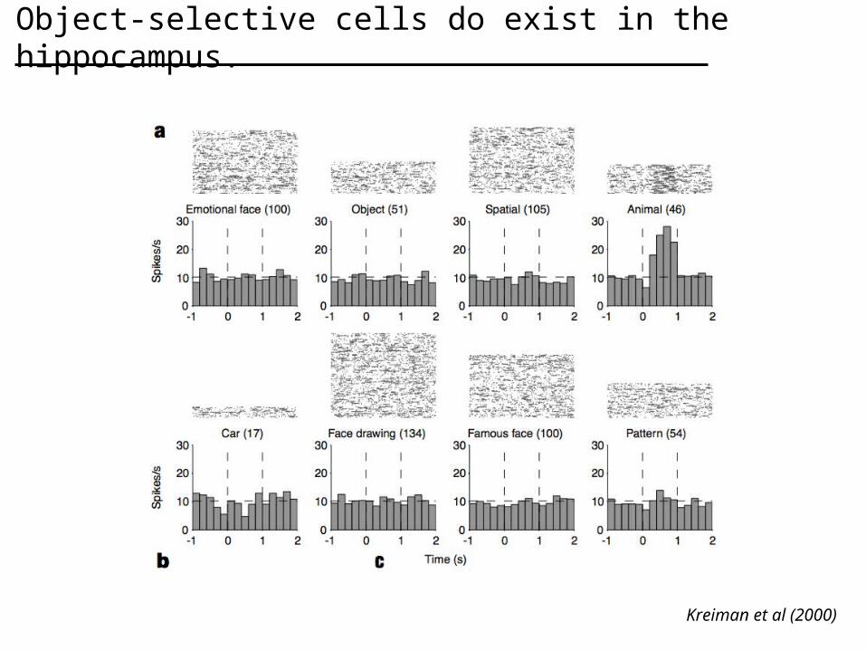
Kreiman et al (2000)
Object-selective cells do exist in the hippocampus.

Outline
1. Introduction
2. Visual processing
3. Memory and plasticity
4. Motor systems

The hippocampus receives input from all sensory modalities.

QuickTime™ and aTIFF (Uncompressed) decompressor
are needed to see this picture.
The basic circuitry of the hippocampus. QuickTime™ and a
TIFF (Uncompressed) decompressorare needed to see this picture.
Ramon y Cajal
QuickTime™ and aTIFF (Uncompressed) decompressor
are needed to see this picture.

QuickTime™ and aTIFF (Uncompressed) decompressor
are needed to see this picture.
Carlson N (2004) Foundations of Physiological Psychology
The basic circuitry of the hippocampus.
QuickTime™ and aTIFF (Uncompressed) decompressor
are needed to see this picture.
Carlson N (2004) Foundations of Physiological Psychology
Synaptic plasticity exists in the hippocampus.
QuickTime™ and aTIFF (Uncompressed) decompressor
are needed to see this picture.
Synaptic plasticity exists in the hippocampus.
Carlson N (2004) Foundations of Physiological Psychology

Robert Mullerhttp://www.bris.ac.uk/depts/Synaptic/research/projects/memory/spatialmem.htm
Neurons in the hippocampus have non-visual receptive fields.
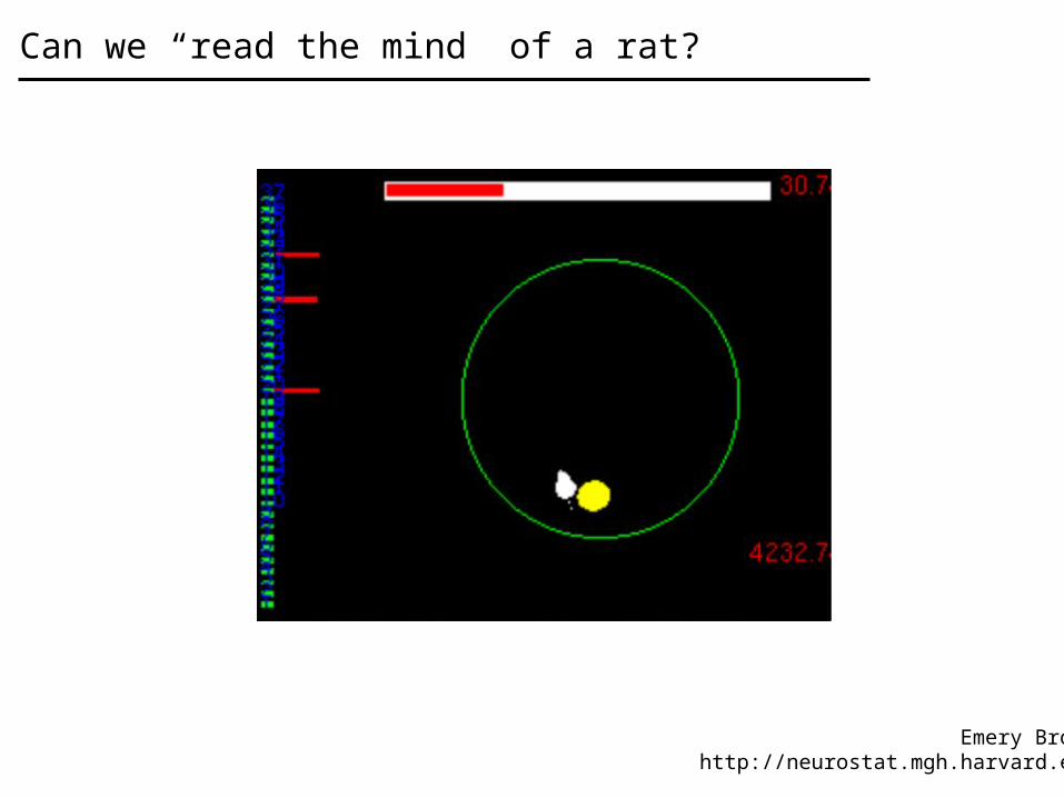
Emery Brownhttp://neurostat.mgh.harvard.edu
Can we “read the mind” of a rat?

QuickTime™ and aTIFF (Uncompressed) decompressor
are needed to see this picture.
www.cyberkinetics.com
Can we “read the mind” of a human?
Outline
1. Introduction
2. Visual processing
3. Memory and plasticity
4. Motor systems

QuickTime™ and aTIFF (Uncompressed) decompressor
are needed to see this picture.
www.brainconnection.com
Motor and decision areas in cortex.

QuickTime™ and aTIFF (Uncompressed) decompressor
are needed to see this picture.
The Brain from Top to Bottomhttp://www.thebrain.mcgill.ca
The primary motor cortex contains a homunculus of body parts.
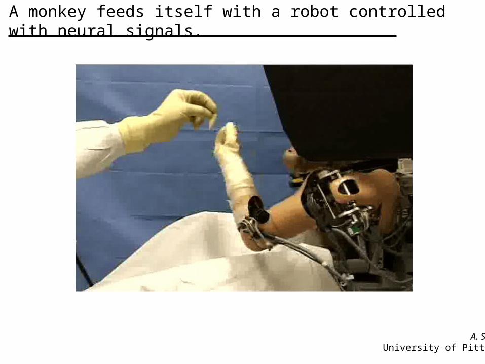
A monkey feeds itself with a robot controlled with neural signals.
A. SchwartzUniversity of Pittsburgh
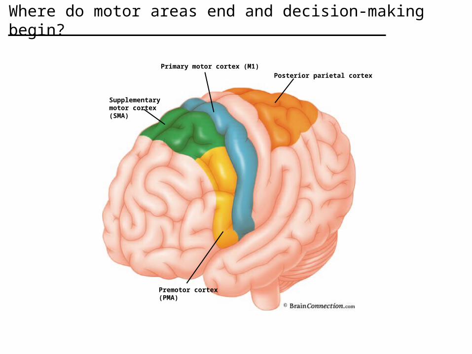
Primary motor cortex (M1)Posterior parietal cortex
Premotor cortex(PMA)
Supplementarymotor cortex(SMA)
Where do motor areas end and decision-making begin?
QuickTime™ and aTIFF (Uncompressed) decompressor
are needed to see this picture.
Richard Andersenhttp://vis.caltech.edu
Designing a neural prosthetic for humans.

QuickTime™ and aTIFF (Uncompressed) decompressor
are needed to see this picture.
www.cyberkinetics.com
Actually, neural prosthetics already exist.

Review
1. Historical perspective• Imhotep
2. Visual processing• Organization of visual cortex
3. Memory and plasticity• Hippocampus and Place Cells
4. Motor systems• Neural Prosthestics
1. A few good classes.
Neuroscience 200A - Cellular NeuroscienceNeuroscience 200B - Systems NeuroscienceNeuroscience 200C - Cognitive Neuroscience
2. A few good books.
Kandel, Schwartz and Jessel (2000) Principles of Neural Science.
Squire et al (2003) Fundamental Neuroscience
3. A few good websites.
“Webvision” http://webvision.med.utah.edu“Neuroscience for Kids”
http://faculty.washington.edu/chudler/neurok.html
Resources

QuickTime™ and aTIFF (Uncompressed) decompressor
are needed to see this picture.
Ventricular system provides cerebrospinal fluid to the brain.