Functional design and evolution of the pharyngeal …glauder/reprints_unzipped/...pharyngeal jaw...
38
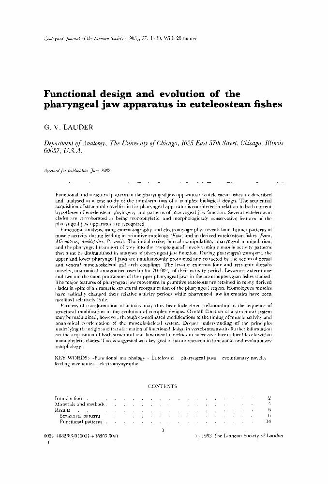
Cuologiral Jorirnal ofthe 1,innmn .COCLU~Y (1983 I, 77: 1 38. \Vith 28 fignres Functional design and evolution of the pharyngeal jaw apparatus in euteleostean fishes G. V. LAUDER Defiaitment of Anatomy, The Unirlersity of Chicago, 1025 East 57th Street, Chicago, Illinois M637, U.S.A. .Icceppledfor publication June 1982 Functional and structural patterns in the pharyngeal jaw apparatus ofeuteleostean fishes are described and analysed as a case study of the transformation of a complex biological design. The sequential acquisition ofstructural novelties in the pharyngeal apparatus is cnnsidered in relation to both current hypotheses of eutrlrostran phylogeny and patterns of pharyngeal jaw function. Several euteleostean cladrs ai-e corroborated as being monophyletic, and morphologically consenzative features of the pharyngeal jaw apparatus are rccognimd. Functional analysis, using cinematography and electromyography, reveals four distinct patterns of muscle activity during feeding in primitive euteleosts (&ox) and In derived euteleostean fishes (Perca, Aficropterus, ilmbloplitrs, Pornoxis). The initial strike, buccal manipulation, pharyngeal manipillation, and the pharyngeal transport of prey into the oesophagus all involve unique muscle activity patterns that must be distinguished in analyses of pharyngeal jaw function. During pharyngeal transport, the upper and lower pharyngeal jaws are simultaneously protracted and retracted by the action of dorsal and ventral musculoskeletal gill arch couplings. The levator externus four and retractor dorsalis muscles, anatomical antagonists, overlap for 70- 90",, of their activity period. Levatores externi one and two are the main protractors of the upper pharyngeal jaws in the acanthopterygian fishes studied. 'Ihe major features of pharyngeal jaw movement in primitive euteleosts are retained in many derived clades in spite of a dramatic structural reorganization of the pharyngeal region. Homologous muscles have radically changed their relative activity periods while pharyngeal jaw kinematics have been modified relatively little. Patterns of transformation of activity may thus bear little direct relationship to the sequence of structural modification in the e\-oliition of complex designs. Overall function of a striicttiral system may he maintained, however. through co-ordinated modifications of the timing of muscle activity and anatomical reorientation of the mnsculoskeletal system. Deeprr rinderstanding of the principles underlying the origin and transformation of function;il dcsian in vertebrates awaits fiirther information on the acquisition of both structural and function;il novelties at successive hicrarchical levels within monophyletic clades. This is suggcrted as a key goal of fiiture research in functional and evolutionary morphology. KEY WORDS :-Functional morphology . Euteleostei pharyngeal jaws evolutionary novelty feeding mechanics electromyography. CONTENTS Introduction . . . . . . . . . . . . . . . . . . . 2 Materials and methods, . . . . . . . . . . . . . . . . 4 Results . . . . . . . . . . . . . . . . . . . . 6 Structural patterns . . . . . . . . . . . . . . . . . 6 Functional patterns . . . . . . . . . . . . . . . . . 14 1 0024- 4082/89!0 10001 + 38503 .00/0 i 1988 The Linnean Society of London 1
Transcript of Functional design and evolution of the pharyngeal …glauder/reprints_unzipped/...pharyngeal jaw...
Cuologiral Jorirnal o f t h e 1,innmn .COCLU~Y (1983 I, 77: 1 38. \Vith 28 fignres
Functional design and evolution of the pharyngeal jaw apparatus in euteleostean fishes
G. V. LAUDER
Defiaitment o f Anatomy, The Unirlersity o f Chicago, 1025 East 57th Street, Chicago, Illinois M637, U.S.A.
.Icceppledfor publication June 1982
Functional and structural patterns in the pharyngeal jaw apparatus ofeuteleostean fishes are described and analysed as a case study of the transformation of a complex biological design. The sequential acquisition ofstructural novelties in the pharyngeal apparatus is cnnsidered in relation to both current hypotheses of eutrlrostran phylogeny and patterns of pharyngeal jaw function. Several euteleostean cladrs ai-e corroborated as being monophyletic, and morphologically consenzative features o f the pharyngeal jaw apparatus are rccognimd.
Functional analysis, using cinematography and electromyography, reveals four distinct patterns of muscle activity during feeding in primitive euteleosts (&ox) and In derived euteleostean fishes (Perca, Aficropterus, ilmbloplitrs, Pornoxis). The initial strike, buccal manipulation, pharyngeal manipillation, and the pharyngeal transport of prey into the oesophagus all involve unique muscle activity patterns that must be distinguished in analyses of pharyngeal jaw function. During pharyngeal transport, the upper and lower pharyngeal jaws are simultaneously protracted and retracted by the action of dorsal and ventral musculoskeletal gill arch couplings. The levator externus four and retractor dorsalis muscles, anatomical antagonists, overlap for 70- 90",, of their activity period. Levatores externi one and two are the main protractors of the upper pharyngeal jaws in the acanthopterygian fishes studied. ' Ihe major features of pharyngeal jaw movement in primitive euteleosts are retained in many derived clades in spite of a dramatic structural reorganization of the pharyngeal region. Homologous muscles have radically changed their relative activity periods while pharyngeal jaw kinematics have been modified relatively little.
Patterns of transformation of activity may thus bear little direct relationship to the sequence of structural modification in the e\-oliition of complex designs. Overall function of a striicttiral system may he maintained, however. through co-ordinated modifications of the timing of muscle activity and anatomical reorientation of the mnsculoskeletal system. Deeprr rinderstanding of the principles underlying the origin and transformation of function;il dcsian in vertebrates awaits fiirther information on the acquisition of both structural and function;il novelties a t successive hicrarchical levels within monophyletic clades. This is suggcrted as a key goal of fiiture research in functional and evolutionary morphology.
KEY WORDS :-Functional morphology . Euteleostei pharyngeal jaws evolutionary novelty feeding mechanics electromyography.
CONTENTS
Introduction . . . . . . . . . . . . . . . . . . . 2 Materials and methods, . . . . . . . . . . . . . . . . 4 Results . . . . . . . . . . . . . . . . . . . . 6
Structural patterns . . . . . . . . . . . . . . . . . 6 Functional patterns . . . . . . . . . . . . . . . . . 14
1 0024- 4082/89!0 10001 + 38503 .00/0 i 1988 The Linnean Society of London
1
2 G. \'. L.4UDER
1)iscussiori . . . . . . . . . Ph>lugcnrtic 1ia"ttnis . . . . . Fiiiirtional morphologv . . .
Fiinction,tl design and c'\ olulion.ir.) Ixtttcriis Chrnparisiiiis . . . . . . .
.\c.kno~~letlgrnients. . . . . . . Kr Frrcnccs . . . . . . . . . \l)ltr.c.\ i i i~ioi is w r d in figures . . . .
. . . . . . . . . . . 21 . . . . . . . . . . . 21 . . . . . . . . . . . 27 . . . . . . . . . . . :I1 . . . . . . . . . . . 32
. . . . . . . . . :35 . . . . . . . . . . . 36 . . . . . . . . . . . :17
I N T K O I ~ U ( ~ 1 I O N
0 1 ' the two major tliemes in the history of morphology, functional design and di\.ersity of' form, fiinctional morphologists have focused most intensively on organismal design. The diversity of morphology and its underlying theme of unity of type has, since the late nineteenth century, been the province mostly of phylogenetic research (Russell, 1916) which has run the gamut fi-om reconstruction of ancestral morphotypes to modern cladistic analysis.
Functional design in organisms h a s been investigated primarily in relation to two goals. First, how do organisms work? For example, what are the mechanisms by which movement occurs during locomotion, feeding, or breathing in \wtebrates? Bioniechanical research of this type has benefited enormously from the infusion of new experimental techniques such as electromyography, cinematography, and pressure and strain recording devices. Experimental analyses are 110 longer limited by inferring muscle activity patterns or fluid pressures from honc kinematic patterns. ;I second goal of research in fiinctional morphology has been to clarify the
relationship between organisms and the environment (Gans, 19743. How do different patterns of actiiity exhibited by organisms relate to the environments thcy inhabit and fluctuations of those environments? :I frequently claimed aim of fiiiictional analyses of organism environment interactions is the inference of selecti\.e pressures that have go\:erned the origin of morphological novelties.
One approach to the imilysis of biological design that has yet to be adeq riately explored concerns the historical origin and transformation of structure arid function. The methodology of structural analysis in historical biology has been iri\,estigated in detail over the last 10 years (Eldredge & Cracraft, 1980; Nelson & Platnick, 1981 ; Wiley, 1981 ; I a i d e r , 1982) and has emphasized the reconstruction of historical changes in morphology and geographic distribution. But the historical relationship between the sequential acquisition of structural and fiinctional novelties remains to be examined. Few investigations have analysed pat terns of striictural and fiinctional no\dties at successive hierarchical levels within ii rnonophyletic clade.
In this paper I present a case study in the transformation of functional design that involves both structural analysis within an explicitly phylogenetic context and ~ l i c experimental determination of fiinctional patterns at se\reral hierarchical le\.els. The phxyngcal jaw apparatus, a complex set of modified gill arches and associated muscles and ligaments in the pharynx of ray-finned fishes, is the structural system chosen for study. A general goal is to provide a foundation for e\.aluating the historical relationship between structural and functional novelties, especially in regard to the origin of complexity of' design.
'Hie phylogenctic hypothesis that serves as the basis for this paper is the cladogram of euteleostean fish relationships first proposed by Rosen ( 1973) and
I~UTELE0STE:IS PH:\RYN(:EhL JA”S 3
subsequently modified by Fink & Weitzman (1982). A summary cladogram is presented in Lauder tk Liem (in press). According to this hypothesis, the euteleostean fishes form a monophyletic lineage of about 17 000 species united by the possession of three derived characters : an adipose fin, breeding tubercles, and the structure of caudal uroneurals (Patterson & Rosen, 1977). The most primitive euteleostean clade is the Esocae (Fink & Weitzman, 1982) containing the pikes and pickerels. The interrelationships of other primitive euteleosteans are still poorly known and the Ostariophysi, Argentinoidei, Osmeroidei, Salmonidae and the neoteleostean fishes are grouped in an unresolved polychotomy near the base of the Euteleostei. The clade Neoteleostei is well-defined and includes the deep-sea stomiatoids (\iperfishes 1, aulopiforms, myctophids (hatchetfishes’), paracan- thopterygians (cods and batrachoid fishes, among others), atherinomorphs, and percomorph fishes (a summary cladogram is presented in Fig. 28). It is important to note that the cladistic branching pattern within the Eiiteleostei is corroborated by the distribution of morphological features other than those found in the pharyngeal region. A non-circular approach to the historical analysis of design requires a cladogram to be corroborated by structural features other than those investigated experimentally.
Given an initial corroborated phylogenetic hypothesis, two fiirther steps are involved in this analysis of pharyngeal design in fishes. First, a comparative morphological in\,estigation of the pharyngeal jaw apparatus (especially branchial myology) in each of the major euteleostean clades is conducted to establish the sequence and nature of structural change during euteleostean evolution. Mapping the structural specializations in each terminal taxon onto the initial phylogenetic hypothesis reveals both features primitive for the Euteleostei and specializations of the pharyngeal jaw apparatus, if any, at each hierarchial level (Lauder, 1981 ). Second, the experimental study of pharyngeal jaw function in terminal taxa within the Euteleostei allows functional specializations (such as a unique muscle activity pattern) to be identified for each clade. Muscle acti\.ity patterns are treated in the same way as structural spccializations and mapped, using parsimony, onto the initial phylogenetic hypothesis. This procedure reveals the distribution of functional novelties within the Euteleostei. Correlations or general relationships between structural and fiinctional novelties at each hierarchical level can then be examined and used to test general explanations for the origin and transformation of ‘character complexes’, or the modification of central neural ‘programmes’ in relation to peripheral structural systems. Higher level historical hypotheses about the design of organisms can best be tested by comparing the predictions of such hypotheses against inferred sequences of structural and fiinctional modification.
The analysis of e\mlutionary patterns in the pharyngeal jaw apparatus of fishes has been hindered both by the lark of a rigorous descriptive framework and by the scarcity of experimental data on generalized taxa. Cichlid pharyngeal jaw mechanisms have been studied in detail (Liem, 1973, 1978) but comparative data are lacking on generalized non-pharyngognath teleosts. Without such data, it is impossible to know which muscle activity patterns, for example, are derived for pharyngognath fishes and which are general acanthopterygian or teleost features.
’The problem of defining a relevant descriptive classification of fiinctional activity is analogous to the difficulties faced by mammalian functional morphologists in dividing the masticatory cycle into biologically relevant segments (Hiiemae, 1978). The aim of any functional classification is to reflect accurately biological
4 G V L4UDER
events and to order these events into natural clusters that facilitate interspecific comparisons. Unless distinct movements are recognized and divided into phases based on consistent kinematic patterns and muscle activities, biologically meaningful events will be hidden in a single highly variable descriptive class. The evaluation of chewing mechanics and the transformation of functional design is crucially dependent on an accurate division of organismal activity into natural units .
In this paper, I will use the classification presented in Lauder (in press), and describe electromyographic patterns that uniquely characterize each movement pattern. Four separate aspects of fish feeding behaviour have been identified: the initial strike, buccal manipulation, pharyngeal manipulation, and pharyngeal transport of prey into the oesophagus. Each of these four movements is distinct and recognizable by either the pattern of jaw bone movement or muscle activity, and the presence of three activity patterns following the initial capture of prey appears to be a primitive feature of euteleostean fishes.
M VIERIALS AND METHODS
Preserved specimens of each of the major euteleost lineages were examined for morphological data on the pharyngeal jaw apparatus. The clades examined include the Esocae, Ostariophysi, Salmonae, Argentinoidei, Osmeroidei, Stomiiformes, Aulopiformes, Myctophiformes, Paracanthopterygii, Atherino- morpha and Percomorpha. In some lineages such as the Ostariophysi and Stomiiformes, species currently hypothesized to be primitive (e.g. Chanos and Dzplophos) were selected for detailed analysis, and only certain characters were examined in more derived species. In lineages such as the Myctophiformes a broad range of comparative material was examined. Only the acanthopterygian species studied experimentally were dissected in detail. The genera, species, and museum numbers for specimens examined are available from the author.
Experimental analysis of feeding behaviour was conducted using high-speed cinematography and electromyography. A Photosonics 16- 1 PL high-speed camera was used in conjunction with Kodak 4X Reversal film to studyjaw bone movement. Electromyographic signals were recorded through fine wire (0.05 1 mm) steel alloy bipolar electrodes as described previously (Lauder, 1980a), and recorded on a six- channel Bell and Howell 4020A FM tape recorder. Grass P511J preamplifers were used with the low pass filter set at either 30Hz or 100 Hz and the high pass filter at SOOOHz. The 60Hz notch filter was used at all times. Electromyograms were recorded at 37.5 cm s-' and played back at 4.7 cm s - ' through a Gould 260 chart recorder.
X-ray cinematographic data were available for the genus Lepomis (discussed in a forthcoming paper) and the results of that analysis in addition to high-speed films were used to establish the kinematic patterns associated with muscle activity.
A wave generator was used to test the frequency response of the entire electromyographic apparatus. Frequencies varying from 100 to 1000 Hz were accurately reproduced on the chart recorder after being amplified, recorded on FM tape and played back. Thus little loss in muscle signal amplitude should occur since the peak power of striated muscle electromyograms lies between 100 and 300 Hz.
Muscle electrical activity was analysed by the following procedure. A reference
EL~l’F.LEOSI‘EAN PHARYNGEAL JAWS 5
muscle was chosen for each of the four categories of feeding behaviour outlined in the Introduction: initial strike and buccal manipulation-sternohyoideus; pharyngeal manipulation-obliquus inferioris; pharyngeal transport-retractor dorsalis. These reference muscles were selected on the basis of preliminary experiments and previous research which showed that they had a clearly definable action, consistent activity pattern, and lacked significant asymmetry in activity between the right and left sides. The onset of activity in the reference muscle was taken as the ‘zero point’ from which the onset and offset of activity in all muscles was determined (see Figs 13, 14 & 16 for examples). Mean times of onset and offset relative to reference muscle onset were calculated as was the standard error for each mean. This procedure is similar to that of,Jenkins & Wei.js (1979). Each bar diagram of muscle activity represents a summary of at least 15 recordings for each muscle, and often considerably more. For the analysis of pharyngeal transport, a repetitive cyclical process, two sequential bursts of activity in the reference muscle were chosen as the standard for comparison (e.g. Fig. 16).
Because choosing the onset of activity in one muscle limits the contribution that \?xiability in the onset time of this muscle makes to the total electromyographic pattern, histograms of burst duration were constructed (e.g. Fig. 19) to illustrate the variability in total activity period of the reference muscles. In the case of the retractor dorsalis, burst duration was very similar in distribution to that of other muscles, while the sternohyoideus and obliquus inferioris showed considerably less variation in activity duration. The summary diagrams presented in this paper include variability between individuals, different prey types, and activity from different times in the chewing cycle.
Electrodes were implanted in the pharyngeal muscles while the experimental subject was anaesthetized as described in Lauder (1980a). Each of the pharyngeal muscles with the exception of the retractor dorsalis could be identified visually and the electrode placed through the thin mucous membrane over the gill arch muscles directly into the muscle belly. At least one of the reference muscles (sternohyoideus, obliquus inferioris, or retractor dorsalis) was always implanted. i2t the end of a series of experiments on a particular fish, the location of the electrode tips was confirmed by dissection.
Six species were studied experimentally : Microperus salmoides (Lacepkde) ( 3 ) , Im6loplite.r rupestrij (Rafinesque) (6) , Pomoxi,r nigromaculatus (Lesueur) and P.
annularis (Rafinesque) ( 2 each) (all in the perciform family Centrarchidae), Perca Jauescens (Mitchill) (2) , and Esox niger (Lesueur) (4). (The number in parentheses represents the number of individuals examined.) ‘4 wide variety of prey was used to assess the dependence of muscle activity pattern on prey type and size: earthworms (Z,umb~icus), crayfish (Orconectes), golden shiner (. \otemigonus cl;vsoleuca.c (Mitchill) , Lthead minnows (Yime/)hale.r fmme1a.r Rafinesque), goldfish (Carassius auratus (L.) j, and emerald shiners (.2btropi.c. atherinoides Rafinesque).
This paper will focus on the pattern of functional activity in the most primitive euteleostean clade, the Esocae (as represented by Esox niger) in comparison to the pattern exhibited by acanthopterygian teleosts (Micropterus, Ambloplites, Pomoxis and Perca) . Several euteleostean clades are not easily analysed experimentally hecause most species 1k.e in relatively inaccessible open ocean or deep sea habitats (e.g. Stomiiformes, Aulopiformes, Myctophiformes). Experimental data on the Salmonidae, Paracanthopterygii and htherinomorpha will not be reported here. Monophyly of the Paracanthopterygii is not well established (Lauder & Liem, in
6 G. V. I,,\UDER
press) and the precise phylogenetic relationships of the Salmonidae and i2therinomorpha have yet to be ascertained.
KESLILTS
Structural patterns
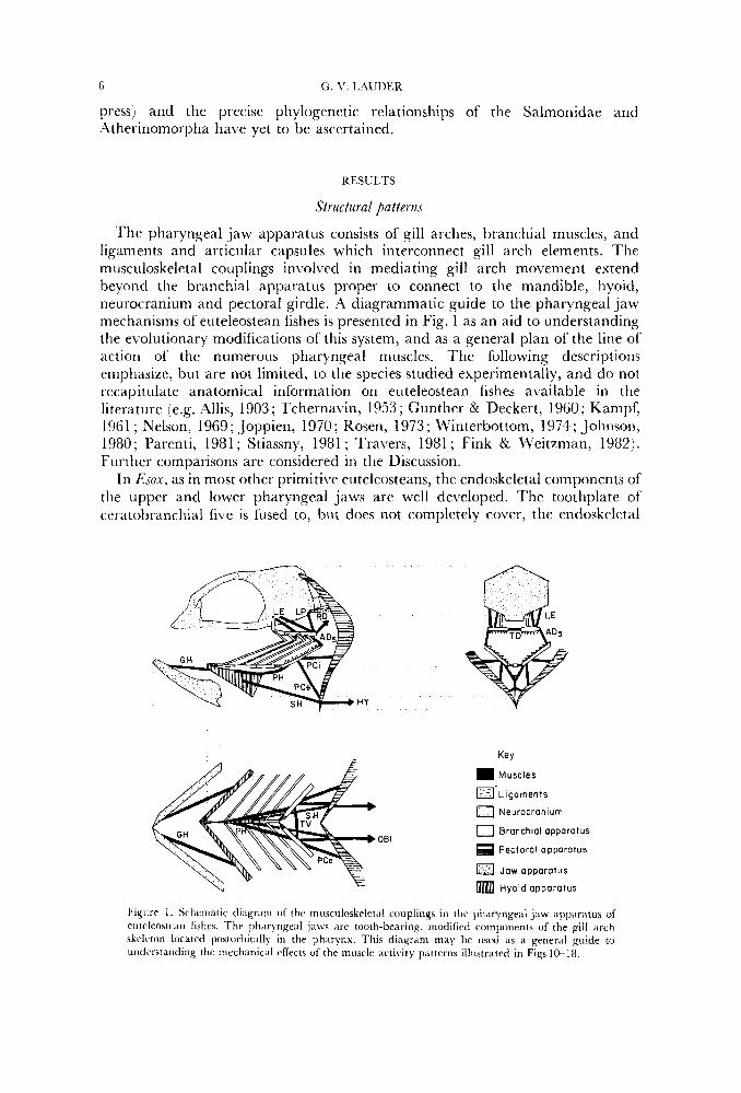
The pharyngeal jaw apparatus consists of gill arches, branchial muscles, and ligaments and articular capsules which interconnect gill arch elements. The musculoskeletal couplings involved in mediating gill arch movement extend beyond the branchial apparatus proper to connect to the mandible, hyoid, neurocranium and pectoral girdle. A diagrammatic guide to the pharyngeal jaw mechanisms of euteleosteari fishes is presented in Fig. 1 as an aid to understanding the evolutionary modifications of h i s system, and as a general plan of the line of action of the numerous pharyngeal muscles. The following descriptions emphasize, but are not limited, to the species studied experimentally, and do not recapitulate anatomical information on euteleostean fishes available in the literature (e.g. Allis, 1903; Tchernavin, 1953; Gunther & Deckert, 1960; Kampf, 1961 ; Nelson, 1969; Joppien, 1970; Rosen, 1973; Winterbottom, 1974; Johnson, 1980; Parenti, 1981; Stiassny, 1981; Travers, 1981; Fink & Weitzman, 1982). Fiirther comparisons are considered in the Discussion.
In Esox, as in most other primitive euteleosteans, the endoskeletal components of the upper and lower pharyngeal jaws are well developed. The toothplate of ceratobranchial five is fused to, but does not completely cover, the endoskeletal
Key
Muscles
='Ligaments
0 Neurocranium
0 Bronchial apparatus
Pectoral apparatus
61 Jaw apparatus
Hyoid apparatus
Figure 1 . Schrm;iLic diagram of t h c musculoskrlct'il couplings in the pharyngeal jaw a p p m t u s of eutelrostean fishes. T h r pharyngeal jaws arc tooth-bearing, modified components of the gill arch skclrton located postorl)itally in thr pharynx. This diagram may he used as a general guide to understanding the rnechanicd r k t s of the muscle activity patterns illustrated in Figs 10-18.
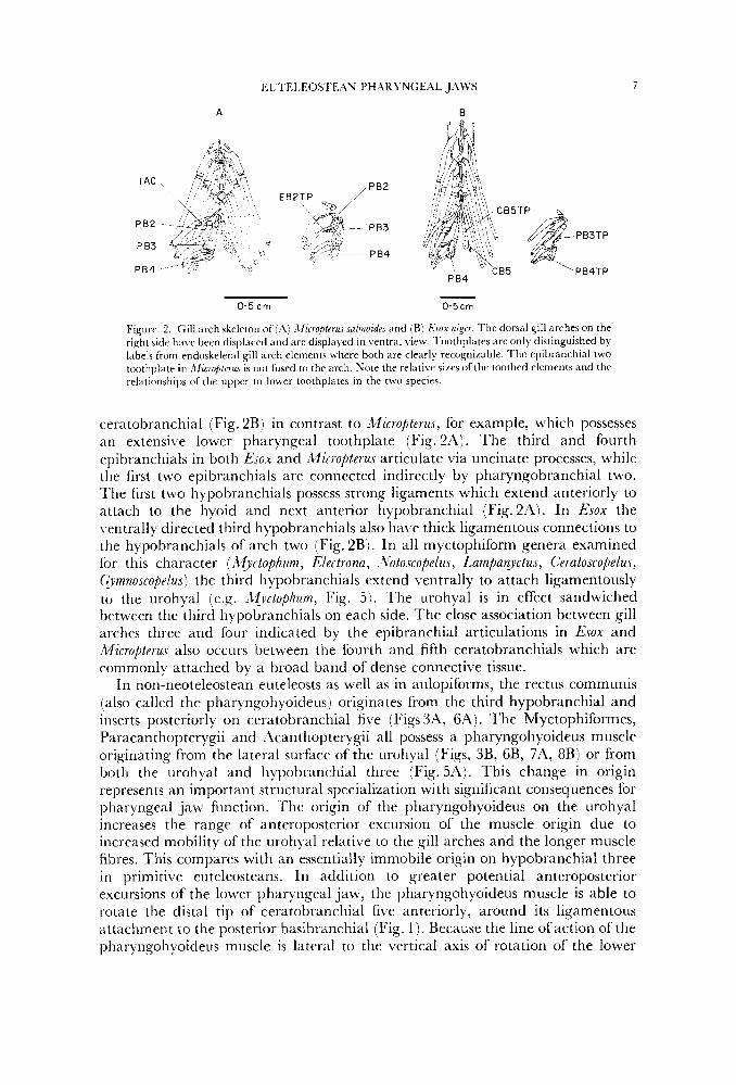
EL1'EI,EOSI'FAN PHhRYNGEAL JAWS
A B
7
- 0.5 cm 0.5cm
Figure 2. Gill arch skeleton of ( A : .\ficrofiterus snlrnoidtr and (B) Esox nigrr. The dorsal gill arches on the right side have been displaced and are displayed in ventral view. l'oothplates are only distinguished by labels from endoskeletal gill arch elements where both are clearly recognizable. The epibranchial two toothplate i n .2fic7ofilrrus is not firsed to the arch. Sote the relative sizes of the toothed elements and the relationships ofthe upper to h e r toothplates in the two species.
ceratobranchial (Fig. 2B) in contrast to Micropterus, for example, which possesses an extensive lower pharyngeal toothplate (Fig. 2A). The third and fourth epibranchials in both Esox and Micropterus articulate via uncinate processes, while the first two epibranchials are connected indirectly by pharyngobranchial two. The first two hypobranchials possess strong ligaments which extend anteriorly to attach to the hyoid and next anterior hypobranchial (Fig.2'4). In Esox the ventrally directed third hypobranchials also have thick ligamentous connections to the hypobranchials of arch two (Fig. 2B). In all myctophiform genera examined for this character (Myctophum, Electrona, ~ Zbtoscopelus, Lampanyctus, C'eratoscopelu.s, (&.noscopelus) the third hypobranchials extend ventrally to attach ligamentously to the urohyal (e.g. ;Wyctophum, Fig. 5 ) . 'The urohyal is in effect sandwiched between the third hypobranchials on each side. The close association between gill arches three and four indicated by the epibranchial articulations in Esox and Micropterus also occurs between the fourth and fifth ceratobranchials which are commonly attached by a broad band of dense connective tissue.
In non-neoteleostean euteleosts as well as in aulopiforms, the rectus communis (also called the pharyngohyoideus) originates from the third hypobranchial and inserts posteriorly on ceratobranchial five (Figs IIA, 6A). The Myctophiformes, Paracanthopterygii and Acanthopterygii all possess a pharyngohyoideus muscle originating from the lateral surface of the urohyal (Figs, 3B, 6B, 7A, 8B) or from both the urohyal and liypobranchial three (Fig. 511). This change in origin represents an important structural specialization with significant consequences for pharyngeal jaw function. The origin of the pharyngohyoideus on the urohyal increases the range of anteroposterior excursion of the muscle origin due to increased mobility of the urohyal relative to the gill arches and the longer muscle fibres. This compares with an essentially immobile origin on hypobranchial three in primitive euteleosteans. In addition to greater potential anteroposterior excursions of the lower pharyngeal jaw, the pharyngohyoideus muscle is able to rotate the distal tip of ceratobranchial five anteriorly, around its ligamentous attachment to the posterior basibranchial (Fig. 1 ) . Because the line ofaction of the pharyngohyoideus muscle is lateral to the vertical axis of rotation of the lower
8 C;. \’. I..\LJL)EK
plinryrigeal jaw at its attdchment to the hasibranchials, anterior rotation of ceratobranchial five will occur.
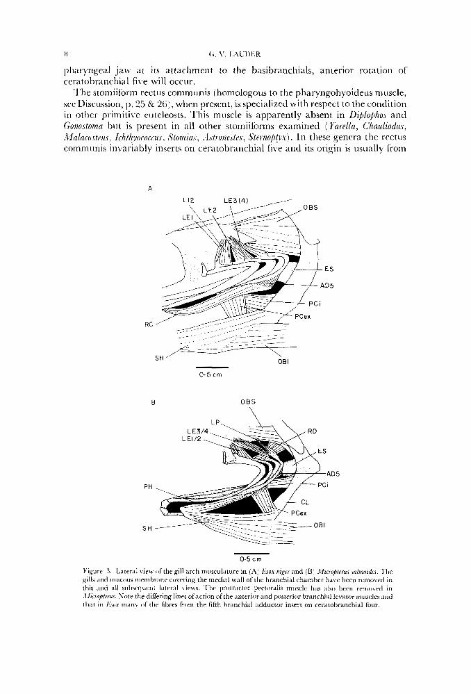
‘l’he stomiiform rect 11s communis [homologol~s to the pharyngohyoideus muscle, ser Discussion, 11.25 & 26), when present, is specialized with respect to the condition in other prirnitiL e euteleosts. This muscle is apparently absent in DiplophoJ and Gonostoma but is present in all other stomiiforms examined ( Yarella, Chauliodus, MnlaeoctPur, Ichthyococcu, StomiaJ, . l ~ t r o n e ~ t e ~ , Sternob!yx). In these genera the rectiis communis invariably inserts on ceratobranchial five and its origin is usually from
A
E S
D5
RC ’ -__ - ,_- __ - - -_ __---
>LJ- ___- --
08 I SH - 0.5 cm
B 00s
PH
S H
0.5 crn
Figure 3. Lateral view of the gill arch musculature in (A) Ksox n Z . y and (B) . lzcroplrrus .sn/moidus. ‘Ihe gills and mucous nimibranr covering the medial wall of the branchial chamber have been removed in this Icnd all sul~acrlr~rnt Iritcral \ icws. Tlrc protractor pectoralis muscle lliis a lso bccrl rcnrovetl in .\fiooptcrus. Note the differing lines ofaction of the anterior ;ind posterior branrhial levator rnusclcs m d t h a t in fi.:~ox n i m y of the fibres from the fifth hranchial adductor insert on ceratobranchial four.
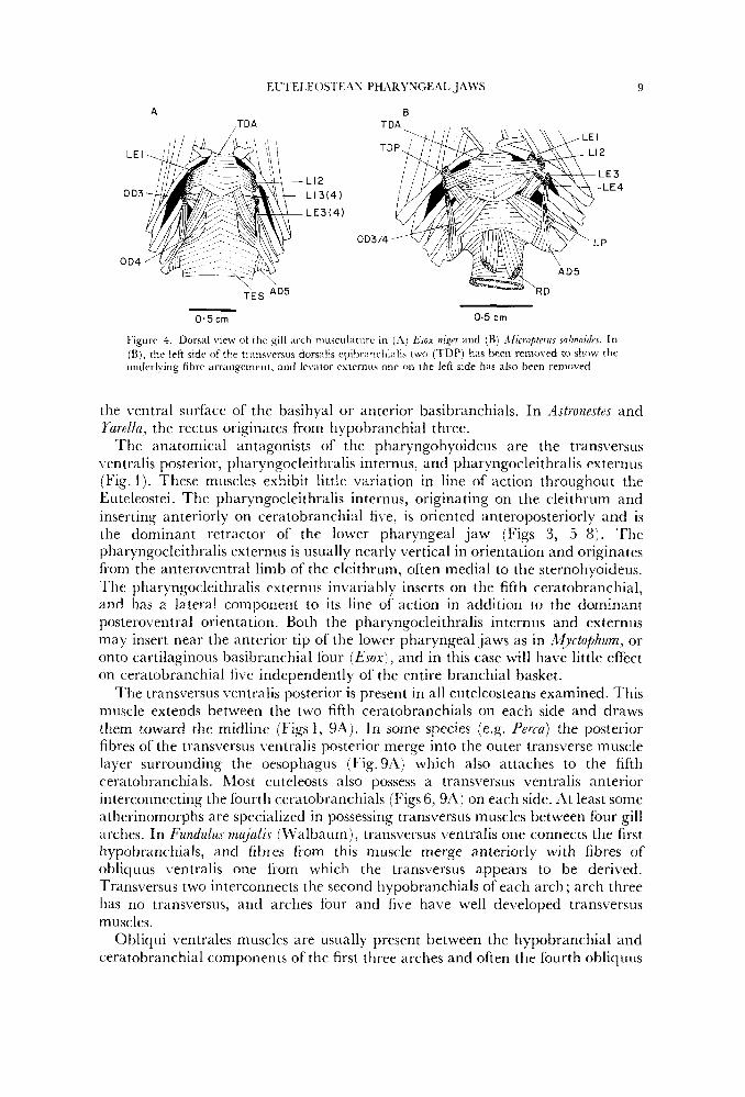
EU 1 EI EOS I E .\U PH.IRYUCt4L JAWS 9
A 8
L12 L13(4)
LE3(4 )
- 0.5 cm 0.5 cm
Figure 1. Dorsal view of the gill arch ttiusciilaturc in (.4) &ox n i p and ( R ) ,Ilicropte7us snl~~nozdus. In (Ki, rhr lrfi side of the tr;iiisverxis dorsilis ~ ~ ~ i t ~ r ~ ~ ~ ~ ~ ~ l l i ~ ~ l i s two (TDP) has been removed to show the underlying fibre arrangement, and Irvdtor externus onc 011 the left side has also been removed.
the ventral surface of the basihyal or anterior basibranchials. In Astrunestes and Yardla, the rectus originates from hypobranchial three.
The anatomical antagonists of the pharyngohyoideus are the transversus \.entralis posterior, pharyngocleithralis internus, and pharyngocleithralis externus (Fig. 1 ) . These muscles exhibit little variation in line of action throughout the Euteleostei. The pharyngocleithralis internus, originating on the cleithrum and inserting anteriorly on ceratobranchial five, is oriented anteroposteriorly and is the dominant retractor of the lower pharyngeal jaw (Figs 3 , 5--8). The pharyngocleithralis externus is usually nearly vertical in orientation and originates from the anteroventral limb of the cleithrum, often medial to the sternohyoideus. The pharyngocleithralis externus invariably inserts on the fifth ceratobranchial, and has a lateral component to its line of action in addition to the dominant posteroventral orientation. Both the pharyngocleithralis internus and externus may insert near the anterior tip of the lower pharyngeal jaws as in Myctuphum, or onto cartilaginous basibranchial four ( E s o x ) , and in this case will have little effect on ceratobranchial five independently of the entire branchial basket.
The transversus ventralis posterior is present in all euteleosteans examined. This muscle extends between the two fifth ceratobranchials on each side and draws them toward the midline (Figs 1 , 9'4). In some specks (e.g. Perm) the posterior fibres of the transversus ventralis posterior merge into the outer transverse muscle layer surrounding the oesophagus (Fig. 9A) which also attaches to the fifth ceratobranchials. Most euteleosts also possess a transversus Lrentralis anterior interconnecting the fourth ceratobranchials (Figs 6, 9Aj on each side. At least some atherinomorphs are specialized in possessing transversus muscles between four gill arches. In Fundulus majalis (Walbaum), transversus ventralis one connects the first hypobranchials, and fibres from this muscle merge anteriorly with fibres of obliquus ventralis one from which the transversus appears to be derived. Transversus two interconnects the second hypobranchials of each arch ; arch three has no transversus, and arches four and five have well de\reloped transversus muscles.
Oblicjui ventrales muscles are usually present between the hypobranchial and ceratobranchial components of the first three arches and often the fourth obliquus
'I..
0.5 cm
OBS
- R D 0 D4
PC I
PCex TES
0 . 5 c m
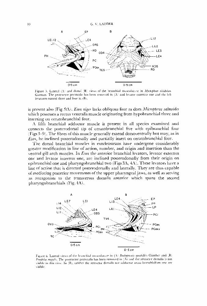
Figure 5. Lateral 1.4; and dorsal (Bi views of the branchial niusculature in .C!vctuphum riitidulurn C,irman. Thr protractor prctor,ilis has tiern rrmmcd in (.I'm arid le\.ator externiis one and the lcft levatores externi three and four in [Bl.
is present also (Fig. 9:Ij. Esox niger lacks obliquus four as does Micropterus sa1moide.r which possesses a rectus ventralis muscle originating from hypobranchial three and inserting on ceratobranchial four.
A fifth liranchial adductor muscle is present in all species examined and connects the posterodorsal tip of ceratobranchial five with epibranchial four (Figs 3-9). The fibres of this muscle generally extend dorsoventrally but may, as in Esox, be inclined posterodorsally and partially insert on ceratobranchial four.
'l'he dorsal branchial muscles in enteleosteans have undergone considerably greater modification in line of action, number, and origin and insertion than the ventral gill arch muscles. In Esox the anterior branchial levators, levator externus onc and levator internus one, are inclined posterodorsally from their origin on cpibranchial one and pharyngobranchial two (Figs 3A, 4A). 'lhese levators have a line of action that is directed posterodorsally and laterally. They are thus capable of mediating posterior movements of the upper pharyngeal jaws, as well as serving as antagonists to the transversus dorsalis anterior which spans the second pharyngobranchials (Fig. 4'4).
F,L'1'F,I,EOSTE;IN PHARYS'GEAL J.4WS 11
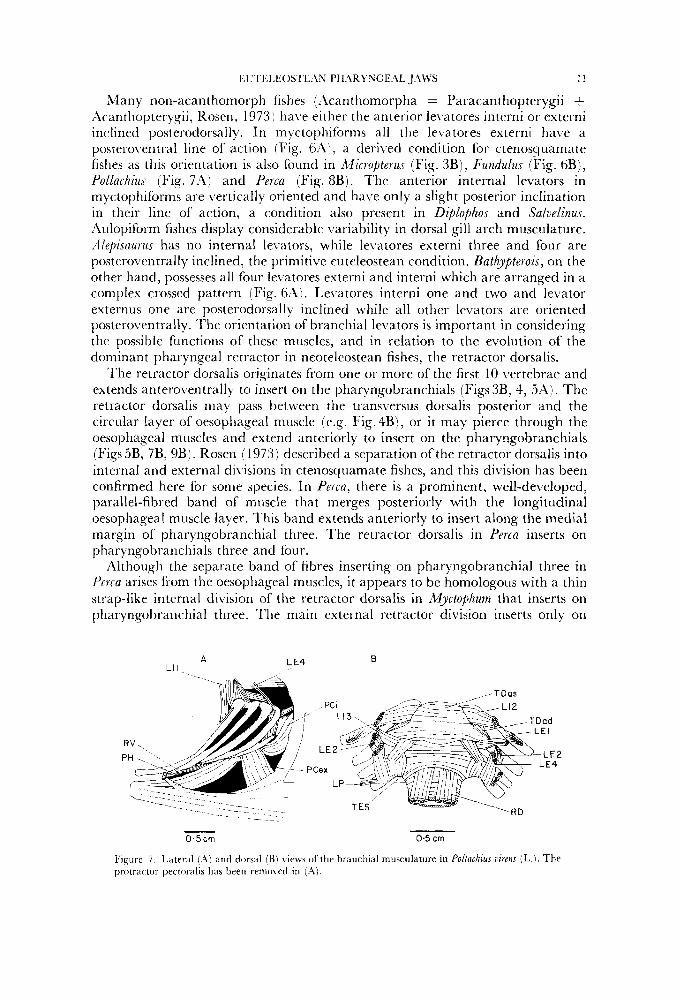
Many non-acanthomorph fishes (A4canthomorpha = Paracanthopterygii + Xcanthopterygii, Rosen, 1973 j 1iaL.e either the anterior le\.atores interni or externi inclined posterodorsally. In myctopliiforms all the levatores externi have a postero\.entral line of action (Fig. 6:1), a derived condition for ctenosquamate fishes as this orientation is also found in Microjiterus (Fig. 3B), Fundulus (Fig. 6B), Pollachius (Fig. 7'4) and Perca (Fig. 8B). The anterior internal levators in myctophiforms are vertically oriented and have only a slight posterior inclination in their line of action, a condition also present in Diplophos and Salvelinus. Aulopiform fishes display considerable \.ariability in dorsal gill arch musculature. :llepisaui-us has no internal levators, while le\.atores externi three and four are posteroventrally inclined, the primitive euteleostean condition. Bathypterois, on the other hand, possesses all four levatores externi and interni which are arranged in a complex crossed pattern (Fig. 6Ai. Lelratores interni one and two and levator externus one are posterodorsally inclined while all other levators are oriented posteroventrally. The orientation of branchial levators is important in considering the possible functions of these muscles, and in relation to the evolution of the dominant pharyngeal retractor in neoteleostean fishes, the retractor dorsalis.
The retractor dorsalis originates from one or more of the first 10 vertebrae and extends antero\rentrally to insert on the pharyngobranchials (Figs 3B, 4, 5A). The retractor dorsalis may pass between the transversus dorsalis posterior and the circular layer of oesophageal muscle (e.g. Fig. 4B), or it may pierce through the oesophageal muscles and extend anteriorly to insert on the pharyngobranchials (Figs 5B, 7B, 9B). Rosen (1973) described a separation of the retractor dorsalis into internal and external divisions in ctenosquamate fishes, and this division has been confirmed here for some species. In Perca, there is a prominent, well-developed, parallel-fibred band of muscle that merges posteriorly with the longitudinal oesophageal muscle layer. This band extends anteriorly to insert along the medial margin of pharyngobranchial three. The retractor dorsalis in Perca inserts on pharyngobranchials three and four.
Although the separate band of fibres inserting on pharyngobranchial three in Perca arises from the oesophageal muscles, it appears to be homologous with a thin strap-like internal division of the retractor dorsalis in Myctophum that inserts on pharyngobranchial three. l 'he main external retractor division inserts only on
A L €4 B LI I.
- 0.5 cm 0.5 crn
Figure i . Lateral (.\) and dorsal (B) views of the branchial muscul;~tiire in Pollachius virens (L.). The protractor pectoralis has been removed in ( .A\.
12 c;. \'. I.,\UDEK
B A LP
PH -..
0.5 cm - 0.5 cm
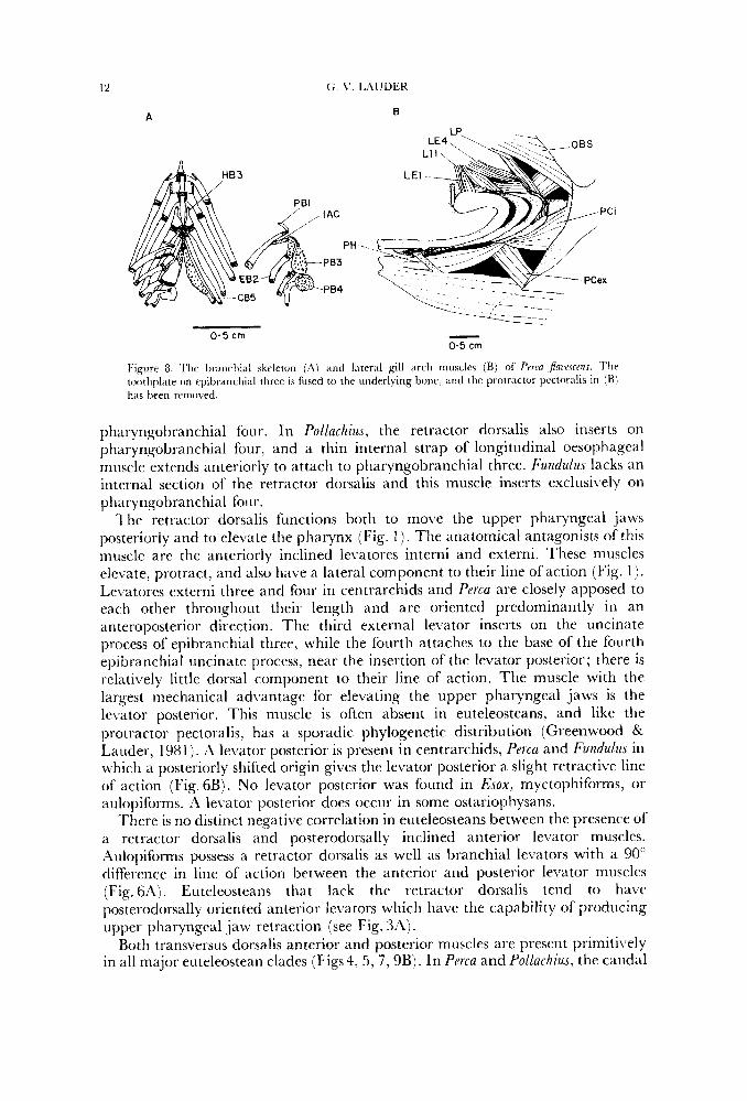
Figure 8 I'hr toothplate on epibranchi,il three IS fused to the underlting bone, nnd the protractor pcctotali? in iB1 has been removed
I tic Ixdnchial 5keleton ( A ) .ind lateral gill nrch Inir5cler (B) 01 Prrro J u w r c m
pharyngobranchial four. In Pollachius, the retractor dorsalis also inserts on pharyngobranchial four, and a thin internal strap of longitudinal oesophageal muscle extends anteriorly to attach to pharyngobranchial three. Fundulus lacks an internal section of the retractor dorsalis and this muscle inserts exclusively on pharyngobranchial four.
'The retractor dorsalis functions both to move the upper pharyngeal jaws posteriorly and to elevate the pharynx (Fig. I ) . The anatomical antagonists of this muscle are the anteriorly inclined levatores interni and externi. These muscles elevate, protract, and also have a lateral component to their line of action (Fig. 1 ) . LeLTatores externi three and four in centrarchids and Perm are closely apposed to each other throughout their length and are oriented predominantly in an anteroposterior direction. The third external levator inserts on the uncinate process o f epibranchial three, while the fourth attaches to the base of the fourth epibranchial uncinate process, near the insertion of the levator posterior; there is relatively little dorsal component to their line of action. The muscle with the largest mechanical advantage for elevating the upper pharyngeal jaws is the levator posterior. This muscle is often absent in euteleosteans, and like the protractor pectoralis, has a sporadic phylogenetic distribution (Greenwood & Lauder, 1981). A levator posterior is present in centrarchids, Perca and Fundulus in which a posteriorly shifted origin gives the levator posterior a slight retractive line of action (Fig. 6B). No levator posterior was found in Esox, myctophiforms, or aulopiforms. A levator posterior does occur in some ostariophysans.
There is no distinct negative correlation in euteleosteans between the presence of a retractor dorsalis and posterodorsally inclined anterior levator muscles. Aulopiforms possess a retractor dorsalis as well as branchial levators with a 90" difference in line of action between the anterior and posterior levator muscles (Fig. 6A). Euteleosteans that lack the retractor dorsalis tend to have posterodorsally oriented anterior levators which have the capability of producing upper pharyngeal jaw retraction (see Fig. 312).
Both transversus dorsalis anterior and posterior muscles are present primitively in all major euteleostean clades (Figs4, 5, 7,9B). In Perca and Pollachius, the caudal
EU7’EI.EOSTE.ZN PH;ZRYNGEAL JAWS 13
A B
- - 0.5 cm 0 - 5 c m
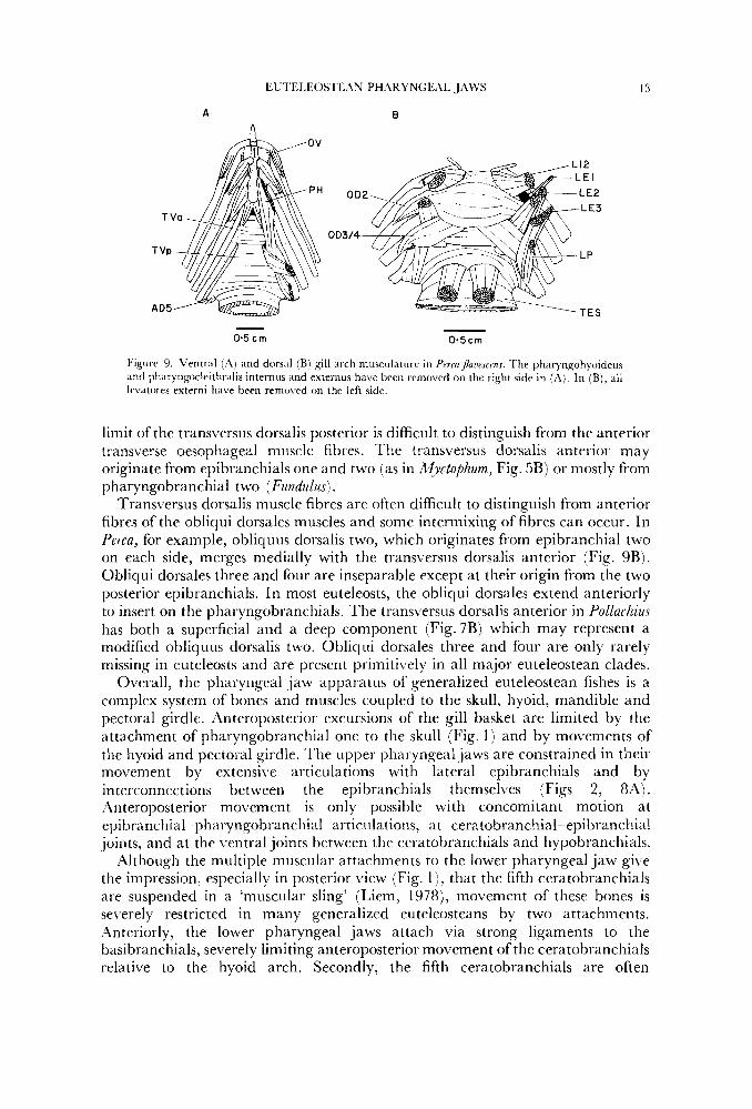
Figure 9. Ventral (A) and dorsal (B) gill arch musculature i n Pmaflawscms. The pharyngohyoideus and phar).n~~cleithtalis internus and externus have been removed on the right side i n ( i l l , In (B), all levatores externi have been removed on the left side.
limit of the transversus dorsalis posterior is difficult to distinguish from the anterior transverse oesophageal muscle fibres. I he transversus dorsalis anterior may originate from epibranchials one and two (as in Myctophum, Fig. 5B) or mostly from pharyngobranchial two (Fundulus) .
Transversus dorsalis muscle fibres are often difficult to distinguish from anterior fibres of the obliqui dorsales muscles and some intermixing of fibres can occur. In Perca, for example, obliquus dorsalis two, which originates from epibranchial two on each side, merges medially with the transversus dorsalis anterior (Fig. 9B). Obliqui dorsales three and four are inseparable except at their origin from the two posterior epibranchials. In most euteleosts, the obliqui dorsales extend anteriorly to insert on the pharyngobranchials. The transversus dorsalis anterior in Pollachius has both a superficial and a deep component (Fig.7B) which may represent a modified obliquus dorsalis two. Obliqui dorsales three and four are only rarely missing in euteleosts and are present primitively in all major euteleostean clades.
Overall, the pharyngeal jaw apparatus of generalized euteleostean fishes is a complex system of bones and muscles coupled to the skull, hyoid, mandible and pectoral girdle. Anteroposterior excursions of the gill basket are limited by the attachment of pharyngobranchial one to the skull (Fig. 1 ) and by movements of the hyoid and pectoral girdle. The upper pharyngeal jaws are constrained in their movement by extensive articulations with lateral epibranchials and by interconnections between the epibranchials themselves (Figs 2, 8.4). Anteroposterior movement is only possible with concomitant motion at epibranchial pharyngobranchial articulations, at ceratobranchial-epibranchial joints, and at the ventral joints between the ceratobranchials and hypobranchials.
Although the multiple muscular attachments to the lower pharyngeal jaw give the impression, especially in posterior L4ew (Fig. 1 ), that the fifth ceratobranchials are suspended in a ‘muscular sling’ (Liem, 1978), movement of these bones is severely restricted in many generalized euteleosteans by two attachments. Anteriorly, the lower pharyngeal jaws attach via strong ligaments to the basibranchials, severely limiting anteroposterior movement of the ceratobranchials relative to the hyoid arch. Secondly, the fifth ceratobranchials are often
7 7
1-1 G \’ L,\CDtR
ligamentously attached to the fourth ceratobranchials. In centrarchids this connection may be Lery broad so that there is little independent movement between these two bones.
Finally, ceratobranchial five is attached to the upper pharyngeal jaws by the relatively short-fibred fifth branchial adductor. ‘This muscle will limit the degree of independent motion between the upper and lower pharyngeal jaws, although some sprcies (c.g., EJOY) havc a relatively long adductor muscle.
Functional patterm
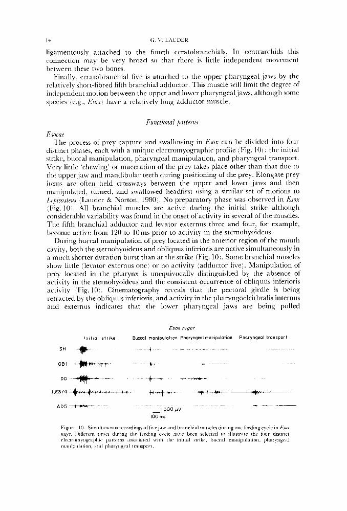
&rap ‘lhe process of prey capture and swallowing in EJOX can be dibided into four
distinct phases, each with a unique electromyographic profile (Fig. 10) : the initial strike, buccal manipulation, pharyngeal manipulation, and pharyngeal transport. F’ery little ‘chewing’ or maceration of the prey takes place other than that due to the upper jaw and mandibular teeth during positioning of the prey. Elongate prey item5 are often held crossways between the upper and lower jaws and then manipulated, turned, and swallowed headfirrt using a similar set of motions to Z,e/izJoJtruJ (1,auder & Norton, 1980). No preparatory phase was observed in EJOX (Fig. 10). .All branchial muscles are active during the initial strike although considerable Lrariability was found in the onset of activity in several of the muscles. The fifth branchial adductor and levator externus three and four, for example, become active fi-om 120 to 10ms prior to activity in the sternohyoideus.
During buccal manipulation of prey located in the anterior region of the mouth c‘i\ity, both the sternohyoideus and obliquus inferioris are active simultaneously in a much shorter duration burst than at the strike (Fig. 101. Some branchial muscles sliow little (le\xtor externus one) or no activity (adductor five). Manipulation of prey located in the pharynx is unequivocally distinguished by the absence of acti\ ity in the sternohyoideus and the consistent occurrence of obliquus inferioris activity (Fig. 101. Cinematography reveals that the pectoral girdle is being retracted by the obliquus inferioris, and activity in the pharyngocleithralis internus and externus indicates that the lower pharyngeal jaws are being pulled
Esox n iger
I n i t i a l s t r i ke Buccal manipulation Pharyngeal rnanlpulatlon Pharyngeal t ransport
t
- SH * OBI -*&++ - t-
DO*--- k - .t* ,,..,*+ -- - -
LE3/44*-++4- b*++-- -+-+&- -&im-- ---- --__ ~~ .~~ ~ - I300 pV A D 5 ~ - ” - -
- 100 ms
Figiirc 10. SiinuIt;ineous rccordings of fivcj<iw and branchial muscles during one feeding c)clc in &ox tzz,yr. Different times during the feeding cycle have been selected to illustrate the four distinct electromyographic patterns associated with the initial strike, buccal manipulation, p h a q n g e d manipiilntion, and phaq ngeal transport,
EU'1 'ELEOSIEhS PHflRYNGEAL JAWS 15
postero\.entrally (confirmed by X-ray cinematography; Lauder, in prep.). The fifth adductor is inactive during pharyngeal manipulation and the upper pharyngeal jaw is elevated by activity in the levatores externi muscles. The overall effect of pharyngeal manipulation is to increase rapidly the distance between the upper and lower pharyngeal jaws allowing repositioning of the prey prior to transport.
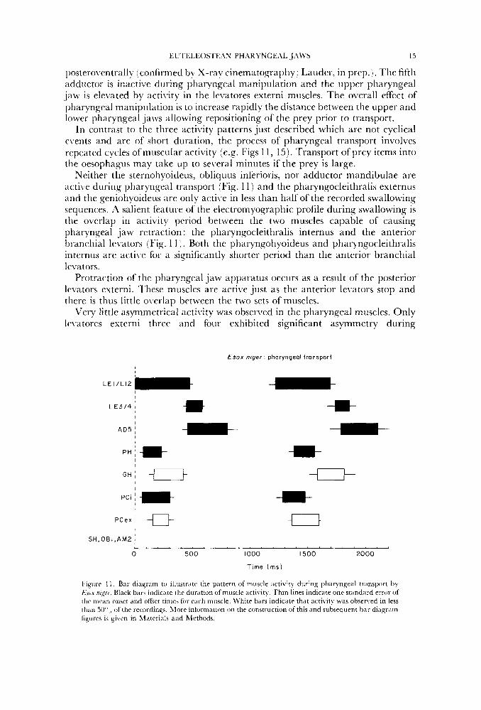
In contrast to the three activity patterns just described which are not cyclical e\.ents and are of short duration, the process of pharyngeal transport involves repeated cycles ofmuscular acti\ity (e.g. Figs 1 1 , 15). Transport ofprey items into the oesophagus may take up to several minutes if the prey is large.
Neither the sternohyoideus, obliquus inferioris, nor adductor mandibulae are acti\:e during pharyngeal transport (Fig. 1 1) and the pharyngocleithralis externus and the geniohyoideus are only active in less than half of the recorded swallowing sequences. A salient feature of the electromyographic profile during swallowing is the overlap in activity period between the two muscles capable of causing pharyngeal jaw retraction : the pharyngocleithralis internus and the anterior branchial levators (Fig. 1 1 ) . Both the pharyngohyoideus and pharyngocleithralis iriternus are acti1.e for a significantly shorter period than the anterior branchial levators.
Protraction of the pharyngeal jaw apparatus occurs as a result of the posterior levators externi. These muscles are active just as the anterior levators stop and there is thus little overlap between the two sets of muscles.
Very little asymmetrical acti\,ity was observed in the pharyngeal muscles. Only le\ratores externi three and four exhibited significant asymmetry during
€sox niger: pharyngeal transport
I S H , O B I , A M 2 ;
0 500 1000 I 5 0 0 2000
T ime ( m s )
Fignre I I . Bar diagram to illustrate. the pattern of muscle activity during phar).ngeal transport by Esux nigcr. Black bars indicate the duration of muscle activity. Thin lines indicate one stiindard error of the tnriin onset and offset times for ench muscle. ll'hite bars indicate that activity was observed in less than W",, of the recordings. hiore inlormation on the construction of this and subsequent bar diagram figures is given in 1I;iteri;ils and Methods.
16 G V LAUDER
pharyngecil transport. The pdttern of muscle activity during transport is relatively stereotyped as defined by the small stdndard error of mean (S.E.) onset and ofI'5et times for pharyngeal muscles (Fig. 1 1 ) relative to burst and interburst duration. 'I'he standdrd error is small even though muscle activities from different " indi\iduals, electrode placements, and prey are all averaged together to produce Fis. 11.
:lcanlho/)terygii 1\11 four of the distinct muscle activity patterns described for Esox were observed
in the acanthopterygian fishes studied experimentally (see Materials and Methods) ; :lmhloplites will be described in the most detail.
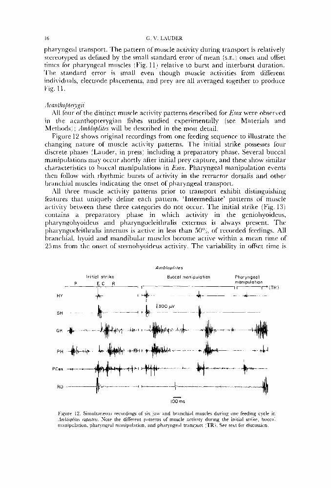
Figure 12 shows original recordings from one feeding sequence to illustrate the changing nature of muscle activity patterns. The initial strike possesses four discrete phases (Lauder, in press) including a preparatory phase. Several buccal manipulations may occur shortly after initial prey capture, and these show similar characteristics to buccal manipulations in Esox. Pharyngeal manipulation events then follow with rhythmic bursts of activity in the retractor dorsalis and other Ix-anchial muscles indicating the onset of pharyngeal transport.
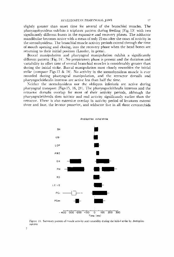
All three muscle activity patterns prior to transport exhibit distinguishing features that uniquely define each pattern. 'Intermediate' patterns of muscle activity between these three categories do not occur. The initial strike (Fig. 13) contains a preparatory phase in which activity in the geniohyoideus, pharyngohyoideus and pharyngocleithralis externus is always present. The pliaryngocleithralis internus is active in less than 50" (, of recorded feedings. All branchial, hyoid and mandibular muscles become active within a mean time of 25ms from the onset of sternohyoideus activity. The variability in offset time is
A mblopli tes
In i t ia l s t r i ke Buccal manipulation Phary ngea I P E C R manipulation
r I I P ( T R )
HY I
SH
Fignre 12. Simultaneons recordings of six jaw and branchial muscles during onc feeding cycle in .Irnhlofi/itri rupestris. Note the diffrrrnt patterns of muscle artivity during the initial strike, biiccal manipulation, pharyngral nrariipulation, and pharyngeal transport (TR). See text fur discussion.
EU I ELEOSTE,ZY PHARYNGEAL JAWS 17
slightly greater than onset time for several of the branchial muscles. The pharyngohyoideus exhibits a triphasic pattern during feeding (Fig. 13) with two significantly different bursts in the expansi\,e and recovery phases. The adductor mandibulae becomes active with a mean of only 25 ms after the onset of activity in the sternohyoideus. The branchial muscle activity periods extend through the time of mouth opening and closing, into the recovery phase when the head bones are returning to their initial position (Lauder, in press).
Buccal manipulation and pharyngeal manipulation exhibit a significantly different pattern (Fig. 14). No preparatory phase is present and the duration and variability in offset time of several branchial muscles is considerably greater than during the initial strike. Buccal manipulation most closely resembles the initial strike (compare Figs 13 & 14). No activity in the sternohyoideus muscle is ever recorded during pharyngeal manipulation, and the retractor dorsalis and pharyngocleithralis internus are active less than half the time.
Neither the sternohyoideus nor the obliquus inferioris are active during pharyngeal transport (Figs 15, 16, 2 1). The pharyngocleithralis internus and the retractor dorsalis overlap for most of their activity periods, although the pharyngocleithralis does initiate and end activity significantly earlier than the retractor. There is also extensive o\,erlap in activity period of levatores externi three and four, the levator posterior, and adductor five in all three centrarchids
Ambloolites init ial strike
SH
O B I
LOP
A M 2
G H * pH - R D
L E 1/2
PCI -+ PCex -m-
m jt i- ** h- *
-400 -300 - 2 0 0 -100 0 100 200 300 Time ( m s )
Figure 13. Summary pattern of muscle activity and variability during the initial strike by AmblopltteJ rupestris.
2
AmbIopliMs : pharyngeal manipulation Ambloplites' buccal manipulation
examined (Figs 15 18). IJei,atores externi one arid two consistently alternate in activity with levatores three and four, and in dmhloplites o\rerlap extensively with acti\,ity in the pharyngohyoideus. This muscle shows \rery different activity patterns in the four acantliopterygians examined, and the differences correlate with patterns in the pharyngocleitlir~ilis externus muscle. In ArnblupLitrs, both the pharyn~ohyoideus arid pharyngocleithralis internus are active for a single burst which alternates with the retractor dorsalis (Fig. 16). Both muscles are active with the retractor dorsalis in Micropteru.r, Pomoxis and Peica although in these last two significant differences in timing do occur. But only in .4mbluplites does pharyngohyoideus and pharyngocleithralis externus activity alternate with the retractor dorsalis.
The grniohyoideus muscle is also \Fariably active during pharyngeal transport, the le\.el of acti\.ity being mostly determined by the size of the prey. During swallowing of large prey, the geniohyoideus is active in concert with the ptiaryngohyoideus arid adductor mandibulae (Fig. 17) to protract the branchial basket.
Retractor dorsalis burst duration was measured for the four species of centrarchids studied experimentally (Fig. 19). 'I'here was no significant difference between the two species of Pomoxis, but each of the three genera did have dilrerent mean burst durations. .Miou,bterus, with the shortest mean of 298 ms, also displayed the least \wiance ;ind thus the most stereotyped pattern. There was no differencc in the range of food types or sizes fed to these species.
Within 21 particular swallowing sequence both the duration of retractor dorsalis activity and the length of time between bursts tends to increase (Fig. 2071. This is more marked in some feedings than in others but the pattern is consistently present regardless of prey type. For small prey, the duration of pharyngeal
19
100 ms Figure 15. Simultaneous recordings fi-om six branchial and hyoid muscles during the phiiqngeal transport stage of feedirig in .4mb/op/iles rupeslrzs. T h e rhythmic cyclical pattern of muscle activity uscd to transport prey into the uesophagus contrasts with the short duration non-repetitive activities i-ecr)rdc.d during buccal and pharyngeal rnanipnlation.
transport may be so short that no increase in either interburst interval or retractor dorsalis activity duration is evident.
While most recordings of pharyngeal transport showed symmetrical activity between right and left muscles, two consistent patterns of asymmetry were found in all species. Figure 22 illtistrates the maximum difference observed between activity
RD
LE 314
L E 1/2
LP
A D 5
PH
GH
PCex
PC I
OBI ,SH
0 500 1000 I500 2000
Time (ms)
Figure 16. Summar) pactern of muscle xt ivi ty during ph;iryngral transport in Amh/op/ila rupstrir. Note the extensive overlap in activity of the retractor dorsalis, levator posterior, levator externus three and four, ;\rid the fifth branchial adductor. Also note the lack of activity in the sternohyoideus and obliquus inferioris.
G. V, IAUDER
Micropterus . pharyngeal transport
R D
LE 314
L E I /2
L P
AD5
P H
GH
PCex
PC i
OBI , SH
AM 2/3
A O P
: * * -m- -- t-
I U 0 500 I000
Time ( m s )
Figure 17. Summary paitrr i i of muscle activity during pharyngeal transport in MicroptuuJ salmozdeJ
in the right and left retractor dorsalis and asymmetry of activity duration and relative timing in the anterior branchial levators. Minor (less than 100ms) differences between right and left muscles were not considered as significant asymmetry because of ( 1 ) variation in electrode location between muscles of each side, (2 ) the lack of a consistent pattern to these small differences within a single swallowing cycle, and (3) these differences in timing are small relative to total burst duration.
The first asymmetrical pattern involves the timing of levator externus one and two activity relative to the retractor dorsalis. Two asymmetrical variations were found. (1) In contrast to the symmetrical activity illuwated in Fig. 16 in which anterior levators alternate with the retractor dorsalis, the anterior levatores interni occasionally showed extended bursts which overlap 50" of the retractor dorsalis activity (Fig. 22:RD(I), LE1/2(1)). Occasionally the anterior levators on both sides show this pattern, but usually one side retains the alternating pattern while the other levators show considerable overlap with the retractor dorsalis. During extended swallowing sequences, the side deviating from the symmetrical pattern may change several times and symmetrical activity usually occurs between a change of side. (2) The second pattern of asymmetry involving the anterior levators produces a complete overlap of levator activity with the retractor dorsalis
I:U'IELF.OSTIXN PH.\RYVGE.AI. JAWS
Pornoxis annutaris pharyngeal transport
21
I , . I . l . l . l l l . l l . . I l l ~
-100 0 200 400 600 800 1000
Time (ms)
Figiirr 18. Summary pattern of muscle activity during pharyngeal transport in Pornoxis annularic
(Fig.23). The symmetrical pattern of activity in four muscles from one side is shown on the left of Fig. 23 and the asymmetrical pattern, 2 s later in the same swallowing sequence, on the right. When asymmetrical activity of this type was occurring, both sides (at different times) showed asymmetry.
l'he second major pattern of asymmetrical pharyngeal muscle activity is a co- ordinated change in the relative timing of muscles in both the upper and lower pharyngeal jaws. A small segment of a long swallowing sequence illustrating correlated asymmetry is shown in Fig. 24. The pharyngohyoideus muscle (PH) is the reference against which the activity patterns in the right and left posterior levators (LE 3/4) and the pharyngocleithralis internus (PCi) can be judged. In the' first set of activity, the left side muscles display the normal symmetrical alternating pattern with the PH while the right side LE 3/4 and PCi begin activity 300ms before the left side. In the very next sequence, on the right in Fig. 24, the relative timing of the two sides is reversed, and the left side muscles are now active throughout the last half of PH activity.
DISCUSSION
Phylogenetir pattarns
The pharyngeal jaw apparatus of teleost fishes has been of particular importance as a source of information for phylogenetic analysis since Nelson focused attention on gill arch morphology with an extensive series of papers in the late 1960s and early 1970s (e.g. Nelson, 1967a, 1969). Most research on
22
70
6 0 .
5 0 .
G. V. IAUDER
Y = 560
Retroctor dorsolis: burst duration
I Am blop It f es Mtcropferus
Pornoxis ornuloris Pornoxis nigromoculoius r
Time (ms)
Figure 19. Histograms tn illustrate the variability in burst duration of the retractor dorsalis muscle in four of the species studied. This muscle was used as the reference muscle for summary diagrams of ptiaryngeal transport [see Materials and Methods).
1
10 15 20 25 30 35 40 45 50 01 1 5
Burst number
Figure 'LO. Scattergram of retractor dorsalis burst duration and the interburst interval vcrsus time [Burst number) in one representative swallowing cycle in Ambloplztrs rupeslrrs.
EUTELEOSTEAN PHARYNGEAL JAWS
Perco. pharyngeal transport
SH
PCex -li J Y -,,
23
- 100 ms
Figure 21. Summary of muscle activity patterns during pharyngeal transport in PvrcaJlai~escens.
Amblopliies: pharyngeal transport
l0o ' ;s
Figure 22. M u ~ l e ;icti\ ity pattern fsirnultaneortsly recorded I durins a portion of a swallowing sequence in Ambiuplitvs rupesfrir t o illustrate one type of asymmetry between right (r) and left (1 j muscles (see text for discussion).
L E 1/2
L P
PP
RD
Figure
Pornoxis onnoloris asymmetrical transport w*-+ ' -&--+ - &q!++--- +*.. ~~-~
23. Pornoxis annularis. Muscle activity pattern in Ibur simultaneously recorded muscles during two separate times of a single swallowina sequence. These recordings illustrate the change in timing oi anterior levator activity relative to the retractor dorsalis.
24 G. v. LrZUDEK
branchial anatomy has emphasized osteology although Nelson ( 1967b, c ) did discuss some qeneral features of teleostean branchial musculature, and Holst\.oogd ( 1965) considered the presence of the retractores arcuum branchialium ( =retractor dorsales) to be a usefiil featurc separating ‘lower’ from ‘higher’ teleosts.
Rosen (1973) utilized gill arch anatomy extensively in his proposed phylogeny of the cuteleostean fishes and defined the major euteleostean clade, the Neoteleostei, by the presence of a retractor dorsalis muscle. Rosen ( 1974) subsequently examined the relationships of the Protacanthopterygii in detail, and this clade has recently been shown to be non-monophyletic by Fink & Weitzman (1982).
Although the interrelationships of primitive euteleosteans are still unsolved, this comparative anatomical analysis ofrepresentatives ofeach ofthe major euteleostean clades, with emphasis on the Neoteleostei, has revealed several new aspects of pharyngeal evolution in euteleosts and has served to define the primitive condition of the euteleostean branchial musculature. Although the features listed below do not uniquely define the Euteleostei, they do characterize the basic structural pattern fi-om which morphological diversification within the Euteleostei has occurred. Primitively, the euteleostean pharyngeal apparatus lacks a retractor dorsalis, possesses a rectus communis originating from hypobranchial three and inserting on ceratobranchial fi\.e, possesses two dorsal and two ventral transversus muscles, a pharyngocleithralis internus and externus, obliqui dorsales three and four, dermal toothplates fLised to the endoskeletal gill arch elements (Nelson, 1969 ; Patterson & Kosen, 1977) and dorsoventrally oriented anterior branchial levators. This pattern is retained in most generalized euteleosteans with two key additions.
Kosen (1973) demonstrated that the occurrence of a retractor dorsalis muscle defincs the Neoteleostei and this result has been confirmed here. Within the Neoteleostei, howe\.er, the retractor dorsalis has undergone several specializations which do not conform to a clear trend toward posterior insertion of the retractor dorsalis. In Perca, for example, the retractor dorsalis inserts on both pharyngobranchials three and four, while in Myctaphum the main external portion of the retractor inserts only on pharyngobranchial four. In myctophiforms, as described by Rosen (1973), the retractor dorsalis appears to be subdivided into an internal diivision inserting on the third pharyngobranchial, and an external division inserting on pharyngobranchial four. In Perca, Mytophum and Pallachius, however, the internal di\ision actually is a direct continuation of the longitudinal
EVTELEOS’I‘E ZN PH.ARYNGE,\I, J‘ZWS 25
oesophageal muscle layer and does not arise from the retractor dors a 1‘ is externus. 1;undulus and Belonesox (Karrer, 1967) lack this anterior extension of the longitudinal oesophageal muscles, but whether this loss is characteristic of all atherinomorphs or not remains to be established. Many acanthopterygians also lack the anterior hand of oesophageal muscle (e.g. the Pharyngognathi, Liem & Greenwood, 198 1 ; Stiassny, 198 1 ) . The occurrence of a thin band of oesophageal muscle pardllel to the retractor dorsalis proper which inserts on the pharyngobranchials provides additional evidence, albeit indirect, for the derivation of the retractor dorsalis from the sphincter oesophagi (Winterbottom, 1974).
In addition to the retractor dorsalis, a second key specialization in the evolution of the euteleostean pharyngeal apparatus is the shift in origin of the rectus communis, which primitively originates from hypobranchial three, to the urohyal. This specialized origin is characteristic of the Ctenosquamata (Myctophiformes + Paracanthopterygii + Acanthopterygii), first defined by Rosen (1973) on the basis of other characters. In some ctenosyuamates, the pharyngohyoideus may originate from both hypobranchial three and the urohyal. Stiassny (in prep.) has found a muscle similar to the pharyngohyoideus with an origin on the urohyal and insertion on ceratobranchial five in some aulopiform taxa. It is not yet established if the Aulopiformes are not monophyletic or if the ‘pharyngohyoideus’ has evolved independently in ctenosquamates and some aulopiforms.
In euteleosteans, the rectus communis primitively inserts on ceratobranchial five. Most ostariophysans examined lacked the rectus communis, and when it was present (e.g. Hepsetus, Hoplias, Brycon, Arius, Aptetonotus) its origin was usually from one of the first three hypobranchials. In other primitive euteleosteans (Alepocephalus, Galaxias, Aplochiton, OsmeruJ, 77pmallus) the rectus communis originates from hypobranchial three and inserts on ceratobranchial five. Occasionally, as in Retrofiinna, the origin of the rectus communis may be from the anterior hypobranchials.
The insertion of the rectus communis on ceratobranchial five appears to be a clupeocephalan character. In EulrumeuJ, Dussumieria and Opisthopterus the rectus communis originates from hypobranchial three and inserts on ceratobranchial five. Kirchhoff (1958) illustrated the rectus of Clupea as inserting on ceratobranchial four with the pharyngocleithralis internus muscle and this appears to be a specialized condition within clupeomorphs. Elopomorphs either lack a rectus communis (Elops, Megalops, rl lbula), or its arises from hypobranchials two and three to insert on ceratobranchial four (pers. obs. ; Nelson, 1967b). In osteoglossomorphs this muscle most commonly originates from hypobranchial two (although this is variable, and some genera lack a rectus communis; Greenwood, 1971), and inserts posteriorly on ceratobranchial four. Only L4nia among non-clupeocephalans convergently possesses a rectus communis inserting on ceratobranchial five (Wiley, 1976, 1979).
This proposed pattern of rectus communis evolution emphasizes that there now exist two names which define different stages in the evolution of the acanthopterygian pharyngohyoideus muscle. Given the longstanding usage of these terms, it is probably best that they both be retained with the understanding that they refer to homologous muscles. Thus, the rectus communis would be used in non-ctenosquamate taxa for the muscle, derived from rectus ventralis four
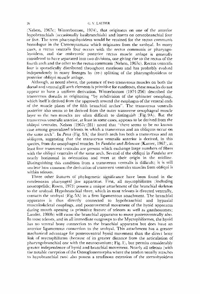
26 C,. \’. LAUDER
(Nelson, 1967c; Winterbottom, 1974), that originates on one of the anterior hypobranchials (occasionally basibranchials) and inserts on ceratobranchial four or five. The term pharyngohyoideus would be retained for the rectus communis hornologue in the Ctenosyuamata which originates from the urohyal. In many cases, a rectus ventralis four occurs with the rectus communis or pharyngo- hyoideus, and the embryonic posterior rectus muscle anlage is generally considered to ha\,e separated into two divisions, one giving rise to the rectus of the fourth arch and the other to the rectus communis (Nelson, 1967~) . Rectus ventralis four is sporadically distributed throughout euteleosts and has probably evolved independently in many lineages by (re-) splitting of the pharyngohyoideus or posterior obliyiii muscle anlage.
Although, as noted above, the presence of two transversus muscles on both the dorsal and ventral gill arch elements is primitive for euteleosts, these muscles do not appear to have a uniform derivation. Winterbottom ( 1974 :256) described the transversus dorsalis as originating “by subdivision of the sphincter oesophagi, which itself is derived from the upgrowth around the esophagus of the ventral ends of the muscle plates of the fifth branchial arches”. The transversus ventralis posterior also seems to be derived from the outer transverse oesophageal muscle layer so the two muscles are often difficult to distinguish (Fig. 9A) . But the transversus \,entralis anterior, at least in some cases, appears to be derived from the obliqui ventrales. Selson (1967c:281) noted that “there seems to be no known case among generalized teleosts in which a transversus and an obliquus occur on the same arch”. In P e m (Fig. 9A) the fourth arch has both a transversus and an obliquus, suggesting that the transversus ventralis anterior is derived, in this species, from the oesophageal muscles. In Fundulus and Befonesox (Karrer, 1967), at least four transversi 1,entrales are present which exchange large numbers of fibres with the obliqui ventrales of the same arch. Several of the obliqui in Fundulus are nearly horizontal in orientation and meet at their origin in the midline. Distinguishing this condition from a transversus ventralis is difficult; it is still unclear how common the derivation of trans\:ersi ventrales muscles from obliqui is wit hi n teleosts.
‘I’hree other features of phylogenetic significance have been found in the euteleostean pharyngeal jaw apparatus. First, all myctophiforms (including neoscopelids; Rosen, 1973) possess a unique attachment of the branchial skeleton to the urohyal. Hypobranchial three, which in most teleosts is directed ventrally, contacts the urohyal (Fig. 5A) in a firm ligamentous attachment. The branchial apparatus is thus directly connected to hypobranchial and hypaxial musculoskeletal couplings, and posteroventral movement of the hyoid apparatus during mouth opening (a primitive feature of teleosts as well as gnathostomes; Lauder, 1980b) will cause the branchial apparatus to move posteroventrally also. In most teleosts, and in all immediate outgroups to the Myctophiformes, the hyoid has no ventral bony connection to the branchial apparatus but does have an anterior ligamentous connection to the urohyal. This attachment has a greater mechanical ad\rantage for posteroventral hyoid movement than the direct bony link of myctophiforms (because of its greater distance from the articulation of pharyngobranchial one with the neurocranium ; Fig. 1 ) , but permits considerably greater independence of hyoid and branchial movement. Nearly all teleosts (with the notable exception of‘the Osteoglossomorpha where the tendon usually attaches to hypobranchial two) also possess a tendinous extension of the sternohyoideus
EUTELE0STE.W PH.ZRYNGEAL JAWS 27
muscle to hypobranchial three. This tendon is illustrated in Figs7‘4 and 8B, but has been removed in the other lateral views of the pharyngeal region to show the ventral gill arch musculature. A tendinous connection between the sternohyoideus and hypobranchial three was present in most members of all the euteleostean clades examined as well as in clupeomorph and elopomorph fishes. It may thus represent an elopocephalan synapomorphy.
A second specialization of the pharyngeal region with phylogenetic implications is the division of the pharyngocleithralis externus into dorsal and ventral sections, both inserting on ceratobranchial five, in Fundulus (Fig. 6B). This condition was also observed in BelonPJox by Karrer (1967) and is not present in any of the numerous outgroup taxa examined. The distribution of this feature within the Atherinomorpha remains to be determined. Sponder & Lauder (1981) described a similar separation of the pharyngocleithralis externus into two muscles in Periophthalmus, but in this species the ventral section inserts anteriorly on ceratobranchial three.
The third remaining feature of phylogenetic interest is the condition of the rectus communis in stomiiforms. In no other teleostean clade has the rectus communis been found to originate on the basihyal or basibranchials. Due to the origin of the rectus from hypobranchial three (the primitive condition) in some stomiiforms and its absence in two other genera examined, the significance of the anterior shift in rectus communis origin within stomiiforms cannot be assessed until more species are examined. The basihyal site of origin may characterize a monophyletic group within stomiiforms and the lack of the rectus communis in Diplophos and Gonostoma may be autapomorphic.
Functional morphology
A fundamental feature of the functional morphology of prey manipulation and swallowing in the euteleostean fishes examined here is the separation of three activities: buccal manipulation, pharyngeal manipulation and pharyngeal transport. The analysis of mean onset and offset times for jaw, hyoid and branchial muscles in Ambloplites (Figs 13, 14, 16) clearly shows that each represents a distinct pattern and that all muscle activity following the initial strike cannot be averaged into one meaningful summary bar diagram. This division of postcapture behaviour is proposed as the basis for comparing functional patterns within the Euteleostei (see below : Comparisons).
Within the context of current hypotheses of euteleostean phylogeny, as corroborated by the characters presented above, the fiinctional analysis of branchial muscle activity in Esox and ‘generalized’ acanthopterygians has revealed several functional attributes of the primitive euteleostean pharyngeal apparatus. These conclusions are presented as tentative and subject to revision when experimental data on gill arch function become available for more euteleostean lineages. Because of the difficulties involved in obtaining and conducting experiments on stomiiform, aulopiform, and myctophiform fishes, future research on euteleostean pharyngeal jaw function will focus on salmoniforms, ostariophysans, paracanthopterygians, atherinomorphs and basal percomorphs.
Several muscle patterns were found in all species studied experimentally and are thus suggested to be primitive for the Euteleostei. ( 1 ) There is a sharp distinction between the pattern of activity in anterior and posterior branchial
28 G. \’, 1,AL‘I)ER
levator muscles. l h e anterior levators in Esox begin and end activity significantly earlier in the chewing cycle than the posterior levators (Fig. 11) and there is little olverlap in activity period. In Ambloplites, the same pattern is observed (Fig. 16) although the function of the anterior levators is reversed : the anterior levators protract the upper pharyngeal jaw, the opposite of their function in &ox. Even though the retractor dorsalis and levatores externi three and four are anatomical antagonists in ilmbloplites these muscles overlap for 70‘j,, or more of their activity period. It is the anterior levators which are active inbetween bursts of the retractor dorsalis. ‘l’he lasator posterior also extensively overlaps retractor acti\.ity (Figs
181, and in Pomoxis nnnzdaTis, there is no significant difyerence between rela- ti\.e activity periods of the retractor dorsalis, levatores externi three and four, and levator posterior. These data indicate that the posterior gill arch levators may func- tion primarily to elevate the upper pharyngeal jaws during the retraction stroke (see below), as the combined action of these dorsal gill arch muscles will move the upper pharyngeal jaws toward the base of the skull. (2 ) The pharyngocleithrah internus is active with those dorsal gill arch muscles causing retraction (either the retractor dorsalis in acanthopterygians or the anterior levators in Esox). In Perca (Fig. 2 1 ) , 21mhlo/dites (Fig. 16) and Pomoxis (Fig. 18) the onset time of the pharyngocleithralis internus is significantly earlier in the swallowing cycle than the onset of the retractor dorsalis. In Micropterus (Fig. 17) , at least half of the activity occurs prior to the onset of retractor dorsalis activity perhaps indicating considerable asynchrony in the protractive and retractive movements of the upper and lower pharyngeal jaws. ( 3 ) The sternohyoideus and obliquus inferioris muscles are not active during pharyngeal transport of prey into the oesophagus. (4) The obliquus inferioris is active during pharyngeal manipulation of prey. Pharyngeal manipulation involves pectoral girdle retraction and posteroventral branchial basket movement mediated by the pharyngocleithralis externus. (5) Both the sternohyoideus and obliquus inferioris are active during the initial strike and buccal manipulation, as are all branchial muscles. The timing of activity in the levatores, retractor dorsalis, and pharyngocleithrales muscles suggests that active expansion of the hranchial basket is occurring, increasing the volume of the posterior portion of the buccal cavity and contributing to negative mouth cavity pressure. (6) Several branchial muscles are active during the preparatory phase of suction feeding. The pharyngohyoideus and geniohyoideus fLinction together to protract the hyoid and reduce buccal volume. In Ambloplites (Fig. 13) the adductor mandibulae is only rarely active but the pharyngocleithrales muscles are active. It is unclear what role these muscles play during the preparatory phase as their major action is to move the lower pharyngeal jaws posteroventrally causing an increase in mouth cavity volume. The anterior levatores and fifth branchial adductor are also frequently active during the preparatory phase.
Although no direct observations of pharyngeal jaw movement were obtained in this study, an analysis of the mechanical relationships of the pharyngeal apparatus, the lines of muscle action, and relative muscle activity periods permit a well founded hypothesis of the pattern of upper and lower pharyngeal jaw movement. This analysis is facilitated by the relatively long durations of muscle activity (up to 1 s ) relative to the rapid sequence of events occurring at the initial strike.
In primitive euteleosteans such as Esox, the upper pharyngeal jaw is able to move relatively little in an anteroposterior direction because the dorsal gill arch muscles have little mechanical advantage (Figs 3A, 25). Upper pharyngeal retraction is
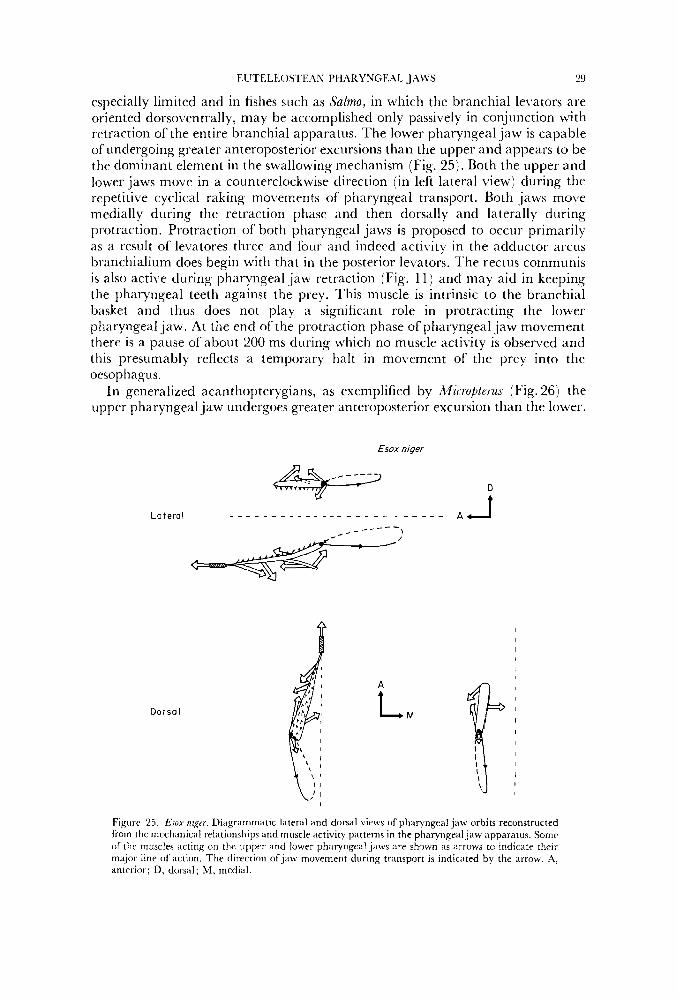
EUTELEOS I E.ZN PHARYNGEAL J.4WS
especially limited and in fishes such as Sulmo, in which the branchial levators are oriented dorsoventrally, may be accomplished only passively in conjunction with retraction of the entire branchial apparatus. The lower pharyngeal jaw is capable of undergoing greater anteroposterior excursions than the upper and appears to be the dominant element in the swallowing mechanism (Fig. 25). Both the upper and loww jaws move in a counterclockwise direction (in left lateral Liew) during the repetitive cyclical raking movements of pharyngeal transport. Both jaws move medially during the retraction phase and then dorsally and laterally during protraction. Protraction of both pharyngeal jaws is proposed to occur primarily as a result of levatores three and four and indeed activity in the adductor arcus branchialium does begin with that in the posterior levators. The rectus communis is also active during pharyngeal jaw retraction (Fig. 11) and may aid in keeping the pharyngeal teeth against the prey. This muscle is intrinsic to the brancliial basket and thus does not play a significant role in protracting the lower pharyngeal jaw. At the end of the protraction phase of pharyngeal jaw movement there is a pause of about 200 ms during which no muscle activity is observed and this presumably reflects a temporary halt in movement of the prey into the oesophagus.
In generalized acanthopterygians, as exemplified by M i c r o p u s (Fig. 26) the uppcr pharyngeal jaw undergoes greater anteroposterior excursion than the lower.
29
Dorsa I
A
L M
Figure 25. EJOX nzger. Diagrammatic hteral and dorsal views of pharyngeal jaw orbits reconstructed from the mcctianical relationships and muscle activity patterns in the pharyngealjaw apparatus. Sonir of tlw muscles acting 011 the upper and lower pharyngeal jaws are shown as arrows to indicate their ma,jor line ofaction. The direction of jaw movement during transport is indicated by the arrow. A, anterior; D, dorsal; M , medial.
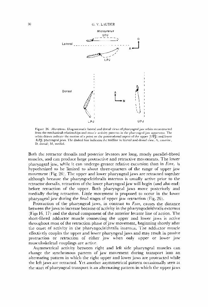
30 G. \’, LhUDER
Micropterus UPJ
4- - - - - - -- &LJ/
Lotera ’ . -. ~. ~ ~ . . . . . . . . - - . .
A A LPJ
L P J U P J
Figure 26. .Clicrojlurns. Diagrammatic lateral and dorsal views of pharyngeal jaw orbits reconstructed from the mechanical relationships and muscle artivity pattrrns in thc pharyngeiil jaw apparatus. l’hr orhits drawn indicate the motion ofa point on the posterodorsal aspect of the upper (UPJ) and lower fLPJj pharyngeal jaws. The dashed line indicates the midline in lateral and dorsal view. A. anterioi-; D, dorsal; 51, mrdial.
Both the retractor dorsalis and posterior levators are long, mostly parallel-fibred muscles, arid can produce large protractive and retractive movements. The lower pharyngeal jaw, while i t can undergo greater relative excursion than in Exox, is hypothesized to be limited to about three-quarters of the range of upper jaw movement (Fig. 26). The upper and lower pharyngeal jaws are retracted together although because the pharyngocleithralis internus is usually active prior to the retractor dorsalis, retraction of the lower pharyngeal jaw will begin (and also end] before retraction of the upper. Both pharyngeal jaws move posteriorly and medially during retraction. Little movement is proposed to occur in the lower pharyngeal jaw during the final stages of upper jaw retraction (Fig. 26).
Protraction of the pharyngeal jaws, in contrast to Esox, causes the distance between the jaws to increase because of activity in the pharyngocleithralis externus (Figs 16, 17) and the dorsal component of the anterior levator line of action. The short-fibred adductor muscle connecting the upper and lower jaws is active throughout most of the retraction phase of jaw movement, beginning shortly after the onset of activity in the pharyngocleithralis internus. The adductor muscle effectively couples the upper and lower pharyngeal jaws and may result in pawive protraction or retraction of either jaw when only upper or lower jaw musculoskeletal couplings are active.
Asymmetrical activity between right and left side pharyngeal muscles can change the synchronous pattern of jaw movement during transport into an alternating pattern in which the right upper and lower jaws are protracted while the left jaws are retracted. Yet another asymmetrical pattern occasionally seen at the start of pharyngeal transport is an alternating pattern in which the upper jaws
EL‘TELEOSTEAN PHARYNGEAL .JALVS 3 1
move anteriorly as the lower jaws retract. These movements are inferred from electromyograms which, instead of‘ fitting the symmetrical pattern of transport (Fig. 16j, display alternating activity between the retractor dorsalis and pharyngocleithralis internus for several cycles.
The general pattern of euteleostean pharyngeal jaw movement as inferred from muscle acti\,ity and pharyngeal mechanics is one of predominately symmetrical and synchronous protraction and retraction. In euteleosts that lack a retractor dorsalis muscle, the lower pharyngeal jaw plays the dominant role in transporting prey into the oesophagus, although the upper jaw is capable of active posterior movement in those taxa with posteriorly inclined levator muscles. The retractor dorsalis muscle mediates extensive posterior movement of the upper pharyngeal jaws, largely because of its length and mechanical advantage. In taxa possessing a retractor dorsalis muscle, the upper pharyngeal jaw contributes more to prey transport than the lower, undergoing greater excursions and continuing to move posteriorly when the lower pharyngeal jaw has stopped. The change from lower to upper pharyngeal jaws as the dominant element of the prey transport mechanism is postulated to ha\re taken place with the origin of the Neoteleostei. The increased use of the upper pharyngeal jaw in prey transport may be related to changes in dentition, pharyngobranchial size, and supporting framework of epibranchials during euteleostean evolution, but the details of the form-function relationship remain to be investigated.
Comparisons Although the conclusions regarding pharyngeal jaw movement in this paper are
based on the mechanical arrangement of the branchial region and on patterns of muscle activity during swallowing, the hypothesized movements of the pharyngeal jaws accord extremely well with previous investigations. Liem ( 1970) investigated deglutition in nandid fishes by examining tooth marks left by pharyngeal teeth on albino, scaleless prey. Sponder & Lauder (1981) directly examined pharyngeal jaw movement in Periophthalmus using cineradiography of terrestrial swallowing so that the small pharyngeal bones could be visualized without X-ray scattering by water. Non-experimental studies of pharyngeal function in euteleosteans include the muscle stimulation research of Vanden Berghe (1928) on C’ottus and Blennius, and the work of Tchernavin j 19.53, Giinther Pr Deckert (1960), Kayser (1962) and Karrer (1967). Experimental research of pharyngeal function in cichlid fishes (Liem, 1973, 1978) has pro\.ided a soiirce of comparative information from pharyngognath fishes.
?’he cycle of pharyngeal jaw mo\.ernent described by Sponder & Lauder (1981 : fig. 4) matches that proposed here Tor generalized acanthopterygians (E’ig.26) almost exactly. The upper pharyngeal jaw is the major element mediating prey transport and the lower pharyngeal jaw pauses at the posterior limit of its movement while the upper pharyngeal jaw completes its retraction stroke. Liem (1970:132-133) emphasized the morphological coupling between the upper and lower pharyngeal jaws and disagreed with the view of Karrer (1967) t h a t protraction of the upper pharyngeal jaws occurs while the lowerjaws are being retracted : “There is strong morphological aridence in favour of the simultaneous protraction and retraction of the upper and lower pharyngeals hypothesis over that of the alternating hypothesis proposed by Karrer (1967)”. Liem also emphasized the mobility of the lower pharyngeal jaws.
'4 2 G. V. I A U D E R
The experimental data presented here, completely congruent with that of Sponder & Lauder (19811, indicate that when both pharyngeal jaws move posteriorly they do so together. But because the lower pharyngeal jaw in the acanthopterygian taxa studied undergoes a significantly smaller range of mo\rcment, i t is not moving during much of pharyngeal retraction and lags behind at the start of the protractive phase. As in Periophthalmus, it is the lower pharyngeal jaw that first begins to move posteriorly. Thus, although most of lower jaw protraction and retraction occur together with the upper jaw, some asynchrony results from the different sizes of the movement orbits. No experimental evidence to date has demonstrated strict alternating movements of the jaws to be frequent during deglutition.
I f the pattern described in this paper for Perca and certain centrarchids is a general one (as comparisons with nandids and Periophthalmus suggest i t is) then the pharyngognath pattern of muscle activity and jaw movement is specialized. Pharyngognaths have added a further activity pattern, pharyngeal mastication, to the three intraoral activities defined here : buccal manipulation, pharyngeal manipulation and pharyngeal transport. Pharyngeal mastication in cichlid fishes involves all pharyngeal muscles as well as the sternohyoideus in a co-ordinated masticatory cycle lasting about 400 ms in piscivorous taxa (Liem, 1978). Prey are crushed and pierced by the pharyngeal teeth during two power strokes. Liem (1978) has presented some electromyographic data on swallowing that show considerable similarity to the proposed general acanthopterygian pattern. Swallowing is accomplished primarily by the upper pharyngeal jaw, and the levator posterior and retractor dorsalis overlap extensively in activity period. Thcse muscles alternate with acti\ity in the first levator internus (the only anterior le\,ator recorded). This is exactly the pattern found in Micropterus, Ambloplites, Pomoxis and Perca and the cichlid upper pharyngeal jaws may have retained the primitive neuromuscular couplings involved in pharyngeal transport.
The lower pharyngeal jaw takes relatively little part in transport (Liem, 1978 :3543 and little activity was found in the ventral branchial musculoskeletal couplings. Liem reported activity in the geniohyoideus, sternohyoideus and pharyngocleithralis externus prior to swallowing and this may represent a pharyngeal manipulation stage in cichlids comparable to that of more generalized percomorphs.
Future studies of euteleostean pharyngeal jaw function will be most usefLil if attention is paid to the basic divisions of feeding behaviour and if muscle activity periods are summarized separately for each behaviour with statistically adequate sample sizes and an indication of the variability (standard error) in mean timing of each muscle. The structural specializations shared by many derived euteleostean clades are relatively well known compared to functional patterns and innovations. The lack of outgroup information on jaw fbnction has seriously hindered our understanding of the origin of evolutionary and ecological trophic specialization in fishes. A general euteleostean pattern now seems to be emerging that can serve as a baseline for future analyses.
Functional design and evolutionary pattern5
Within the last 10 years, research on cladistic methods has provided a n extensive body of literature on patterns of structure in organisms and the ways in which
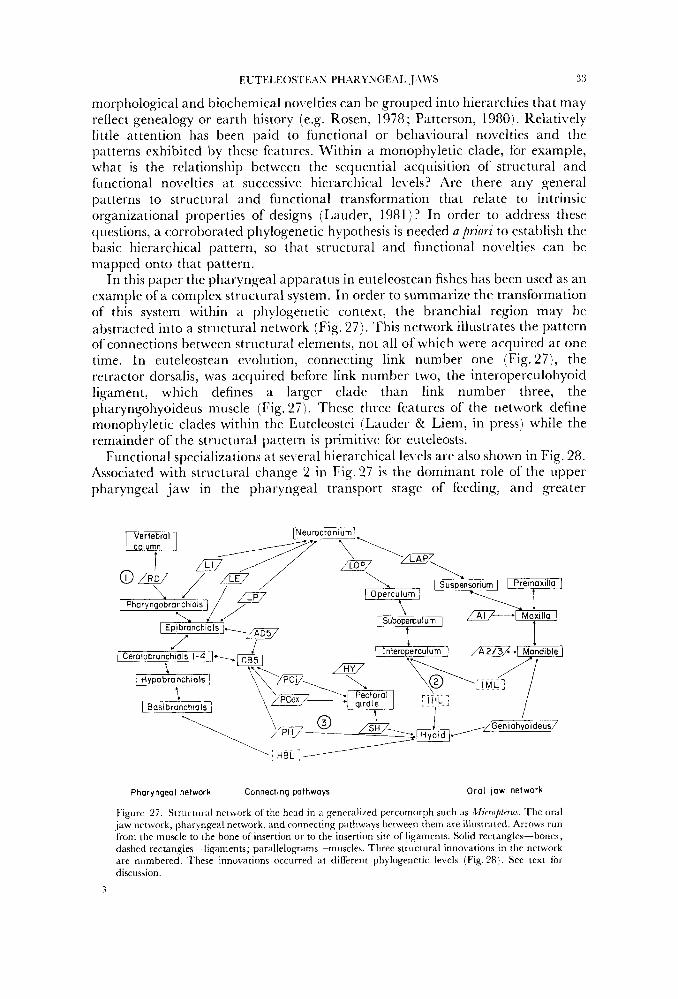
EU?’EI,EOSTE:\S PHARYNGEAI. J/\LVS 3 3
morphological and biochemical novelties can be grouped into hierarchies that may reflect genealogy or earth history (e.g. Rosen, 1978; Patterson, 1980). Relatively little attention has been paid to functional or beliavioural novelties and the patterns exhibited by these features. Within a monophyletic clade, for example, what is the relationship between the sequential acquisition of structural and fiinctional novelties at successive hierarchical levels? Are there any general patterns to structural and functional transformation that relate to intrinsic organizational properties of designs (Lauder, 1981 ) ? In order to address these questions, a corroborated phylogenetic hypothesis is needed a priori to establish the basic hierarchical pattern, so that structural and functional novelties can be mapped onto that pattern.
In this paper the pharyngeal apparatus in euteleostean fishes has been used as an example of a complex structural system. In order to summarize the transformation of this system within a phylogenetic context, the branchial region may be abstracted into a structural network (Fig. 27). This network illustrates the pattern of connections between structural elements, not all of which were acquired at one time. In euteleostean evolution, connecting link number one (Fig. 27), the retractor dorsalis, was acquired before link number two, the interoperculohyoid ligament, which defines a larger clade than link number three, the pharyngohyoideus muscle (Fig. 27). These three features of the network define monophyletic clades within the Euteleostei (Lauder 81 Liem, in press) while the remainder of the structural pattern is primitive for euteleosts.
Functional specializations at several hierarchical levels are also shown in Fig. 28. Associated with structural change 2 in Fig.27 is the dominant role of the upper pharyngeal jaw in the pharyngeal transport stage of feeding, and greater
Pharyngeal network Connecling palhways Oral law network
Figure 27. Structnral network of the head in a gencraliLecl percomorph such as Mirruplrms. The oral ,jaw network. phxyngeal network, and connecting pithways between them are illtistratcd. Arrows rim frorn the muscle to the bone of insertion or to the insertion site of ligaments. Solid rectangles-bones; dashed rectang1es~-ligaments; paralleloRrams-miiscles. Three structural innovations in the network are numbered. ’These innovations occurred at different phylogenetic levels (Fig. 28). See text for discussion.
3
Clupeocephala I 1
Eute leos fe i I I
Neoteleostei I Ctenosquomata '
Figiirc'LH. Ph) Iogcn) o f t h e Eiiteleoatci I m c d on the work of'Koscri (197:5 1 , Fink &k \ t 'ei tzmm (19821, ;ind chmrctcrs preseiitcd in L'iiider &k Lirm ( i n press), T h e three structural novelties 1 ii~i1)iitii~iri~Iiiesi froin Fig. 27 are (h.itclicd h r s l : ( 1 \ the prcscrice of;i r r t r x t o r dorsalis muscle, (2 ) the presencc of i i i i i i t trr i~~~crrirlohyoid liganiciit (Iioniolog~ius to the in;tndihuioiiyoiti l ig i tn ient l , and , '; tile ch,iiigc. i i i origin of thr icctiis communis n~risclc 11) t l i c i iroliyiil i some iiiilopiforms may i i l s o possrss .I p l r a r ~ ngohyoidens- -see text I . Two other synaponiorphics corroboratinF: the initial ph) Iogeiietic hypotlicsis haw 1)ceti discovered (black birrs1 : (;I) the rectiis commonis rriiiscle inserts Ixisteriorly on rerntot~rarichi;ll five (see text for discussion), and (b] the urohyal is at tached firmly to hvpotxanchinl tlircc on e x h siclr. T h r clndugriirri shown i s corroborated by many other chiiracters (see 1 ~ 1 1 d r r S: I.iem, in press). Foiir levels ( A D) have been i d e n t i f k l with sprcific function;tl i i inowitioiis. '1: 'interior 1~r;iiichi~iI Irvators e l e w t c and/or retriirt the upper pharyngeal j aw; anterior hfiinchial lc \a torh active with the I'1i;iryiigocIeithrnlis internus; the lowcr pharyngeal j aw plays the d~i in in~int role i r i p r q t imspor[ . U: thc upper ~ h i r y i i g c ~ i l ,j;i\v is the major etiector of prcy trmsport. C : hyoid i-ctr;iction is trariarnitted dircctly to t h e interoperculuni. I>: the lower phitrynge;il jaw is couplcd t o the h!riitl . ip i ) , i r i i t i i s ;ind undergoes grc;itt>r ;internposterior excursions a s 21 reatilt of the shift in ph;iryrigohyi)itlciis iii-igiti; the anterior 1ev:rtorcs Ii:it,c ;I ~ i r o t r a c t i \ ~ function and iilteriiiitr ui th activity i n the ph;irynRocleithr;ilic irrterniis and retriictw dorsiilis; acti\Jity of the fifth branchial adductor .ind lc\iitiires rxtrriii three and four shifta into the retraction stroke of the iipppr pharyngeal jaw. F'iiiictioii;il nitvrltics iii-c associated with structuriil innovations in a hierarchical Ixi t t iwi. The initial ph)Iogrnetic hypothesia o f t l ic F,ritrlrostei can thus be used 21s 'I guide to examine patterns in the : ic(l i i iGti i i i i of both strtictiiral arid ftinctioii;il novelties (see text tiir fiirther diaciission).
iinteroposterior excursions of the upper pharyngeal ,jaw as compared with the lower. 'I'hc anterior branchial levator muscles at this phylogenetic level are inferrecl not to overlap activity of die pharyngocleithralis internus, in contrast to the primitive condition a t l e \ d ,2 (Fig. 28) where these dorsal and \ventral I)ranchial muscles arc active synchronously (as in Fig. 1 1 ) . At level D, the shift in
F.1 ~TEL,F:OSI‘E.\S PH.ARYNGE.\I. J.\\VS 3.5
origin of the rectus commiinis to the urohyal couples the lower pharvngeal Jaw directly to liyoid movement and, \.ia the geniohyoideus muscle, to the mandible (Fig. 1 ) . This greatly increases the absolute anteroposterior mobility of the lower pharyngeal Jaw, although because of its anterior attachment to the basibranchials it still undergoes less movement than the upper pharyngeal jaw.
‘l’his analysis reveals two key steps in the evolution of the acanthopterygian pharyngeal jaw apparatus. First, at the neoteleostean level, the upper pharyngeal jaw acquired the capability of significant prey transport functions and this ei’ent is inferred to have been accompanied by a shift in timing of contraction in a large number of gill arch muscles into the retraction stroke (Fig. 28 : level B). Second, at thc ctenosquamatc Ic\rel (Fig. 28:le\rel D) , the lower pharyngeal Jaw beame coupled to the hyoid apparatus and is inferred to have been capable of a greater range of movement than in prectenosquamate taxa. Only with the origin of the Ctenosquamata, then, did the pharyngeal jaw apparatus acquire the general acanthopterygian configuration shown in Fig. 1. And i t is important to emphasize that the mechanical linkages and acti\,ity patterns of muscles in Fig. 1 were acquired in a definite historical sequence only discernable by a phylogenetic analysis of structural and functional nolselties (Fig. 28).
‘The pattern of structural and functional transformation discovered for the euteleostean pharyngeal jaw apparatus has several important implications for the e\.olution of functional design in \.ertebrates. Homologous muscles have been proposed to possess evolutionarily conservative action patterns which are maintained in the facc of significant reorganizations of muscle line of action and liinction (Bramble, 1980). In eriteleostean fislies this appears not to be the case a s homologous muscles radically altered their acti\ity patterns with the advent of the rrtractor dorsalis. Howe\rer, the o\rerall kinematic pattern of pharyngeal jaw mo\rement was changed relatively little a s in both primitive and deriised euteleosts the retraction stroke of the upper and lower jaws largely coincides. It thus appears that as structural and functional noxrelties in the pharyngeal region were acquired, the overall pattern of jaw mo\.cment was maintained, although, to he sure, each novel feature modified the primitive kinematic pattern to some degree. Data are not yet a\ailable from other case studies in the historical analysis of form to show whether co-ordinated modification in structural and functional patterns and the consequent maintenance of the primi1iL.e movement sequence, arc general features in the transformation of \.ertebrate design (Fig. 28).
A key aim of future research in the transformation of design is the relationship between the arrangement of connecting pathways in structural networks (Fig. 27 I and patterns of morphological and fimctional diversity in monophyletic clades.
.2(:KNO\L’I.EnGESiENTS
This research was supported by a Block Fund award from The Uni\,ersity of Chicago, the .Andrew M’. hlellon Foundation, and NSF DEB 81 -15048. Preliminary research was conducted under a Junior Fellowship from the Society of Fellows, Har1w-d University. I thank the British Museum (Natural History) for iise oftheir facilities and P. H . Greenwood, K. Banister, G. Howes, R. Travers and M. Stiassny for many conversations about euteleostean evolution during my visit. P. H. Greenwood, M’. L. Fink and K. Hartel kindly provided many specimens for dissection. A Penrose Fund award from the ilmerican Philosophical Society
36 G. \', I.AUDEK
provided partial fiinding for travel to the British Museum (Natural History). I thank D. Rosen, W. I,. Fink, S. Emerson, J. Cracraft and K. F. Liem for many conversations on the issucs discussed in this paper. M. L. J. Stiassny and K. F. Liem criticized the manuscript in great depth. Special thanks to Joan Hives for her assistance with all phases of the research and manuscript preparation.
.\LI,IS, E. P., 1903. T h r sku l l and cranial and first spinal muscles and nerves of Scomber icombvr. Jouinal of
BRAMBLE, D., 1980. Feeding in tortoises and mammals: why so similar? (Abstract). American zoulugist, 20: 981. ELDREDGE, N. & CRACRAFI', J., 1980. P/ylogenetic Pattenis and the &o/utionayv Proms. New York: Colunhia
FINK, W. I,. & WEITZhL\N, S. H. , 1982. Relationships of the Stoiniiform fishes (Teler)steii, with a description
.tlnrpho/u,~s. 18: 45- 328.
University Press.
of Dipluphos. Ilullrtin of the .\fust~um n / Cinnparntiric <onlngy, Hnrriard C'niuersi@, 150: 31--93. ., 1974. Biomechanic.r, :In .lppromh to T'rrtdmlv Biology. Philadelphia: J. B. Lippincott. 1'0OD. P. H. , 1971. Hyoid and ventral gill arch musculature in osteoglossomorph fishes. Bullrtirt (!/the .Ifustum (.\'atura/ History), <ooln,<v, 22: 1-55.
GREENWOOD, P. H. & LAL'DER, G. V., 1981. The protractor pectoralis muscle and the classification of tclcost fishes. ~ullut i i t oj'the British Musrum (.Va:atural Histocy), ,Soology, 41: 213 234.
(;['STHER, K. Lyi DECKERT, K., 1960. Neue Untersuchengen uber die funktionellen Auswirkungen der raud;ilventralen Verlangcrung des Kieferstiels bei Knochenfischen. IV. Das Viscerocranium von Harpodnn nrhrreus Ham. Buch. (Iniomi, Harpodontidae). <oulugischr Britrage ( , V . F . ) , 5: 1 280.
HIIEblAE, K. M., 1978. Mammalian mastication: a reiview ofthe activity of the jaw muscles and the movemrnts they produce in chewing. In P. M. Butlrr & K. '4. Joysey (Eds), Studies on lhr Deuelupment and Fundiort qf Terth: 360~398. London: Academic Press.
HOISTVOOGD, C., 1965. The pharyngeal bones and muscles in Telrostei, a taxonomic study. Prncredings oJ'thr XoninklijAe .Z;derlaridsr .Ikademie unit Wetenschappen (C ~ 68: 209-2 18.
JENKINS, F. A. & b'EIJS, W. A,, 1979. The functional anatomy of the shoulder in the Virginia opossum (Dide1phi.r virginianal. Journal of zuulog)', 188: 379-410.
JOHNSON, G. D., 1980. The limits and relationships of the Lutjanidae and associated fiirnilies. Bulletin o f the SrippJ fnrtifutinii cfOreanograph_v, 24: 1 114.
,JOPPIEN, H., 1970. Vergleichend-anatoinische und frinktions analytische Untcrsuchungen an drr Kiefer- und Kiemenapparaten der raubrrischen knochenfische Aphaiiopus und ,tfrrluccius. ,Soofogischr Heitrage (.V.F.j, 16: 263 385.
KAhlPF, 1%'. I ) , , 196 1. \TergIeichende funktionsmorphologische Untersuchungen an den Viscerocranien einiger r;liibcrisch lebrnder Knochenfisrhe. <un/ugiJchr Beitrap ( .V.F.), 6: 391496.
KARRER, C., 1967. Funktionell-r\natomische und vergleichende Untersuchung des Schadels von Hechtkarpfling Belonvrnx. <ou/ogische J a h ~ hitcher .Inntomie, #4: 19 1 ~ 2.18.
K.ZYSER, H., 1962. L'ergleichende Untersiichting uber Vorsticckmechanismen der Oberkiefer bei Fischen. Der Bau und die Function dcs Kiefer- und Kiemenapparates von Knochenfschen der Cattung ,4mmo&tr~ iind (.'allini~vmus. ;oulogiJchr flritragr (, V . F . ) , 7: 32 I 445.
KIKCHHOFF, H. , 1958. E'unktionell-iinatoinische Untersuchung des Visceral apparates von Clitpra harengus L. ~oolnqische Jahrbuchn .Inatomie. 76: 461 ,540.
L:\UI)ER, C. \',, l980a. The suction feeding mechmism in sunfishes Lepomisj : an exprrimental analysis. Journn/ OJ Experimeiital Uiolug,'. 88: 49 72.
LZITDER, G. \'., 19801). E\.olution of the feeding mechanism in primitive actinopterygian fishes: a functional anatomical analysis of PoIyptfvxi, f .up im/rus , and .4mia. ,7uuma/ of .2lorphology, 16.9: 283-3 17.
L:\UDER, G. V, , 1981. Form and function: structural analysis in evolutionary morphology. Paleobiologp, 7: 430 442.
I,.\UDF,R, G. \~., 1982. Historic;il biology and the problem o f design. Joutnal 01' 'Theoretical Biolo,py 97: 5 7 ~ 67. LAUDER, G , V., (in press). Food capture. In P. W. Webb & D. Weihs (Eds), Fi.rh Hzomrchanics. New York:
Praeger.
ocirlatus. Journal UJ l~,'x/~~rimrntal Biuln,cv, 84: 17 32.
Bulletin a/ the 12duseurn of Compam1zr:e zuology, Haroard 1 'niriersiQ.
I,.\t ' l)ER, C. 1'. K: NORTON, S. F,, 1980. Asymmetrical muscle activity feedirlg in the gar, I.(/ z ? l . i l ) . ~ t m Y
LAUDER, G. V. & LIKM, K. F., (in press). The evolution and interrelationships of the actinoptengian fishes.
I.1F.M. K. F.. 1070. Compiriitive functional ;in:ctomy of the Sandidae (Pisces: Teleostei). Firfdiana. zoooofog~,. 5(;:
LIEhI, I(. F.. 1973. Lvoliitionar~ strategies and morphological innovations: cichlid pharyngeal jaws. Systematic I -. 1 66.
<oolugv, 22: 425 441.
IIUTELEOSI'EAS PH;\RYNGIL\I. J)\WS 37
I,IEkl, K. F., 1978. hlodnlatory multiplicity in the fiinctional repertoire of the feeding mechanism in richlid
l,lF,hl, K. F. 6r GREENWOOD, P. H. , 1981. .\ 1iiiiction;iI ,ippro:irh to tlir phylogmy of thr pti;iryngogn;tth
NF,I,SON, G. J., 19(j7;1 Gill ;irches of sonic trlmsre;in fishes of the families Girellidae, Pomarmtridae,
S I - I S O N , G . J . , l967b. Hr;inchial Inr1srlt-s in represcnt;itives of five eel families. Pacific Science, .?/: 348 363. NI:.I,SON, G. J , , 1 9 6 7 ~ . Branchid muscles in some gener;ilizcd telcostean fishes. .4cta zoologica, Stockholm. 48:
NELSON, G . J., 1969. Gill ;irchcs and the phylogen) of lislics, wi th notcs on the classification of ertcbrates.
NELSON, G. J . Oi PLA'I'NICK, N . I . , 1881. Systematics and flio,qro,qraphy, Cladi.ttics and I'icariance. New York:
P,\REN'I'I, I.. R., 1981. .\ phylogenetir m d biogeographir analysis of cyprinodontiform fishes (Teleostei:
P;\TTERSON, C . , 1980. Cladistirs. Riulogist, 27: 234-240. P;\'T'I'ERSON, C:. Kr ROSEN, D. E., 1877. Review ofirhthyodectilbrm ,ind other hlesozoic teleost fishes and the
theory and practice of classifying fossils. Bul[etiii of thr .4rnrric.an .2fusrurn uf.Vatura/ Histoy, 1.58: 81 172. KOSEN, D. E., 1973. I~itcrrrl;~tionships of higher cuteleostean fishrs. In P. H. Greenwood, R . S. Miles & C.
Patterson (Eds), Intmulatiunships of Fishu~: 397 -51 3 . Imidon: Academic Press. ROSES, D. E. , 1974. Phylogeny and zoogeography of s;tlmoniform fishes and relationships of f.e,bidogalnxzas
ra1nmandroideJ. Rul le t i i~ of the Amrricnn .Ifitsrum of,Vatural HiJtorv, 1.5.1: 267 325. ROSEN, D. E., 1978. Vicariant patterns xnd historical explmation in biogeography. Swternutzc <oologv, 27:
159 180. RUSSELL, E. S . , 1916. Form and Func/ion. '4 (&itrihufioti to fhu Hi~t07v ofdii ima/ ,2lorpholo,gy. London : John k1urr.iy.
(Rcprintrd, 1982, by thr University of Chicago Press.) Sl'ONDEK, D. & LAVDER, C . V., 1981. Terrestrial feeding in the mudskipper Periophthalmus (Pisces:
Trlrostri) : ii cineradiographic analysis. Jonrm7i o f ~ o o o l o ~ ~ v . I SI'IXSSNY. hl. L. J.. 1981. ' I h c phylctir s t a t u s of ttic Lirnil)
anatomical investigation. ,2bthPdands Journal oJ ,~w lugv . 31: 'I'C:HF.RNh\'lN, \'. V.. 1953. The freding merhanisms of ii drcp sca fish Chauliodus .tlonni Srhneider. I n i d o n :
British Museum (Natural History\. TRAL'ERS, R. A,, 1981, The interarcual rartilage; :I review of its development, distribution and value iis an
indicator of phyletir relationships i n ruteleostem fishes. J o i m ~ ~ / of . I 'aturn/ HiJtoTy, 1.5: 853-871, \'.\NDEN BERGHE, L., 1928. Kecherches stir la dCglutitioli chrL les Poissons '1i.ICosti.ens. Hulkt in du I'ilcadmiu
Royalr dr Rrl~&ur. Cla.rse d(j< Scirnces, 14: 322 332. \VII.EY, E. O., 1976. The phylogeny and biogeography of fossil and rerent gars (rlctinopterygii: Lepisosteidae).
l 'niiwtitv of Kansas .2luJeum of .'lhlural History .\discrllanruus Publicatiom, 64: 1 1 1 1 . \VlI,F,Y, E. O., 1979. \'rntr;il gill arch muscles and the intrrrelationships of gnathostomes, with a new
c1;issification of the Vertcbrata. <oolo,qicnl Journal u/thr /.znriuan .Soci?ty, 67: 149 179. WII,EY, F.. 0.. 11181. ~ t ! ~ / i J , ~ P l / < h " , thr ThmrT a i d Pinr/ict ?f ~ ' / y I o g m / i c SwtemccticJ. New York: John \Vile). \~IN'I'E;RH~I''I'OI\.1. K., 197 1. ,\ drsciipti\.e
.Iro&?r!y of . $h/uinI Scimci,J of Pii//a(f(,l~~/iio. /
fishes. , journal of .ii'orphology, 1.58: 323 360.
trlrosts. .Im&can <ooki,gkt, 2/: 83 101.
Embiotoridae, Labridar and Scaridae. Journal of . I 'a tu~a/ Hzstorv. I : 289 293.
277 288.
Hul l r l in ofthr z l ~ ~ ~ r ~ ~ c ~ ? / .Ifu.\runi o / . ! i r t u r n / tfistrity, 141: 175 5.52.
Colinnbia University Press.
Atherinomorpha). Hullrtin of thr . h r r i c a r t IZliirriini 1 ~ / . 2 a / u r n / Histurv. 168: 33.5 -557.
the. stri;ited rnusclcs of tkic Teleostei. pr(i(?udiitgA o f l h r
\HHRE\'I ZTIONS [JSCD IY FIGURES
, \ I , :\2/3, A M 2 , Ah1213 divisions of t h e adductor
.\D5 ;iddrictor arrus br;incIii;ilinm mitsclr AOP adductor opcrculi nitiscle C coniprrsaivc phase of the initial strike (:H5 ceratobr;mchi;il five CHjTP toothpl;ite of reratnbr;inrhi,il five C I , clcitliriim I)() dilator oprrculi muscle
rnnndil)ular muscle
xp'insive phase of the initial strike
'17'P tooth patch, unfused, on epilxinchi;il three epixial rniisrles oesophagus
2 cpil~r'lnchi;ll two
GH geniohyoideus muscle HHS hypol,r;inrhia~ thrrr HBL ligament between nrohyil and 11ypohy;ils
HY hyp;ixi,il miisrulature, synonymous ;is used here
IAC interarcu;il cartilage IHI. liqament from the interoperculuni to the liyoid Ih4L ligament from the mandible to the hyoid I>AP levator 'irciis palatini muscle LE, LEI -4 levatores externi muscles 1.1, 1.11 1 Ie\atores intri-ni miisrlcs I .0P 1ev;itor opcrculi muscle LP Ic\rator posterior 1,PJ lowrr pharyngeal jaw OBI obliquus inferioris muscle OBS ohliqtrus superioris miiscle OD, OD1 -4 obliqui dorsales muscles OV, OV1-4 obliqui ventrales muscles P prrlmratory phase of the initial strike PBI ,2,3,4 pharyngobranchial bones
with the obliquus inferioris (OBI)

3 H G. \’. I..IUDF.R
PH31’P tvothplate (fused! to ~,liarvngoblanchial thrw PU41’P toothplatr ffiised, to pharyngohranrhial Lxir PCe, PCex ph;iryngorleithrr\lis externtis PCexv phar!ngorleitliralis externus, ventral division PII ~ ~ t i ~ r y i i g ~ r l i ~ ~ ~ i d e u s muscle PCi I’haryngorleithralis internus muscle PP protractor pectoralis muscle R reco\wy phase of the initial strike RC rrctiis cvmmiinis inusclr RD retr<ictor dorsalis muscle K\’ rcctus ventralis mitsrle SH s t r I ~ ~ r ~ l i ~ ~ ~ i d c ~ t a muscle
’ID transversus dorsalis rnusclc TDad deep division of the transversus dorsalis anterior
TDas superficial division of the transversus dorsalis
TDP transversus dorsalis epibranchialis two muscle TES transverse muscle layer of the oesophagus ‘ I R pharyngeal transport phase of feeding T\!a,p anterior and posterior transwrsi ventralec
TV, transversi ventrales of arches onc to four CP-J upper ph<iryngedl jaw
muscle
anterior musrlr
rnllsclr\


![ANIMAL ROBOTS Copyright © 2019 Tuna robotics: A high ...glauder/reprints_unzipped/Zhu.etal.2019[18987].pdftail, resulting directly in faster swimming speed. In this way, fish swimming](https://static.fdocuments.in/doc/165x107/60e1110c70aefb5c785877df/animal-robots-copyright-2019-tuna-robotics-a-high-glauderreprintsunzippedzhuetal201918987pdf.jpg)