
Functional Characterization of Pneumocystis carinii ... · It is well appreciated that the absence...
11
Functional Characterization of Pneumocystis carinii Inositol Transporter 1 Melanie T. Cushion, a,b Margaret S. Collins, a,b Thomas Sesterhenn, a,b Aleksey Porollo, a,c Anish Kizhakkekkara Vadukoot, d Edward J. Merino d University of Cincinnati College of Medicine, Cincinnati, Ohio, USA a ; Cincinnati VAMC, Cincinnati, Ohio, USA b ; Cincinnati Children’s Hospital Medical Center, Cincinnati, Ohio, USA c ; Department of Chemistry, University of Cincinnati, Cincinnati, Ohio, USA d ABSTRACT Fungi in the genus Pneumocystis live in the lungs of mammals, where they can cause a fatal pneumonia (PCP [Pneu- mocystis pneumonia]) in hosts with compromised immune systems. The absence of a continuous in vitro culture system for any species of Pneumocystis has led to limited understanding of these fungi, especially for the discovery of new therapies. We recently reported that Pneumocystis carinii, Pneumocystis murina, and most significantly, Pneumocystis jirovecii lack both enzymes nec- essary for myo-inositol biosynthesis but contain genes with homologies to fungal myo-inositol transporters. Since myo-inositol is essential for eukaryotic viability, the primary transporter, ITR1, was functionally and structurally characterized in P. carinii. The predicted structure of P. carinii ITR1 (PcITR1) contained 12 transmembrane alpha-helices with intracellular C and N ter- mini, consistent with other inositol transporters. The apparent K m was 0.94 0.08 (mean standard deviation), suggesting that myo-inositol transport in P. carinii is likely through a low-affinity, highly selective transport system, as no other sugars or inosi- tol stereoisomers were significant competitive inhibitors. Glucose transport was shown to use a different transport system. The myo-inositol transport was distinct from mammalian transporters, as it was not sodium dependent and was cytochalasin B resis- tant. Inositol transport in these fungi offers an attractive new drug target because of the reliance of the fungi on its transport, clear differences between the mammalian and fungal transporters, and the ability of the host to both synthesize and transport this critical nutrient, predicting low toxicity of potential inhibitors to the fungal transporter. IMPORTANCE myo-Inositol is a sugarlike nutrient that is essential for life in most organisms. Humans and microbes alike can ob- tain it by making it, which involves only 2 enzymes, by taking it from the environment by a transport process, or by recycling it from other cellular constituents. Inspection of the genomes of the pathogenic fungi of the genus Pneumocystis showed that these pneumonia-causing parasites could not make myo-inositol, as they lacked the 2 enzymes. Instead, we found evidence of inositol transporters, which would import the sugar from the lungs where the fungi reside. In the present report, we characterized the transport of myo-inositol in the fungus and found that the transporter was highly selective for myo-inositol and did not trans- port any other molecules. The transport was distinct from that in mammalian cells, and since mammals can both make and transport myo-inositol, while Pneumocystis fungi must transport it, this process offers a potential new drug target. Received 5 October 2016 Accepted 14 November 2016 Published 13 December 2016 Citation Cushion MT, Collins MS, Sesterhenn T, Porollo A, Vadukoot AK, Merino EJ. 2016. Functional characterization of Pneumocystis carinii inositol transporter 1. mBio 7(6):e01851-16. doi:10.1128/mBio.01851-16. Editor Louis M. Weiss, Albert Einstein College of Medicine Copyright © 2016 Cushion et al. This is an open-access article distributed under the terms of the Creative Commons Attribution 4.0 International license. Address correspondence to Melanie T. Cushion, [email protected]. F ungi belonging to the genus Pneumocystis live in the lungs of most mammals, where they can cause a fatal pneumonia in hosts with compromised immune systems (1). Pneumocystis ji- rovecii is the species that causes a lethal pneumonia (PCP [Pneu- mocystis pneumonia]) in humans, especially those with primary or acquired immune deficiencies (2). PCP continues to be a prob- lem worldwide, even in the context of combined antiretroviral therapy in patients infected with HIV (3). Unexpectedly, the mor- tality rate of PCP after highly active antiretroviral therapy (HAART) remains unchanged, at about 15%, in HIV-positive populations in developed countries like the United States and England (2). However, the mortality rates in the developing world and certain urban areas of the United States underserved by the health care establishment approach 80% (4). Recent evidence cor- relating P. jirovecii as a comorbidity agent in respiratory condi- tions like chronic obstructive pulmonary disease (COPD) (5) or pneumonia in association with anti-tumor necrosis factor (TNF) antibody therapies (6) or with lung cancers (7) indicates a broadening of the population susceptible to PCP. In addition, patients with chronic inflammatory conditions are also showing increased infections due to concomitant corticosteroid therapy and other immunomodulators (8). Patients in these non-HIV- positive categories fare worse than those with HIV (9–11). The mainstay of PCP treatment and prophylaxis has long been trimethoprim-sulfamethoxazole (TMP-SMX). This treatment regimen can lead to severe allergic reactions (12), while P. jirovecii resistance (3) is being increasingly observed in the clinical man- agement of these infections. Importantly, the lack of new therapies with which to treat colonized patients or those with PCP is a crit- ical shortcoming. RESEARCH ARTICLE crossmark November/December 2016 Volume 7 Issue 6 e01851-16 ® mbio.asm.org 1 on February 27, 2019 by guest http://mbio.asm.org/ Downloaded from
Transcript of Functional Characterization of Pneumocystis carinii ... · It is well appreciated that the absence...

Functional Characterization of Pneumocystis carinii InositolTransporter 1
Melanie T. Cushion,a,b Margaret S. Collins,a,b Thomas Sesterhenn,a,b Aleksey Porollo,a,c Anish Kizhakkekkara Vadukoot,d
Edward J. Merinod
University of Cincinnati College of Medicine, Cincinnati, Ohio, USAa; Cincinnati VAMC, Cincinnati, Ohio, USAb; Cincinnati Children’s Hospital Medical Center, Cincinnati,Ohio, USAc; Department of Chemistry, University of Cincinnati, Cincinnati, Ohio, USAd
ABSTRACT Fungi in the genus Pneumocystis live in the lungs of mammals, where they can cause a fatal pneumonia (PCP [Pneu-mocystis pneumonia]) in hosts with compromised immune systems. The absence of a continuous in vitro culture system for anyspecies of Pneumocystis has led to limited understanding of these fungi, especially for the discovery of new therapies. We recentlyreported that Pneumocystis carinii, Pneumocystis murina, and most significantly, Pneumocystis jirovecii lack both enzymes nec-essary for myo-inositol biosynthesis but contain genes with homologies to fungal myo-inositol transporters. Since myo-inositolis essential for eukaryotic viability, the primary transporter, ITR1, was functionally and structurally characterized in P. carinii.The predicted structure of P. carinii ITR1 (PcITR1) contained 12 transmembrane alpha-helices with intracellular C and N ter-mini, consistent with other inositol transporters. The apparent Km was 0.94 � 0.08 (mean � standard deviation), suggesting thatmyo-inositol transport in P. carinii is likely through a low-affinity, highly selective transport system, as no other sugars or inosi-tol stereoisomers were significant competitive inhibitors. Glucose transport was shown to use a different transport system. Themyo-inositol transport was distinct from mammalian transporters, as it was not sodium dependent and was cytochalasin B resis-tant. Inositol transport in these fungi offers an attractive new drug target because of the reliance of the fungi on its transport,clear differences between the mammalian and fungal transporters, and the ability of the host to both synthesize and transportthis critical nutrient, predicting low toxicity of potential inhibitors to the fungal transporter.
IMPORTANCE myo-Inositol is a sugarlike nutrient that is essential for life in most organisms. Humans and microbes alike can ob-tain it by making it, which involves only 2 enzymes, by taking it from the environment by a transport process, or by recycling itfrom other cellular constituents. Inspection of the genomes of the pathogenic fungi of the genus Pneumocystis showed that thesepneumonia-causing parasites could not make myo-inositol, as they lacked the 2 enzymes. Instead, we found evidence of inositoltransporters, which would import the sugar from the lungs where the fungi reside. In the present report, we characterized thetransport of myo-inositol in the fungus and found that the transporter was highly selective for myo-inositol and did not trans-port any other molecules. The transport was distinct from that in mammalian cells, and since mammals can both make andtransport myo-inositol, while Pneumocystis fungi must transport it, this process offers a potential new drug target.
Received 5 October 2016 Accepted 14 November 2016 Published 13 December 2016
Citation Cushion MT, Collins MS, Sesterhenn T, Porollo A, Vadukoot AK, Merino EJ. 2016. Functional characterization of Pneumocystis carinii inositol transporter 1. mBio7(6):e01851-16. doi:10.1128/mBio.01851-16.
Editor Louis M. Weiss, Albert Einstein College of Medicine
Copyright © 2016 Cushion et al. This is an open-access article distributed under the terms of the Creative Commons Attribution 4.0 International license.
Address correspondence to Melanie T. Cushion, [email protected].
Fungi belonging to the genus Pneumocystis live in the lungs ofmost mammals, where they can cause a fatal pneumonia in
hosts with compromised immune systems (1). Pneumocystis ji-rovecii is the species that causes a lethal pneumonia (PCP [Pneu-mocystis pneumonia]) in humans, especially those with primaryor acquired immune deficiencies (2). PCP continues to be a prob-lem worldwide, even in the context of combined antiretroviraltherapy in patients infected with HIV (3). Unexpectedly, the mor-tality rate of PCP after highly active antiretroviral therapy(HAART) remains unchanged, at about 15%, in HIV-positivepopulations in developed countries like the United States andEngland (2). However, the mortality rates in the developing worldand certain urban areas of the United States underserved by thehealth care establishment approach 80% (4). Recent evidence cor-relating P. jirovecii as a comorbidity agent in respiratory condi-
tions like chronic obstructive pulmonary disease (COPD) (5) orpneumonia in association with anti-tumor necrosis factor(TNF) antibody therapies (6) or with lung cancers (7) indicatesa broadening of the population susceptible to PCP. In addition,patients with chronic inflammatory conditions are also showingincreased infections due to concomitant corticosteroid therapyand other immunomodulators (8). Patients in these non-HIV-positive categories fare worse than those with HIV (9–11). Themainstay of PCP treatment and prophylaxis has long beentrimethoprim-sulfamethoxazole (TMP-SMX). This treatmentregimen can lead to severe allergic reactions (12), while P. jiroveciiresistance (3) is being increasingly observed in the clinical man-agement of these infections. Importantly, the lack of new therapieswith which to treat colonized patients or those with PCP is a crit-ical shortcoming.
RESEARCH ARTICLE
crossmark
November/December 2016 Volume 7 Issue 6 e01851-16 ® mbio.asm.org 1
on February 27, 2019 by guest
http://mbio.asm
.org/D
ownloaded from

It is well appreciated that the absence of a continuous in vitroculture system for any species of Pneumocystis has led to limitedunderstanding of the life cycles, transmission, and natural histo-ries of Pneumocystis fungi. These shortcomings have in turn led toa dearth of new therapies for treatment and prophylaxis of PCP.Since Pneumocystis fungi, and in particular P. jirovecii, lack ergos-terol as the primary sterol component, the standard antifungalagents, such as the azoles and amphotericin B, are ineffective (13).The echinocandins, which target �-1,3-D-glucan synthesis, inhibitthe formation of asci (cysts) but spare the more numerous prolif-erative life cycle stage, the trophic form (14). In previous work, weshowed that after withdrawal of anidulafungin therapy, asci re-turned to the levels seen in untreated, Pneumocystis murina-in-fected mice, rendering this line of drugs ineffective as monothera-pies (15). Thus, new targets are critically needed to treat thisfungal pneumonia.
Recent advances in high-throughput sequencing techniqueshave facilitated investigation of the Pneumocystis and other fastid-ious microbes. We recently used comparative genomics to revealthat Pneumocystis carinii, P. murina, and most significantly, P. ji-rovecii lack both of the enzymes necessary for myo-inositol bio-synthesis, inositol-3-phosphate synthase and inositol monophos-phatase, which use glucose-6-phosphate as the initial substrate(16). The lack of these two enzymes was later upheld by anothercomparative genomics study of the three Pneumocystis species(17). This discovery is significant, as myo-inositol is a cyclic polyolthat is critical for the pathogen’s survival. Thus, these fungi areinositol auxotrophs, and it follows that they must rely on seques-tration of the host’s inositol. During scanning of the three Pneu-mocystis genomes, we identified sequences in all three that hadhomology to fungal inositol transporters (ITRs) (16). In P. cariniiand P. murina, the species that infect rodents, two inositol trans-porter homologs (ITR1 and ITR2) were found, but the genome ofP. jirovecii contained only a single such transporter, ITR1. Wewent on to show that both of the rodent ITRs were not only ex-pressed during PCP but upregulated in comparison to the expres-sion of other single-copy genes (16). Moreover, several othergenes involved in inositol metabolism were also upregulated, in-dicating the importance of these pathways (16).
In the present study, we explored the predicted structure ofP. carinii ITR1 (PcITR1), characterized the substrate specificity,pharmacology, and ion specificity of myo-inositol transport inP. carinii, and compared these to myo-inositol transport functionin other fungi, protozoan parasites, and mammals. P. carinii or-ganisms were used to characterize myo-inositol transport, as theyare readily available from immunosuppressed rats. P. jirovecii isavailable in only limited numbers, and such studies could not beconducted using it at present.
We report that myo-inositol in P. carinii is likely obtainedthrough a low-affinity, highly selective transport system. Thetransport is sufficiently distinct from mammalian transport sys-tems that inhibitors which interrupt transport in these fungalpathogens will not likely affect transport in the host. In addition,since mammals and humans can synthesize myo-inositol, poten-tial toxicity would be further reduced. Since Pneumocystis fungican only obtain this essential nutrient through transport and thefungus infecting humans has but a single transporter to performthis function, myo-inositol transport presents an ideal new drugtarget.
RESULTSStructure annotation of the Pneumocystis carinii inositol trans-porter. At the time of manuscript preparation, Protein Databankdid not contain resolved structures of any inositol transporters.Phyre2, with multiple templates, was therefore used to model thestructures of the selected ITR1s. The resulting PcITR1 model(Fig. 1) has 93% of the residues modeled with over 90% confi-dence and is based on six combined templates (PDB identifiers[IDs] 4ybq_B, 5c65_A, 4gbz_A, 4pyp_A, 4lds_B, and 3j20_T; A, B,and T designate protein chains; the latter template was primarilyused to model the C terminus only). The model indicates thepresence of 12 transmembrane alpha-helical regions, forming atransport channel that is capped by nonmembrane-spanningalpha-helices (Fig. 1, blue) from one side, suggesting their possiblerole in regulating the permeability of the transporter. This sugges-tion is in line with the publications detailing the resolved struc-tures used as templates to model PcITR1. GLUT5 (PDB ID 4ybq)and GLUT1 (PDB ID 4pyp) were proposed to have a gated-pore-type transport mechanism, where the intracellular helix bundlemay work as a latch to keep the transporter gate in the outward-facing conformation (18, 19). Comparison with 3-dimensional(3-D) models of other selected ITRs (listed in Materials and Meth-ods) supports the topology of the inositol transporter with 12membrane regions. All models have the transmembrane regionswell aligned, with root mean square deviation (RMSD) valuesranging from 2.6 to 3.3 Å, showing differences only in their ter-mini beyond the membrane domain (see Fig. S1 in the supple-mental material).
A number of sequence-based and one structure/sequenceconsensus-based prediction methods were employed to evaluatethe membrane topology of PcITR1 and to determine cytosolic andextracellular parts of the membrane protein. All methods agree onthe 12-alpha-helix topology of the transporter, and most indicatethat both N and C termini are located in the cytoplasm (see Fig. S2in the supplemental material). The NCBI Conserved Domain Da-
FIG 1 A 3-D model of PcITR1 in three projections. Colored yellow areresidues predicted to be within the membrane-spanning regions. Residuespredicted to be facing extracellular space are colored red. Residues coloredblue are predicted to be cytosolic (for details on predictions, refer to Fig. S2 inthe supplemental material).
Cushion et al.
2 ® mbio.asm.org November/December 2016 Volume 7 Issue 6 e01851-16
on February 27, 2019 by guest
http://mbio.asm
.org/D
ownloaded from

tabase (CDD) indicates that the sequence has the highest homol-ogy to the following domains: sugar (and other) transporter (Evalue � 1.53 � 10�103), major facilitator superfamily (MSF)transporter (E value � 3.36 � 10�93), and D-xylose transporter (Evalue � 2.94 � 10�60). The other selected inositol transportersshare only 25 to 27% sequence identity with PcITR1. Nevertheless,CDD shows that they all yield top homology to the same con-served domains as does PcITR1.
The Kmapp of P. carinii myo-inositol uptake myo-Inositol up-
take by P. carinii was time dependent and saturable. The apparentMichaelis constant (Km
app) was calculated from the linear uptakeof myo-inositol by P. carinii at varying concentrations and at dif-fering time points, following a modification of the methods of Jinand Seyfang (20). Nonlinear regression analysis was used on theaveraged results of six biological replicates. The inositol concen-trations ranged from 10 �M to 1 mM to mimic the physiologicalrange and avoid the higher concentrations where passive diffusionis possible. The Km
app was found to be 0.94 � 0.08 mM (mean �standard deviation), with a Vmax of 8.3 � 0.5 pmol min�1 per 5 �107 P. carinii cells (Fig. 2). The Km
app is within four- to twofoldthose of myo-inositol transport in other fungi. Candida albicanshas a reported Km of 240 � 15 �M (20), Schizosaccharomy-ces pombe a Km of 260 � 4 �M (21), the protozoan parasite Leish-mania donovani a Km of 250 � 5 �M (22), and Saccharomy-ces cerevisiae a reported Km range of 100 to 470 �M (23).Linearization of the data by Hanes plot transformation revealed arelatively good fit with an R value of 0.92 within the substraterange tested (data not shown), demonstrating that myo-inositoluptake in P. carinii occurs by a single or dominating high-affinitytransport system, since a value of 1 would indicate the absence ofcooperativity. Thus, ITR1 is the major protein responsible for ino-sitol uptake and has slightly weaker affinity than other inositoltransporters. To further investigate this transporter, we decided toinvestigate its selectivity.
myo-Inositol transport in P. carinii is proton dependent andsodium independent. As most drugs used in humans for control
of diabetes target the sodium-dependent transporters, it is impor-tant to characterize the ion specificity of the myo-inositol trans-port in Pneumocystis fungi to better target potential inhibitors(24). myo-Inositol uptake was evaluated in the presence of differ-ent proton concentrations (pH) and other inhibitors to identifythe ion specificity of its transporter, using uptake of myo-[2-3H]inositol as the readout (Fig. 3A). The sodium independence ofP. carinii myo-inositol transport was clearly demonstrated by up-take in a sodium-free buffer and replacement of NaCl by KCl orcholine chloride without a difference in transport capability. Incontrast, myo-inositol transport was shown to be pH dependent,as illustrated by a significant difference at pH 8.5, where the H�
proton concentration is 3.2 nM, compared to 316 nM at pH 6.5.This further differentiates the P. carinii myo-inositol transportfrom the sodium-dependent myo-inositol transporters (SMITs)in mammalian cells and shows that it has the same characteristicsas the high-affinity myo-inositol transporter in C. albicans (20).
Pharmacology of myo-inositol transport in P. carinii differsfrom that of myo-inositol transport in mammals. P. carinii up-take of radiolabeled myo-inositol was measured in the absence orpresence of the protonophores carbonilcyanide and dinitrophe-nol, which are thought to exert their effects by collapsing mem-
FIG 2 Substrate saturation kinetics of myo-inositol uptake in P. carinii. Up-take of myo-[2-3H]inositol in P. carinii cells was monitored at concentrationsof between 50 �M and 1.5 mM myo-inositol at pH 7.0 in PBS. Nonlinearregression was used to calculate an apparent Km of 0.94 mM, Vmax of 8.32pmol/min, and R of 0.99. Error bars show standard deviations.
FIG 3 Ion specificity and pharmacology of myo-inositol transport in P. cari-nii. (A) Ion specificity. myo-[2-3H]inositol uptake was measured in the pres-ence of different ion buffers and at pHs ranging from acidic to basic. Data areexpressed as the percentage of the result for the PBS control. Significance wasdetermined by one-way analysis of variance (ANOVA). ***, P � 0.001. (B)Pharmacology. myo-[2-3H]inositol uptake was measured in the absence (PBScontrol) or presence of various inhibitors. Data are expressed as counts perminute (cpm). Significance was determined by one-way ANOVA. *, P � 0.05;***, P � 0.001. Cyto B, cytochalasin B; DNP, dinitrophenol; Carb Cy, carbo-nilcyanide; PBS, phosphate-buffered saline. Error bars show standard devia-tions.
Pneumocystis myo-Inositol Transport
November/December 2016 Volume 7 Issue 6 e01851-16 ® mbio.asm.org 3
on February 27, 2019 by guest
http://mbio.asm
.org/D
ownloaded from

brane proton gradients. Both exhibited a significant effect on myo-inositol transport in P. carinii (43.5 and 21%, respectively),strongly suggesting that the transport is proton coupled (Fig. 3B).The inhibition of transport was pronounced with sodium azide(~50%), indicating that the process is energy dependent, as azidereduces ATP levels by disruption of ion pumps that maintain aelectrochemical proton gradient across membranes. CytochalasinB and phloridzin (also called phlorizin), which act by inhibitingtransporters that operate by facilitated diffusion, like the mamma-lian SGLT1 and SGLT2, had no or little effect on uptake (5.4 and18% increase, respectively), suggesting that this mechanism oftransport does not affect myo-inositol transport. Phloridzin is themore potent glycoside of phloretin, which has been found to in-hibit GLUT2 and a variety of urea transporters.
P. carinii ITR1 is highly specific at physiological concentra-tions of competing substrates. Substrate specificity is a criticaldefining attribute that characterizes different transporters, and itsdetermination is necessary for future drug design efforts. Bio-chemical characterization was also critical to perform here, as thespecificity of these transporters in Pneumocystis fungi is in ques-tion (17). The substrate specificity of P. carinii myo-inositol trans-port was determined by testing 8 stereoisomers, including myo-inositol, 4 inositol derivatives, and 14 sugars for their ability tocompete with myo-inositol uptake. Stereoisomer assays were per-formed with 5 �M radiolabeled inositol and 500 �M competingsubstrate; sugars were added at 10 mM.
Stereoisomer specificity. To define the substrate specificityof P. carinii myo-inositol transport, we explored the abilities ofthe eight stereoisomers of myo-inositol, as well as 4 inositolderivatives, to compete with myo-[2-3H]inositol in uptake inthe absence or presence of a 100-fold excess of the unlabeledisomers (Fig. 4A). myo-Inositol is a polyol in which each of thesix carbons is hydroxylated. The stereoisomers of myo-inositoldiffer in the configuration of their hydroxyl groups above (cis)or below (trans) the carbon ring, with myo-inositol as cis-1,2,3,5-trans-4,6-cyclohexanehexol, scyllo-inositol as 1,3,5/2,4,6-hexahydroxycyclohexane, neo-inositol as 1,2,3/4,5,6-cyclo-hexanehexol, muco-inositol as 1,2,4,5/3,6-cyclohexanehexol, D-chiro-inositol as 1,2,5/3,4,6 hexahydroxycyclohexane, L-chiro-inositol as1,4,5/2,3,6 hexahydroxycyclohexane, epi-inositol as 1,2,3,4,5/6-hexahydroxycyclohexane, and allo-inositol as 1,2,3,4/5,6 hexahy-droxycyclohexane.
Our studies found that the only isomer able to compete fortransport with radioactive myo-inositol uptake by P. carinii wasthe unlabeled myo-inositol. neo-Inositol and scyllo-inositolshowed some inhibition, though none of the results reachedstatistical significance, indicating that even slight changes in ste-reochemistry are not well tolerated. In addition, none of thederivatives, including viburnitol (1-D-3-deoxy-myo-inositol),L-quebrachitol (2-O-methyl-L-chiro-inositol), phytic acid (myo-inositol-1,2,3,4,5,6-hexakisphosphate), and D-pinitol (3-O-methyl-D-chiro-inositol) (data not shown), were able to significantly in-hibit the uptake. This striking selectivity under physiological con-ditions is very similar to that of the Trypanosoma brucei HMIT,which tolerates only a single modification on the inositol ring byelimination of the hydroxyl group at the C-1 or C-6 position, asdetermined by electrophysiological assessment (25). Inversion ofthe hydroxyl groups was partially tolerated, as evidenced by someactivity remaining with scyllo-, epi-, L-chiro-, and D-chiro-inositolin the same study. However, an inversion with a substitution (de-
oxy or methyl ether) suppressed all activity, as was observed withquebrachitol. The only stereoisomer recognized by the C. albicansmyo-inositol transport system was neo-inositol, suggesting thatthe hydroxyl at carbon 5 is not important for recognition of sub-strate. No inhibition was observed for scyllo- or D-chiro-inositol,which differ from myo-inositol only by the hydroxyl at carbon 2 or3, respectively, suggesting that both of these hydroxyls are criticalfor recognition and binding (20). Likewise, the hydroxyl in trans
FIG 4 Isomer and sugar specificity of myo-inositol transport. (A) Isomerspecificity. Competition for uptake of myo-inositol and 5 inositol derivativeswas tested. Concentrations of 0.5 �M myo-[2-3H]inositol and 100� the unla-beled inositol isomers were used. Data are the average results from 5 separateanalyses. Data are expressed as percentages of the results for the PBS control(no competitor). Significance was determined by the paired-sample two-tailedt test. ***, P � 0.001. (B) Sugar specificity. Competition for uptake of myo-inositol and 14 common sugars was tested. Concentrations of 0.5 �M myo-[2-3H]inositol and 100� the unlabeled inositol isomers and sugars were used.Significance was determined by the paired-sample t test with two-tailed P val-ues. *, P � 0.05; ***, P � 0.001. Error bars show standard deviations.
Cushion et al.
4 ® mbio.asm.org November/December 2016 Volume 7 Issue 6 e01851-16
on February 27, 2019 by guest
http://mbio.asm
.org/D
ownloaded from

at carbon 4 in the C. albicans system appears to be important forrecognition and binding, since weak or no inhibition was ob-served with epi-inositol or allo-inositol. Based on the studies pre-sented here, no changes in stereoisomer structure or substitutionwould allow competition with the radiolabeled myo-inositol, sug-gesting very high affinity of myo-inositol transport in P. carinii.
Sugar substrate specificity. When evaluating myo-inositoltransport, it is necessary to consider monosaccharides, hexoses,and pentose sugars because they are also present in high concen-trations in the host. Sugar substrate selectivity was probed in thesame manner described for stereoisomers and in the presence of10 mM of unlabeled sugars as potential competitors (Fig. 4B). Thefour hexose sugars tested were D-glucose, D-mannose, D-galactose,and D-fructose. Slightly decreased effects on uptake were observedwith the latter 3 sugars, but these did not reach significance. Thepentose sugar D-xylose is structurally closest to myo-inositol andpotentially the closest competitor, but it had no significant effecton uptake, nor did its isomer or the other pentose sugar isomersribose, lyxose, and arabinose (all aldopentoses) or the deoxy-aldohexoses, D- and L-fucose. In contrast, the addition of unla-beled myo-inositol significantly inhibited the uptake, by ~60%,attesting to the specificity of this transporter. The stereoisomerand sugar findings are congruent in their identification of the highsubstrate specificity of myo-inositol transport in P. carinii underthese test conditions. It is notable that the pyranose L-fucose or itsstereoisomer D-fucose did not have any effect, because L-fucose isincreased in diabetic patients and is a strong competitor and sub-strate for the human SMIT. L-fucose and D-xylose are inhibitors ofthe Na�-dependent inositol transport system in humans. Thus,the Na� independence of myo-inositol transport (Fig. 3A) and thelack of effect of L-fucose on the viability of P. carinii (Table 1)underscore the fact that the P. carinii myo-inositol transport sys-tem is highly selective and Na� independent.
To further establish the specificity of myo-inositol transport inP. carinii, the uptake of 14C-D-glucose was evaluated in the contextof monosaccharide inhibitors in much the same manner used forassessing the specificity of myo-inositol uptake. As shown by theresults in Fig. 5, transport of the radiolabeled glucose was notinhibited by the addition of unlabeled myo-inositol, but thehexose sugars glucose (~70%), mannose (~50%), and galactose(~50%), as well as D-fucose (15%), were all able to inhibit theuptake, Interestingly, D-arabinose significantly stimulated glu-cose uptake, by about 15%.
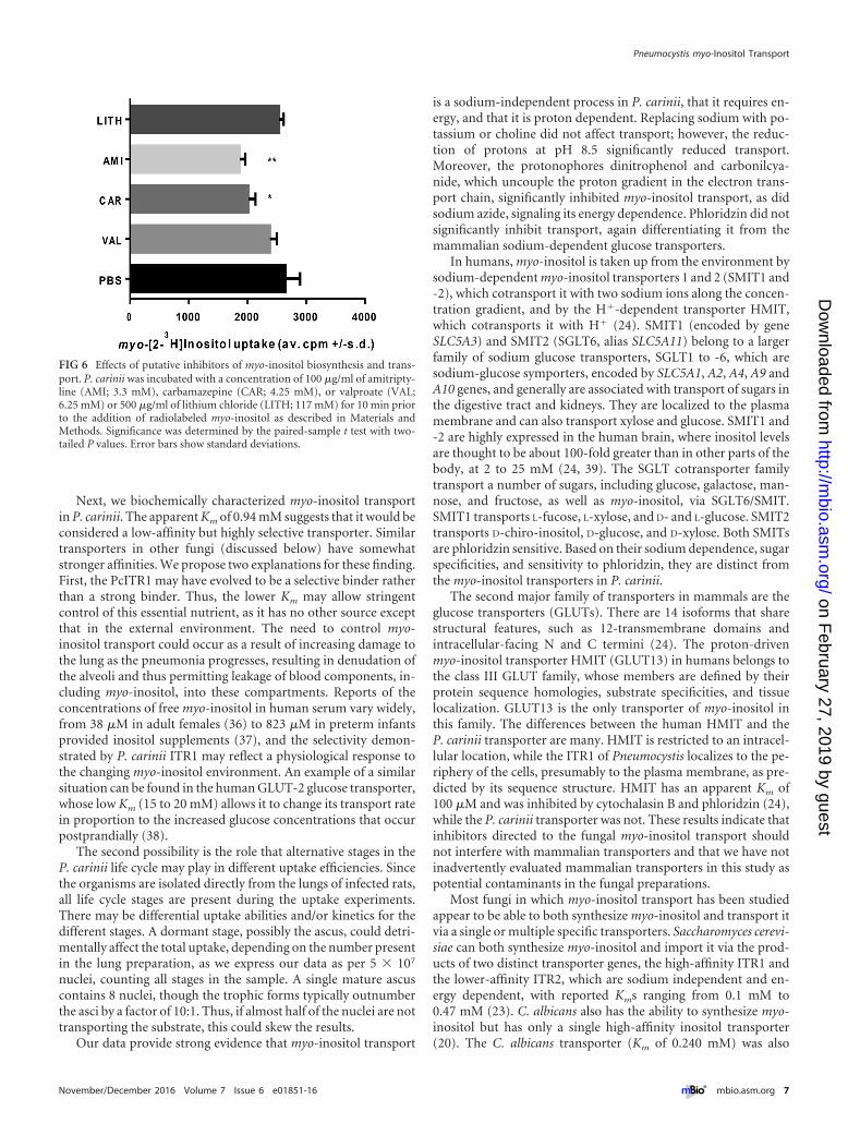
Effects of putative inhibitors of myo-inositol synthesis andtransport on viability of P. carinii. Studies on the inhibition ofmyo-inositol transport and interruption of myo-inositol biosyn-thesis have explored them as potential therapies for bipolar dis-ease. Standard treatments of this neurological disease include lith-ium salts, (e.g., lithium chloride), carbamazepine, and valproate(26). Studies have hypothesized both that lithium can inhibit myo-inositol biosynthesis and that it can decrease the activity of high-affinity myo-inositol transport, along with 2 other bipolar thera-pies, valproate and carbamazepine, based on studies in culturedastrocytes (26). We evaluated the effects of these drugs on theuptake of radiolabeled myo-inositol (Fig. 6) and on the viability ofcultured P. carinii using an ATP-driven assay (Table 1) (27). Asshown by the results in Fig. 6, lithium and valproate had no effecton uptake, while amitriptyline and carbamazepine reduced up-take by about 30% and 20%, respectively.
To evaluate whether these compounds had a correlative effect
on the viability of P. carinii, an in vitro assessment using live or-ganisms was then conducted. Table 1 shows the results of theinhibitors after 3 days of exposure to P. carinii in a cell-free system,with the readout of ATP measured by relative light units (RLU) ina luciferine-luciferase-based assay (28). Included in these assayswas the sugar L-fucose, which has been shown to inhibit the hu-man SMIT transporters. P. carinii was unaffected by exposure tolithium chloride at concentrations as high as 500 �g/ml, whichwould be consistent with the lack of synthetic activity in P. carinii.Likewise, valproate and L-fucose had no significant effects on ATPlevels. In contrast, carbamazepine reduced the ATP content by70% at the highest concentration, though lower levels did notexert any influence. Carbamazepine was shown to reduce radiola-beled myo-inositol transport in astrocyte cell cultures (29). Ofinterest was the effect of amitriptyline, a tricyclic antidepressantwhich acts primarily as a serotonin-norepinephrine reuptake in-hibitor, with strong actions on the serotonin transporter andmoderate effects on the norepinephrine transporter (29). Be-sides identifying potential new anti-PCP therapies, these stud-ies showed the correlation between the 2 assay systems. Lith-ium and valproate were unable to block radiolabeled myo-inositol uptake (Fig. 6) and also had no effect on the viability ofP. carinii in the ATP assay system (Table 1), while amitriptylineand carbamazepine did block uptake (Fig. 6) and were able toreduce viability as assessed by ATP levels (Table 1). However,the normal therapeutic target levels for humans are 120 to150 ng/ml for amitriptyline and 4 to 12 �g/ml for carbamaz-epine. These levels are much lower than those used in these
TABLE 1 In vitro assessment of potential transport inhibitors
Compound tested,concn (�g/ml)
% reduction in ATP vs.untreated control after 72 h
Lithium chloride500 0250 0125 062.5 0
L-Fucose1,000 0100 010 01 2.550.1 3.37
Valproate100 11.6810 1.911 20.620.1 13.73
Amitriptyline100 99.6110 98.761 4.370.1 0
Carbamazepine100 69.3010 6.411 0.070.1 0
Pneumocystis myo-Inositol Transport
November/December 2016 Volume 7 Issue 6 e01851-16 ® mbio.asm.org 5
on February 27, 2019 by guest
http://mbio.asm
.org/D
ownloaded from

short-term experiments, and the inhibition observed couldhave been due to other, off-target effects. Nonetheless, therewas some selectivity exhibited by these 2 drugs.
DISCUSSION
Eukaryotic cells typically obtain myo-inositol from three sources:they can make it from glucose-6-phosphate using just 2 enzymes,1-D-myo-inositol-phosphate synthase and inositol monophos-phatase, they can sequester it from extracellular sources by spe-cialized myo-inositol transporters, or it can be obtained by de-phosphorylation or recycling of inositol-containing membrane orcellular phospholipids (30). myo-Inositol is involved in at leastthree essential cellular functions. As a constituent of phosphati-dylinositol, it is a component of biological membranes, and whenphosphorylated, it participates in exocytosis, cytoskeletal restruc-turing, and intracellular membrane trafficking; it is critical in sig-nal transduction pathways that control many cellular functions;and lastly, it is a precursor of glycosylphosphatidylinositol (GPI)membrane anchors, which affix glycolipids to cell surfaces (31). IfPneumocystis cannot obtain its myo-inositol by de novo synthesis,we hypothesized that these fungi must transport it from its envi-ronment within the mammalian lung. In keeping with that sup-position, we identified 2 potential inositol transporters in the ge-nomes of P. carinii and P. murina and a single one in P. jirovecii(16).
Characterization of P. carinii myo-inositol transport andcomparisons to mammalian and fungal transport systems.PcITR1, as well as other ITRs analyzed in this work, appears tobelong to the major facilitator superfamily (MFS) of membraneproteins that are expressed ubiquitously across all phyla and areinvolved in the transport of various small-molecule compounds(32). The superfamily comprises at least 17 distinct families thatgenerally show specificity to a single class of compounds, in-cluding simple sugars (hexoses, pentoses, and disaccharides),inositols, amino acids, nucleosides, various organic and inor-ganic cations and anions, and even some drugs. The MFS foldconsists of two 6-transmembrane helix bundles that are con-nected by intracellular helices or extended loops. These two6-helix bundles can adopt a clamshell-like outward openingwith a hydrophilic cavity, where the amino acids lining thiscavity determine the transporter’s specificity for a certain classof substrates (19, 33-35). Once a substrate is docked into thecavity, the MFS transporter changes the conformation of the6-helix bundles to open the cavity inward and simultaneouslyclose the outward cavity through a so-called “rocker switch”mechanism (also known as the alternating access mode of op-eration), which prevents a continuous permeability across themembrane (34, 35). In this work, we characterized the kinetics,substrate specificity, and possible inhibition of PcITR1 to helpdefine the function of this essential system.
FIG 5 D-[U-14C]glucose uptake by P. carinii. Competition for uptake of myo-inositol and 14 common sugars was tested. Concentrations of 5 �M D-[U-14C]glucose and 100� the unlabeled inositol isomers and sugars were used. Significance was determined by the paired-sample t test with two-tailed P values.Error bars show standard deviations.
Cushion et al.
6 ® mbio.asm.org November/December 2016 Volume 7 Issue 6 e01851-16
on February 27, 2019 by guest
http://mbio.asm
.org/D
ownloaded from
Next, we biochemically characterized myo-inositol transportin P. carinii. The apparent Km of 0.94 mM suggests that it would beconsidered a low-affinity but highly selective transporter. Similartransporters in other fungi (discussed below) have somewhatstronger affinities. We propose two explanations for these finding.First, the PcITR1 may have evolved to be a selective binder ratherthan a strong binder. Thus, the lower Km may allow stringentcontrol of this essential nutrient, as it has no other source exceptthat in the external environment. The need to control myo-inositol transport could occur as a result of increasing damage tothe lung as the pneumonia progresses, resulting in denudation ofthe alveoli and thus permitting leakage of blood components, in-cluding myo-inositol, into these compartments. Reports of theconcentrations of free myo-inositol in human serum vary widely,from 38 �M in adult females (36) to 823 �M in preterm infantsprovided inositol supplements (37), and the selectivity demon-strated by P. carinii ITR1 may reflect a physiological response tothe changing myo-inositol environment. An example of a similarsituation can be found in the human GLUT-2 glucose transporter,whose low Km (15 to 20 mM) allows it to change its transport ratein proportion to the increased glucose concentrations that occurpostprandially (38).
The second possibility is the role that alternative stages in theP. carinii life cycle may play in different uptake efficiencies. Sincethe organisms are isolated directly from the lungs of infected rats,all life cycle stages are present during the uptake experiments.There may be differential uptake abilities and/or kinetics for thedifferent stages. A dormant stage, possibly the ascus, could detri-mentally affect the total uptake, depending on the number presentin the lung preparation, as we express our data as per 5 � 107
nuclei, counting all stages in the sample. A single mature ascuscontains 8 nuclei, though the trophic forms typically outnumberthe asci by a factor of 10:1. Thus, if almost half of the nuclei are nottransporting the substrate, this could skew the results.
Our data provide strong evidence that myo-inositol transport
is a sodium-independent process in P. carinii, that it requires en-ergy, and that it is proton dependent. Replacing sodium with po-tassium or choline did not affect transport; however, the reduc-tion of protons at pH 8.5 significantly reduced transport.Moreover, the protonophores dinitrophenol and carbonilcya-nide, which uncouple the proton gradient in the electron trans-port chain, significantly inhibited myo-inositol transport, as didsodium azide, signaling its energy dependence. Phloridzin did notsignificantly inhibit transport, again differentiating it from themammalian sodium-dependent glucose transporters.
In humans, myo-inositol is taken up from the environment bysodium-dependent myo-inositol transporters 1 and 2 (SMIT1 and-2), which cotransport it with two sodium ions along the concen-tration gradient, and by the H�-dependent transporter HMIT,which cotransports it with H� (24). SMIT1 (encoded by geneSLC5A3) and SMIT2 (SGLT6, alias SLC5A11) belong to a largerfamily of sodium glucose transporters, SGLT1 to -6, which aresodium-glucose symporters, encoded by SLC5A1, A2, A4, A9 andA10 genes, and generally are associated with transport of sugars inthe digestive tract and kidneys. They are localized to the plasmamembrane and can also transport xylose and glucose. SMIT1 and-2 are highly expressed in the human brain, where inositol levelsare thought to be about 100-fold greater than in other parts of thebody, at 2 to 25 mM (24, 39). The SGLT cotransporter familytransport a number of sugars, including glucose, galactose, man-nose, and fructose, as well as myo-inositol, via SGLT6/SMIT.SMIT1 transports L-fucose, L-xylose, and D- and L-glucose. SMIT2transports D-chiro-inositol, D-glucose, and D-xylose. Both SMITsare phloridzin sensitive. Based on their sodium dependence, sugarspecificities, and sensitivity to phloridzin, they are distinct fromthe myo-inositol transporters in P. carinii.
The second major family of transporters in mammals are theglucose transporters (GLUTs). There are 14 isoforms that sharestructural features, such as 12-transmembrane domains andintracellular-facing N and C termini (24). The proton-drivenmyo-inositol transporter HMIT (GLUT13) in humans belongs tothe class III GLUT family, whose members are defined by theirprotein sequence homologies, substrate specificities, and tissuelocalization. GLUT13 is the only transporter of myo-inositol inthis family. The differences between the human HMIT and theP. carinii transporter are many. HMIT is restricted to an intracel-lular location, while the ITR1 of Pneumocystis localizes to the pe-riphery of the cells, presumably to the plasma membrane, as pre-dicted by its sequence structure. HMIT has an apparent Km of100 �M and was inhibited by cytochalasin B and phloridzin (24),while the P. carinii transporter was not. These results indicate thatinhibitors directed to the fungal myo-inositol transport shouldnot interfere with mammalian transporters and that we have notinadvertently evaluated mammalian transporters in this study aspotential contaminants in the fungal preparations.
Most fungi in which myo-inositol transport has been studiedappear to be able to both synthesize myo-inositol and transport itvia a single or multiple specific transporters. Saccharomyces cerevi-siae can both synthesize myo-inositol and import it via the prod-ucts of two distinct transporter genes, the high-affinity ITR1 andthe lower-affinity ITR2, which are sodium independent and en-ergy dependent, with reported Kms ranging from 0.1 mM to0.47 mM (23). C. albicans also has the ability to synthesize myo-inositol but has only a single high-affinity inositol transporter(20). The C. albicans transporter (Km of 0.240 mM) was also
FIG 6 Effects of putative inhibitors of myo-inositol biosynthesis and trans-port. P. carinii was incubated with a concentration of 100 �g/ml of amitripty-line (AMI; 3.3 mM), carbamazepine (CAR; 4.25 mM), or valproate (VAL;6.25 mM) or 500 �g/ml of lithium chloride (LITH; 117 mM) for 10 min priorto the addition of radiolabeled myo-inositol as described in Materials andMethods. Significance was determined by the paired-sample t test with two-tailed P values. Error bars show standard deviations.
Pneumocystis myo-Inositol Transport
November/December 2016 Volume 7 Issue 6 e01851-16 ® mbio.asm.org 7
on February 27, 2019 by guest
http://mbio.asm
.org/D
ownloaded from

shown to be sodium independent, energy dependent, and insen-sitive to inhibitors of facilitated diffusion, like the transporters inP. carinii. Remarkably, Cryptococcus neoformans possesses 10 to11 inositol transporters, depending on the variety of the fungus(40), which is thought to result from the requirement for inositolfrom plants in its native environment. Two of these, ITR1A andITR3C, are suggested to be the primary inositol transporterswithin this large family (41). These transporters were functionallycharacterized in yeast mutants (41) but remain to be analyzed forsubstrate specificity. Schizosaccharomyces pombe, which like Pneu-mocystis fungi, is a myo-inositol auxotroph, has 2 transporters,ITR1 and ITR2, with a Km of 0.260 mM (20).
The myo-inositol transport in some protozoan parasites hasbeen characterized. In Trypanosoma cruzi, the etiologic agent ofChagas disease or American trypanosomiasis, myo-inositoltransport is facilitated by an energy-dependent, phloridzin-sensitive but cytochalasin B-insensitive system, suggesting thatthere are at least 2 transporters, a sodium-dependent and asodium-independent one, that are operational in these parasites(42). Leishmania donovani, the agent of leishmaniasis, transportsmyo-inositol with a single transporter, L. donovani MIT (LdMIT),with a Km of 0.08 mM (43). Like the fungal transporters, LdMIT isa myo-inositol proton symporter driven by a proton gradient andis expressed at the plasma membrane (22).
C. albicans has a number of opportunities to salvage inositolfrom its human host. As an intestinal commensal, it has access tothe dietary intake of myo-inositol, which is about 1 g/day, makingfor a reliable source (20). In the disseminated state, it can be ex-posed to 15 to 70 �M in human serum and up to 200 to 270 �M inliver and lymphatic tissue. In infections with C. albicans andC. neoformans, myo-inositol is associated with pathogenesis andvirulence (20). In the case of C. neoformans, myo-inositol secretedby plants stimulates mating, as does exogenous myo-inositol im-ported by transporters in S. pombe (21). In Trypanosoma brucei,the H�-linked myo-inositol transporter T. brucei HMIT (TbH-MIT) was recently shown to be essential for viability in its blood-stream form (25). In L. donovani, a cytotoxic myo-inositol ana-logue was shown to dramatically reduce viability (44).
Within the lung, Pneumocystis is exposed to rich sources ofmyo-inositol in its free form (45) or as cleavage products ofinositol-containing molecules, such as phosphatidylinositol orphospholipids (45). Thus, these fungi may have additional meansto import inositol-containing molecules that could then be me-tabolized to release the inositol, entering into further metabolicpathways. The upregulation of genes associated with inositol me-tabolism described in our previous paper provides some supportfor this contention (16). The connection of inositol with mating inS. pombe and C. neoformans is additionally intriguing, as Pneumo-cystis species reproduce sexually within the lung (1). All of thesestudies attest to the essentiality of this simple sugar and its poten-tial as a new target.
Pneumocystis myo-inositol transport is highly selective. Incontrast to myo-inositol transport in C. albicans, L. donovani, andT. brucei, the Pneumocystis myo-inositol transport system was se-lective for myo-inositol only and did not transport other stereo-isomers or sugars that were evaluated in several experiments. Totest this specificity in another way, we conducted transport studieswith radiolabeled D-glucose. Transport was significantly inhibitedby D-mannose, D-glucose, D-galactose, and D-fucose but not myo-
inositol, providing evidence that there is a hexoselike transporterthat is separate and distinct from the transport of myo-inositol.
Inositol and the management of its levels in the human brainare a focus of investigation for the treatment of neurological dis-orders like bipolar disease, where it is hypothesized that reductionof cellular signaling via inhibition of myo-inositol biosynthesis orreduction of its cellular concentrations reduces or prevents symp-toms (29, 43). Inositol monophosphatase is one putative target forlithium therapy, one of the primary treatments for bipolar disease.Valproate and other mood stabilizers, such as carbamazepine,have also been investigated for their mechanisms of action, whichappear to act in shared and disparate cellular processes. SMIT1activity was inhibited by lithium, valproate, and carbamazepine,but it is not likely to be the primary target of these mood stabilizers(29). We report here that the uptake of radiolabeled myo-inositolby P. carinii was unaffected by the addition of lithium salt, nor waslithium salt effective in reducing the viability of the fungus after3 days of exposure, even at high levels. Such results would beexpected if the target is biosynthesis of myo-inositol. Valproatewas also ineffective at blocking transport and reducing viability.The inhibition of uptake and reduction of viability by amitripty-line and carbamazepine were unexpected, though both had mod-est effects in each assay and were present at levels exceeding targettherapeutic doses. The mechanisms of action of both compoundsare under active investigation, but a role for inhibition of myo-inositol transport has not been reported, and the effects on P. cari-nii may be due to a downstream or heretofore-unidentified mech-anism. Nonetheless, further investigations of the effects of thesecompounds on Pneumocystis infection in an animal model of PCPseem warranted.
MATERIALS AND METHODSSource of Pneumocystis. Stocks of P. carinii were prepared from individ-ual rats, assessed for microbial contamination, ATP content, and organ-ism numbers, and then cryopreserved in liquid nitrogen, as we have de-scribed previously (46). For use in uptake studies, cryopreservedorganisms were quickly thawed at 37°C, plated in RPMI 1640 with 20%horse serum and other nutritional supplements (47), and treated withantibiotics and antimycotics for 24 h to further reduce the possibility ofmicrobial contamination.
Suppression of the immune system of rats was induced by intraperi-toneal injections of methylprednisolone (0.01 to 0.02 mg/kg of bodyweight subcutaneously once per week) for up to 8 to 10 weeks. Water wasacidified to prevent any secondary bacterial infections. Rats were used as asource of the organisms, as they produce higher organism burdens withlarger Pneumocystis numbers than mice and fewer rats are needed tomaintain stock levels. All procedures were conducted according toIACUC-approved protocols. P. carinii organisms were purified from lungtissue by a series of centrifugations and a lytic step to reduce erythrocytesand host lung cells (46).
The studies involving animals were conducted in compliance withapplicable laws, regulations, and institutional policies and with approvalby the institutional animal care and use committees of the University ofCincinnati and the Cincinnati Veterans Affairs Medical Center (M. T.Cushion holds the protocols).
Reagents. myo-[2-3H]inositol (specific activity of 21 to 24 Ci mmol�1;777 to 888 GBq mmol�1) was purchased from PerkinElmer Life Sciences.Inositol isomers and derivatives were obtained from Sigma Aldrich (St.Louis, MO) (allo-, D-chiro-, L-chiro-, epi-, muco-, and myo-inositol, phyticacid, and neo-inositol, and quebrachitol), Calbiochem (scyllo- and myo-inositol), or Industrial Research Ltd. (Lower Hutt, New Zealand) (L-chiro-inositol, D-ononitol, D-pinitol, and viburnitol). Monosaccharides, iono-
Cushion et al.
8 ® mbio.asm.org November/December 2016 Volume 7 Issue 6 e01851-16
on February 27, 2019 by guest
http://mbio.asm
.org/D
ownloaded from

phores, and other inhibitors were purchased from Sigma Aldrich and wereof the highest grade available.
Inositol uptake assays. For uptake assays, overnight cultures ofP. carinii (as described above) (5 � 10 7 to 1 � 108 cells ml�1) wereharvested by centrifugation at 2,000 � g for 10 min, washed twice inphosphate-buffered saline (PBS; 135 mM NaCl, 1.3 mM KCl, 3.2 mMNa2HPO4, 0.5 mM KH2PO4, pH 7.4) at 4°C, and resuspended in PBS,unless otherwise stated. After 10 min of preincubation at 37°C, the uptakeof radiolabeled myo-inositol was initiated by the addition of 100 �l of cellsuspension (5 � 107) to 100 �l of myo-[2-3H]inositol (3 �Ci ml�1;110 kBq ml�1) at 0.5 �M final concentration in PBS at 37°C. At varioustime points (1, 3, 5, 7, and 10 min), uptake was terminated by the additionof ice-cold PBS, followed by centrifugation at 2,000 � g. The pelletedorganisms were snap frozen and then lysed with 0.1 M NaOH and addedto 3 ml scintillation fluid (Scintiverse BD; Fisher Scientific). Radioactivitywas measured by scintillation counting using a Packard TriCarb 2100 TRliquid scintillation analyzer (PerkinElmer Life Sciences). The uptake ofradiolabeled myo-inositol at each time point was calculated and plotted asa function of time. The initial myo-inositol uptake rate was determined bylinear regression analysis of the plotted data points for each assay withinthe first 10 min of myo-inositol uptake. Determination of picomoles per5 � 107 P. carinii cells was based on a standard curve of varying concen-trations of myo-inositol and the resultant counts per minute (cpm) (R2 �0.9989).
Substrate saturation kinetics. Kinetic studies for myo-inositol trans-port in P. carinii were performed in PBS, pH 7.4, within the linear uptakerange of each concentration of myo-inositol, which ranged from 50 �M to3 mM. Six biological replicates were used, based on an initial power anal-ysis. The apparent Km and Vmax values were determined by the Michaelis-Menten equation V � Vmax[S]/(Km � [S]), where S is the myo-inositolconcentration. The data were background corrected and fitted using a4-parameter regression in Kaleidagraph (Synergy Software, Reading, PA).The regression coefficient for the Michaelis-Menten equation fit was0.9677.
Ion specificity and pharmacology studies. The ion specificity of myo-inositol transport in P. carinii was determined by first washing 5 � 107
P. carinii cells in each of the uptake buffers of 140 mM NaCl, KCl, orcholine chloride in 25 mM HEPES, pH 7.3, prior to the addition of theradiolabeled myo-inositol as described above. For assessment of the effectof pH (H� concentration), the P. carinii cells were washed in each of threepH buffers representing differing proton concentrations, 316 nM H�
(pH 6.5), 31 nM H� (pH 7.5), and 3.2 nM H� (pH 8.5).For the inhibitor (pharmacological profile) studies, the drugs car-
bonilcyanide, dinitrophenol, sodium azide, cytochalasin B, and phlo-ridzin were incubated with 5 � 107 ml�1 P. carinii at their final con-centrations (noted in Fig. 3B) for 10 min at 37°C prior to the start ofthe uptake assays.
Substrate specificity. The substrate specificities of P. carinii myo-inositol and glucose transport were examined by testing 8 stereoisomers ofmyo-inositol, 5 inositol derivatives, and 15 sugars for their ability to com-pete with myo-inositol and glucose uptake. Cryopreserved P. carinii or-ganisms were prepared as described above. Radiolabeled myo-inositol orradiolabeled glucose was added as described above, followed by incuba-tion at 37°C for 10 min. The final concentrations of isotopes were 5 �MD-[14C]glucose and 0.5 �M [3H]myo-inositol. To stop uptake, iced PBSwas added. Samples were washed twice in cold PBS and centrifuged at2,000 � g at 4°C. Pellets were suspended in 100 �l 0.1 M NaOH, and thelysate was transferred to 3 ml scintillation fluid. The cpm were determinedwith a Beckman LS6500 liquid scintillation counter.
In vitro ATP assay. Lithium chloride, amitriptyline, carbamazepine,valproate, and L-fucose were evaluated for their ability to reduce the via-bility of P. carinii using the ATP bioluminescence assay established in ourlaboratory (28). All were also evaluated for their ability to inhibit theuptake of myo-[2-3H]inositol, as described above. The ATP assay wasinitiated by rapidly thawing cryopreserved P. carinii organisms at 37°C
and resuspending them at 5 � 107 nuclei/ml in RPMI 1640 containing10% horse serum with or without a test drug. Each drug concentrationwas assayed in triplicate in 12-well plates. Negative controls includedmedium with P. carinii and without drug, medium with P. carinii and10 �g of ampicillin/ml (negative control), and medium with P. cariniiwithout drug and with any vehicles used. Pentamidine at 1 �g/mlserved as the positive control for drug activity. The plates were incu-bated at 5% CO2, 37°C, and ATP content was measured at 24, 48, and72 h postinoculation.
ATP was measured using a bioluminescence assay as previously de-scribed (48). The linear range of the ATP assay is 1 �M to 100 fM(~20,000,000 to 2,000 RLU). Fifty-microliter samples were lysed, trans-ferred to opaque white plates, and assessed for ATP content usingATPlite-M for light emission at 562 nm, measured with a PolarStar Op-tima spectrophotometer (BMG, Inc.). A quench control to evaluate effectson the enzyme-substrate reaction was run for every drug tested.
Protein structure prediction and annotation. Protein structure mod-eling and annotation are based on the P. carinii ITR1 sequence (RefSeqaccession number AIU34725.1). For comparison, ITR1 3-D structureswere modeled from Saccharomyces cerevisiae (RefSeq accession numberNP_010785.1; ScITR1), Schizosaccharomyces pombe (RefSeq accessionnumber NP_593858.2; SpITR1), Candida tropicalis (RefSeq accessionnumber XP_002547329.1; CtITR1), and Fusarium oxysporum (GenBankaccession number ENH74095.1; FoITR1). 3-D structures were modeledusing the Phyre2 server (49). The advantage of the server is that its searchfor templates includes both sequence homology and similarity in the sec-ondary structure profile, enabling the identification of similar folds evenwithout high sequence identity. Prediction of the transmembrane regionsand membrane topology was conducted using HMMTop (50), TMPred(http://www.ch.embnet.org/software/TMPRED_form.html), MINNOU(51), CCTOP (52), and Phyre2 (49). Protein structure classification wasperformed using the NCBI Conserved Domain Database (33).
SUPPLEMENTAL MATERIALSupplemental material for this article may be found at http://mbio.asm.org/lookup/suppl/doi:10.1128/mBio.01851-16/-/DCSupplemental.
Figure S1, PDF file, 0.03 MB.Figure S2, PDF file, 0.1 MB.
FUNDING INFORMATIONThis work, including the efforts of Melanie T. Cushion, was funded byNational Heart, Lung, and Blood Institute (NHLBI) (HL119190) and theVeterans Affairs BLR&D through a Merit Review award.
REFERENCES1. Cushion MT, Stringer JR. 2010. Stealth and opportunism: alternative
lifestyles of species in the fungal genus Pneumocystis. Annu Rev Microbiol64:431– 452. http://dx.doi.org/10.1146/annurev.micro.112408.134335.
2. Carmona EM, Limper AH. 2011. Update on the diagnosis and treatmentof Pneumocystis pneumonia. Ther Adv Respir Dis 5:41–59. http://dx.doi.org/10.1177/1753465810380102.
3. Huang L, Cattamanchi A, Davis JL, den Boon S, Kovacs J, Meshnick S,Miller RF, Walzer PD, Worodria W, Masur H. 2011. HIV-associatedPneumocystis pneumonia. Proc Am Thorac Soc 8:294 –300. http://dx.doi.org/10.1513/pats.201009-062WR.
4. Tellez I, Barragán M, Franco-Paredes C, Petraro P, Nelson K, Del RioC. 2008. Pneumocystis jiroveci pneumonia in patients with AIDS in theinner city: a persistent and deadly opportunistic infection. Am J Med Sci335:192–197. http://dx.doi.org/10.1097/MAJ.0b013e318152004b.
5. Morris A, Sciurba FC, Norris KA. 2008. Pneumocystis: a novel pathogenin chronic obstructive pulmonary disease? COPD 5:43–51. http://dx.doi.org/10.1080/15412550701817656.
6. Murdaca G, Spanò F, Contatore M, Guastalla A, Penza E, Magnani O,Puppo F. 2015. Infection risk associated with anti-TNF-alpha agents: areview. Expert Opin Drug Saf 14:571–582. http://dx.doi.org/10.1517/14740338.2015.1009036.
7. Estébanez-Muñoz M, Soto-Abánades CI, Ríos-Blanco JJ, Arribas JR.2012. Updating our understanding of pulmonary disease associated with
Pneumocystis myo-Inositol Transport
November/December 2016 Volume 7 Issue 6 e01851-16 ® mbio.asm.org 9
on February 27, 2019 by guest
http://mbio.asm
.org/D
ownloaded from
HIV infection. Arch Bronconeumol 48:126 –132. (In Spanish.) http://dx.doi.org/10.1016/j.arbres.2011.12.001.
8. Reid AB, Chen SC, Worth LJ. 2011. Pneumocystis jirovecii pneumonia innon-HIV-infected patients: new risks and diagnostic tools. Curr OpinInfect Dis 24:534 –544. http://dx.doi.org/10.1097/QCO.0b013e32834cac17.
9. Ceron I, Rabagliati R, Langhaus J, Silva F, Guzman AM, Lagos M. 2014.Pneumocystis jiroveci pneumonia: comparative study of cases in HIV-infected patients and immunocompromised non-HIV-infected patients.Rev Chilena Infectol 31:417– 424. (In Spanish.) http://dx.doi.org/10.4067/S0716-10182014000400007.
10. Fillatre P, Revest M, Belaz S, Robert-Gangneux F, Zahar JR, Roblot F,Tattevin P. 2016. Pneumocystosis in non-HIV-infected immunocompro-mised patients. Rev Med Interne 37:327–336. (In French.) http://dx.doi.org/10.1016/j.revmed.2015.10.002.
11. Guo F, Chen Y, Yang SL, Xia H, Li XW, Tong ZH. 2014. Pneumocystispneumonia in HIV-infected and immunocompromised non-HIV in-fected patients: a retrospective study of two centers in China. PLoS One9:e101943. http://dx.doi.org/10.1371/journal.pone.0101943.
12. Koopmans PP, Burger DM. 1998. Managing drug reactions to sulfon-amides and other drugs in HIV infection: desensitization rather than re-challenge? Pharm World Sci 20:253–257.
13. Porollo A, Meller J, Joshi Y, Jaiswal V, Smulian AG, Cushion MT. 2012.Analysis of current antifungal agents and their targets within the Pneumo-cystis carinii genome. Curr Drug Targets 13:1575–1585. http://dx.doi.org/10.2174/138945012803530107.
14. Powles MA, Liberator P, Anderson J, Karkhanis Y, Dropinski JF,Bouffard FA, Balkovec JM, Fujioka H, Aikawa M, McFadden D,Schmatz D. 1998. Efficacy of MK-991 (L-743,872), a semisynthetic pneu-mocandin, in murine models of Pneumocystis carinii. Antimicrob AgentsChemother 42:1985–1989.
15. Cushion MT, Linke MJ, Ashbaugh A, Sesterhenn T, Collins MS, LynchK, Brubaker R, Walzer PD. 2010. Echinocandin treatment of pneumo-cystis pneumonia in rodent models depletes cysts leaving trophic burdensthat cannot transmit the infection. PLoS One 5:e8524. http://dx.doi.org/10.1371/journal.pone.0008524.
16. Porollo A, Sesterhenn TM, Collins MS, Welge JA, Cushion MT. 2014.Comparative genomics of pneumocystis species suggests the absence ofgenes for myo-inositol synthesis and reliance on inositol transport andmetabolism. mBio 5(6):e01834-14. http://dx.doi.org/10.1128/mBio.01834-14.
17. Ma L, Chen Z, Huang da W, Kutty G, Ishihara M, Wang H, AbouelleilA, Bishop L, Davey E, Deng R, Deng X, Fan L, Fantoni G, Fitzgerald M,Gogineni E, Goldberg JM, Handley G, Hu X, Huber C, Jiao X, Jones K,Levin JZ, Liu Y, Macdonald P, Melnikov A, Raley C, Sassi M, ShermanBT, Song X, Sykes S, Tran B, Walsh L, Xia Y, Yang J, Young S, Zeng Q,Zheng X, Stephens R, Nusbaum C, Birren BW, Azadi P, Lempicki RA,Cuomo CA, Kovacs JA. 2016. Genome analysis of three Pneumocystisspecies reveals adaptation mechanisms to life exclusively in mammalianhosts. Nat Commun 7:10740. http://dx.doi.org/10.1038/ncomms10740.
18. Nomura N, Verdon G, Kang HJ, Shimamura T, Nomura Y, Sonoda Y,Hussien SA, Qureshi AA, Coincon M, Sato Y, Abe H, Nakada-NakuraY, Hino T, Arakawa T, Kusano-Arai O, Iwanari H, Murata T, Ko-bayashi T, Hamakubo T, Kasahara M, Iwata S, Drew D. 2015. Structureand mechanism of the mammalian fructose transporter GLUT5. Nature526:397– 401. http://dx.doi.org/10.1038/nature14909.
19. Deng D, Xu C, Sun P, Wu J, Yan C, Hu M, Yan N. 2014. Crystalstructure of the human glucose transporter GLUT1. Nature 510:121–125.http://dx.doi.org/10.1038/nature13306.
20. Jin JH, Seyfang A. 2003. High-affinity myo-inositol transport in Candidaalbicans: substrate specificity and pharmacology. Microbiology 149(Pt12):3371–3381. http://dx.doi.org/10.1099/mic.0.26644-0.
21. Niederberger C, Graub R, Schweingruber AM, Fankhauser H, Rusu M,Poitelea M, Edenharter L, Schweingruber ME. 1998. Exogenous inositoland genes responsible for inositol transport are required for mating andsporulation in Shizosaccharomyces pombe. Curr Genet 33:255–261.
22. Drew ME, Langford CK, Klamo EM, Russell DG, Kavanaugh MP,Landfear SM. 1995. Functional expression of a myo-inositol/H� sym-porter from Leishmania donovani. Mol Cell Biol 15:5508 –5515. http://dx.doi.org/10.1128/MCB.15.10.5508.
23. Nikawa J, Tsukagoshi Y, Yamashita S. 1991. Isolation and characteriza-tion of two distinct myo-inositol transporter genes of Saccharomycescerevisiae. J Biol Chem 266:11184 –11191.
24. Augustin R, Mayoux E. 2014. Mammalian sugar transporters. In Sza-blewski L (ed.), Glucose homeostasis. InTech, Rijeka, Croatia. http://dx.doi.org/10.5772/58325.
25. González-Salgado A, Steinmann M, Major LL, Sigel E, Reymond JL,Smith TK, Bütikofer P. 2015. Trypanosoma brucei bloodstream formsdepend upon uptake of myo-inositol for Golgi complex phosphatidylino-sitol synthesis and normal cell growth. Eukaryot Cell 14:616 – 624. http://dx.doi.org/10.1128/EC.00038-15.
26. Aouameur R, Da Cal S, Bissonnette P, Coady MJ, Lapointe JY. 2007.SMIT2 mediates all myo-inositol uptake in apical membranes of rat smallintestine. Am J Physiol Gastrointest Liver Physiol 293:G1300 –G1307.http://dx.doi.org/10.1152/ajpgi.00422.2007.
27. Cushion MT, Chen F, Kloepfer N. 1997. A cytotoxicity assay for evalu-ation of candidate anti-Pneumocystis carinii agents. Antimicrob AgentsChemother 41:379 –384.
28. Cushion MT, Walzer PD. 2009. Preclinical drug discovery for new anti-pneumocystis compounds. Curr Med Chem 16:2514 –2530. http://dx.doi.org/10.2174/092986709788682038.
29. Lubrich B, van Calker D. 1999. Inhibition of the high affinity myo-inositol transport system: a common mechanism of action of antibipolardrugs? Neuropsychopharmacology 21:519 –529. http://dx.doi.org/10.1016/S0893-133X(99)00037-8.
30. Deranieh RM, Greenberg ML. 2009. Cellular consequences of inositoldepletion. Biochem Soc Trans 37:1099 –1103. http://dx.doi.org/10.1042/bst0371099.
31. Simonsen A, Wurmser AE, Emr SD, Stenmark H. 2001. The role ofphosphoinositides in membrane transport. Curr Opin Cell Biol 13:485– 492. http://dx.doi.org/10.1016/S0955-0674(00)00240-4.
32. Pao SS, Paulsen IT, Saier MH, Jr. 1998. Major facilitator superfamily.Microbiol Mol Biol Rev 62:1–34.
33. Marchler-Bauer A, Derbyshire MK, Gonzales NR, Lu S, Chitsaz F, GeerLY, Geer RC, He J, Gwadz M, Hurwitz DI, Lanczycki CJ, Lu F, MarchlerGH, Song JS, Thanki N, Wang Z, Yamashita RA, Zhang D, Zheng C,Bryant SH. 2015. CDD: NCBI’s Conserved Domain Database. NucleicAcids Res 43:D222–D226. http://dx.doi.org/10.1093/nar/gku1221.
34. Abramson J, Smirnova I, Kasho V, Verner G, Kaback HR, Iwata S. 2003.Structure and mechanism of the lactose permease of Escherichia coli. Sci-ence 301:610 – 615. http://dx.doi.org/10.1126/science.1088196.
35. Huang Y, Lemieux MJ, Song J, Auer M, Wang DN. 2003. Structure andmechanism of the glycerol-3-phosphate transporter from Escherichia coli.Science 301:616 – 620. http://dx.doi.org/10.1126/science.1087619.
36. Chiu TT, Rogers MS, Law EL, Briton-Jones CM, Cheung LP, Haines CJ.2002. Follicular fluid and serum concentrations of myo-inositol in pa-tients undergoing IVF: relationship with oocyte quality. Hum Reprod 17:1591–1596. http://dx.doi.org/10.1093/humrep/17.6.1591.
37. Hallman M, Arjomaa P, Hoppu K. 1987. Inositol supplementation inrespiratory distress syndrome: relationship between serum concentration,renal excretion, and lung effluent phospholipids. J Pediatr 110:604 – 610.
38. Lachaal M, Spangler RA, Jung CY. 1993. High Km of GLUT-2 glucosetransporter does not explain its role in insulin secretion. Am J Physiol265(6 Pt 1):E914 –E919.
39. Croze ML, Soulage CO. 2013. Potential role and therapeutic interests ofmyo-inositol in metabolic diseases. Biochimie 95:1811–1827. http://dx.doi.org/10.1016/j.biochi.2013.05.011.
40. Xue C. 2012. Cryptococcus and beyond—inositol utilization and its im-plications for the emergence of fungal virulence. PLoS Pathog 8:e1002869.http://dx.doi.org/10.1371/journal.ppat.1002869.
41. Wang Y, Liu TB, Delmas G, Park S, Perlin D, Xue C. 2011. Two majorinositol transporters and their role in cryptococcal virulence. EukaryotCell 10:618 – 628. http://dx.doi.org/10.1128/EC.00327-10.
42. Einicker-Lamas M, Almeida AC, Todorov AG, de Castro SL, Caruso-Neves C, Oliveira MM. 2000. Characterization of the myo-inositol trans-port system in Trypanosoma cruzi. Eur J Biochem 267:2533–2537. http://dx.doi.org/10.1046/j.1432-1327.2000.01302.x.
43. Schneider S. 2015. Inositol transport proteins. FEBS Lett 589:1049 –1058.http://dx.doi.org/10.1016/j.febslet.2015.03.012.
44. Mongan TP, Ganapasam S, Hobbs SB, Seyfang A. 2004. Substrate spec-ificity of the Leishmania donovani myo-inositol transporter: critical roleof inositol C-2, C-3 and C-5 hydroxyl groups. Mol Biochem Parasitol135:133–141. http://dx.doi.org/10.1016/j.molbiopara.2004.01.015.
45. Holub BJ. 1986. Metabolism and function of myo-inositol and inositolphospholipids. Annu Rev Nutr 6:563–597. http://dx.doi.org/10.1146/annurev.nu.06.070186.003023.
Cushion et al.
10 ® mbio.asm.org November/December 2016 Volume 7 Issue 6 e01851-16
on February 27, 2019 by guest
http://mbio.asm
.org/D
ownloaded from

46. Collins MS, Cushion MT. 2001. Standardization of an in vitro drugscreening assay by use of cryopreserved and characterized Pneumocystiscarinii populations. J Eukaryot Microbiol 48:178S–179S. http://dx.doi.org/10.1111/j.1550-7408.2001.tb00509.x.
47. Cushion MT, Collins MS. 2011. Susceptibility of Pneumocystis to echi-nocandins in suspension and biofilm cultures. Antimicrob Agents Che-mother 55:4513– 4518. http://dx.doi.org/10.1128/AAC.00017-11.
48. Maciejewska D, Zabinski J, Kazmierczak P, Rezler M, Krassowska-Swiebocka B, Collins MS, Cushion MT. 2012. Analogs of pentamidine aspotential anti-Pneumocystis chemotherapeutics. Eur J Med Chem 48:164 –173. http://dx.doi.org/10.1016/j.ejmech.2011.12.010.
49. Kelley LA, Sternberg MJ. 2009. Protein structure prediction on the Web:
a case study using the Phyre server. Nat Protoc 4:363–371. http://dx.doi.org/10.1038/nprot.2009.2.
50. Tusnady GE, Simon I. 1998. Principles governing amino acid composi-tion of integral membrane proteins: application to topology prediction. JMol Biol 283:489 –506. http://dx.doi.org/10.1006/jmbi.1998.2107.
51. Cao B, Porollo A, Adamczak R, Jarrell M, Meller J. 2006. Enhancedrecognition of protein transmembrane domains with prediction-basedstructural profiles. Bioinformatics 22:303–309. http://dx.doi.org/10.1093/bioinformatics/bti784.
52. Dobson L, Remenyi I, Tusnady GE. 2015. CCTOP: a Consensus Con-strained TOPology prediction web server. Nucleic Acids Res 43:W408 –W412. http://dx.doi.org/10.1093/nar/gkv451.
Pneumocystis myo-Inositol Transport
November/December 2016 Volume 7 Issue 6 e01851-16 ® mbio.asm.org 11
on February 27, 2019 by guest
http://mbio.asm
.org/D
ownloaded from


