Functional Anatomy Section One: The Skeleton. Functions of a Skeleton FUNCTIONEXPLANATIONEXAMPLES...
64
Functional Anatomy Section One: The Skeleton
-
Upload
steven-austin -
Category
Documents
-
view
230 -
download
0
Transcript of Functional Anatomy Section One: The Skeleton. Functions of a Skeleton FUNCTIONEXPLANATIONEXAMPLES...

Functional Anatomy
Section One: The Skeleton
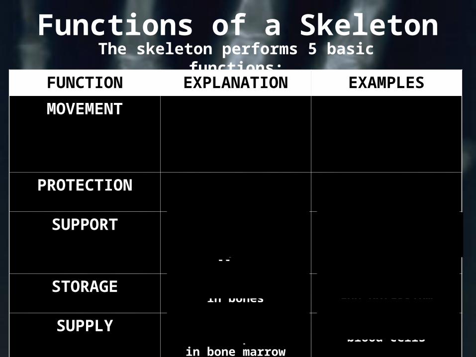
Functions of a Skeleton
FUNCTION EXPLANATION EXAMPLES
MOVEMENT Where bones meet we form joints. In combination with
muscles, we create movement.
Bones move because of joints and
muscles, e.g. elbow moved by biceps
PROTECTION Provide protection to vital organs
Scapula – lungsSkull – brain
SUPPORT Give support for organs and tissue so they do not collapse.
Spine supports the head and trunk
STORAGE Minerals are stored in bones
Storage of calcium and potassium
SUPPLY Red and white blood cells are produced in
bone marrow
Femur produces red blood cells
The skeleton performs 5 basic functions:
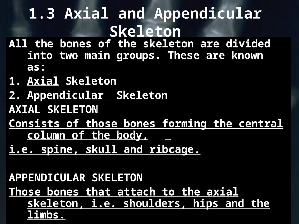
1.3 Axial and Appendicular Skeleton
All the bones of the skeleton are divided into two main groups. These are known as:
1. Axial Skeleton2. Appendicular SkeletonAXIAL SKELETONConsists of those bones forming the central
column of the body, i.e. spine, skull and ribcage.
APPENDICULAR SKELETONThose bones that attach to the axial
skeleton, i.e. shoulders, hips and the limbs.
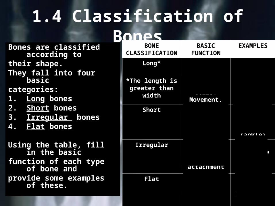
1.4 Classification of Bones
Bones are classified according to
their shape.They fall into four basiccategories:1. Long bones2. Short bones3. Irregular bones4. Flat bones
Using the table, fill in the basic
function of each type of bone and
provide some examples of these.
BONE CLASSIFICATI
ON
BASIC FUNCTION
EXAMPLES
Long*
*The length is greater than
width
Production of red blood
cells and white blood
cells. Movement.
Humerus, femur,
radius and ulna
Short Small of fine movements
Carpals (wrist)Tarsals (ankle)
Irregular Movement support and
muscle attachment
Face and vertebrae
Flat Protection and
attachment
Shoulder blade and breastbon
e
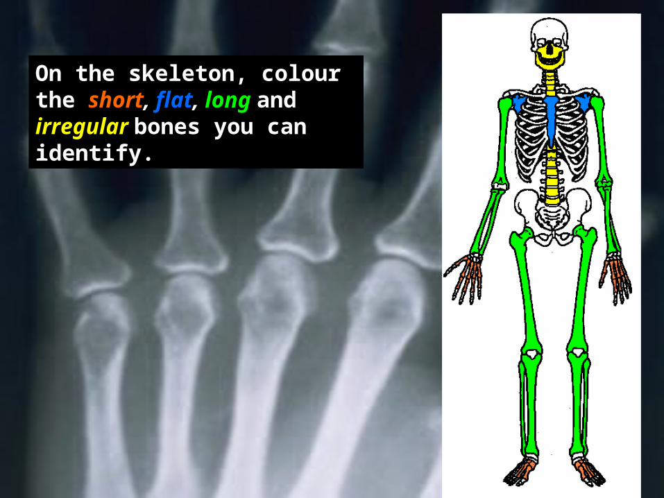
On the skeleton, colour the short, flat, long and irregular bones you can identify.
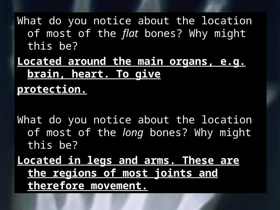
What do you notice about the location of most of the flat bones? Why might this be?
Located around the main organs, e.g. brain, heart. To give
protection.
What do you notice about the location of most of the long bones? Why might this be?
Located in legs and arms. These are the regions of most joints and therefore movement.
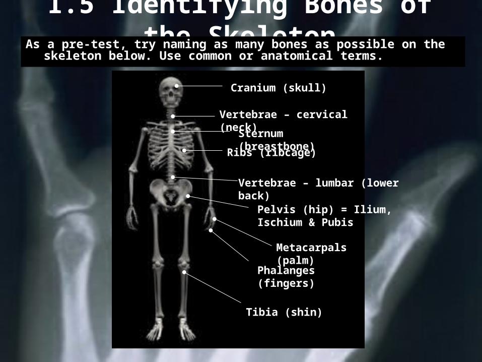
1.5 Identifying Bones of the SkeletonAs a pre-test, try naming as many bones as possible on the
skeleton below. Use common or anatomical terms.
Cranium (skull)
Vertebrae – cervical (neck)Sternum
(breastbone)Ribs (ribcage)
Vertebrae – lumbar (lower back)
Pelvis (hip) = Ilium, Ischium & Pubis
Metacarpals (palm)
Phalanges (fingers)
Tibia (shin)
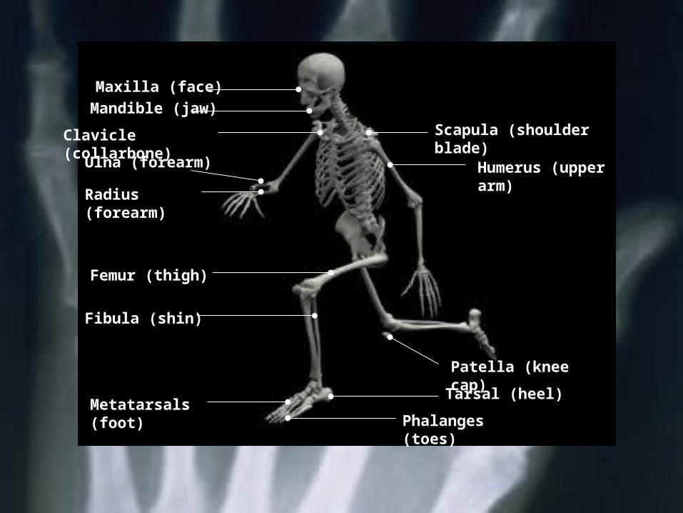
Maxilla (face)
Humerus (upper arm)
Scapula (shoulder blade)
Fibula (shin)
Metatarsals (foot)
Patella (knee cap)Tarsal (heel)
Phalanges (toes)
Mandible (jaw)
Ulna (forearm)
Clavicle (collarbone)
Radius (forearm)
Femur (thigh)
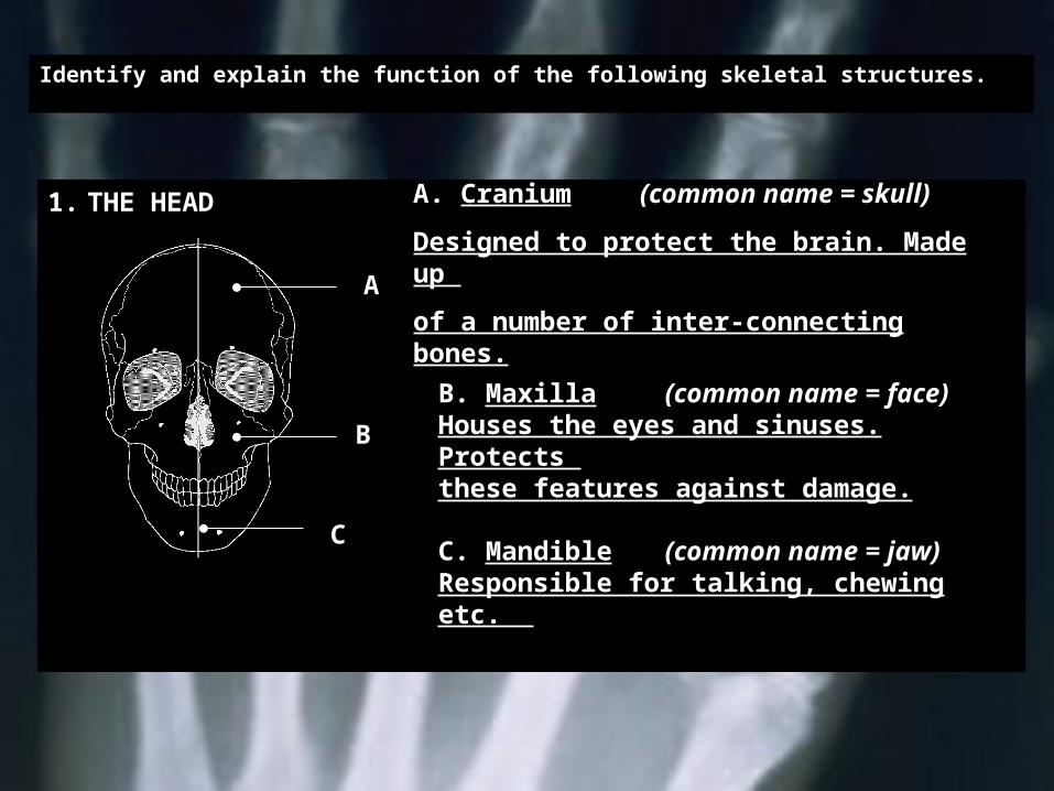
Identify and explain the function of the following skeletal structures.
1. THE HEAD
A
B
C
A. Cranium (common name = skull)
Designed to protect the brain. Made up
of a number of inter-connecting bones.
C. Mandible (common name = jaw)Responsible for talking, chewing etc.
B. Maxilla (common name = face)Houses the eyes and sinuses. Protects these features against damage.
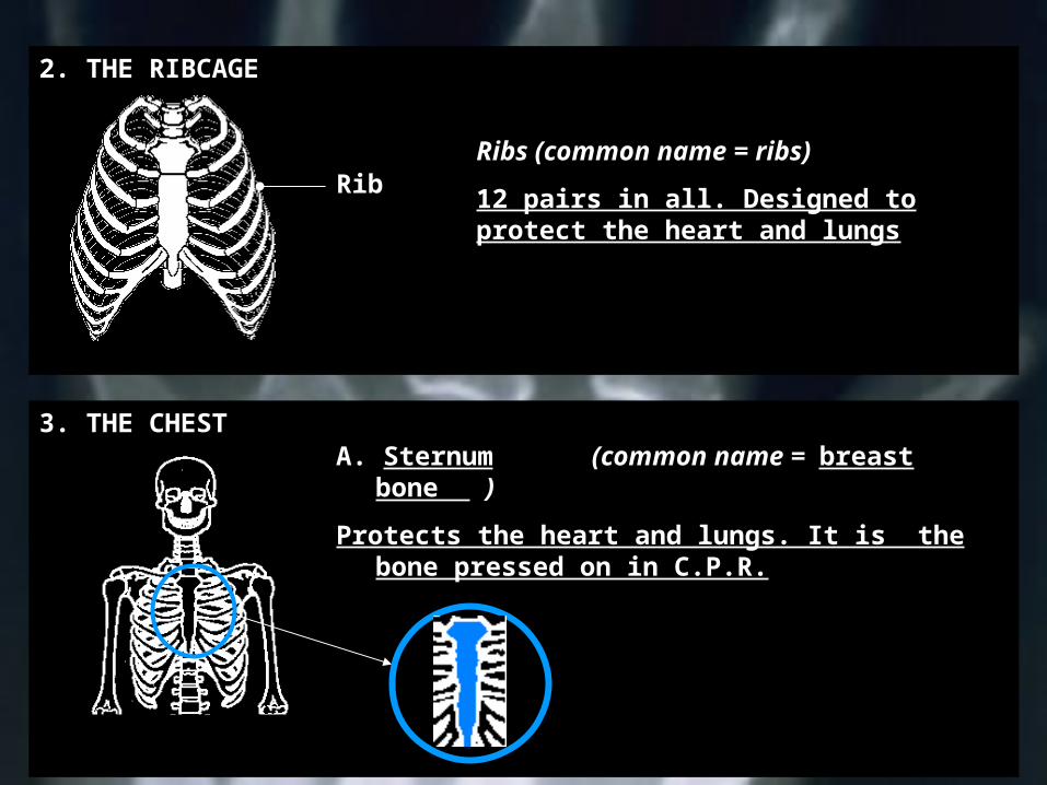
2. THE RIBCAGE
Ribs (common name = ribs)
12 pairs in all. Designed to protect the heart and lungs
Rib
3. THE CHESTA. Sternum (common name = breast
bone )
Protects the heart and lungs. It is the bone pressed on in C.P.R.
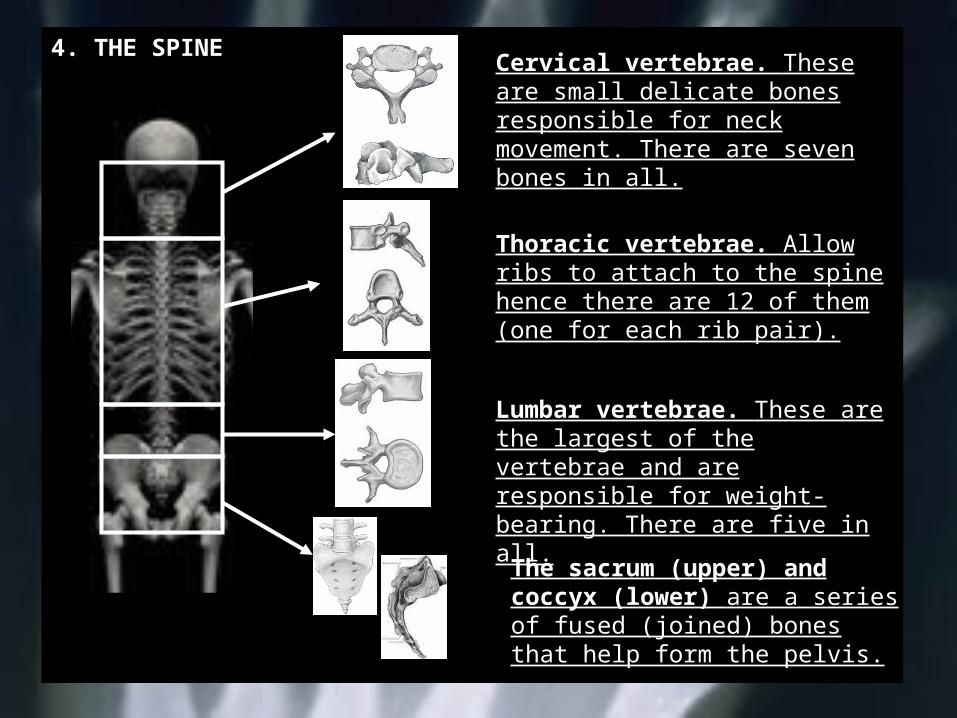
4. THE SPINECervical vertebrae. These are small delicate bones responsible for neck movement. There are seven bones in all.
Thoracic vertebrae. Allow ribs to attach to the spine hence there are 12 of them (one for each rib pair).
Lumbar vertebrae. These are the largest of the vertebrae and are responsible for weight-bearing. There are five in all.
The sacrum (upper) and coccyx (lower) are a series of fused (joined) bones that help form the pelvis.
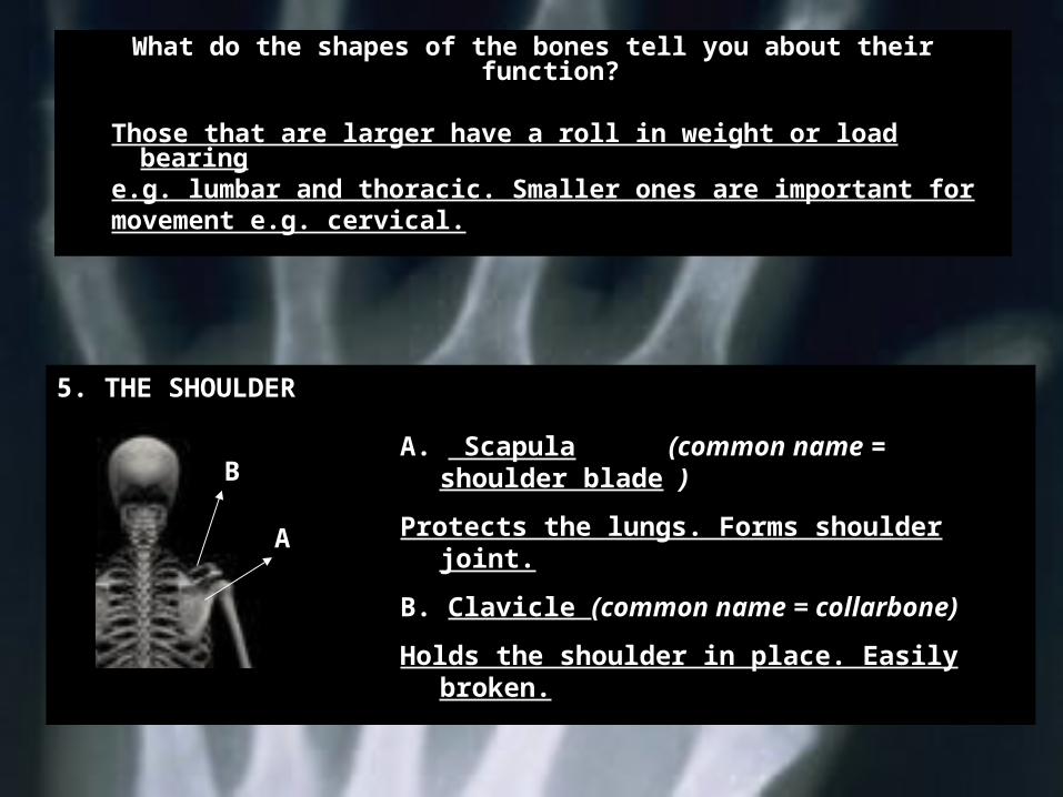
What do the shapes of the bones tell you about their function?
Those that are larger have a roll in weight or load bearing
e.g. lumbar and thoracic. Smaller ones are important formovement e.g. cervical.
5. THE SHOULDER
A. Scapula (common name = shoulder blade )
Protects the lungs. Forms shoulder joint.
B. Clavicle (common name = collarbone)
Holds the shoulder in place. Easily broken.
B
A
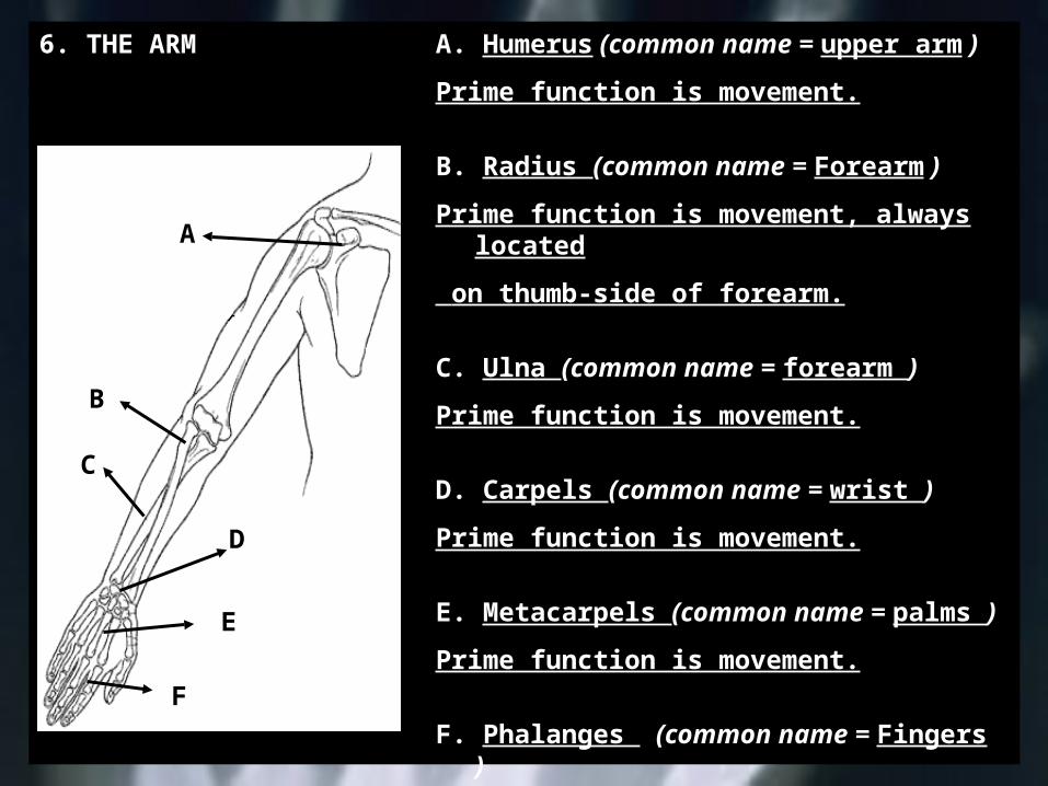
6. THE ARM A. Humerus (common name = upper arm )
Prime function is movement.
B. Radius (common name = Forearm )
Prime function is movement, always located
on thumb-side of forearm.
C. Ulna (common name = forearm )
Prime function is movement.
D. Carpels (common name = wrist )
Prime function is movement.
E. Metacarpels (common name = palms )
Prime function is movement.
F. Phalanges (common name = Fingers )
Prime function is movement.
D
A
E
F
B
C
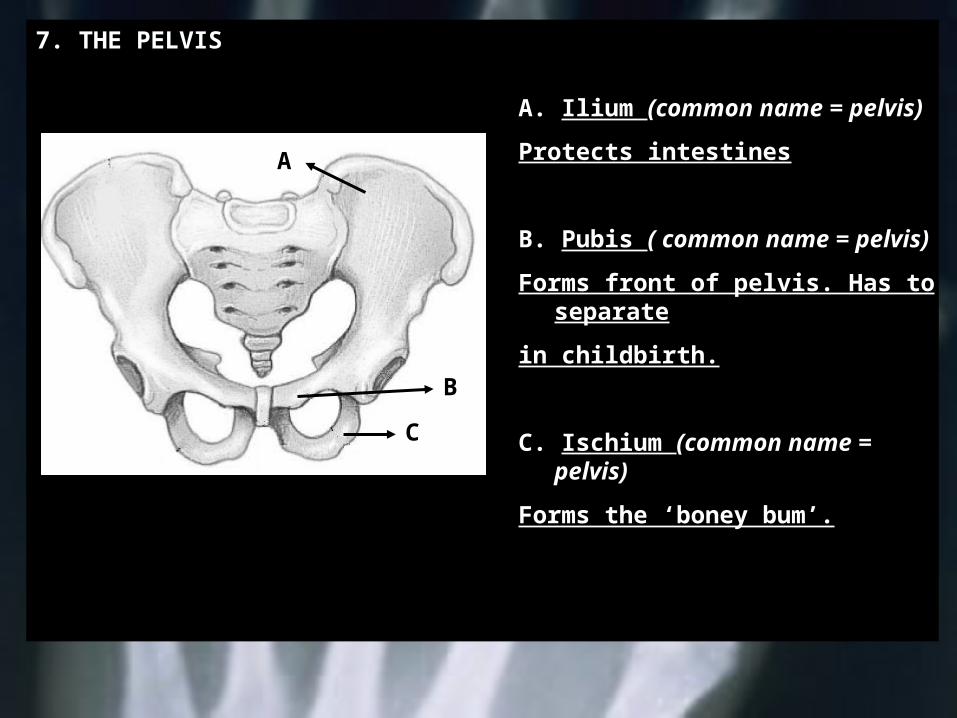
7. THE PELVIS
A. Ilium (common name = pelvis)
Protects intestines
B. Pubis ( common name = pelvis)
Forms front of pelvis. Has to separate
in childbirth.
C. Ischium (common name = pelvis)
Forms the ‘boney bum’.
A
B
C
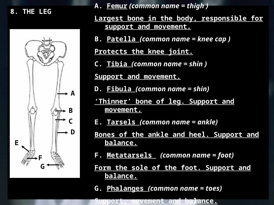
8. THE LEG
A
B
C
F
D
G
E
A. Femur (common name = thigh )
Largest bone in the body, responsible for support and movement.
B. Patella (common name = knee cap )
Protects the knee joint.
C. Tibia (common name = shin )
Support and movement.
D. Fibula (common name = shin)
‘Thinner’ bone of leg. Support and movement.
E. Tarsels (common name = ankle)
Bones of the ankle and heel. Support and balance.
F. Metatarsels (common name = foot)
Form the sole of the foot. Support and balance.
G. Phalanges (common name = toes)
Support, movement and balance.
Functional Anatomy
Section Two: Terms of Direction
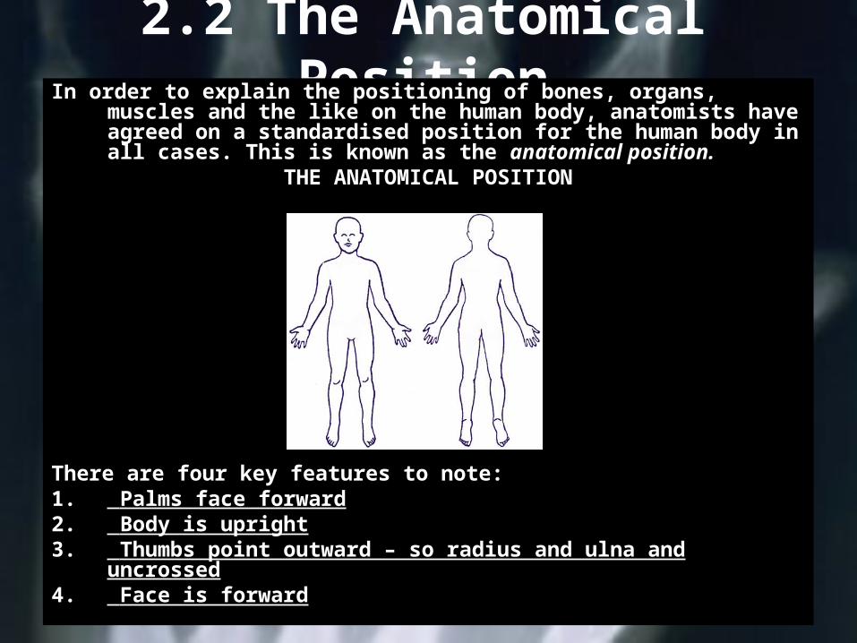
2.2 The Anatomical PositionIn order to explain the positioning of bones, organs, muscles
and the like on the human body, anatomists have agreed on a standardised position for the human body in all cases. This is known as the anatomical position.
THE ANATOMICAL POSITION
There are four key features to note:1. Palms face forward2. Body is upright3. Thumbs point outward – so radius and ulna and
uncrossed4. Face is forward
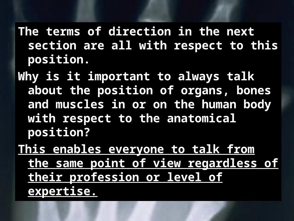
The terms of direction in the next section are all with respect to this position.
Why is it important to always talk about the position of organs, bones and muscles in or on the human body with respect to the anatomical position?
This enables everyone to talk from the same point of view regardless of their profession or level of expertise.
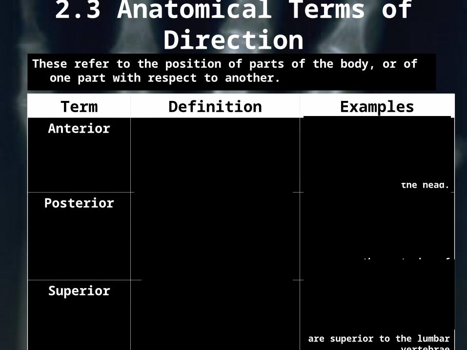
2.3 Anatomical Terms of Direction
These refer to the position of parts of the body, or of one part with respect to another.
Term Definition ExamplesAnterior On the FRONT of the
body or limb
1. The chest is on the anterior of the
body2. The face is an
anterior aspect of the head.
Posterior On the BACK of the body or limb
1. The buttocks are on the posterior of the
body2. The calf muscles are on the posterior of the
leg
Superior Above or on top of 1. The cervical vertebrae are superior to the thoracic
vertebrae2. The thoracic vertebrae are superior to the lumbar
vertebrae

Term Definition Examples
InferiorBelow or beneath
1. Thoracic vertebrae are inferior to the cervical
vertebrae2. The lumbar vertebrae
are inferior to the thoracic vertebrae
MedialNearer the midline
of the body
1. The big toe is on the medial aspect
of the foot 2. The little finger is
on the medial aspect of the hand
LateralFurther away from the midline of the
body
1. Little toe is on the lateral aspect of
the foot.2. The thumb is on the lateral aspect
of the hand
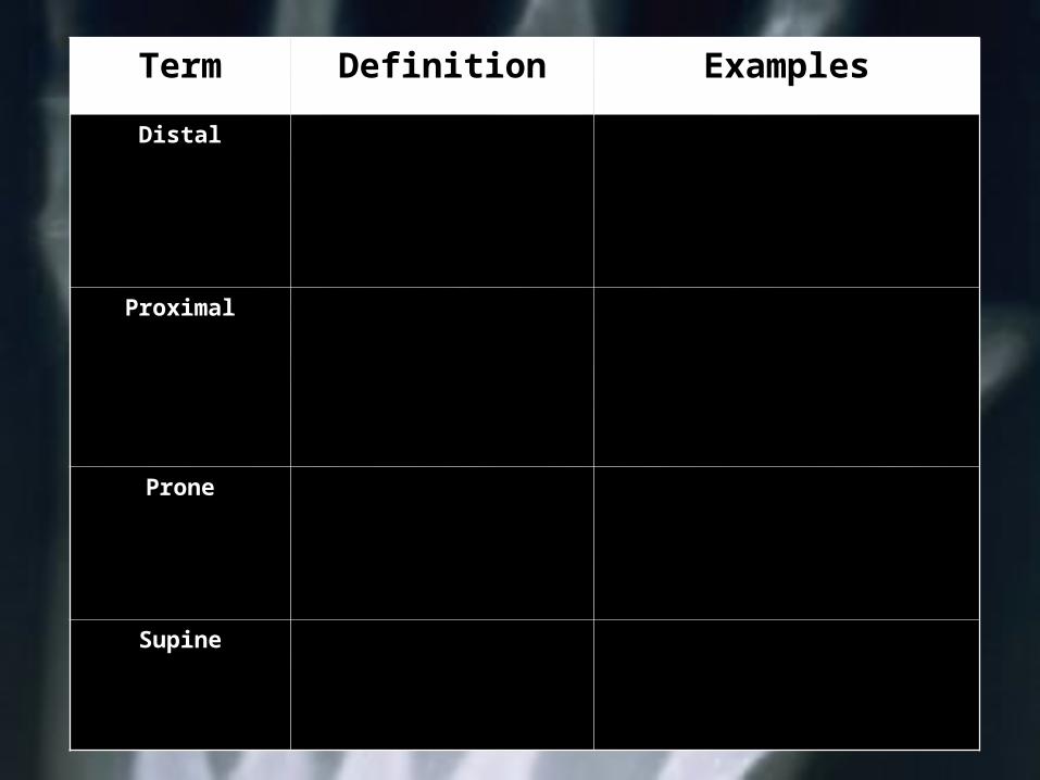
Term Definition Examples
DistalFurther away from the body. Usually
refers to the limbs.
1. Wrist is distal to the elbow
2. Elbow joint is distal to the shoulder
joint
ProximalNearer the body.
Usually refers to the limbs.
1. Elbow is proximal to the wrist joint
2. Shoulder joint is proximal to the
elbow
ProneFace down
A press-up is on the PRONE position
SupineFace up
A sit-up is on the SUPINE position
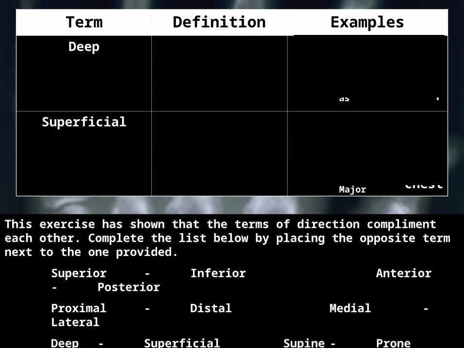
Term Definition ExamplesDeep When a muscle
is BENEATH another with
respect to the skin surface
The ilio psoas is a deep muscle
of the hip
Superficial On the surface of the body or
limb
Pectoralis major is a
superficial muscle of the
chest
Iliopsoas
Pectoralis Major
This exercise has shown that the terms of direction compliment each other. Complete the list below by placing the opposite term next to the one provided.
Superior - Inferior Anterior- Posterior
Proximal - Distal Medial -Lateral
Deep - Superficial Supine - Prone

Functional Anatomy
Section Three: The Joints
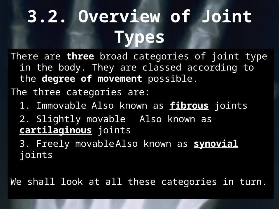
3.2. Overview of Joint Types
There are three broad categories of joint type in the body. They are classed according to the degree of movement possible.
The three categories are:1. Immovable Also known as fibrous joints2. Slightly movable Also known as cartilaginous joints3. Freely movable Also known as synovial joints
We shall look at all these categories in turn.
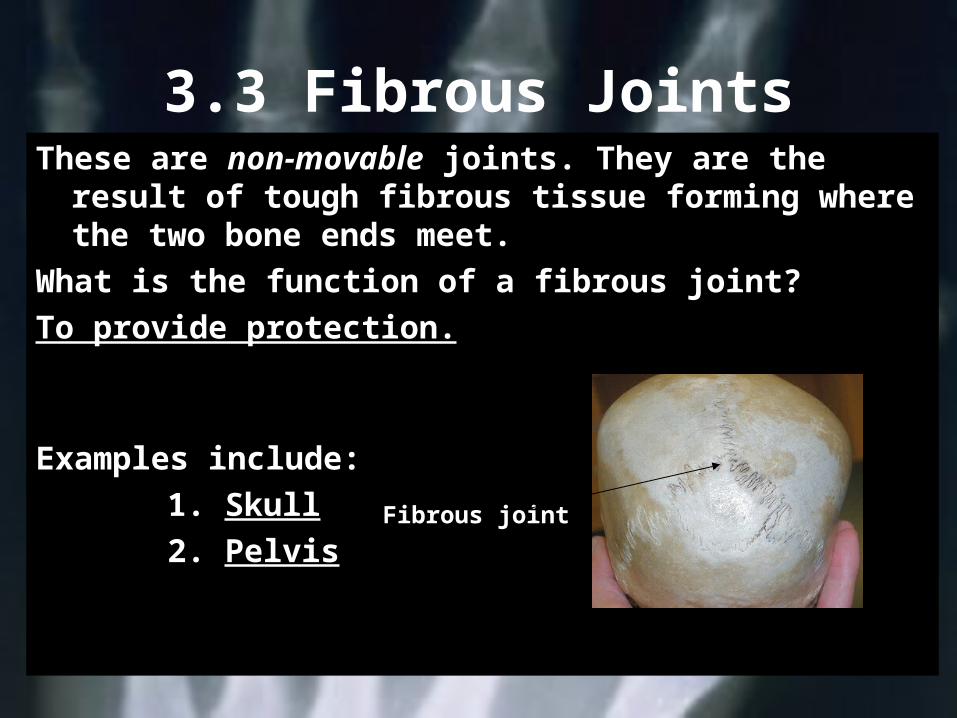
3.3 Fibrous JointsThese are non-movable joints. They are the result
of tough fibrous tissue forming where the two bone ends meet.
What is the function of a fibrous joint?To provide protection.
Examples include:1. Skull2. Pelvis Fibrous joint
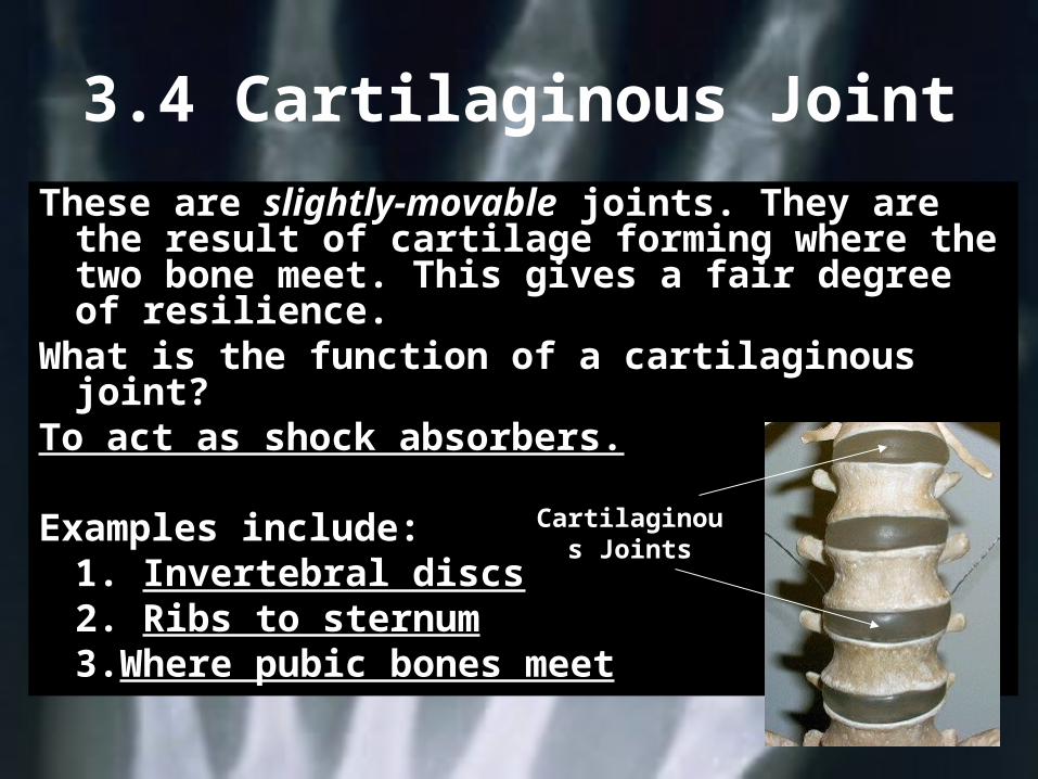
3.4 Cartilaginous Joint
These are slightly-movable joints. They are the result of cartilage forming where the two bone meet. This gives a fair degree of resilience.
What is the function of a cartilaginous joint?
To act as shock absorbers.
Examples include:1. Invertebral discs2. Ribs to sternum3.Where pubic bones meet
Cartilaginous Joints
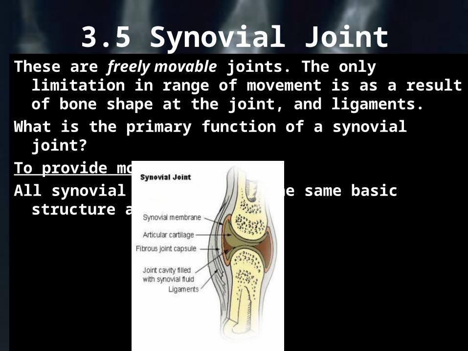
3.5 Synovial JointThese are freely movable joints. The only
limitation in range of movement is as a result of bone shape at the joint, and ligaments.
What is the primary function of a synovial joint?To provide movement.All synovial joints follow the same basic structure
as shown
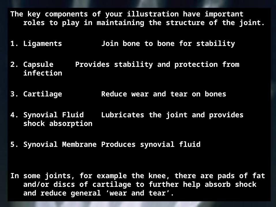
The key components of your illustration have important roles to play in maintaining the structure of the joint.
1. Ligaments Join bone to bone for stability
2. Capsule Provides stability and protection from infection
3. Cartilage Reduce wear and tear on bones
4. Synovial Fluid Lubricates the joint and provides shock absorption
5. Synovial MembraneProduces synovial fluid
In some joints, for example the knee, there are pads of fat and/or discs of cartilage to further help absorb shock and reduce general ‘wear and tear’.
3.6 Types of Synovial Joints
Synovial joints can be divided into six basic types. The types are governed by the type of movement or movements they allow.
The six basic types are:1.Gliding2.Hinge3.Pivot4.Condyloid5.Saddle6.Ball and Socket
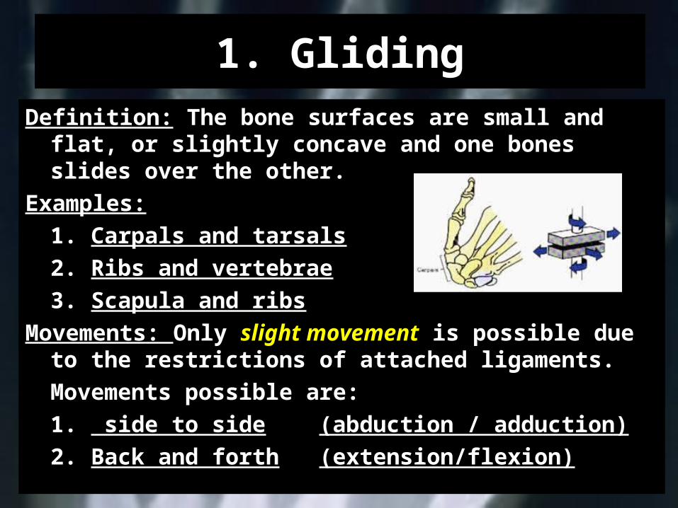
1. GlidingDefinition: The bone surfaces are small and flat, or
slightly concave and one bones slides over the other.
Examples:1. Carpals and tarsals2. Ribs and vertebrae3. Scapula and ribs
Movements: Only slight movement is possible due to the restrictions of attached ligaments.Movements possible are:1. side to side (abduction / adduction)2. Back and forth (extension/flexion)
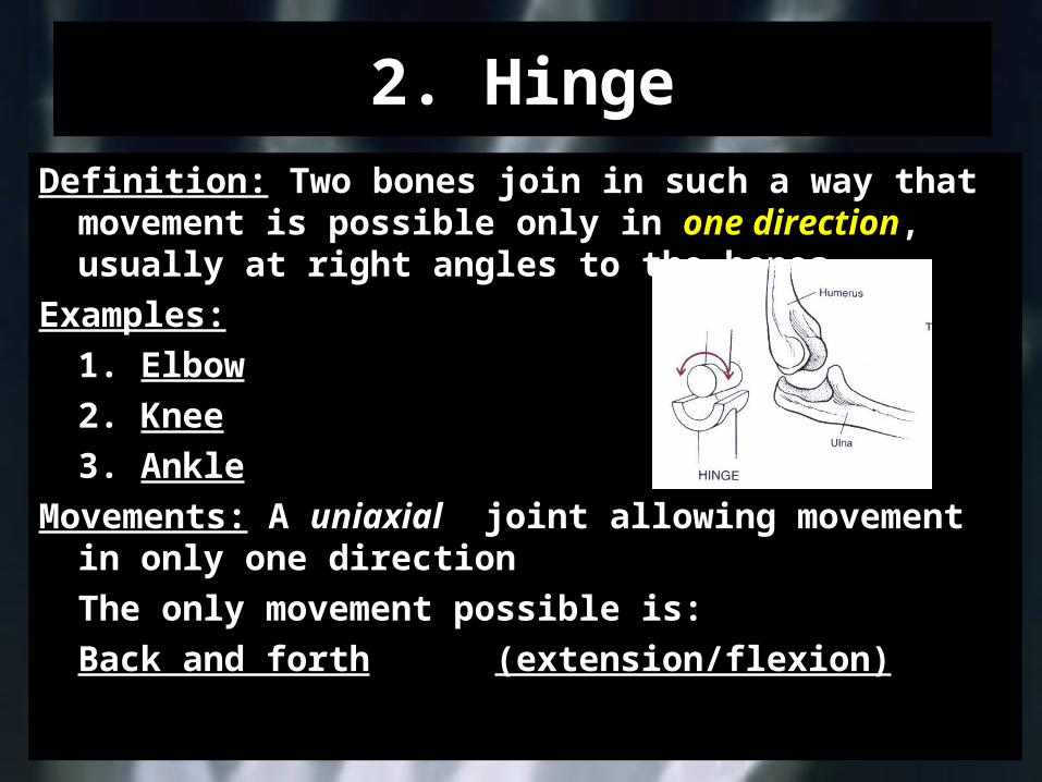
2. HingeDefinition: Two bones join in such a way that
movement is possible only in one direction, usually at right angles to the bones.
Examples:1. Elbow2. Knee3. Ankle
Movements: A uniaxial joint allowing movement in only one directionThe only movement possible is:Back and forth (extension/flexion)
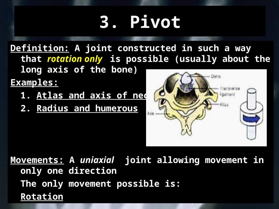
3. PivotDefinition: A joint constructed in such a way that
rotation only is possible (usually about the long axis of the bone)
Examples:1. Atlas and axis of neck2. Radius and humerous
Movements: A uniaxial joint allowing movement in only one directionThe only movement possible is:Rotation
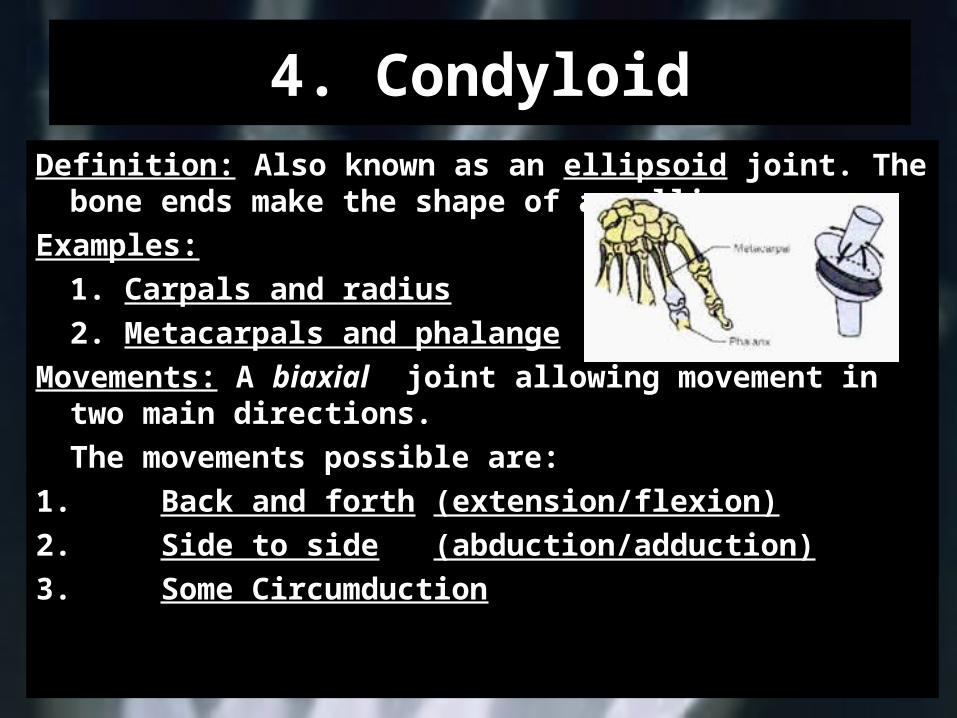
4. CondyloidDefinition: Also known as an ellipsoid joint. The
bone ends make the shape of an ellipse.Examples:
1. Carpals and radius2. Metacarpals and phalange
Movements: A biaxial joint allowing movement in two main directions.The movements possible are:
1. Back and forth (extension/flexion)2. Side to side
(abduction/adduction)3. Some Circumduction
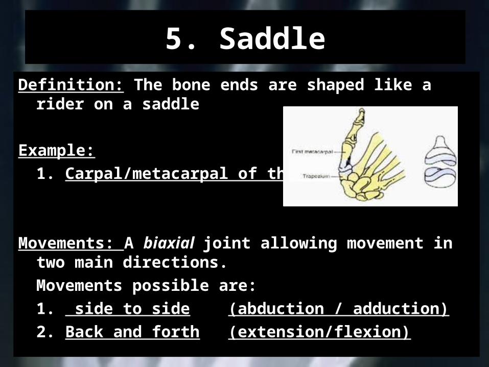
5. SaddleDefinition: The bone ends are shaped like a rider
on a saddle
Example:1. Carpal/metacarpal of thumb
Movements: A biaxial joint allowing movement in two main directions.Movements possible are:1. side to side (abduction / adduction)2. Back and forth (extension/flexion)
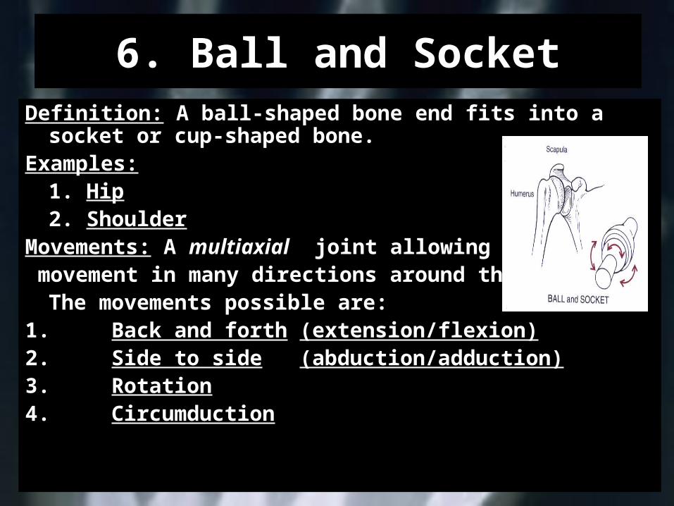
6. Ball and SocketDefinition: A ball-shaped bone end fits into a
socket or cup-shaped bone.Examples:
1. Hip2. Shoulder
Movements: A multiaxial joint allowing movement in many directions around the joint.
The movements possible are:1. Back and forth (extension/flexion)2. Side to side
(abduction/adduction)3. Rotation4. Circumduction
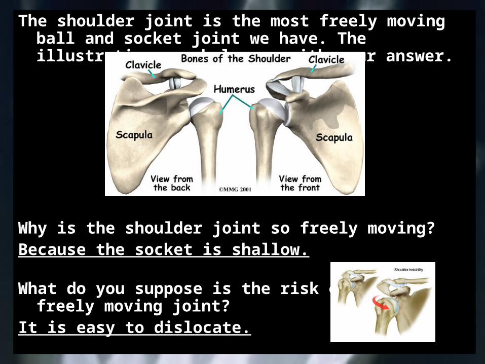
The shoulder joint is the most freely moving ball and socket joint we have. The illustration may help you with your answer.
Why is the shoulder joint so freely moving?Because the socket is shallow.
What do you suppose is the risk of such a freely moving joint?
It is easy to dislocate.
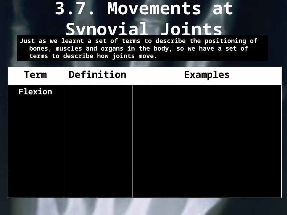
3.7. Movements at Synovial Joints
Just as we learnt a set of terms to describe the positioning of bones, muscles and organs in the body, so we have a set of terms to describe how joints move.
Term Definition Examples
Flexion Bending or decreasing the angle
between two bones
Bending at the knee6. Knee
Raising the thigh towards the trunk
5. Hip
Bringing the palm towards the forearm
4. Wrist
Bending at the elbow3. Arm
Moving the arm forward2. Shoulder
Bending forwards (sideways – lateral flexion)
1. Trunk
Bending at the knee6. Knee
Raising the thigh towards the trunk
5. Hip
Bringing the palm towards the forearm
4. Wrist
Bending at the elbow3. Arm
Moving the arm forward2. Shoulder
Bending forwards (sideways – lateral flexion)
1. Trunk
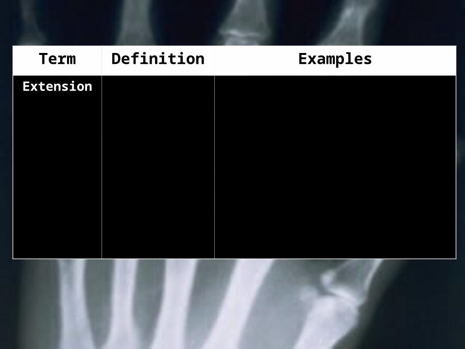
Term Definition Examples
Extension Straightening or increasing
the angle between two
bones
Straightening the knee6. Knee
Moving the leg backward5. Hip
Straightening the wrist4. Wrist
Straightening the elbow3. Arm
Moving the arm backward2. Shoulder
Straightening up1. Trunk
Straightening the knee6. Knee
Moving the leg backward5. Hip
Straightening the wrist4. Wrist
Straightening the elbow3. Arm
Moving the arm backward2. Shoulder
Straightening up1. Trunk
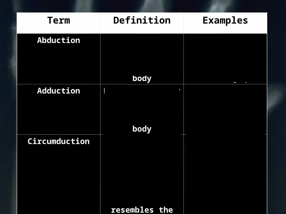
Term Definition ExamplesAbduction Moving a limb or
part of a limb away from the midline of the
bodyMoving outwards on
a star jump
Adduction Moving a limb or part of a limb towards the
midline of the body
Bringing the limbs back together in
a star jump
Circumduction A combination of flexion,
extension, abduction and
adduction.The movement
of the limb resembles the
shape of a cone
The arm stroke in Butterfly
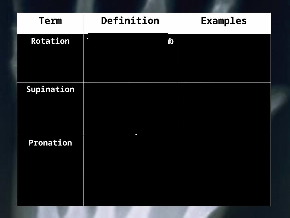
Term Definition Examples
Rotation Twisting of a limb about its long axis
Turning the headTwisting the trunk
Supination Movement of the hand into a palm-
up positionHolding a bowl of
soup
Turning a card overTurning a page in a
book
PronationMovement of the hand into a palm-
down position tipping the soup
out
Turning a card face down
Closing a book
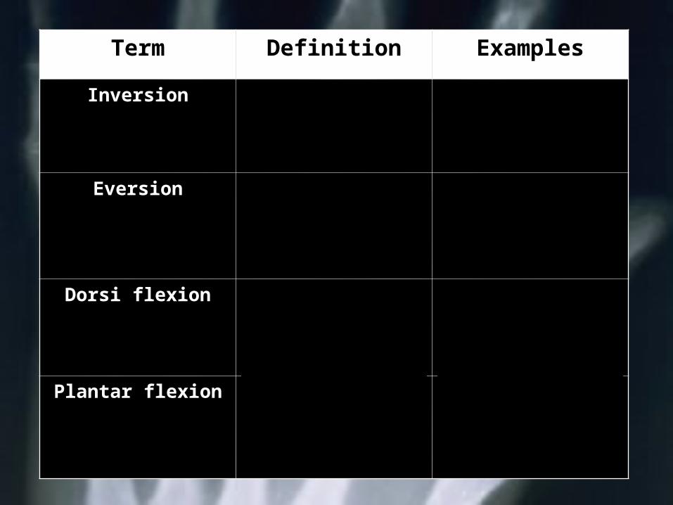
Term Definition Examples
Inversion Movement of the sole of the foot
inward
Eversion Movement of the sole of the foot
outward
Dorsi flexion Movement of the top of the foot upward, closer
to the shin
Plantar flexion Movement of the sole of the foot
downward
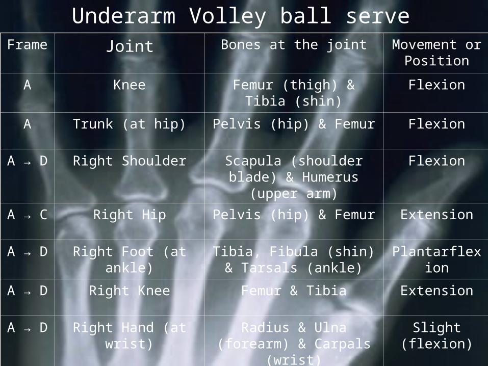
Underarm Volley ball serveFrame Joint Bones at the joint Movement or
Position
A Knee Femur (thigh) & Tibia (shin)
Flexion
A Trunk (at hip) Pelvis (hip) & Femur Flexion
A → D Right Shoulder Scapula (shoulder blade) & Humerus
(upper arm)
Flexion
A → C Right Hip Pelvis (hip) & Femur Extension
A → D Right Foot (at ankle) Tibia, Fibula (shin) & Tarsals (ankle)
Plantarflexion
A → D Right Knee Femur & Tibia Extension
A → D Right Hand (at wrist)
Radius & Ulna (forearm) & Carpals (wrist)
Slight (flexion)
D Right Elbow Radius, Ulna & Humerus Extension

Functional Anatomy
Section Four: The Muscles
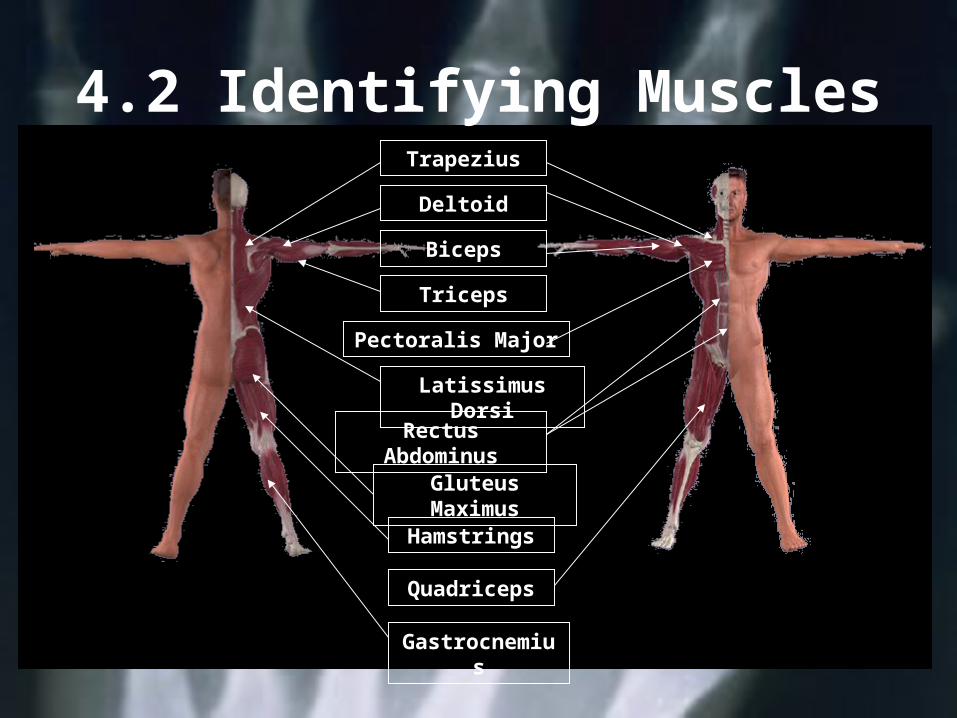
4.2 Identifying MusclesTrapezius
Deltoid
Biceps
Triceps
Pectoralis Major
Latissimus Dorsi
Rectus Abdominus
Gluteus Maximus
Hamstrings
Quadriceps
Gastrocnemius
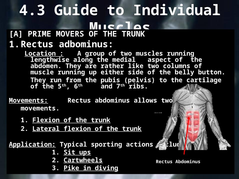
4.3 Guide to Individual Muscles[A] PRIME MOVERS OF THE TRUNK
1. Rectus adbominus: Location : A group of two muscles running
lengthwise along the medial aspect of the abdomen. They are rather like two columns of
muscle running up either side of the belly button.
They run from the pubis (pelvis) to the cartilage of the 5th, 6th and 7th ribs.
Movements: Rectus abdominus allows two basic movements.
1. Flexion of the trunk2. Lateral flexion of the trunk
Application: Typical sporting actions include:1. Sit ups2. Cartwheels3. Pike in diving
Rectus Abdominus
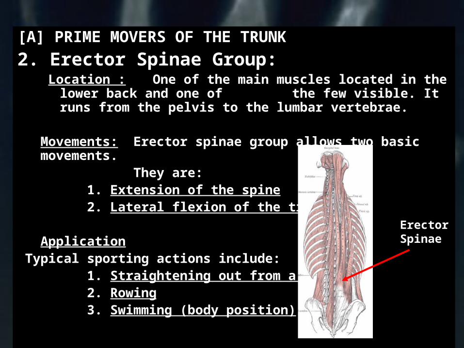
[A] PRIME MOVERS OF THE TRUNK
2. Erector Spinae Group: Location : One of the main muscles located in the
lower back and one of the few visible. It runs from the pelvis to the lumbar vertebrae.
Movements: Erector spinae group allows two basic movements.
They are:1. Extension of the spine2. Lateral flexion of the trunk
Application Typical sporting actions include:
1. Straightening out from a pike2. Rowing3. Swimming (body position)
Erector Spinae
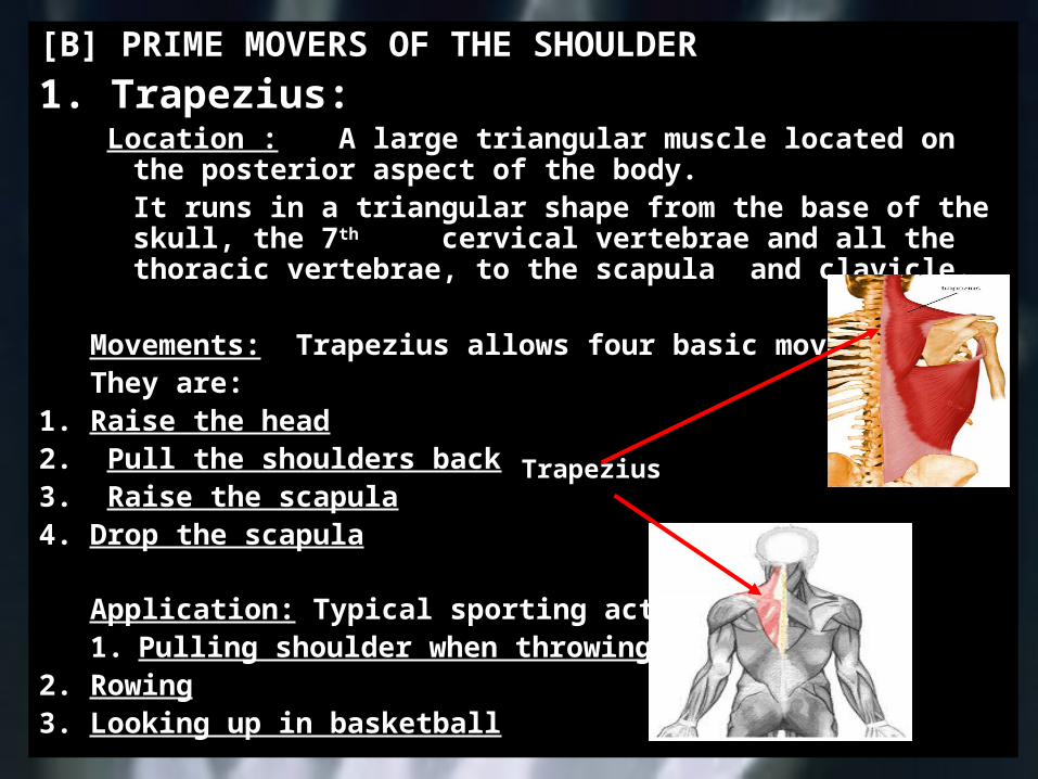
[B] PRIME MOVERS OF THE SHOULDER
1. Trapezius: Location : A large triangular muscle located on the
posterior aspect of the body.It runs in a triangular shape from the
base of the skull, the 7th cervical vertebrae and all the thoracic vertebrae, to the scapula
and clavicle.
Movements: Trapezius allows four basic movements.They are:
1. Raise the head2. Pull the shoulders back3. Raise the scapula4. Drop the scapula
Application: Typical sporting actions include:
1. Pulling shoulder when throwing2. Rowing3. Looking up in basketball
Trapezius

[B] PRIME MOVERS OF THE SHOULDER2. Latissimus Dorsi:
Location : The broadest muscle of the back. It forms the back of the armpit.This is a large triangular muscle which covers the lumbar and lower thoracic region of the back.It runs from the lower thoracic and lumber
regions, to the anterior aspect of the humerus
Movements: Latissimus dorsi allows three basic movements.They are:
1. Adduction of the upper arm2. Extension of the shoulder3. Internal rotation of the shoulder
How can latissimus dorsi allow internal rotation of the shoulder to occur if it is a muscle located on the back?Because it attaches to the humerus
Application: Typical sporting actions include:1. Recovery in breaststroke2. Ten-pin bowling3. Drawing arm back to punch
Latissimus dorsi
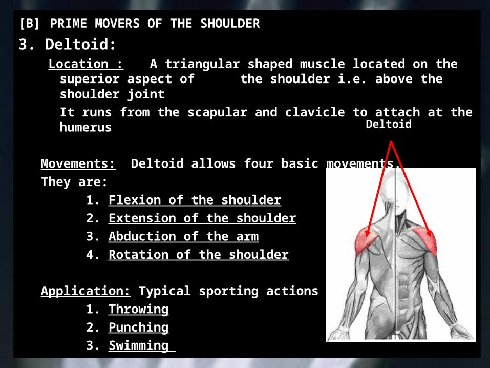
[B] PRIME MOVERS OF THE SHOULDER
3. Deltoid: Location : A triangular shaped muscle located on the
superior aspect of the shoulder i.e. above the shoulder joint
It runs from the scapular and clavicle to attach at the humerus
Movements: Deltoid allows four basic movements.They are:
1. Flexion of the shoulder2. Extension of the shoulder3. Abduction of the arm4. Rotation of the shoulder
Application: Typical sporting actions include:1. Throwing2. Punching3. Swimming
Deltoid
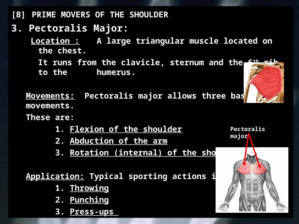
[B] PRIME MOVERS OF THE SHOULDER
3. Pectoralis Major: Location : A large triangular muscle located on the
chest.It runs from the clavicle, sternum and
the 6th rib, to the humerus.
Movements: Pectoralis major allows three basic movements.
These are:1. Flexion of the shoulder2. Abduction of the arm3. Rotation (internal) of the shoulder
Application: Typical sporting actions include:1. Throwing2. Punching3. Press-ups
Pectoralis major
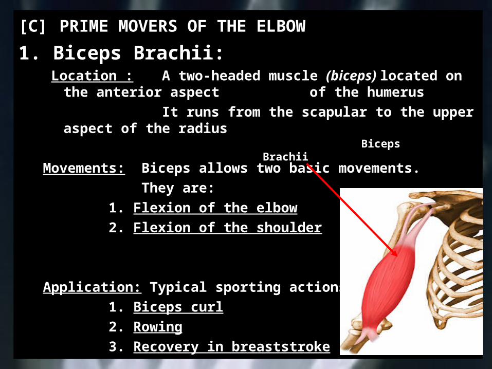
[C] PRIME MOVERS OF THE ELBOW
1. Biceps Brachii: Location : A two-headed muscle (biceps) located on
the anterior aspect of the humerusIt runs from the scapular to the upper
aspect of the radius
Movements: Biceps allows two basic movements.They are:
1. Flexion of the elbow2. Flexion of the shoulder
Application: Typical sporting actions include:1. Biceps curl2. Rowing3. Recovery in breaststroke
Biceps Brachii
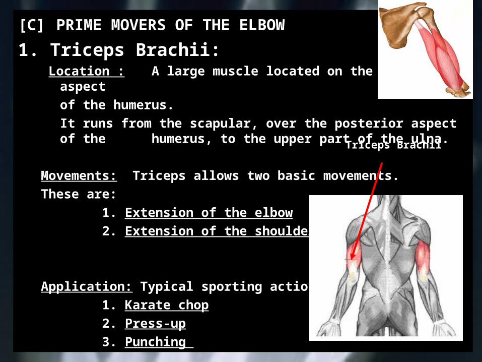
[C] PRIME MOVERS OF THE ELBOW
1. Triceps Brachii: Location : A large muscle located on the posterior
aspect of the humerus.It runs from the scapular, over the posterior
aspect of the humerus, to the upper part of the ulna.
Movements: Triceps allows two basic movements.These are:
1. Extension of the elbow2. Extension of the shoulder
Application: Typical sporting actions include:1. Karate chop2. Press-up3. Punching
Triceps Brachii
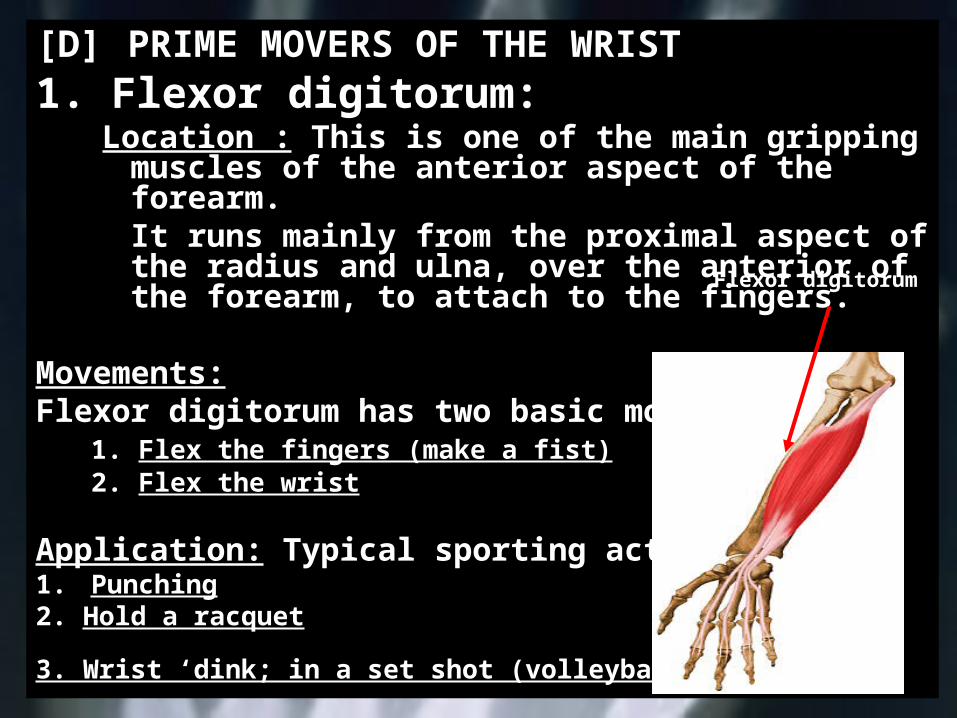
[D] PRIME MOVERS OF THE WRIST1. Flexor digitorum:
Location : This is one of the main gripping muscles of the anterior aspect of the forearm.It runs mainly from the proximal aspect of the radius and ulna, over the anterior of the forearm, to attach to the fingers.
Movements:Flexor digitorum has two basic movements.
1. Flex the fingers (make a fist)2. Flex the wrist
Application: Typical sporting actions include: 1. Punching2. Hold a racquet
3. Wrist ‘dink; in a set shot (volleyball)
Flexor digitorum

[D] PRIME MOVERS OF THE WRIST
2. Extensor digitorum: Location : This is one of the muscles in direct
opposition to the flexor digitorum. It is located on the posterior aspect of the forearm.It runs over the posterior of the forearm, to attach to the fingers.
Movements: Extensor digitorum allows two basic movements.
1. Extend the fingers 2. Extend the wrist
Application: Typical sporting actions :1. Karate chop2. Fending in tackling3. Set shot in (volleyball)
Extensor
digitorum

[E] PRIME MOVERS OF THE HIP AND KNEE
1. Illiopsoas: Location : This is a group of three muscles located
deep inside the hip region.It runs from the lumbar vertebrae and pelvis to the
upper femur.
Movements: Illiopsoas allows one basic movement.This is:1. Flexion of the hip
Application: Typical sporting actions include:1. Kicking2. Running3. Cycling
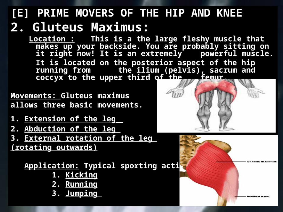
[E] PRIME MOVERS OF THE HIP AND KNEE
2. Gluteus Maximus: Location : This is a the large fleshy muscle that
makes up your backside. You are probably sitting on it right now! It is an extremely powerful muscle.
It is located on the posterior aspect of the hip running from the ilium (pelvis), sacrum and coccyx to the upper third of the femur.
Movements: Gluteus maximus allows three basic movements.
1. Extension of the leg 2. Abduction of the leg 3. External rotation of the leg (rotating outwards)
Application: Typical sporting actions include:1. Kicking2. Running3. Jumping
[E] PRIME MOVERS OF THE HIP AND KNEE
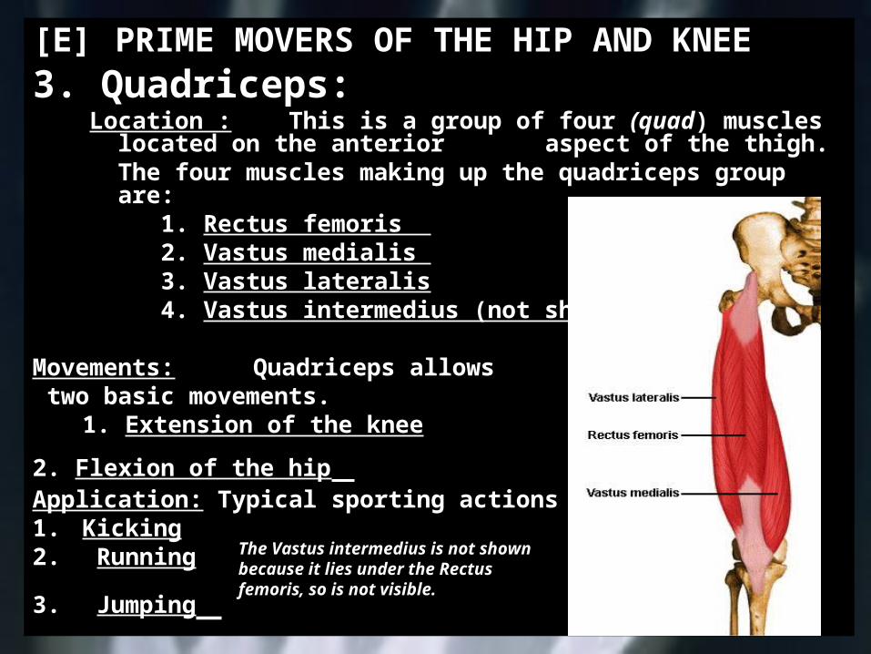
3. Quadriceps: Location : This is a group of four (quad) muscles
located on the anterior aspect of the thigh.The four muscles making up the
quadriceps group are:1. Rectus femoris 2. Vastus medialis 3. Vastus lateralis4. Vastus intermedius (not shown)
Movements: Quadriceps allows two basic movements.
1. Extension of the knee
2. Flexion of the hip Application: Typical sporting actions include:1. Kicking2. Running
3. Jumping
The Vastus intermedius is not shown because it lies under the Rectus femoris, so is not visible.
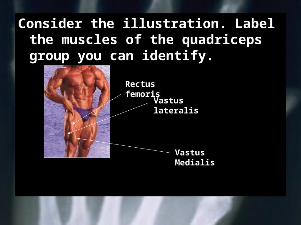
Consider the illustration. Label the muscles of the quadriceps group you can identify.
Rectus femoris
Vastus lateralis
Vastus Medialis
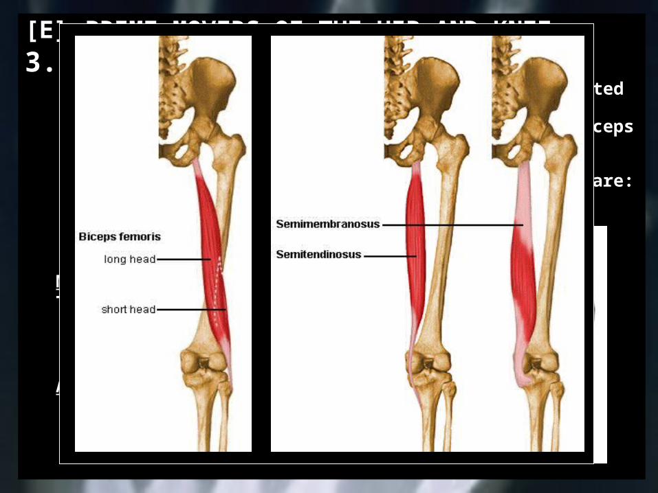
[E] PRIME MOVERS OF THE HIP AND KNEE3. Hamstrings:
Location : This is a group of three muscles located on the posterior aspect of the thigh.
Since they are in direct opposition to the quadriceps and are generally weaker they are prone to injury.
The three muscles making up the hamstrings group are:
1. Semitendinosus 2. Biceps femoris3. Semimembranosis
Movements: Hamstrings allows two basic movements.These are:
1. Extension of the hip2. Flexion of the knee
Application: Typical sporting actions include:1. Kicking2. Running3. Jumping
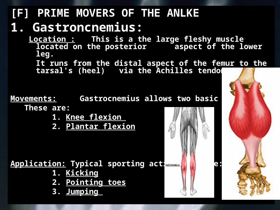
[F] PRIME MOVERS OF THE ANLKE1. Gastroncnemius:
Location : This is a the large fleshy muscle located on the posterior aspect of the lower leg.It runs from the distal aspect of the femur to the tarsal's (heel) via the Achilles tendon.
Movements: Gastrocnemius allows two basic movements.These are:
1. Knee flexion 2. Plantar flexion
Application: Typical sporting actions include:1. Kicking2. Pointing toes3. Jumping
[F] PRIME MOVERS OF THE ANLKE
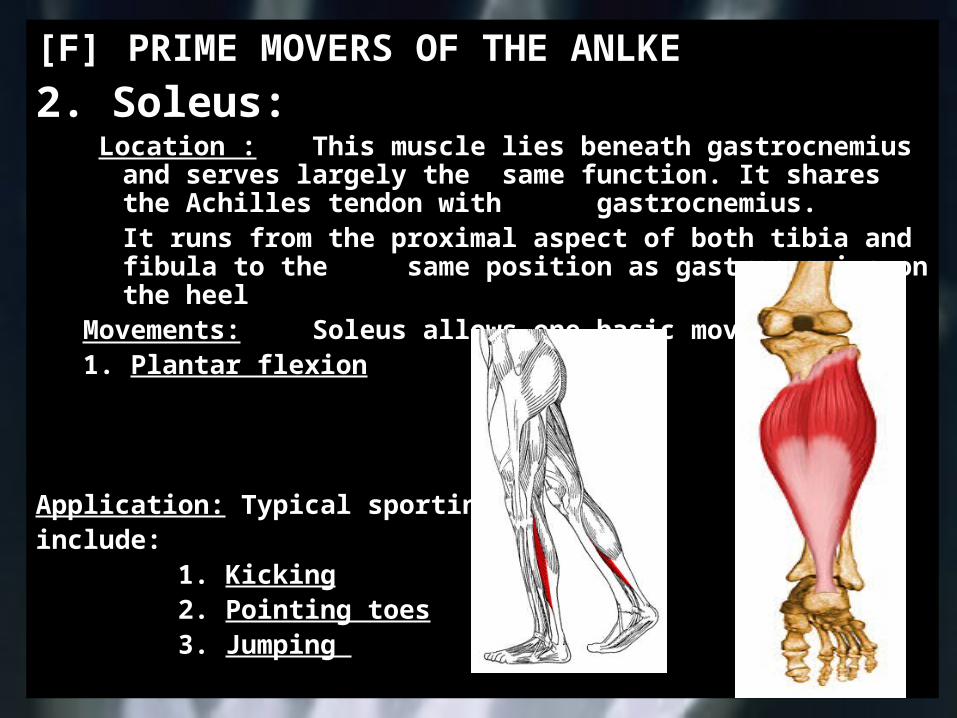
2. Soleus: Location : This muscle lies beneath gastrocnemius
and serves largely the same function. It shares the Achilles tendon with
gastrocnemius.It runs from the proximal aspect of both
tibia and fibula to the same position as gastrocnemius on the heel
Movements: Soleus allows one basic movements.1. Plantar flexion
Application: Typical sporting actions include:
1. Kicking2. Pointing toes3. Jumping
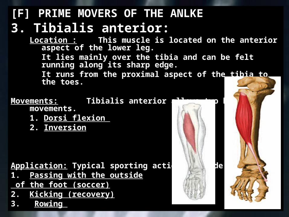
[F] PRIME MOVERS OF THE ANLKE3. Tibialis anterior:
Location : This muscle is located on the anterior aspect of the lower leg.It lies mainly over the tibia and can be felt running along its sharp edge.It runs from the proximal aspect of the tibia to the toes.
Movements: Tibialis anterior allows two basic movements.1. Dorsi flexion 2. Inversion
Application: Typical sporting actions include:1. Passing with the outside of the foot (soccer)2. Kicking (recovery)3. Rowing
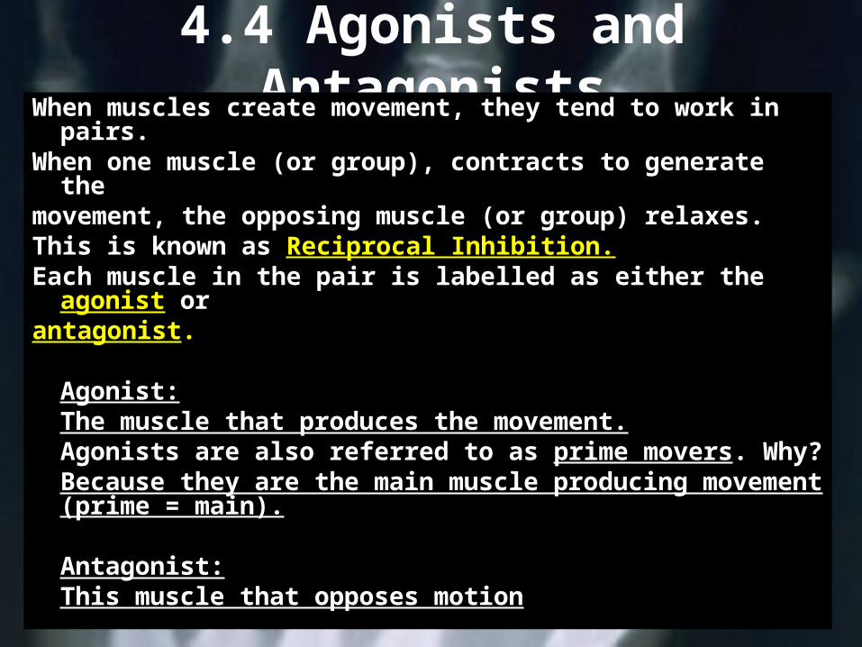
4.4 Agonists and AntagonistsWhen muscles create movement, they tend to work in
pairs.When one muscle (or group), contracts to generate
themovement, the opposing muscle (or group) relaxes.This is known as Reciprocal Inhibition.Each muscle in the pair is labelled as either the
agonist orantagonist.
Agonist:The muscle that produces the movement.
Agonists are also referred to as prime movers. Why?
Because they are the main muscle producing movement (prime = main).
Antagonist:This muscle that opposes motion
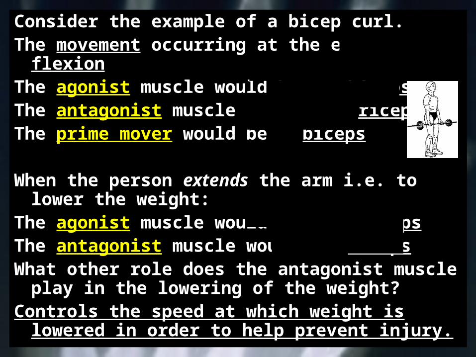
Consider the example of a bicep curl.The movement occurring at the elbow is
flexionThe agonist muscle would be bicepsThe antagonist muscle would be tricepsThe prime mover would be biceps
When the person extends the arm i.e. to lower the weight:
The agonist muscle would be tricepsThe antagonist muscle would be bicepsWhat other role does the antagonist muscle
play in the lowering of the weight?Controls the speed at which weight is
lowered in order to help prevent injury.