Functional Anatomy of the Respiratory System Original PPT made by Pearson, Modified by Lindsey Sweis...
43
Functional Anatomy of the Respiratory System Original PPT made by Pearson, Modified by Lindsey Sweis and Jones A&P, 2015.
-
Upload
candice-bradford -
Category
Documents
-
view
218 -
download
0
Transcript of Functional Anatomy of the Respiratory System Original PPT made by Pearson, Modified by Lindsey Sweis...
Functional Anatomy of the Respiratory System
Original PPT made by Pearson, Modified by Lindsey Sweis and Jones A&P, 2015.

Why is this possible?

Organs of the Respiratory System
• Nose• Pharynx• Larynx• Trachea• Bronchi• Lungs—alveoli

Nasal cavity
Nostril
Larynx
Right main(primary)bronchus
Trachea
Right lung
Oral cavityPharynx
Left main (primary) bronchusLeft lung
Diaphragm
Figure 13.1

Functions of the Respiratory System
• Gas exchanges between the blood and external environment – Occurs in the alveoli of the lungs
• Passageways to the lungs purify, humidify, and warm the incoming air
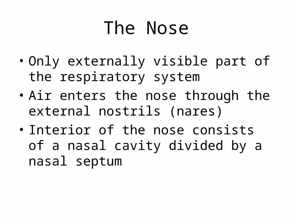
The Nose
• Only externally visible part of the respiratory system
• Air enters the nose through the external nostrils (nares)
• Interior of the nose consists of a nasal cavity divided by a nasal septum

Cribriform plateof ethmoid bone
Sphenoidal sinusPosterior nasalaperture
Nasopharynx• Pharyngeal tonsil
• Opening ofpharyngotympanictube
• Uvula
Oropharynx• Palatine tonsil
• Lingual tonsil
Laryngopharynx
Esophagus
Trachea
Frontal sinus
Nasal cavity• Nasal conchae (superior,
middle and inferior)
• Nasal meatuses (superior,middle, and inferior)
• Nasal vestibule• Nostril
Hard palate
Soft palate
Tongue
Hyoid bone
Larynx• Epiglottis• Thyroid cartilage• Vocal fold • Cricoid cartilage
(b) Detailed anatomy of the upper respiratory tract
Figure 13.2b

Anatomy of the Nasal Cavity
• Olfactory receptors are located in the mucosa on the superior surface
• The rest of the cavity is lined with respiratory mucosa that– Moisten air– Trap incoming foreign particles

Anatomy of the Nasal Cavity
• Lateral walls have projections called conchae– Increase surface area– Increase air turbulence within the nasal cavity
• The nasal cavity is separated from the oral cavity by the palate– Anterior hard palate (bone)– Posterior soft palate (muscle)

Paranasal Sinuses
• Cavities within bones surrounding the nasal cavity are called sinuses
• Sinuses are located in the following bones– Frontal bone– Sphenoid bone– Ethmoid bone– Maxillary bone
http://www.hopkinsmedicine.org/healthlibrary/GetImage.aspx?ImageId=129342

Cribriform plateof ethmoid bone
Sphenoidal sinusPosterior nasalaperture
Nasopharynx• Pharyngeal tonsil
• Opening of pharyngotympanic
tube• Uvula
Oropharynx• Palatine tonsil
• Lingual tonsil
Laryngopharynx
Esophagus
Trachea
Frontal sinus
Nasal cavity• Nasal conchae (superior,
middle and inferior)
• Nasal meatuses (superior,middle, and inferior)
• Nasal vestibule• Nostril
Hard palate
Soft palate
Tongue
Hyoid bone
Larynx• Epiglottis• Thyroid cartilage• Vocal fold • Cricoid cartilage
(b) Detailed anatomy of the upper respiratory tract
Figure 13.2b

Paranasal Sinuses
• Function of the sinuses– Lighten the skull– Act as resonance chambers for speech– Produce mucus that drains into the nasal cavity
http://www.hopkinsmedicine.org/healthlibrary/GetImage.aspx?ImageId=129342

If you laugh hard when you’re drinking something and you keep your mouth
closed, why does your drink sometimes come out of your nose?

Pharynx (Throat)
• Muscular passage from nasal cavity to larynx• Three regions of the pharynx– Nasopharynx—superior region behind nasal cavity– Oropharynx—middle region behind mouth– Laryngopharynx—inferior region attached to
larynx
• The oropharynx and laryngopharynx are common passageways for air and food

Pharynx• Nasopharynx• Oropharynx• Laryngopharynx
(a) Regions of the pharynxFigure 13.2a

Structures of the Pharynx• Pharyngotympanic tubes (a.k.a. Eustachian tubes; auditory tubes) open into the nasopharynx• Tonsils of the pharynx
• Pharyngeal tonsil (adenoid) is located in the nasopharynx• Palatine tonsils are located in the oropharynx• Lingual tonsils are found at the base of the tongue

Structures of the Pharynx

Cribriform plateof ethmoid bone
Sphenoidal sinusPosterior nasalaperture
Nasopharynx• Pharyngeal tonsil
• Opening of pharyngotympanic
tube• Uvula
Oropharynx• Palatine tonsil
• Lingual tonsil
Laryngopharynx
Esophagus
Trachea
Frontal sinus
Nasal cavity• Nasal conchae (superior,
middle and inferior)
• Nasal meatuses (superior,middle, and inferior)
• Nasal vestibule• Nostril
Hard palate
Soft palate
Tongue
Hyoid bone
Larynx• Epiglottis• Thyroid cartilage• Vocal fold • Cricoid cartilage
(b) Detailed anatomy of the upper respiratory tract
Figure 13.2b

Larynx (Voice Box)
• Routes air and food into proper channels• Plays a role in speech• Made of eight rigid hyaline cartilages and a
spoon-shaped flap of elastic cartilage (epiglottis)
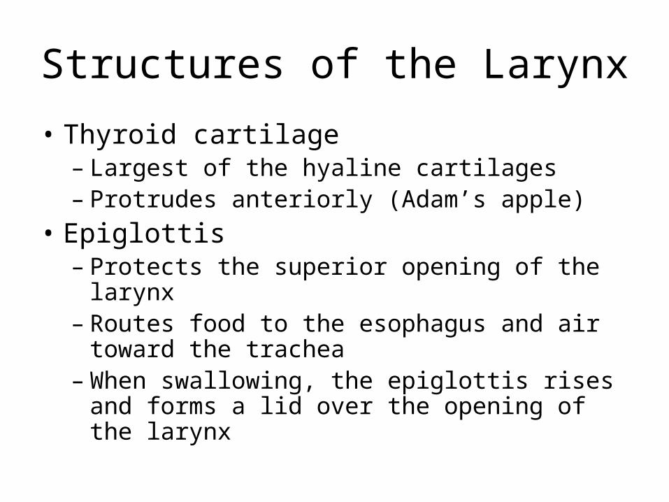
Structures of the Larynx
• Thyroid cartilage– Largest of the hyaline cartilages– Protrudes anteriorly (Adam’s apple)
• Epiglottis– Protects the superior opening of the larynx– Routes food to the esophagus and air toward the
trachea– When swallowing, the epiglottis rises and forms a
lid over the opening of the larynx

Structures of the Larynx
• Vocal folds (true vocal cords)– Vibrate with expelled air to create sound (speech)
• Glottis—opening between vocal cords

Cribriform plateof ethmoid bone
Sphenoidal sinusPosterior nasalaperture
Nasopharynx• Pharyngeal tonsil
• Opening of pharyngotympanic
tube• Uvula
Oropharynx• Palatine tonsil
• Lingual tonsil
Laryngopharynx
Esophagus
Trachea
Frontal sinus
Nasal cavity• Nasal conchae (superior,
middle and inferior)
• Nasal meatuses (superior,middle, and inferior)
• Nasal vestibule• Nostril
Hard palate
Soft palate
Tongue
Hyoid bone
Larynx• Epiglottis• Thyroid cartilage• Vocal fold • Cricoid cartilage
(b) Detailed anatomy of the upper respiratory tract
Figure 13.2b

Trachea (Windpipe)
• Four-inch-long tube that connects larynx with bronchi
• Walls are reinforced with C-shaped hyaline cartilage
• Lined with ciliated mucosa– Beat continuously in the opposite direction of
incoming air– Expel mucus loaded with dust and other debris
away from lungs

PosteriorMucosa
Submucosa
Seromucousgland insubmucosa
Hyalinecartilage
AdventitiaAnterior
Lumen of trachea
Trachealismuscle
Esophagus
Figure 13.3a
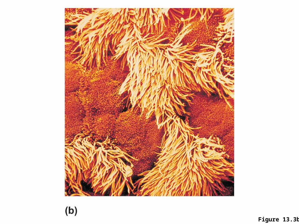
Figure 13.3b

Main (Primary) Bronchi
• Formed by division of the trachea• Enters the lung at the hilum (medial
depression)• Right bronchus is wider, shorter, and
straighter than left• Bronchi subdivide into smaller and smaller
branches

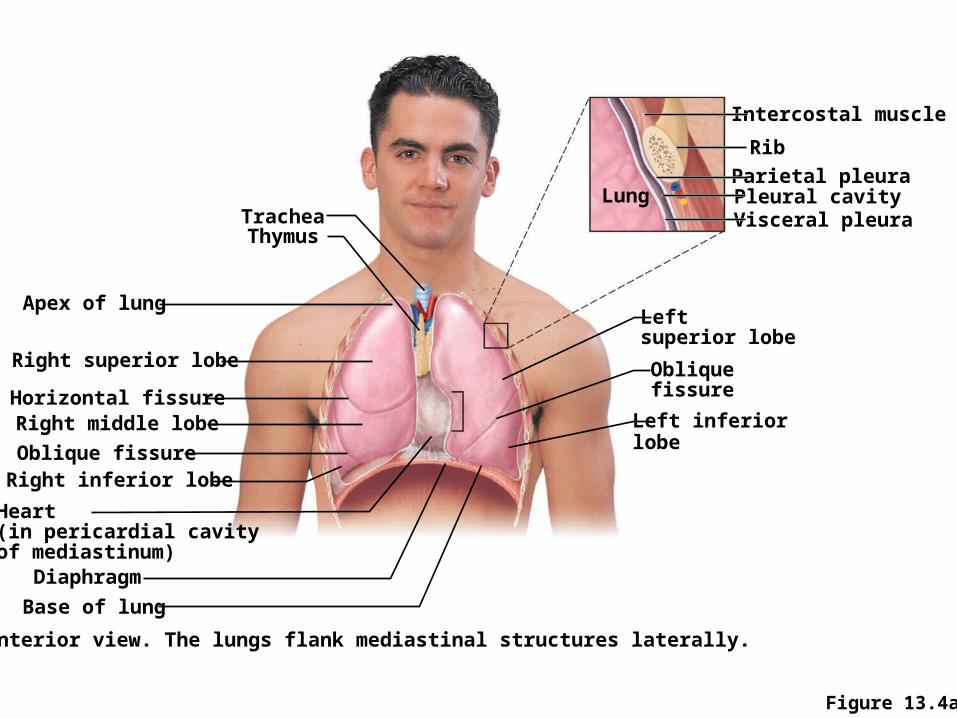
TracheaThymus
Apex of lung
Right superior lobe
Horizontal fissureRight middle lobeOblique fissureRight inferior lobe
Heart(in pericardial cavity of mediastinum)
DiaphragmBase of lung
(a) Anterior view. The lungs flank mediastinal structures laterally.
Left inferiorlobe
Obliquefissure
Leftsuperior lobe
Visceral pleuraPleural cavityParietal pleura
Rib
Intercostal muscle
Lung
Figure 13.4a

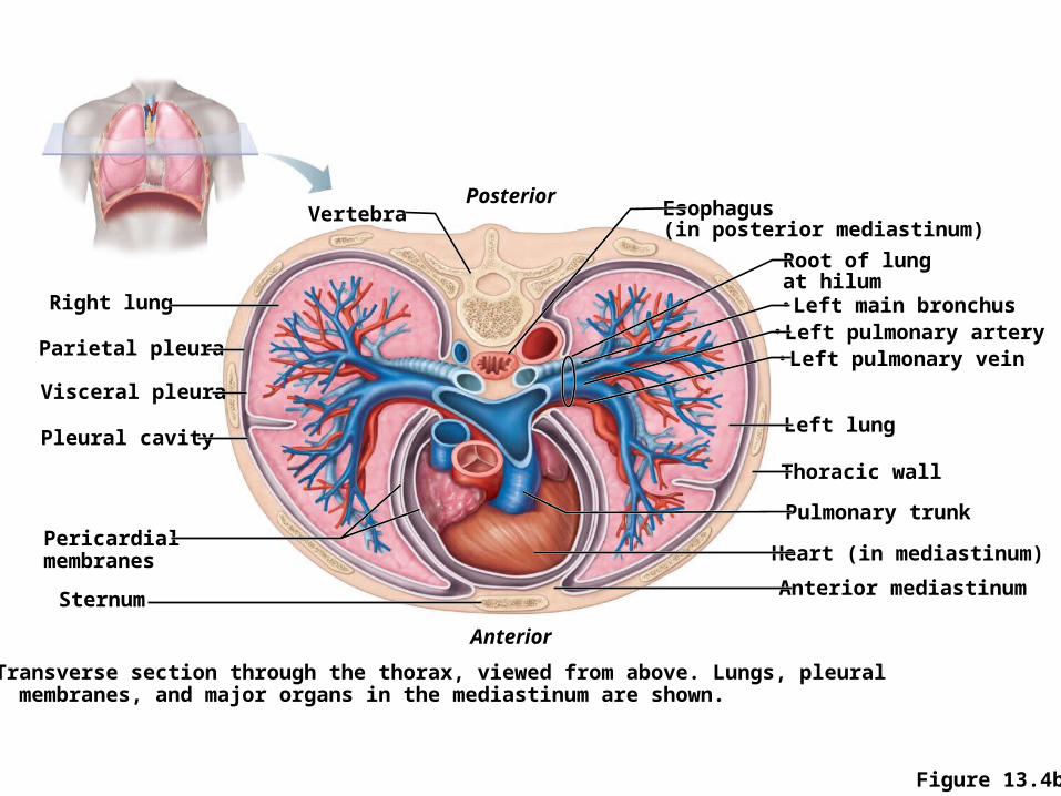
Posterior Esophagus(in posterior mediastinum)
Root of lungat hilum• Left main bronchus• Left pulmonary artery• Left pulmonary vein
Left lung
Thoracic wall
Pulmonary trunk
Anterior mediastinum
Anterior
(b) Transverse section through the thorax, viewed from above. Lungs, pleural membranes, and major organs in the mediastinum are shown.
Sternum
Pericardial membranes
Pleural cavity
Visceral pleura
Parietal pleura
Right lung
Vertebra
Heart (in mediastinum)
Figure 13.4b

Lungs
• Occupy most of the thoracic cavity– Heart occupies central portion called mediastinum
• Apex is near the clavicle (superior portion)• Base rests on the diaphragm (inferior portion)• Each lung is divided into lobes by fissures– Left lung—two lobes– Right lung—three lobes
TracheaThymus
Apex of lung
Right superior lobe
Horizontal fissureRight middle lobeOblique fissureRight inferior lobe
Heart(in pericardial cavity of mediastinum)
DiaphragmBase of lung
(a) Anterior view. The lungs flank mediastinal structures laterally.
Left inferiorlobe
Obliquefissure
Leftsuperior lobe
Visceral pleuraPleural cavityParietal pleura
Rib
Intercostal muscle
Lung
Figure 13.4a

Posterior Esophagus(in posterior mediastinum)
Root of lungat hilum• Left main bronchus• Left pulmonary artery• Left pulmonary vein
Left lung
Thoracic wall
Pulmonary trunk
Anterior mediastinum
Anterior
(b) Transverse section through the thorax, viewed from above. Lungs, pleural membranes, and major organs in the mediastinum are shown.
Sternum
Pericardial membranes
Pleural cavity
Visceral pleura
Parietal pleura
Right lung
Vertebra
Heart (in mediastinum)
Figure 13.4b

Coverings of the Lungs
• Serosa covers the outer surface of the lungs– Pulmonary (visceral) pleura covers the lung
surface– Parietal pleura lines the walls of the thoracic
cavity
• Pleural fluid fills the area between layers of pleura to allow gliding
• These two pleural layers resist being pulled apart

TracheaThymus
Apex of lung
Right superior lobe
Horizontal fissureRight middle lobeOblique fissureRight inferior lobe
Heart(in pericardial cavity of mediastinum)
DiaphragmBase of lung
(a) Anterior view. The lungs flank mediastinal structures laterally.
Left inferiorlobe
Obliquefissure
Leftsuperior lobe
Visceral pleuraPleural cavityParietal pleura
Rib
Intercostal muscle
Lung
Figure 13.4a
Posterior Esophagus(in posterior mediastinum)
Root of lungat hilum• Left main bronchus• Left pulmonary artery• Left pulmonary vein
Left lung
Thoracic wall
Pulmonary trunk
Anterior mediastinum
Anterior
(b) Transverse section through the thorax, viewed from above. Lungs, pleural membranes, and major organs in the mediastinum are shown.
Sternum
Pericardial membranes
Pleural cavity
Visceral pleura
Parietal pleura
Right lung
Vertebra
Heart (in mediastinum)
Figure 13.4b

Bronchial (Respiratory) Tree Divisions
• All but the smallest of these passageways have reinforcing cartilage in their walls– Primary bronchi– Secondary bronchi– Tertiary bronchi– Bronchioles– Terminal bronchioles

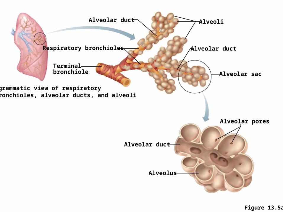
Alveolar duct Alveoli
Alveolar duct
Alveolar sac
Alveolar pores
Alveolar duct
Alveolus
(a) Diagrammatic view of respiratory bronchioles, alveolar ducts, and alveoli
Terminalbronchiole
Respiratory bronchioles
Figure 13.5a

Respiratory Zone
• Structures– Respiratory bronchioles– Alveolar ducts– Alveolar sacs– Alveoli (air sacs)
• Site of gas exchange = alveoli only
Alveolar duct Alveoli
Alveolar duct
Alveolar sac
Alveolar pores
Alveolar duct
Alveolus
(a) Diagrammatic view of respiratory bronchioles, alveolar ducts, and alveoli
Terminalbronchiole
Respiratory bronchioles
Figure 13.5a
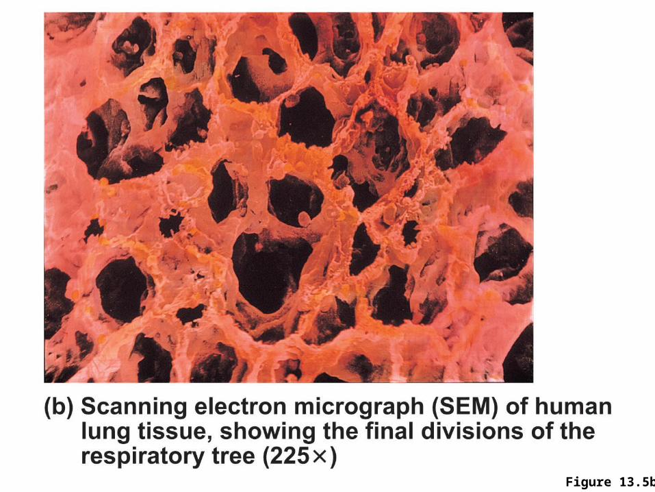
Figure 13.5b

Respiratory Membrane (Air-Blood Barrier)
• Thin squamous epithelial layer lines alveolar walls
• Alveolar pores connect neighboring air sacs• Pulmonary capillaries cover external surfaces
of alveoli• On one side of the membrane is air and on the
other side is blood flowing past

Figure 13.6 (1 of 2)
Endothelial cellnucleus
Alveolar pores
Capillary
Macrophage
Nucleus ofsquamousepithelial cell
Respiratorymembrane
Alveoli (gas-filled airspaces)
Red bloodcell incapillary
Surfactant-secreting cell
Squamousepithelial cellof alveolar wall

Figure 13.6 (2 of 2)
Endothelial cellnucleus
Alveolar pores
Capillary
Macrophage
Nucleus ofsquamousepithelial cell
Respiratorymembrane
Capillary endothelium
Fused basementmembranes
Alveolar epithelium
Alveolus
CO2O2
Capillary
Red blood cell
O2 CO2

Gas Exchange
• Gas crosses the respiratory membrane by diffusion– Oxygen enters the blood– Carbon dioxide enters the alveoli
• Alveolar macrophages (“dust cells”) add protection by picking up bacteria, carbon particles, and other debris
• Surfactant (a lipid molecule) coats gas-exposed alveolar surfaces