Fully human broadly neutralizing monoclonal...
9
Fully human broadly neutralizing monoclonal antibodies against influenza A viruses generated from the memory B cells of a 2009 pandemic H1N1 influenza vaccine recipient Weibin Hu a,1 , Aizhong Chen b,1 , Yi Miao c,1 , Shengli Xia d,1 , Zhiyang Ling a , Ke Xu a , Tongyan Wang a , Ying Xu b , Jun Cui b , Hongqiang Wu b , Guiyu Hu b , Lin Tian b , Lingling Wang b , Yuelong Shu e , Xiaowei Ma f , Bianli Xu d , Jin Zhang d , Xiaojun Lin f,n , Chao Bian b,n , Bing Sun a,b,n a Molecular Virus Unit, Key Laboratory of Molecular Virology & Immunology, Institut Pasteur of Shanghai, Chinese Academy of Sciences, Shanghai 200025, China b Key Laboratory of Molecular Cell Biology, Institute of Biochemistry and Cell Biology, Shanghai Institutes for Biological Sciences, Chinese Academy of Sciences, Shanghai 200031, China c Shanghai Xuhui Central Hospital, Shanghai 200031, China d Center for Disease Control and Prevention of Henan Province, Zhengzhou 450016, China e Chinese Center for Disease Control and Prevention, Beijing 102206, China f Hualan Biological Bacterin Company, Xinxiang 453003, China article info Article history: Received 1 August 2012 Returned to author for revisions 21 September 2012 Accepted 27 September 2012 Available online 16 October 2012 Keywords: Influenza H1N1 Fully human monoclonal antibody Neutralization Epitope HA2 Single-cell RT-PCR Fusion peptide abstract Whether the 2009 pandemic H1N1 influenza vaccine can induce heterosubtypic cross-protective anti- hemagglutinin (HA) neutralizing antibodies is an important issue. We obtained a panel of fully human monoclonal antibodies from the memory B cells of a 2009 pandemic H1N1 influenza vaccine recipient. Most of the monoclonal antibodies targeted the HA protein but not the HA1 fragment. Among the analyzed antibodies, seven mAbs exhibited neutralizing activity against several influenza A viruses of different subtypes. The conserved linear epitope targeted by the neutralizing mAbs (FIEGGWTGMVDG- WYGYHH) is part of the fusion peptide on HA2. Our work suggests that a heterosubtypic neutralizing antibody response primarily targeting the HA stem region exists in recipients of the 2009 pandemic H1N1 influenza vaccine. The HA stem region contains various conserved neutralizing epitopes with the fusion peptide as an important one. This work may aid in the design of a universal influenza A virus vaccine. & 2012 Elsevier Inc. All rights reserved. Introduction Because of their highly flexible genomes, influenza A viruses cause annual epidemics and sometimes pandemics around the world. For nearly 100 years, influenza A viruses have been a global threat to humans (Palese, 2004). Based on the antigenicity of the hemagglutinin (HA) protein, influenza A viruses are classified into two groups and at least 16 different subtypes (H1-H16). The HA protein is the functional protein that mediates the entry of influenza viruses into susceptible host cells and thus contains various epitopes that are recognized by neutralizing antibodies (Skehel and Wiley, 2000). However, heterosubtypic neutralizing or protective antibody responses are rarely observed in the general population, largely because of the high mutation rate of the HA protein, especially in the globular head (HA1) region, which is the primary target of the humoral immune response. Consequently, when a new reassortant influenza virus emerges that the human immune system has not previously encountered, a pandemic may occur. The 2009 swine-origin H1N1 influenza is an example of such a pandemic. The 2009 pandemic H1N1 influenza virus contains gene segments that are in both the American and the Eurasia swine genetic linkages (Garten et al., 2009). Nucleotide sequence align- ment has shown that the HA sequence of the 2009 pandemic H1N1 influenza virus is divergent from the sequences of the seasonal H1 influenza viruses that have previously been Contents lists available at SciVerse ScienceDirect journal homepage: www.elsevier.com/locate/yviro Virology 0042-6822/$ - see front matter & 2012 Elsevier Inc. All rights reserved. http://dx.doi.org/10.1016/j.virol.2012.09.034 Abbreviations: mAb, monoclonal antibody; TCID 50 , 50% tissue culture infective dose n Correspondence to: 320 Yueyang Road, Shanghai 200031, China. Fax: þ86 21 54920520. E-mail addresses: [email protected] (X. Lin), [email protected] (C. Bian), [email protected] (B. Sun). 1 These authors contributed equally to this work. Virology 435 (2013) 320–328
Transcript of Fully human broadly neutralizing monoclonal...

Virology 435 (2013) 320–328
Contents lists available at SciVerse ScienceDirect
Virology
0042-68
http://d
Abbre
dosen Corr
Fax: þ8
E-m
bsun@s1 Th
journal homepage: www.elsevier.com/locate/yviro
Fully human broadly neutralizing monoclonal antibodies against influenza Aviruses generated from the memory B cells of a 2009 pandemic H1N1influenza vaccine recipient
Weibin Hu a,1, Aizhong Chen b,1, Yi Miao c,1, Shengli Xia d,1, Zhiyang Ling a, Ke Xu a, Tongyan Wang a,Ying Xu b, Jun Cui b, Hongqiang Wu b, Guiyu Hu b, Lin Tian b, Lingling Wang b, Yuelong Shu e,Xiaowei Ma f, Bianli Xu d, Jin Zhang d, Xiaojun Lin f,n, Chao Bian b,n, Bing Sun a,b,n
a Molecular Virus Unit, Key Laboratory of Molecular Virology & Immunology, Institut Pasteur of Shanghai, Chinese Academy of Sciences, Shanghai 200025, Chinab Key Laboratory of Molecular Cell Biology, Institute of Biochemistry and Cell Biology, Shanghai Institutes for Biological Sciences, Chinese Academy of Sciences, Shanghai 200031,
Chinac Shanghai Xuhui Central Hospital, Shanghai 200031, Chinad Center for Disease Control and Prevention of Henan Province, Zhengzhou 450016, Chinae Chinese Center for Disease Control and Prevention, Beijing 102206, Chinaf Hualan Biological Bacterin Company, Xinxiang 453003, China
a r t i c l e i n f o
Article history:
Received 1 August 2012
Returned to author for revisions
21 September 2012
Accepted 27 September 2012Available online 16 October 2012
Keywords:
Influenza
H1N1
Fully human monoclonal antibody
Neutralization
Epitope
HA2
Single-cell RT-PCR
Fusion peptide
22/$ - see front matter & 2012 Elsevier Inc. A
x.doi.org/10.1016/j.virol.2012.09.034
viations: mAb, monoclonal antibody; TCID50,
espondence to: 320 Yueyang Road, Shanghai
6 21 54920520.
ail addresses: [email protected] (X. Lin)
ibs.ac.cn (B. Sun).
ese authors contributed equally to this work
a b s t r a c t
Whether the 2009 pandemic H1N1 influenza vaccine can induce heterosubtypic cross-protective anti-
hemagglutinin (HA) neutralizing antibodies is an important issue. We obtained a panel of fully human
monoclonal antibodies from the memory B cells of a 2009 pandemic H1N1 influenza vaccine recipient.
Most of the monoclonal antibodies targeted the HA protein but not the HA1 fragment. Among the
analyzed antibodies, seven mAbs exhibited neutralizing activity against several influenza A viruses of
different subtypes. The conserved linear epitope targeted by the neutralizing mAbs (FIEGGWTGMVDG-
WYGYHH) is part of the fusion peptide on HA2. Our work suggests that a heterosubtypic neutralizing
antibody response primarily targeting the HA stem region exists in recipients of the 2009 pandemic
H1N1 influenza vaccine. The HA stem region contains various conserved neutralizing epitopes with the
fusion peptide as an important one. This work may aid in the design of a universal influenza A virus
vaccine.
& 2012 Elsevier Inc. All rights reserved.
Introduction
Because of their highly flexible genomes, influenza A virusescause annual epidemics and sometimes pandemics around theworld. For nearly 100 years, influenza A viruses have been aglobal threat to humans (Palese, 2004). Based on the antigenicityof the hemagglutinin (HA) protein, influenza A viruses areclassified into two groups and at least 16 different subtypes(H1-H16). The HA protein is the functional protein that mediates
ll rights reserved.
50% tissue culture infective
200031, China.
, [email protected] (C. Bian),
.
the entry of influenza viruses into susceptible host cells and thuscontains various epitopes that are recognized by neutralizingantibodies (Skehel and Wiley, 2000). However, heterosubtypicneutralizing or protective antibody responses are rarely observedin the general population, largely because of the high mutationrate of the HA protein, especially in the globular head (HA1)region, which is the primary target of the humoral immuneresponse. Consequently, when a new reassortant influenza virusemerges that the human immune system has not previouslyencountered, a pandemic may occur. The 2009 swine-originH1N1 influenza is an example of such a pandemic.
The 2009 pandemic H1N1 influenza virus contains genesegments that are in both the American and the Eurasia swinegenetic linkages (Garten et al., 2009). Nucleotide sequence align-ment has shown that the HA sequence of the 2009 pandemicH1N1 influenza virus is divergent from the sequences of theseasonal H1 influenza viruses that have previously been

W. Hu et al. / Virology 435 (2013) 320–328 321
circulating in humans. The antigenicity of the HA in this strain isalso highly distinct from that of the previously circulating H1influenza viruses (Garten et al., 2009; Hancock et al., 2009).People, especially young people, generally lacked protectionagainst this new virus (Hancock et al., 2009), and the 2009pandemic H1N1 influenza vaccines have been proven effectivein inducing neutralizing antibody responses against the pandemicinfluenza virus (Liang et al., 2010; Zhu et al., 2009).
It is important to determine whether cross-reactive neutralizingantibodies against both seasonal and pandemic influenza viruses arepresent in individuals who were infected with or vaccinated against2009 pandemic H1N1 influenza. Recently, Wrammert et al. discov-ered that plasmablasts from 2009 pandemic H1N1 influenza patientsproduced cross-subtype neutralizing antibodies that targeted boththe HA stalk and the head domain (Wrammert et al., 2011). Weexamined whether such antibodies existed in individuals vaccinatedagainst pandemic influenza.
In this study, we used the full-length HA protein from the 2009pandemic H1N1 influenza virus to raise fully human neutralizingmAbs. We obtained 19 monoclonal antibodies from the memory Bcells of a 2009 pandemic H1N1 influenza vaccine recipient andconfirmed that all 19 of the monoclonal antibodies recognized thelysates of both the pandemic virus and the recently circulatingseasonal H1N1 influenza virus. Seven of the human monoclonalantibodies were further found to have apparent neutralizingeffects against different subtypes of influenza A viruses, includingviruses belonging to both group 1 and group 2 and the pandemicinfluenza virus. Interestingly, we found that most of the mono-clonal antibodies, including the seven neutralizing mAbs, boundto the HA stem region (HA2), which is relatively conserved among
Fig. 1. The isolation of pandemic H1N1 HA-specific memory B cells. (A) Small lymph
(B) Memory B cells were sorted using CD19 and IgG as markers. (C) Negative control: ce
(D) Cells were sorted using the pandemic H1N1 HA protein. HA-specific memory B cel
different influenza A virus strains. These findings indicate that abroad cross-subtype neutralizing antibody response targeting theHA stem region exists in individuals vaccinated against 2009pandemic H1N1 influenza and that these broadly reactive mem-ory B cells may be important for protecting humans frominfection with different influenza A viruses. A functional analysisrevealed that the HA2 region contained several (at least four)conserved neutralizing epitopes that could be recognized by theraised mAbs. Further experiments showed that one of them was alinear epitope (FIEGGWTGMVDGWYGYHH), which was in theregion of the fusion peptide on HA2. These results may be helpfulin the design of universal influenza vaccines.
Results
Generation of fully human mAbs and their gene usage study
A 27-year-old healthy female adult volunteer who had beenvaccinated with a 2009 pandemic H1N1 influenza split-virionvaccine for one month was enrolled in this study. We used flowcytometry to separate pandemic H1N1 HA-specific memory Bcells with three surface markers: CD19, IgG, and HA-specific BCR.A baculovirus-expressed HA protein was used for cell sorting. Asshown in Fig. 1, the memory B cells accounted for approximately0.6% of the total peripheral blood small lymphocytes, and lessthan 1% of the selected memory B cells were HA-specific .
The antibody variable genes of these memory B cells wereidentified with single-cell RT-PCR and nested PCR (Smith et al.,2009; Wrammert et al., 2008). Nineteen human monoclonal
ocytes were sorted from the human peripheral blood of a vaccinated individual.
lls stained with an unrelated protein (RBD, receptor-binding domain of SARS-CoV).
ls accounted for approximately 0.61% of the total memory B cells.

W. Hu et al. / Virology 435 (2013) 320–328322
antibodies (constructed using the IgG1 and k framework) wereobtained that bound to the HA protein (Fig. 2A). These mAbs useddifferent V, D, and J gene segments in their heavy and light chainvariable genes; the V, D, and J usage is presented in detail inTable 1. The presence of somatic mutations in the 19 mAbsrevealed that most of the B cells producing these mAbs werefrom the germinal center. Notably, five of the mAbs used the VH1-
69 gene; the use of this gene is considered to be a non-exclusivecharacteristic of heterosubtypic binding to the HA stem region,with a neutralizing effect (Ekiert et al., 2009; Sui et al., 2009).These five mAbs used different DH, VL, and JL genes, indicatingthat they are from different B cell clones.
Biological features of the mAbs
Further experiments showed that after the HA protein wasdenatured with heat, four mAbs (1E1, 3D2, 3E2, and 5A2) fullymaintained their binding abilities to the antigen, one mAb (3E1)completely lost its binding ability to the antigen, and the rest ofthe mAbs partially retained their binding abilities to the HAprotein (Fig. 2A). All of the mAbs bound to the 2008–09 seasonalH1 influenza (A/Brisbane/59/2007) virus lysate and the 2009pandemic H1N1 influenza virus lysate, but their binding aviditieswere different (Fig. 2B). An insect cell-expressed HA1 domainprotein from the 2009 pandemic H1N1 influenza virus was alsoused to analyze the binding activity between HA1 and the mAbs.
Fig. 2. An ELISA to determine the binding activities of the naturally isolated human mo
of the 2009 pandemic H1N1 influenza virus was used to coat the wells of the plates for
lysates from both the pandemic and the seasonal H1N1 viruses were used for binding a
lysate with HA from A/California/7/2009 (H1N1), Bri59: seasonal H1 influenza virus
antibody recognizing a specific conformational epitope in the HA1 domain of the 200
regions encoded by the antibody expression plasmid. OD 450 nm: optical density mea
As shown in Fig. 2A, only one mAb, 3D2, exhibited bindingactivity to the HA1 domain, whereas all of the other mAbs didnot bind to HA1. These results suggest that the other mAbs maybind to the HA2 domain. These results indicate that memory Bcells from people vaccinated with the 2009 pandemic H1N1influenza vaccine can secrete antibodies that primarily targetthe HA2 region, and these antibodies are cross-reactive with the2008–09 seasonal H1 influenza virus.
To investigate the neutralizing effect of all of the fully humanmAbs, a microneutralization assay was adopted to screen the mAbneutralizing activities. The experiment demonstrated that sevenmAbs (1C4, 1E1, 1F2, 1F4, 1G1, 3C4, and 3E1) showed relativelyhigher neutralizing activities against the 2009 pandemic H1N1influenza virus. The IC50 of the seven neutralizing mAbs against thepandemic influenza virus SC09 were about 1 mg/ml or less (Table 2),while the IC50 of the other mAbs against SC09 were more than200 mg/ml (data not shown). Interestingly, all seven of the mAbs alsoshowed neutralizing activities against several other influenza virusstrains, including subtypes of H3 (H3N2), H5 (PR8-H5), H7 (PR8-H7),and H9 (H9N2) (Fig. 3 and Table 2). Sequence analysis revealed thatthree mAbs (1F2, 1F4, and 1G1) used the VH3-23 and VL3-15 genesand one mAb, 3C4, used the VH1-69 gene (Table 1).
Next, we analyzed the neutralizing mechanism of the broadneutralizing mAbs. A cell–cell fusion experiment was adopted.The results showed that all the seven broad neutralizing mAbsexhibited apparent inhibition activities against syncytia forma-tion of HEK293T cells transfected with the HA expressing
noclonal antibodies. (A) HA, heat-denatured (100 1C, 5 min) HA or the HA1 domain
binding activity tests of the 19 human monoclonal antibodies. (B) Influenza virus
ctivity tests of the 19 human monoclonal antibodies. Cal07: pandemic H1N1 virus
lysate with HA from A/Brisbane/59/2007 (H1N1). S-95-7: a mouse monoclonal
9 pandemic H1N1 influenza virus. Vector: human antibody IgG and Igk constant
sured at 450 nm.

Table 1V gene usage of the cross-strain-reactive human monoclonal antibodies. The
germline usage of the heavy and light chain variable genes was defined using the
IMGT database.
mAb VH DH JH aa
mutations
VL JL aa
mutations
1C4a 3-30n04 5-24n01 6n02 5/121 3-20n01 2n01 2/108
1C6 3-11n01 6-25n01 4n01 9/121 3-15n01 2n01 8/109
1D2 1-69n09 4-17n01 4n02 7/121 3-15n01 2n01 4/108
1E1a 3-23n04 1-1n01 4n02 7/121 3-11n01 5n01 0/108
1F2a 3-23n04 5-5n01 5n02 6/121 3-15n01 5n01 2/108
1F4a 3-23n04 2-8n02 3n02 6/121 3-15n01 4n01 0/108
1G1a 3-23n04 3-16n01 4n02 9/121 3-15n01 4n01 1/108
1G6 3-23n04 6-19n01 5n02 7/123 2D-40n01 4n01 3/108
3A4 3-33n01 5-24n01 3n02 6/127 3-15n01 3n01 3/108
3A6 1-69n09 3-22n01 4n02 5/125 3-11n01 1n01 2/108
3B6 1-69n09 5-24n01 4n02 5/121 3-11n01 1n01 6/108
3C4a 1-69n09 1-7n01 4n02 4/121 3-20n01 2n01 7/108
3D2 4-34n01 3-9n01 4n02 0/127 3-15n01 1n01 0/108
3D3 1-69n09 3-9n01 4n02 4/121 3-11n01 2n01 1/108
3E1a 4-4n07 3-16n02 4n02 4/121 1-5n03 1n01 0/108
3E2 1-3n01 3-9n01 4n02 1/127 3-15n01 4n01 3/108
5A2 4-59n01 1-1n01 4n02 3/123 4-1n01 2n01 4/110
5A4 3-23n04 3-3n01 6n02 5/128 1-9n01 3n01 1/110
5A6 3-30n14 2-8n02 4n02 6/121 4-1n01 2n01 3/108
a mAbs with good neutralizing capacity against the 2009 pandemic H1N1
influenza virus.
Table 2IC50 of the seven neutralizing mAbs.
mAb IC50 (mg/ml)a
SC09 H3N2 PR8-H5 PR8-H7 H9N2
1C4 0.795 33.9 57.8 21.2 7.80
1E1 1.26 7.51 84.2 11.7 10.1
1F2 0.0935 27.9 132 15.0 17.1
1F4 1.20 17.5 62.7 10.2 11.9
1G1 o0.1 49.9 130 24.7 19.1
3C4 0.972 27.5 111 29.8 2.94
3E1 0.383 56.4 41000 41.5 32.8
a IC50 was calculated by the GraphPad Prism software.
W. Hu et al. / Virology 435 (2013) 320–328 323
plasmids (Fig. 4), which probably indicated that these mAbs tookeffect in the virus-cell membrane fusion phase and hindered thefunction of HA2. The broad neutralizing effect probably resultedfrom the relatively conservation of HA2.
Epitope mapping of the broad neutralizing mAbs
To map the epitopes recognized by the tested mAbs, acompetitive ELISA was used. As Fig. 5 shows, 1F2, 1F4, and 1G1,which used the same VH and VL genes, bound to HA at the samesite. It is consistent with the sequence analysis results (Table 1).1C4 and 3C4 were derived from different variable genes butrecognized the same epitope. As shown in Fig. 2A, the neutralizingepitope recognized by 1E1 was linear (epit-3), whereas theepitope recognized by 3E1 was completely conformational (epit-4).The other two neutralizing epitopes (recognized by 1F2/1F4/1G1 and1C4/3C4, respectively) were partially conformational epitopes (epit-1and epit-2). These results revealed that the HA2 region containedseveral (at least four) conserved neutralizing epitopes that co-exist indifferent strains of the influenza virus.
To further confirm whether the selected neutralizing mAbs arecapable of binding to HA2, a series of synthetic peptides (from G345to P504 of HA) that cover the entire HA2 region with ten-amino-acidoverlaps were used to detect the binding sites of the seven mAbs(Table 3). As shown in Fig. 6, 1F2, 1F4, and 1E1 could specifically
recognize peptide no. 2 (FIEGGWTGMVDGWYGYHH) in the ELISAassay, which is in the location of the fusion peptide that is believed tobe very conserved among different strains of influenza A viruses(Skehel and Wiley, 2000). Sequence alignment shows that someamino acid residuals in the linear epitope are very conserved amongthe tested viruses of different subtypes (Fig. 7). The conserved aminoacid residuals may play critical roles in recognition of the epitope bythe cross-subtype neutralizing mAbs. Based on this observation, it isconcluded that 1F2, 1F4, and 1E1 recognize a linear epitope in thefusion peptide region. The other mAbs (1G1, 1C4, 3C4, and 3E1)showed no significant reaction with the peptides, suggesting thatthey target conformational epitopes. We will analyze the complex ofthe HA protein together with the mAbs and use structural biologymethods to reveal the characteristics of the epitopes recognized bythe mAbs (1G1, 1C4, 3C4, and 3E1).
Discussion
In this study, we demonstrated that memory B cells thatsecrete broadly neutralizing heterosubtypic antibodies that targetHA2 were present in a recipient of the 2009 pandemic H1N1influenza vaccine. Corti et al. reported that cross-subtype neu-tralizing mAbs from immortalized B cells were raised in indivi-duals vaccinated against seasonal influenza, and they confirmedthat almost all of the heterosubtypic mAbs recognized epitopeswithin the HA stem region (Corti et al., 2010). Our results are inline with the observation of Corti et al. (2010) and Wrammertet al. (2011). They show that broadly reactive cross-subtypeneutralizing antibody responses are induced in influenza virusinfected patients and in individuals who accept vaccinationagainst the pandemic or seasonal influenza viruses.
The influenza HA stem region is crucial for recognition bybroadly neutralizing cross-subtype antibodies, and this region canbe a promising target for cross-subtype influenza vaccines. Itcontains a linear B-cell neutralizing epitope and several confor-mational ones, as shown by this study. Importantly, the fusionpeptide is a critical target for the binding of broad neutralizingmAbs. We believe that studies on these broad neutralizingmonoclonal antibodies would be beneficial for the developmentof a broadly effective heterosubtypic influenza vaccine.
We generated seven neutralizing monoclonal antibodies inthis study. It is interesting to note that partial V gene differencescan influence the neutralizing activity without changing thebinding activity; for example, the 3A6, 3B6, and 3C4 mAbs showsimilar antigen-binding but different neutralizing activities. Thereason may be that the neutralizing mAbs target the post-adhesion phase and interfering with HA2 function during thevirus-cell membrane fusion process. The binding activity alonemay be not sufficient to prevent viral entry into susceptible cells.Additionally, 1C4 and 3C4 used completely different VH genes,but they recognized the same site in the HA2 region.
Our work also indicates that the broadly reactive humanneutralizing monoclonal antibodies obtained in this study maybe potentially used for the treatment of severe cases of influenzaA virus infection (Hung et al., 2011). Several neutralizing mono-clonal antibodies targeting different epitopes can be used in acocktail that covers as many influenza subtypes as possible.
So far, many neutralizing mAbs with the target of the HA stemregion have been generated and carefully studied. These anti-bodies are incompletely enumerated in Table 4. The informationobtained is useful for designing broadly protective influenzavaccines. Nevertheless, the antigenic character of the HA stemregion is complicated. Including the fusion peptide, there are anumber of linear or conformational neutralizing epitopes withinthis region. Therefore, further research is needed to obtain a

Fig. 3. Microneutralization assay of the human monoclonal antibodies. The neutralizing abilities of the seven selected human mAbs against several influenza virus strains
of different subtypes, including SC09, H3N2, PR8-H5, PR8-H7, and H9N2, as listed in the ‘‘Viruses’’ section.
W. Hu et al. / Virology 435 (2013) 320–328324
deeper and more comprehensive understanding of the interactionbetween the neutralizing antibodies and the HA stem region.Minimizing the interference of non-neutralizing antibodies willaid in the successful development of a heterosubtypic broadlyprotective influenza vaccine.
Single-cell RT-PCR is a rapid method for the production of fullyhuman neutralizing monoclonal antibodies. The CD19, IgG, and HA-specific BCR markers used in this work are suitable for selectingantigen-specific memory B cells, and this screening strategysignificantly increased the efficiency of obtaining high-affinity

Fig. 4. Inhibition of cell–cell fusion by the broadly neutralizing mAbs. HEK293T cells were transfected with plasmids expressing the pandemic H1N1 HA0, then treated
with trypsin and pH 5.0. (A) Treatment with no antibody added. (B–H) Treatment in the presence of 100 mg/ml of broadly neutralizing mAbs: 1C4, 3C4, 1E1, 3E1, 1F2, 1F4,
and 1G1 represented by B to H in sequence. (I) Cells transfected with plasmids expressing enhanced green fluorescent protein following with the same treatment.
(J) Treatment in the presence of 100 mg/ml of a control mAb.
Fig. 5. A competitive ELISA to detect the binding epitopes of the seven neutraliz-
ing mAbs. The percentage of competition between two mAbs in the ELISA binding
assay is shown. The percentage of competition was calculated as the reduction in
the absorbance when mAb binding competition occurred. Shadowed grids:
complete competition (percentage of competition 490%). epit-1 to epit-4: four
neutralizing epitopes. The vertical row: non-conjugated mAbs. The horizontal
row: biotin-conjugated mAbs.
Table 3Overlapping peptides covering the HA2 (G345
to P504).
1 GLFGAIAGFIEGGWTGMVa
2 FIEGGWTGMVDGWYGYHH
3 MVDGWYGYHHQNEQGSGY
4 HHQNEQGSGYAADLKSTQ
5 GYAADLKSTQNAIDEITN
6 TQNAIDEITNKVNSVIEK
7 TNKVNSVIEKMNTQFTAV
8 EKMNTQFTAVGKEFNHLE
9 AVGKEFNHLEKRIENLNK
10 LEKRIENLNKKVDDGFLD
11 NKKVDDGFLDIWTYNAEL
12 LDIWTYNAELLVLLENER
13 ELLVLLENERTLDYHDSN
14 ERTLDYHDSNVKNLYEKV
15 SNVKNLYEKVRSQLKNNA
16 KVRSQLKNNAKEIGNGCF
17 NAKEIGNGCFEFYHKCDN
18 CFEFYHKCDNTCMESVKN
19 DNTCMESVKNGTYDYP
a The peptides are overlapped with 10 amino acid
residuals with a total length of 18 amino acids, except
peptide no. 19 with a length of 16 amino acids.
W. Hu et al. / Virology 435 (2013) 320–328 325
antigen-specific human monoclonal antibodies. The platform estab-lished in this study can be applied to the development of fullyhuman mAbs that target other infectious agents, such as HIV, HCV,and HBV.
Materials and methods
Volunteer
This study was approved by the Biomedical Research EthicsCommittee of the Shanghai Institutes for Biological Sciences, CAS
(ER-SIBS-211101). A 27-year-old healthy female adult who hadreceived the pandemic influenza vaccine was recruited for this study.
Viruses
The influenza virus strains used in this study were as follows:A/Sichuan/1/2009 (H1N1) (referred to as SC09), A/Jiangxi-Donghu/312/2006 (H3N2) (referred to as H3N2), and A/Guangzhou/333/99(H9N2) (referred to as H9N2) were kindly provided by Prof. YuelongShu from the Chinese Center for Disease Control and Prevention,

Fig. 6. A peptide ELISA to detect the binding epitopes of the seven neutralizing mAbs. A total of 19 overlapping peptides covering the HA2, as listed in Table 3, were tested.
No. 1 to 19: the overlapping peptides. 0: biotin without peptide conjugation. vac: the 2009 pandemic H1N1 influenza vaccine. OD 450 nm: optical density measured at
450 nm.
Fig. 7. Sequence alignment of the linear peptide epitope (FIEGGWTGMVDGWY-
GYHH). SC09, H3N2, PR8-H5, PR8-H7, and H9N2 referred to the influenza viruses
tested in the microneutralization assay as listed in the ‘‘Viruses’’ section. Under-
lined letters represent the conserved amino acids among the viruses.
W. Hu et al. / Virology 435 (2013) 320–328326
Beijing. PR8-H7 was rescued by reverse genetics from HA and NAgenes of low pathogenic A/turkey/Italy/1265/1999 (H7N1) (kindlyprovided by Prof. H.D. Klenk from Marburg University, Germany)combining with 6 other viral genes of A/Puerto Rico/8/34 inpHW2000 plasmids (kindly provided by Prof. Robert G. Websterfrom the St. Jude Children’s Research Hospital, University ofTennessee). The A/Anhui/1/2005 (H5N1)–PR8-IBCDC-RG5 virus(referred to as PR8-H5) was kindly provided by Prof. Yuelong Shufrom the Chinese Center for Disease Control and Prevention, Beijing,for which the four basic amino acid residues at the HA cleavage sitewere removed and has been confirmed to be non-pathogenic inmammalian (ferrets) and avian (chickens) hosts (Dong et al., 2009).
The H1 subtype virus lysates used in this study were fromthe 2009 pandemic H1N1 split-virus vaccine (HA from strainA/California/7/2009 (H1N1)) and the 2008–09 trivalent seasonalinfluenza vaccine (HA from strain A/Brisbane/59/2007 (H1N1)).Both of the vaccines were produced by Hualan Biological BacterinCompany, China.
Protein expression
The HA gene was synthesized by Generay China with thesequence from the influenza strain A/California/4/2009 (H1N1).The full-length HA and the HA1 sub-domain (18–344 aa) wereexpressed using the bac-to-bac baculovirus expression system(Invitrogen) according to the manufacturer’s protocol. HA waspurified according to a previously described method (Wang et al.,2006). HA1 (C-terminal fusion with a human IgG Fc tag) waspurified using a Protein G Sepharose 4 Fast Flow column (GE)according to the manufacturer’s instructions.
Cell sorting
Venous blood was collected from the volunteer in a tube contain-ing 0.4% sodium citrate as an anticoagulant. Lymphocytes and
monocytes were prepared using the density separation mediumLympholytes-H (Cedarlane) according to the manufacturer’s instruc-tions. Single-cell sorting was performed using an ARIA II (BD) cellsorter. A FITC-labeled mouse anti-human CD19 antibody and an APC-labeled mouse anti-human IgG antibody were purchased from BD.A streptavidin-Cy3 conjugate was purchased from Sigma-Aldrich .The HA protein was labeled with biotin using the EZ-Link Sulfo-NHS-LC-Biotin biotinylation reagent (Thermo Scientific) according to themanufacturer’s instructions. The HA-specific memory B cells weregated and isolated as CD19þ/IgGþ/HA-specific BCRþ cells. The flowcytometry data were analyzed with FlowJo software.
The generation of human monoclonal antibodies
The generation of human monoclonal antibodies was per-formed using single-cell RT-PCR as previously described (Smithet al., 2009; Wrammert et al., 2008). Briefly, HA-specific memoryB cells were sorted into 96-well PCR plates containing an RNaseinhibitor (Promega) at one cell per well. The antibody variablegenes (VH and Vk) were amplified from each cell with RT-PCRand nested PCR using panels of primers as previously described(Smith et al., 2009). Restriction sites were incorporated with PCRwith specific primers designed according to the characteristics ofthe V and J gene families to which the amplified genes belonged.The VH and Vk genes were cloned into IgG and Igk expressionvectors, respectively. The IgG and Igk expression plasmids werethen co-transfected into HEK293T cells using Lipofectaminetransfection reagent (Invitrogen) according to the manufacturer’sinstructions. The supernatants were harvested, and the humanmonoclonal antibodies were purified using an rmp Protein ASepharose Fast Flow column (GE) according to the manufacturer’sinstructions.
ELISA
Ninety-six microwell plates (Nunc) were coated with virallysate or recombinant protein at a concentration of 10 mg/ml in0.1 M sodium carbonate-bicarbonate buffer (pH 9.6) at 41 Covernight. Phosphate-buffered saline (PBS, pH 7.4) containing10% bovine serum albumin and 0.1% Tween-20 was used as theblocking and dilution buffer. After blocking at 37 1C for two hours,the plates were washed, and human monoclonal antibodies wereadded. The plates were then incubated at 37 1C for another twohours. Next, a horseradish peroxidase-conjugated goat anti-human IgG (Fc-specific) antibody (Sigma-Aldrich) was added,and the plates were incubated at 37 1C for one hour. Tetramethyl-benzidine (Sigma-Aldrich) was used as the substrate, and the
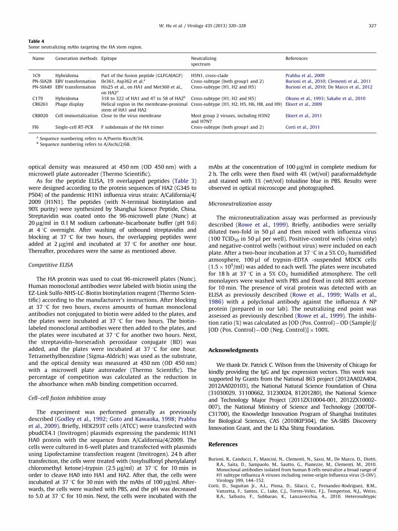
Table 4Some neutralizing mAbs targeting the HA stem region.
Name Generation methods Epitope Neutralizing
spectrum
References
1C9 Hybridoma Part of the fusion peptide (GLFGAIAGF) H5N1, cross-clade Prabhu et al., 2009
PN-SIA28 EBV transformation Ile361, Asp362 et al.a Cross-subtype (both group1 and 2) Burioni et al., 2010; Clementi et al., 2011
PN-SIA49 EBV transformation His25 et al., on HA1 and Met360 et al.,
on HA2a
Cross-subtype (H1, H2 and H5) Burioni et al., 2010; De Marco et al., 2012
C179 Hybridoma 318 to 322 of HA1 and 47 to 58 of HA2b Cross-subtype (H1, H2 and H5) Okuno et al., 1993; Sakabe et al., 2010
CR6261 Phage display Helical region in the membrane-proximal
stem of HA1 and HA2
Cross-subtype (H1, H2, H5, H6, H8, and H9) Ekiert et al., 2009
CR8020 Cell immortalization Close to the virus membrane Most group 2 viruses, including H3N2
and H7N7
Ekiert et al., 2011
FI6 Single-cell RT-PCR F subdomain of the HA trimer Cross-subtype (both group1 and 2) Corti et al., 2011
a Sequence numbering refers to A/Puerto Rico/8/34.b Sequence numbering refers to A/Aichi/2/68.
W. Hu et al. / Virology 435 (2013) 320–328 327
optical density was measured at 450 nm (OD 450 nm) with amicrowell plate autoreader (Thermo Scientific).
As for the peptide ELISA, 19 overlapped peptides (Table 3)were designed according to the protein sequences of HA2 (G345 toP504) of the pandemic H1N1 influenza virus strain: A/California/4/2009 (H1N1). The peptides (with N-terminal biotinylation and90% purity) were synthesized by Shanghai Science Peptide, China.Streptavidin was coated onto the 96-microwell plate (Nunc) at20 mg/ml in 0.1 M sodium carbonate–bicarbonate buffer (pH 9.6)at 4 1C overnight. After washing of unbound streptavidin andblocking at 37 1C for two hours, the overlapping peptides wereadded at 2 mg/ml and incubated at 37 1C for another one hour.Thereafter, procedures were the same as mentioned above.
Competitive ELISA
The HA protein was used to coat 96-microwell plates (Nunc).Human monoclonal antibodies were labeled with biotin using theEZ-Link Sulfo-NHS-LC-Biotin biotinylation reagent (Thermo Scien-tific) according to the manufacturer’s instructions. After blockingat 37 1C for two hours, excess amounts of human monoclonalantibodies not conjugated to biotin were added to the plates, andthe plates were incubated at 37 1C for two hours. The biotin-labeled monoclonal antibodies were then added to the plates, andthe plates were incubated at 37 1C for another two hours. Next,the streptavidin–horseradish peroxidase conjugate (BD) wasadded, and the plates were incubated at 37 1C for one hour.Tetramethylbenzidine (Sigma-Aldrich) was used as the substrate,and the optical density was measured at 450 nm (OD 450 nm)with a microwell plate autoreader (Thermo Scientific). Thepercentage of competition was calculated as the reduction inthe absorbance when mAb binding competition occurred.
Cell–cell fusion inhibition assay
The experiment was performed generally as previouslydescribed (Godley et al., 1992; Goto and Kawaoka, 1998; Prabhuet al., 2009). Briefly, HEK293T cells (ATCC) were transfected withpbudCE4.1 (Invitrogen) plasmids expressing the pandemic H1N1HA0 protein with the sequence from A/California/4/2009. Thecells were cultured in 6-well plates and transfected with plasmidsusing Lipofectamine transfection reagent (Invitrogen). 24 h aftertransfection, the cells were treated with (tosylsulfonyl phenylalanylchloromethyl ketone)-trypsin (2.5 mg/ml) at 37 1C for 10 min inorder to cleave HA0 into HA1 and HA2. After that, the cells wereincubated at 37 1C for 30 min with the mAbs of 100 mg/ml. After-wards, the cells were washed with PBS, and the pH was decreasedto 5.0 at 37 1C for 10 min. Next, the cells were incubated with the
mAbs at the concentration of 100 mg/ml in complete medium for2 h. The cells were then fixed with 4% (wt/vol) paraformaldehydeand stained with 1% (wt/vol) toluidine blue in PBS. Results wereobserved in optical microscope and photographed.
Microneutralization assay
The microneutralization assay was performed as previouslydescribed (Rowe et al., 1999). Briefly, antibodies were seriallydiluted two-fold in 50 ml and then mixed with influenza virus(100 TCID50 in 50 ml per well). Positive-control wells (virus only)and negative-control wells (without virus) were included on eachplate. After a two-hour incubation at 37 1C in a 5% CO2 humidifiedatmosphere, 100 ml of trypsin–EDTA -suspended MDCK cells(1.5�105/ml) was added to each well. The plates were incubatedfor 18 h at 37 1C in a 5% CO2 humidified atmosphere. The cellmonolayers were washed with PBS and fixed in cold 80% acetonefor 10 min. The presence of viral protein was detected with anELISA as previously described (Rowe et al., 1999; Walls et al.,1986) with a polyclonal antibody against the influenza A NPprotein (prepared in our lab). The neutralizing end point wasassessed as previously described (Rowe et al., 1999). The inhibi-tion ratio (%) was calculated as [OD (Pos. Control)�OD (Sample)]/[OD (Pos. Control)�OD (Neg. Control)]�100%.
Acknowledgments
We thank Dr. Patrick C. Wilson from the University of Chicago forkindly providing the IgG and Igk expression vectors. This work wassupported by Grants from the National 863 project (2012AA02A404,2012AA020103), the National Natural Science Foundation of China(31030029, 31100662, 31230024, 81201280), the National Scienceand Technology Major Project (2011ZX10004-001, 2012ZX10002-007), the National Ministry of Science and Technology (2007DF-C31700), the Knowledge Innovation Program of Shanghai Institutesfor Biological Sciences, CAS (2010KIP304), the SA-SIBS DiscoveryInnovation Grant, and the Li Kha Shing Foundation.
References
Burioni, R., Canducci, F., Mancini, N., Clementi, N., Sassi, M., De Marco, D., Diotti,R.A., Saita, D., Sampaolo, M., Sautto, G., Pianezze, M., Clementi, M., 2010.Monoclonal antibodies isolated from human B cells neutralize a broad range ofH1 subtype influenza A viruses including swine-origin Influenza virus (S-OIV).Virology 399, 144–152.
Corti, D., Suguitan Jr., A.L., Pinna, D., Silacci, C., Fernandez-Rodriguez, B.M.,Vanzetta, F., Santos, C., Luke, C.J., Torres-Velez, F.J., Temperton, N.J., Weiss,R.A., Sallusto, F., Subbarao, K., Lanzavecchia, A., 2010. Heterosubtypic

W. Hu et al. / Virology 435 (2013) 320–328328
neutralizing antibodies are produced by individuals immunized with aseasonal influenza vaccine. J. Clin. Invest. 120, 1663–1673.
Clementi, N., De Marco, D., Mancini, N., Solforosi, L., Moreno, G.J., Gubareva, L.V.,Mishin, V., Di Pietro, A., Vicenzi, E., Siccardi, A.G., Clementi, M., Burioni, R.,2011. A human monoclonal antibody with neutralizing activity against highlydivergent influenza subtypes. PLoS One 6, e28001.
Corti, D., Voss, J., Gamblin, S.J., Codoni, G., Macagno, A., Jarrossay, D., Vachieri, S.G.,Pinna, D., Minola, A., Vanzetta, F., Silacci, C., Fernandez-Rodriguez, B.M., Agatic,G., Bianchi, S., Giacchetto-Sasselli, I., Calder, L., Sallusto, F., Collins, P., Haire,L.F., Temperton, N., Langedijk, J.P., Skehel, J.J., Lanzavecchia, A., 2011. Aneutralizing antibody selected from plasma cells that binds to group 1 andgroup 2 influenza A hemagglutinins. Science 333, 850–856.
De Marco, D., Clementi, N., Mancini, N., Solforosi, L., Moreno, G.J., Sun, X., Tumpey,T.M., Gubareva, L.V., Mishin, V., Clementi, M., Burioni, R., 2012. A non-VH1-69heterosubtypic neutralizing human monoclonal antibody protects miceagainst H1N1 and H5N1 viruses. PLoS One 7, e34415.
Dong, J., Matsuoka, Y., Maines, T.R., Swayne, D.E., O’Neill, E., Davis, C.T., Van-Hoven,N., Balish, A., Yu, H.J., Katz, J.M., Klimov, A., Cox, N., Li, D.X., Wang, Y., Guo, Y.J.,Yang, W.Z., Donis, R.O., Shu, Y.L., 2009. Development of a new candidate H5N1avian influenza virus for pre-pandemic vaccine production. Influenza OtherRespir. Viruses 3, 287–295.
Ekiert, D.C., Bhabha, G., Elsliger, M.A., Friesen, R.H., Jongeneelen, M., Throsby, M.,Goudsmit, J., Wilson, I.A., 2009. Antibody recognition of a highly conservedinfluenza virus epitope. Science 324, 246–251.
Ekiert, D.C., Friesen, R.H., Bhabha, G., Kwaks, T., Jongeneelen, M., Yu, W., Ophorst,C., Cox, F., Korse, H.J., Brandenburg, B., Vogels, R., Brakenhoff, J.P., Kompier, R.,Koldijk, M.H., Cornelissen, L.A., Poon, L.L., Peiris, M., Koudstaal, W., Wilson, I.A.,Goudsmit, J., 2011. A highly conserved neutralizing epitope on group 2influenza A viruses. Science 333, 843–850.
Garten, R.J., Davis, C.T., Russell, C.A., Shu, B., Lindstrom, S., Balish, A., Sessions,W.M., Xu, X., Skepner, E., Deyde, V., Okomo-Adhiambo, M., Gubareva, L.,Barnes, J., Smith, C.B., Emery, S.L., Hillman, M.J., Rivailler, P., Smagala, J., deGraaf, M., Burke, D.F., Fouchier, R.A., Pappas, C., Alpuche-Aranda, C.M., Lopez-Gatell, H., Olivera, H., Lopez, I., Myers, C.A., Faix, D., Blair, P.J., Yu, C., Keene,K.M., Dotson Jr., P.D., Boxrud, D., Sambol, A.R., Abid, S.H., St George, K.,Bannerman, T., Moore, A.L., Stringer, D.J., Blevins, P., Demmler-Harrison, G.J.,Ginsberg, M., Kriner, P., Waterman, S., Smole, S., Guevara, H.F., Belongia, E.A.,Clark, P.A., Beatrice, S.T., Donis, R., Katz, J., Finelli, L., Bridges, C.B., Shaw, M.,Jernigan, D.B., Uyeki, T.M., Smith, D.J., Klimov, A.I., Cox, N.J., 2009. Antigenicand genetic characteristics of swine-origin 2009 A(H1N1) influenza virusescirculating in humans. Science 325, 197–201.
Godley, L., Pfeifer, J., Steinhauer, D., Ely, B., Shaw, G., Kaufmann, R., Suchanek, E.,Pabo, C., Skehel, J.J., Wiley, D.C., Wharton, S., 1992. Introduction of intersubunitdisulfide bonds in the membrane-distal region of the influenza hemagglutininabolishes membrane fusion activity. Cell 68, 635–645.
Goto, H., Kawaoka, Y., 1998. A novel mechanism for the acquisition of virulence bya human influenza A virus. Proc. Natl. Acad. Sci. U S A 95, 10224–10228.
Hancock, K., Veguilla, V., Lu, X., Zhong, W., Butler, E.N., Sun, H., Liu, F., Dong, L.,DeVos, J.R., Gargiullo, P.M., Brammer, T.L., Cox, N.J., Tumpey, T.M., Katz, J.M.,2009. Cross-reactive antibody responses to the 2009 pandemic H1N1 influ-enza virus. N. Engl. J. Med. 361, 1945–1952.
Hung, I.F., To, K.K., Lee, C.K., Lee, K.L., Chan, K., Yan, W.W., Liu, R., Watt, C.L., Chan,W.M., Lai, K.Y., Koo, C.K., Buckley, T., Chow, F.L., Wong, K.K., Chan, H.S., Ching,C.K., Tang, B.S., Lau, C.C., Li, I.W., Liu, S.H., Chan, K.H., Lin, C.K., Yuen, K.Y., 2011.Convalescent plasma treatment reduced mortality in patients with severe
pandemic influenza A (H1N1) 2009 virus infection. Clin. Infect. Dis. 52,447–456.
Liang, X.F., Wang, H.Q., Wang, J.Z., Fang, H.H., Wu, J., Zhu, F.C., Li, R.C., Xia, S.L.,Zhao, Y.L., Li, F.J., Yan, S.H., Yin, W.D., An, K., Feng, D.J., Cui, X.L., Qi, F.C., Ju, C.J.,Zhang, Y.H., Guo, Z.J., Chen, P.Y., Chen, Z., Yan, K.M., Wang, Y., 2010. Safety andimmunogenicity of 2009 pandemic influenza A H1N1 vaccines in China: amulticentre, double-blind, randomised, placebo-controlled trial. Lancet 375,56–66.
Okuno, Y., Isegawa, Y., Sasao, F., Ueda, S., 1993. A common neutralizing epitopeconserved between the hemagglutinins of influenza A virus H1 and H2 strains.J. Virol. 67, 2552–2558.
Palese, P., 2004. Influenza: old and new threats. Nat. Med. 10, S82–87.Prabhu, N., Prabakaran, M., Ho, H.T., Velumani, S., Qiang, J., Goutama, M., Kwang, J.,
2009. Monoclonal antibodies against the fusion peptide of hemagglutininprotect mice from lethal influenza A virus H5N1 infection. J. Virol. 83,2553–2562.
Rowe, T., Abernathy, R.A., Hu-Primmer, J., Thompson, W.W., Lu, X., Lim, W.,Fukuda, K., Cox, N.J., Katz, J.M., 1999. Detection of antibody to avian influenzaA (H5N1) virus in human serum by using a combination of serologic assays.J. Clin. Microbiol. 37, 937–943.
Skehel, J.J., Wiley, D.C., 2000. Receptor binding and membrane fusion in virusentry: the influenza hemagglutinin. Annu. Rev. Biochem. 69, 531–569.
Smith, K., Garman, L., Wrammert, J., Zheng, N.Y., Capra, J.D., Ahmed, R., Wilson,P.C., 2009. Rapid generation of fully human monoclonal antibodies specific to avaccinating antigen. Nat. Protoc. 4, 372–384.
Sui, J., Hwang, W.C., Perez, S., Wei, G., Aird, D., Chen, L.M., Santelli, E., Stec, B.,Cadwell, G., Ali, M., Wan, H., Murakami, A., Yammanuru, A., Han, T., Cox, N.J.,Bankston, L.A., Donis, R.O., Liddington, R.C., Marasco, W.A., 2009. Structuraland functional bases for broad-spectrum neutralization of avian and humaninfluenza A viruses. Nat. Struct. Mol. Biol. 16, 265–273.
Sakabe, S., Iwatsuki-Horimoto, K., Horimoto, T., Nidom, C.A., Le, M.Q., Takano, R.,Kubota-Koketsu, R., Okuno, Y., Ozawa, M., Kawaoka, Y., 2010. A cross-reactiveneutralizing monoclonal antibody protects mice from H5N1 and pandemic(H1N1) 2009 virus infection. Antiviral Res. 88, 249–255.
Walls, H.H., Harmon, M.W., Slagle, J.J., Stocksdale, C., Kendal, A.P., 1986. Char-acterization and evaluation of monoclonal antibodies developed for typinginfluenza A and influenza B viruses. J. Clin. Microbiol. 23, 240–245.
Wang, K., Holtz, K.M., Anderson, K., Chubet, R., Mahmoud, W., Cox, M.M., 2006.Expression and purification of an influenza hemagglutinin—one step closer toa recombinant protein-based influenza vaccine. Vaccine 24, 2176–2185.
Wrammert, J., Smith, K., Miller, J., Langley, W.A., Kokko, K., Larsen, C., Zheng, N.Y.,Mays, I., Garman, L., Helms, C., James, J., Air, G.M., Capra, J.D., Ahmed, R.,Wilson, P.C., 2008. Rapid cloning of high-affinity human monoclonal anti-bodies against influenza virus. Nature 453, 667–671.
Wrammert, J., Koutsonanos, D., Li, G.M., Edupuganti, S., Sui, J., Morrissey, M.,McCausland, M., Skountzou, I., Hornig, M., Lipkin, W.I., Mehta, A., Razavi, B.,Del Rio, C., Zheng, N.Y., Lee, J.H., Huang, M., Ali, Z., Kaur, K., Andrews, S., Amara,R.R., Wang, Y., Das, S.R., O’Donnell, C.D., Yewdell, J.W., Subbarao, K., Marasco,W.A., Mulligan, M.J., Compans, R., Ahmed, R., Wilson, P.C., 2011. Broadly cross-reactive antibodies dominate the human B cell response against 2009pandemic H1N1 influenza virus infection. J. Exp. Med. 208, 181–193.
Zhu, F.C., Wang, H., Fang, H.H., Yang, J.G., Lin, X.J., Liang, X.F., Zhang, X.F., Pan, H.X.,Meng, F.Y., Hu, Y.M., Liu, W.D., Li, C.G., Li, W., Zhang, X., Hu, J.M., Peng, W.B.,Yang, B.P., Xi, P., Wang, H.Q., Zheng, J.S., 2009. A novel influenza A (H1N1)vaccine in various age groups. N. Engl. J. Med. 361, 2414–2423.