Full thickness wound healing and safety of Reactive Skin ...
14
Defence Research and Development Canada Reference Document DRDC-RDDC-2020-D042 May 2020 CAN UNCLASSIFIED CAN UNCLASSIFIED Full thickness wound healing and safety of Reactive Skin Decontamination Lotion (RSDL) application in rabbits Jessica Franken John Mikler DRDC – Suffield Research Centre The body of this CAN UNCLASSIFIED document does not contain the required security banners according to DND security standards. However, it must be treated as CAN UNCLASSIFIED and protected appropriately based on the terms and conditions specified on the covering page.
Transcript of Full thickness wound healing and safety of Reactive Skin ...

Defence Research and Development Canada Reference Document
DRDC-RDDC-2020-D042
May 2020
CAN UNCLASSIFIED
CAN UNCLASSIFIED
Full thickness wound healing and safety of Reactive Skin Decontamination Lotion (RSDL) application in rabbits
Jessica Franken John Mikler DRDC – Suffield Research Centre
The body of this CAN UNCLASSIFIED document does not contain the required security banners according to DND security standards. However, it must be treated as CAN UNCLASSIFIED and protected appropriately based on the terms and conditions specified on the covering page.

CAN UNCLASSIFIED
Template in use: EO Publishing App for SR-RD-EC Eng 2018-12-19_v1 (new disclaimer).dotm © Her Majesty the Queen in Right of Canada (Department of National Defence), 2020
© Sa Majesté la Reine en droit du Canada (Ministère de la Défense nationale), 2020
CAN UNCLASSIFIED
IMPORTANT INFORMATIVE STATEMENTS
This document was reviewed for Controlled Goods by Defence Research and Development Canada (DRDC) using the Schedule to the Defence Production Act.
Disclaimer: This publication was prepared by Defence Research and Development Canada an agency of the Department of National Defence. The information contained in this publication has been derived and determined through best practice and adherence to the highest standards of responsible conduct of scientific research. This information is intended for the use of the Department of National Defence, the Canadian Armed Forces (“Canada”) and Public Safety partners and, as permitted, may be shared with academia, industry, Canada’s allies, and the public (“Third Parties”). Any use by, or any reliance on or decisions made based on this publication by Third Parties, are done at their own risk and responsibility. Canada does not assume any liability for any damages or losses which may arise from any use of, or reliance on, the publication.
In conducting the research described in this report, the investigators adhered to the “Guide to the Care and Use of Experimental Animals, Vol. I, 2nd Ed.” published by the Canadian Council on Animal Care.
Endorsement statement: This publication has been published by the Editorial Office of Defence Research and Development Canada, an agency of the Department of National Defence of Canada. Inquiries can be sent to: [email protected].

DRDC-RDDC-2020-D042 i
Abstract
Reactive skin decontamination lotion (RSDL) was developed to remove chemical warfare agents (CWA),
including vesicants and organophosphorus nerve agents, from the epidermis. RSDL acts by breaking
down some agents and to decontaminate the surface more effectively than soapy water. Other products
have been developed but RSDL has proven to be superior in each case, except when open wounds are
present. Safety studies are lacking for how wound healing is effected if RSDL is used to decontaminate a
burn, is rinsed off, and then allowed to heal. This review set out to determine whether a proposed rabbit
chemical burn study is the most appropriate way to model wound healing.

ii DRDC-RDDC-2020-D042
Résumé
La lotion neutralisante pour la décontamination de la peau (LNDP) a été mise au point pour éliminer de
l’épiderme les agents de guerre chimique (CWA), notamment les agents vésicants et les agents
neurotoxiques organophosphorés. La LNDP agit en dégradant certains agents et est plus efficace que
l’eau savonneuse pour décontaminer les surfaces exposées. D’autres produits ont été mis au point, mais la
LNDP s’est révélée supérieure dans chaque cas, sauf en présence de plaies ouvertes. Il n’existe aucune
étude d’innocuité sur le processus de cicatrisation des plaies lorsqu’on utilise la LNDP pour décontaminer
une brûlure, puis que l’on rince la peau et qu’on la laisse cicatriser. Cet examen visait à déterminer si une
étude proposée sur les brûlures chimiques chez le lapin constitue le moyen le plus approprié de modéliser
la guérison des plaies.

DRDC-RDDC-2020-D042 iii
Table of Contents
Abstract . . . . . . . . . . . . . . . . . . . . . . . . . . . . . . . . . . . i
Résumé . . . . . . . . . . . . . . . . . . . . . . . . . . . . . . . . . . . ii
Table of Contents . . . . . . . . . . . . . . . . . . . . . . . . . . . . . . . iii
List of Tables . . . . . . . . . . . . . . . . . . . . . . . . . . . . . . . . iv
1 Background . . . . . . . . . . . . . . . . . . . . . . . . . . . . . . . . 1
2 Statement of Results . . . . . . . . . . . . . . . . . . . . . . . . . . . . . 2
3 Conclusion . . . . . . . . . . . . . . . . . . . . . . . . . . . . . . . . . 4
References . . . . . . . . . . . . . . . . . . . . . . . . . . . . . . . . . . 5
List of Symbols/Abbreviations/Acronyms/Initialisms . . . . . . . . . . . . . . . . . . . 6

iv DRDC-RDDC-2020-D042
List of Tables
Table 1: Cytokine function and relation to wound healing phases. . . . . . . . . . . . . . 3

DRDC-RDDC-2020-D042 1
1 Background
Reactive Skin Decontamination Lotion (RSDL) was developed for the purpose of decontaminating
epidermal tissue exposed to chemical warfare agents (CWA), specifically vesicants and
organophosphorus nerve agents. Destruction and decontamination of CWA’s on the epidermis by RSDL
has been shown to be superior to other products developed for the same purpose. In order for Canada to
universalize use of RSDL by civilian first responders in emergency chemical exposures (including
accidental spills of a strong acid or base), further studies investigating safety of use in open wounds need
to be conducted. The proposed test system to investigate this question is rabbits and wound healing of full
thickness sulfuric acid burns that undergo decontamination with either RSDL or soapy water is to be
evaluated. A literature review was conducted to determine whether rabbits are the best choice and what
parameters ought to be measured and scored for the most accurate comparison of healing.
2 DRDC-RDDC-2020-D042
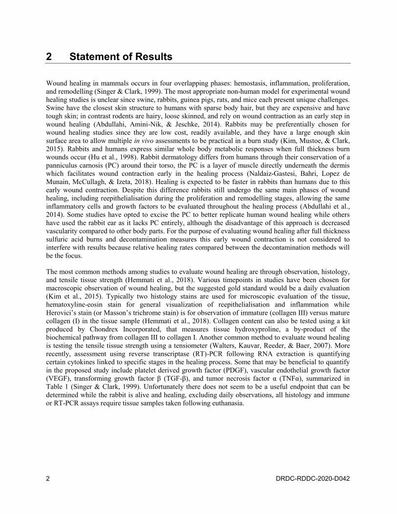
2 Statement of Results
Wound healing in mammals occurs in four overlapping phases: hemostasis, inflammation, proliferation,
and remodelling (Singer & Clark, 1999). The most appropriate non-human model for experimental wound
healing studies is unclear since swine, rabbits, guinea pigs, rats, and mice each present unique challenges.
Swine have the closest skin structure to humans with sparse body hair, but they are expensive and have
tough skin; in contrast rodents are hairy, loose skinned, and rely on wound contraction as an early step in
wound healing (Abdullahi, Amini-Nik, & Jeschke, 2014). Rabbits may be preferentially chosen for
wound healing studies since they are low cost, readily available, and they have a large enough skin
surface area to allow multiple in vivo assessments to be practical in a burn study (Kim, Mustoe, & Clark,
2015). Rabbits and humans express similar whole body metabolic responses when full thickness burn
wounds occur (Hu et al., 1998). Rabbit dermatology differs from humans through their conservation of a
panniculus carnosis (PC) around their torso, the PC is a layer of muscle directly underneath the dermis
which facilitates wound contraction early in the healing process (Naldaiz-Gastesi, Bahri, Lopez de
Munain, McCullagh, & Izeta, 2018). Healing is expected to be faster in rabbits than humans due to this
early wound contraction. Despite this difference rabbits still undergo the same main phases of wound
healing, including reepithelialisation during the proliferation and remodelling stages, allowing the same
inflammatory cells and growth factors to be evaluated throughout the healing process (Abdullahi et al.,
2014). Some studies have opted to excise the PC to better replicate human wound healing while others
have used the rabbit ear as it lacks PC entirely, although the disadvantage of this approach is decreased
vascularity compared to other body parts. For the purpose of evaluating wound healing after full thickness
sulfuric acid burns and decontamination measures this early wound contraction is not considered to
interfere with results because relative healing rates compared between the decontamination methods will
be the focus.
The most common methods among studies to evaluate wound healing are through observation, histology,
and tensile tissue strength (Hemmati et al., 2018). Various timepoints in studies have been chosen for
macroscopic observation of wound healing, but the suggested gold standard would be a daily evaluation
(Kim et al., 2015). Typically two histology stains are used for microscopic evaluation of the tissue,
hematoxyline-eosin stain for general visualization of reepithelialisation and inflammation while
Herovici’s stain (or Masson’s trichrome stain) is for observation of immature (collagen III) versus mature
collagen (I) in the tissue sample (Hemmati et al., 2018). Collagen content can also be tested using a kit
produced by Chondrex Incorporated, that measures tissue hydroxyproline, a by-product of the
biochemical pathway from collagen III to collagen I. Another common method to evaluate wound healing
is testing the tensile tissue strength using a tensiometer (Walters, Kauvar, Reeder, & Baer, 2007). More
recently, assessment using reverse transcriptase (RT)-PCR following RNA extraction is quantifying
certain cytokines linked to specific stages in the healing process. Some that may be beneficial to quantify
in the proposed study include platelet derived growth factor (PDGF), vascular endothelial growth factor
(VEGF), transforming growth factor β (TGF-β), and tumor necrosis factor α (TNFα), summarized in
Table 1 (Singer & Clark, 1999). Unfortunately there does not seem to be a useful endpoint that can be
determined while the rabbit is alive and healing, excluding daily observations, all histology and immune
or RT-PCR assays require tissue samples taken following euthanasia.

DRDC-RDDC-2020-D042 3
Table 1: Cytokine function and relation to wound healing phases.
Cytokine Function Healing stage
PDGF Chemotaxis (fibroblast and smooth muscle cells) Early inflammatory to early
proliferation
TGF-β Chemotaxis (fibroblast, smooth muscle, PDGF,
TNFα), collagen/collagenase expression
Early inflammatory to early
proliferation
TNFα Chemotaxis, fibroblast mitogen Proliferation
VEGF Neovascularization and angiogenesis Reepithelialization
During hemostasis, PDGF and TGF-β content in tissue will be rising and remain high throughout the
inflammatory period which is generally around 3 days (Singer & Clark, 1999). As the inflammatory
period ends and the proliferation period begins collagen formation has begun, TNFα will become
detectable. VEGF becomes prominent further into the proliferation phase. Complete reeplithelialization of
excisional wounds in most studies reviewed occurred by day 21. This research indicates that histology
and molecular biology would be most useful to detect possible differences in healing between
decontamination variables at days 3 and 21. RSDL has been shown to be safe to use on unbroken skin but
research is limited regarding RSDL’s effect on wound healing. As, (Walters et al., 2007) found RSDL
treated wounds did not differ from control wounds histologically but were weaker in the tensile strength
test and contained less collagen. There were many limitations in this study, particularly the method of
treating the wound with RSDL and neglecting to wash it off.

4 DRDC-RDDC-2020-D042
3 Conclusion
The decision to use rabbits as the test system for this decontamination and wound healing study is
appropriate. One cm2 acid burn wounds on rabbits would be expected to first undergo clotting and
achieve hemostasis, then inflammation and contraction of PC musculature starts healing the wound, lastly
reeipithelialization will occur. This process will likely take around three weeks. Histology must be
conducted to evaluate the healing stage, an additional stain specific for type III vs type I collagen may
want to be considered. Tissue tensile strength evaluation is extremely important to compare with
(Walters’ et al., 2007) results. Further analysis using RT-PCR of healing factors PDGF, TGF-β, TNFα,
and VEGF may be beneficial if part of the wound tissue can be used for RNA extraction.
RSDL is currently carried by CAF and several other armies to serve as the primary decontaminant for
exposures to CWAs since it is the most effective. Threat to civilian populations is possible and first
responders would benefit from carrying RSDL as their primary decontaminant. Currently
decontamination by first responders involves soapy water, which if the patient is suffering strong
acid/base burns does not affect wound healing. Scientific evidence that RSDL does not hinder wound
healing would be imperative to the decision to add RSDL to the kit used by civilian first responders.

DRDC-RDDC-2020-D042 5
References
Abdullahi, A., Amini-Nik, S., and Jeschke, M. G. (2014). Animal models in burn research. Cell Mol Life
Sci, 71(17), pp. 3241–3255. doi:10.1007/s00018-014-1612-5.
Hemmati, A. A., Larki-Harchegani, A., Shabib, S., Jalali, A., Rezaei, A., and Housmand, G. (2018).
Wound healing property of milk in full thickness wound model of rabbit. Int J Surg, 54(Pt A),
pp. 133–140. doi:10.1016/j.ijsu.2018.04.030.
Hu, R. H., Yu, Y. M., Costa, D., Young, V. R., Ryan, C. M., Burke, J. F., and Tompkins, R. G. (1998). A
rabbit model for metabolic studies after burn injury. J Surg Res, 75(2), pp. 153–160.
doi:10.1006/jsre.1998.5274.
Kim, D. J., Mustoe, T., and Clark, R. A. (2015). Cutaneous wound healing in aging small mammals: a
systematic review. Wound Repair Regen, 23(3), pp. 318–339. doi:10.1111/wrr.12290.
Naldaiz-Gastesi, N., Bahri, O. A., Lopez de Munain, A., McCullagh, K. J. A., and Izeta, A. (2018). The
panniculus carnosus muscle: an evolutionary enigma at the intersection of distinct research fields. J Anat.
doi:10.1111/joa.12840.
Singer, A. J., and Clark, R. A. (1999). Cutaneous wound healing. N Engl J Med, 341(10), pp. 738–746.
doi:10.1056/NEJM199909023411006.
Walters, T. J., Kauvar, D. S., Reeder, J., and Baer, D. G. (2007). Effect of reactive skin decontamination
lotion on skin wound healing in laboratory rats. Mil Med, 172(3), pp. 318–321.

6 DRDC-RDDC-2020-D042
List of Symbols/Abbreviations/Acronyms/Initialisms
CWA Chemical Warfare Agents
DND Department of National Defence
DRDC Defence Research and Development Canada
PC Panniculus Carnosis
PCR Polymerase Chain Reaction
PDGF Platelet Derived Growth Factor
R&D Research & Development
RSDL Reactive Skin Decontamination Lotion
RT Reverse Transcriptase
TGF-β Transforming Growth Factor β
TNFα Tumor Necrosis Factor α
VEGF Vascular Endothelial Growth Factor

DOCUMENT CONTROL DATA *Security markings for the title, authors, abstract and keywords must be entered when the document is sensitive
1. ORIGINATOR (Name and address of the organization preparing the document. A DRDC Centre sponsoring a contractor's report, or tasking agency, is entered in Section 8.)
DRDC – Suffield Research Centre Defence Research and Development Canada P.O. Box 4000, Station Main Medicine Hat, Alberta T1A 8K6 Canada
2a. SECURITY MARKING (Overall security marking of the document including special supplemental markings if applicable.)
CAN UNCLASSIFIED
2b. CONTROLLED GOODS
NON-CONTROLLED GOODS DMC A
3. TITLE (The document title and sub-title as indicated on the title page.)
Full thickness wound healing and safety of Reactive Skin Decontamination Lotion (RSDL) application in rabbits
4. AUTHORS (Last name, followed by initials – ranks, titles, etc., not to be used)
Franken, J.; Mikler, J.
5. DATE OF PUBLICATION (Month and year of publication of document.)
May 2020
6a. NO. OF PAGES
(Total pages, including Annexes, excluding DCD, covering and verso pages.)
10
6b. NO. OF REFS
(Total references cited.)
7
7. DOCUMENT CATEGORY (e.g., Scientific Report, Contract Report, Scientific Letter.)
Reference Document
8. SPONSORING CENTRE (The name and address of the department project office or laboratory sponsoring the research and development.)
DRDC – Suffield Research Centre Defence Research and Development Canada P.O. Box 4000, Station Main Medicine Hat, Alberta T1A 8K6 Canada
9a. PROJECT OR GRANT NO. (If appropriate, the applicable research and development project or grant number under which the document was written. Please specify whether project or grant.)
06da
9b. CONTRACT NO. (If appropriate, the applicable number under which the document was written.)
10a. DRDC PUBLICATION NUMBER (The official document number by which the document is identified by the originating activity. This number must be unique to this document.)
DRDC-RDDC-2020-D042
10b. OTHER DOCUMENT NO(s). (Any other numbers which may be assigned this document either by the originator or by the sponsor.)
11a. FUTURE DISTRIBUTION WITHIN CANADA (Approval for further dissemination of the document. Security classification must also be considered.)
Public release
11b. FUTURE DISTRIBUTION OUTSIDE CANADA (Approval for further dissemination of the document. Security classification must also be considered.)
12. KEYWORDS, DESCRIPTORS or IDENTIFIERS (Use semi-colon as a delimiter.)
RSDL; CWA

13. ABSTRACT (When available in the document, the French version of the abstract must be included here.)
Reactive skin decontamination lotion (RSDL) was developed to remove chemical warfare agents (CWA), including vesicants and organophosphorus nerve agents, from the epidermis. RSDL acts by breaking down some agents and to decontaminate the surface more effectively than soapy water. Other products have been developed but RSDL has proven to be superior in each case, except when open wounds are present. Safety studies are lacking for how wound healing is effected if RSDL is used to decontaminate a burn, is rinsed off, and then allowed to heal. This review set out to determine whether a proposed rabbit chemical burn study is the most appropriate way to model wound healing.
La lotion neutralisante pour la décontamination de la peau (LNDP) a été mise au point pour éliminer de l’épiderme les agents de guerre chimique (CWA), notamment les agents vésicants et les agents neurotoxiques organophosphorés. La LNDP agit en dégradant certains agents et est plus efficace que l’eau savonneuse pour décontaminer les surfaces exposées. D’autres produits ont été mis au point, mais la LNDP s’est révélée supérieure dans chaque cas, sauf en présence de plaies ouvertes. Il n’existe aucune étude d’innocuité sur le processus de cicatrisation des plaies lorsqu’on utilise la LNDP pour décontaminer une brûlure, puis que l’on rince la peau et qu’on la laisse cicatriser. Cet examen visait à déterminer si une étude proposée sur les brûlures chimiques chez le lapin constitue le moyen le plus approprié de modéliser la guérison des plaies.