Frontostriatal System in Planning Complexity: A Parametric Functional Magnetic Resonance Version of...
9
Frontostriatal system in planning complexity: a parametric functional magnetic resonance version of Tower of London task Odile A. van den Heuvel, a,b, * Henk J. Groenewegen, c Frederik Barkhof, d Richard H.C. Lazeron, e Richard van Dyck, a and Dick J. Veltman a,b a Department of Psychiatry, VU Medical Centre, Amsterdam, The Netherlands b Clinical PET Centre, VU Medical Centre, Amsterdam, The Netherlands c Department of Anatomy, VU Medical Centre, Amsterdam, The Netherlands d Department of Radiology, VU Medical Centre, Amsterdam, The Netherlands e Department of Neurology, VU Medical Centre, Amsterdam, The Netherlands Received 1 April 2002; revised 27 June 2002; accepted 6 August 2002 Abstract In the present study, we sought to investigate which brain structures are recruited in planning tasks of increasing complexity. For this purpose, a parametric self-paced pseudo-randomized event-related functional MRI version of the Tower of London task was designed. We tested 22 healthy subjects, enabling assessment of imaging results at a second (random effects) level of analysis. Compared with baseline, planning activity was correlated with increased blood oxygenation level-dependent (BOLD) signal in the dorsolateral prefrontal cortex, striatum, premotor cortex, supplementary motor area, and visuospatial system (precuneus and inferior parietal cortex). Task load was associated with increased activity in these same regions. In addition, increasing task complexity was correlated with activity in the left ante rior prefrontal corte x, a regio n supposed to be speci fical ly involve d in third-order higher cognitive functionin g. © 2003 Elsevier Science (USA). All rights reserved. Keywords: Planning; Tower of London; Functional magnetic resonance imaging; Parametric; Prefrontal cortex; Basal ganglia Introduction Pla nni ng, i.e., the abi lit y to achieve a goa l thr ough a series of intermediate steps, is an essential component of higher-order cognitive processes, such as problem solving. Central to the concept of the neural substrate for cognitive planning is the position of the dorsolateral prefrontal cortex, but other brain regions, like the premotor, cingulate, and insul ar cortic es and the striatum, have also been implic ated in co gniti ve pl anni ng (Owen, 1997; Robbins, 1998). Whereas until recently insight into the role of brain struc- tures in planning processes was based primarily on lesion studies in patients, contemporary studies are able to employ human brain imaging techniques. The results of such func- tional neuroimaging studies have suggested that a distrib- uted corticocortical and corticosubcortical network is being activated during complex planning tasks, although there is yet no complete agreement about the contribution of the various components of such neuronal networks. A direct and very frequently used test to probe planning processes is the Tower of London task, originally developed by Shallice (1982). The Tower of London task has been modified in var iou s ways to make it suitable for testing different components of cognitive planning in clinical and experimental psychological settings. The complexity of the tas k can range from trivial, requiring only one obviou s move, to highly complex. The more complex versions of the task require the planning of intermediate, in many instances counterintuitive, steps to successfully solve a problem. In- div iduals with decreased pre fron tal functionin g, such as those with neurosurgical lesions (Shallice, 1982), neurode- generative diseases like Huntington’s and Parkinson’s dis- * Corresponding author: Clinical PET Centre, VU Medical Centre, PO Box 7057, 1007 MB Amsterdam, The Netherlands. Fax: 31-20-4449636. E-mail address: [email protected] (O.A. van den Heuvel). R Available online at www.sciencedirect.com NeuroImage 18 (2003) 367–374 www.elsevier.com/locate/ynimg 1053-8119/03/$ – see front matter © 2003 Elsevier Science (USA). All rights reserved. PI I: S1053-8119(02)00010-1
-
Upload
ammonluckart -
Category
Documents
-
view
219 -
download
0
Transcript of Frontostriatal System in Planning Complexity: A Parametric Functional Magnetic Resonance Version of...

8/12/2019 Frontostriatal System in Planning Complexity: A Parametric Functional Magnetic Resonance Version of Tower of London Task
http://slidepdf.com/reader/full/frontostriatal-system-in-planning-complexity-a-parametric-functional-magnetic 1/9
Frontostriatal system in planning complexity: a parametric functionalmagnetic resonance version of Tower of London task
Odile A. van den Heuvel,a,b,* Henk J. Groenewegen,c Frederik Barkhof,d
Richard H.C. Lazeron,e Richard van Dyck,a and Dick J. Veltmana,b
a Department of Psychiatry, VU Medical Centre, Amsterdam, The Netherlandsb Clinical PET Centre, VU Medical Centre, Amsterdam, The Netherlands
c Department of Anatomy, VU Medical Centre, Amsterdam, The Netherlandsd Department of Radiology, VU Medical Centre, Amsterdam, The Netherlandse Department of Neurology, VU Medical Centre, Amsterdam, The Netherlands
Received 1 April 2002; revised 27 June 2002; accepted 6 August 2002
Abstract
In the present study, we sought to investigate which brain structures are recruited in planning tasks of increasing complexity. For this
purpose, a parametric self-paced pseudo-randomized event-related functional MRI version of the Tower of London task was designed. We
tested 22 healthy subjects, enabling assessment of imaging results at a second (random effects) level of analysis. Compared with baseline,
planning activity was correlated with increased blood oxygenation level-dependent (BOLD) signal in the dorsolateral prefrontal cortex,
striatum, premotor cortex, supplementary motor area, and visuospatial system (precuneus and inferior parietal cortex). Task load was
associated with increased activity in these same regions. In addition, increasing task complexity was correlated with activity in the left
anterior prefrontal cortex, a region supposed to be specifically involved in third-order higher cognitive functioning.
© 2003 Elsevier Science (USA). All rights reserved.
Keywords: Planning; Tower of London; Functional magnetic resonance imaging; Parametric; Prefrontal cortex; Basal ganglia
Introduction
Planning, i.e., the ability to achieve a goal through a
series of intermediate steps, is an essential component of
higher-order cognitive processes, such as problem solving.
Central to the concept of the neural substrate for cognitive
planning is the position of the dorsolateral prefrontal cortex,
but other brain regions, like the premotor, cingulate, and
insular cortices and the striatum, have also been implicated
in cognitive planning (Owen, 1997; Robbins, 1998).
Whereas until recently insight into the role of brain struc-
tures in planning processes was based primarily on lesion
studies in patients, contemporary studies are able to employ
human brain imaging techniques. The results of such func-
tional neuroimaging studies have suggested that a distrib-
uted corticocortical and corticosubcortical network is being
activated during complex planning tasks, although there is
yet no complete agreement about the contribution of the
various components of such neuronal networks.
A direct and very frequently used test to probe planning
processes is the Tower of London task, originally developed
by Shallice (1982). The Tower of London task has been
modified in various ways to make it suitable for testingdifferent components of cognitive planning in clinical and
experimental psychological settings. The complexity of the
task can range from trivial, requiring only one obvious
move, to highly complex. The more complex versions of the
task require the planning of intermediate, in many instances
counterintuitive, steps to successfully solve a problem. In-
dividuals with decreased prefrontal functioning, such as
those with neurosurgical lesions (Shallice, 1982), neurode-
generative diseases like Huntington’s and Parkinson’s dis-
* Corresponding author: Clinical PET Centre, VU Medical Centre, PO
Box 7057, 1007 MB Amsterdam, The Netherlands. Fax: 31-20-4449636.
E-mail address: [email protected] (O.A. van den Heuvel).
R
Available online at www.sciencedirect.com
NeuroImage 18 (2003) 367–374 www.elsevier.com/locate/ynimg
1053-8119/03/$ – see front matter © 2003 Elsevier Science (USA). All rights reserved.
PII: S 1 0 5 3 -8 1 1 9 (0 2 )0 0 0 1 0 -1
8/12/2019 Frontostriatal System in Planning Complexity: A Parametric Functional Magnetic Resonance Version of Tower of London Task
http://slidepdf.com/reader/full/frontostriatal-system-in-planning-complexity-a-parametric-functional-magnetic 2/9
eases (Dagher et al., 1999; Watkins et al., 2000; Hodgson et
al., 2002), or psychiatric disorders like obsessive– compul-
sive disorder (Veale et al., 1996) and schizophrenia (Pan-
telis et al., 1997), are impaired on the task, in particular
when complexity increases.
The Tower of London task is also very suitable for
studying in healthy subjects the patterns of brain activationin the context of planning tasks using positron emission
tomography (PET) (Owen et al., 1996; Baker et al., 1996;
Dagher et al., 1999; Rowe et al., 2001) or single-photon
emission computed tomography (SPECT) (Rezai et al.,
1993; Morris et al., 1993). The results of these imaging
studies agree on the involvement of the dorsolateral pre-
frontal cortex and parieto-occipital regions (visuospatial
system) during planning. However, activation of other brain
regions, such as the cingulate and insular cortices and the
striatum, has not been found across all mentioned studies.
Furthermore, the results of a functional MRI study using the
Tower of London task of Lazeron et al. (2000) confirmed
activation of the dorsolateral prefrontal, parietal, cingulate,
and insular cortices but failed to show striatal activation.
Several methodological differences between these studies
may explain the inconsistencies in the results. First, differ-
ent baseline conditions have been used, ranging from low-
level conditions (watching a blank screen) to conditions
involving motor activity (moving beads without a goal) or
cognitive functioning (counting beads). Second, differences
in the execution of the task exist: whereas in some studies
subjects used a touch screen to perform the task (Owen et
al., 1996; Dagher et al., 1999; Rowe et al., 2001), in other
designs subjects were requested to execute their moves
mentally (Baker et al., 1996; Lazeron et al., 2000). A thirdlikely source of inconsistency concerns the differences in
task load. Because in subtraction designs the specificity of
active versus baseline differences for the (cognitive) func-
tion under study is often questionable (Friston et al., 1996;
Sidtis et al., 1999), several researchers have used designs in
which task load during performance of the Tower of Lon-
don task was manipulated. For example, Baker et al.,
(1996), in comparing “dif ficult” (four or five moves) and
“easy” trials (two or three moves), found task load to be
correlated with increased regional cerebral blood flow
(rCBF) in rostrolateral and dorsolateral prefrontal cortex,
and the parietal and cingulate cortices. Owen et al. (1996)reported increased activation in striatum and thalamus, but
not in cortical areas. Lazeron et al. (2000) did not find any
difference between their “easy” and “dif ficult” versions, but
they used a more dif ficult design comparing two to four
moves with five to seven moves. The only study employing
a parametric approach in studying task load (Dagher et al.,
1999) used five levels of planning and showed a correlation
between planning complexity and increased rCBF in the
right caudate nucleus and prefrontal and cingulate areas.
The involvement of visuospatial areas in the parietal lobe,
however, seemed not to be related to the task load. A
general methodological drawback in the studies performed
so far is the lack of control over performance differences,
due to the use of blocked designs. Since performance is
likely to deteriorate at higher complexity, this is especially
relevant for parametric tasks.
It must be assumed that the various cortical and subcor-
tical brain structures that have been demonstrated to be
involved in planning processes execute these functions as“nodes” in distributed neuronal networks (Owen, 1997).
Employing a neuronal network model for planning pro-
cesses, Dehaene and Changeux (1997) proposed multiple
hierarchic levels with ascending and descending streams for
execution of planning and evaluation of this behavior, re-
spectively. This model also stresses that planning behavior
cannot be related to a single region, but rather relies on
multiple neuronal circuits coding for specialized subpro-
cesses such as working memory, plan generation, and in-
ternal reward. These subprocesses may differentially con-
tribute to different levels of planning behavior; some
subprocesses may be relatively independent of task load,while other subprocesses are involved mainly at higher
levels of planning behavior. The main aspect in increasing
planning complexity is the increasing amount of subgoals
and counterintuitive moves, while holding in mind the over-
all main goal. Koechlin et al. (2000) introduced the term
branching for this process of integrating working memory
with attentional resource allocation, a third-order higher
cognitive function. They found, using a multitask experi-
ment, branching to rely on activation of the rostral part of
the prefrontal cortex. To be able to investigate these specific
subprocesses and to differentiate between different brain
regions in their contribution to different levels of planning
behavior, parameterization of the task is essential. There-
fore, the main aim of the present study was to determine the
effects of task load in planning execution.
The present study employs event-related functional MRI
as a method to study activation patterns on a trial-by-trial
basis. This enabled us (i) to randomize items with respect to
task complexity, and (ii) to include correct responses only.
A large number of subjects (n 22) were included, with the
aim of performing second-level or random effects analyses,
allowing for generalization of our results. On the basis of
the considerations above, we hypothesized that increased
planning complexity would be associated with increased
activity in dorsolateral prefrontal, visuospatial, and striatalregions. In addition, we expected rostral areas of the pre-
frontal cortex to be specially involved in the higher levels of
planning execution.
Materials and methods
Subjects
Twenty-two healthy right-handed subjects (11 men and
11 women; mean age 29.9 years, range 21– 49 years) per-
368 O.A. van den Heuvel et al. / NeuroImage 18 (2003) 367–374
8/12/2019 Frontostriatal System in Planning Complexity: A Parametric Functional Magnetic Resonance Version of Tower of London Task
http://slidepdf.com/reader/full/frontostriatal-system-in-planning-complexity-a-parametric-functional-magnetic 3/9
formed the Tower of London task, while functional MRI
data were collected. Subjects were recruited among univer-
sity students and staff. The ethical review board of the VU
Medical Centre approved of the study and all participants
provided written informed consent.
Task paradigm
To ensure that participants were familiar with the proce-
dure, the test was explained and practiced outside the scan-
ner before MR imaging was performed. The present version
of the Tower of London task consisted of six conditions: a
baseline condition and five planning conditions ranging
from one to five moves. In the planning condition, subjects
were presented, on a single screen, a starting configuration
and a target configuration with the instruction “count the
amount of steps” displayed at the top and two answers at the
bottom (Fig. 1B). In both configurations, three colored
beads are placed on three vertical rods, which can accom-modate one, two, and three beads, respectively. One bead
can be moved at a time and only when there is no other bead
on top. Subjects were requested to determine the minimum
number of moves necessary to reach the target configuration
and to press the button corresponding to the side of the
screen (left or right) where the correct answer was pre-
sented. In the baseline condition, subjects simply had to
count the total of yellow and blue beads, a task that does not
require any planning activity, the display being similar to
the planning condition, but the instruction now being “count
the total amount of yellow and blue beads” (Fig. 1A).
Moreover, the numbers of beads of each color in the twoconfigurations, used for the baseline condition, were un-
equal, with the aim of preventing planning activity. We used
a pseudo-randomized, self-paced design with a maximum
response duration of 30 s for each trial. We adopted a
pseudo-randomized design to control for any overflow ef-
fects (i.e., persevering of task-related cognitive processes
after a dif ficult trial). Therefore, each trial of three or more
moves was followed by a baseline trial. No feedback re-
garding the answers was provided during the task.
Data acquisition
Imaging was performed on a 1.5-T Sonata MR system
(Siemens, Erlangen, Germany) with a standard circularly
polarized head coil. Stimuli were generated by a Pentium
PC and projected on a screen at the end of the scanner table,
which was seen through a mirror mounted above the sub-
ject’s head. Two magnet-compatible four-key response
boxes were used to record subject’s performance and reac-
tion times (RTs). To reduce motion artifacts, the subject’s
head was immobilized using foam pads; in addition, we
used automatic online motion correction.
Anatomic imaging included a coronal 3D gradient-echo
T1-weighted sequence (matrix 256 160, voxel size 1 1
1.5 mm, 160 sections). For functional MRI, an echo
planar imaging sequence (TR 3.045 s, TE 45 ms, matrix 64
64, field of view 192 192 mm, flip angle 90°) was used,
creating transversal whole-brain acquisitions (35 slices, 3
3-mm in-plane resolution, 2.5-mm slice thickness, 0.5-mm
interslice gap). In total, 433 EPI volumes per subject were
scanned. The distribution frequency of event types wasbased on RT data of a pilot study, so that a similar amount
of scans (around 70 EPI volumes) was acquired per subject
for each of the six conditions.
Data analysis
Psychometric data were analyzed using a standard sta-
tistical package. Imaging data were analyzed using SPM99
(Wellcome Departmentof Cognitive Neurology, http://www.
fil.ion.ucl.ac.uk/spm). After the first four volumes were dis-
carded, time series were corrected for differences in sliceacquisition times and realigned. Spatial normalization into
approximate Talairach and Tournoux space was performed
using a standard SPM EPI template. Data were resliced to 2
2 2-mm voxels and spatially smoothed using a Gauss-
ian kernel of 6 mm.
Next, data were analyzed in the context of the general
linear model, using delta functions convolved with a canon-
ical hemodynamic response function to model responses of
varying length to each type of stimulus. In addition, error
trials were modeled separately as a condition of no interest.
For each subject, weighted contrasts were computed for
simple main effects (all active conditions vs baseline) andfor task load. Contrast images containing parameter esti-
mates for main effects and task load were entered into a
second level (random effects) analysis. All results are re-
ported at P 0.05 ( z score 5.00) corrected for multiple
comparisons.
Results
Task performance
Mean performance scores were 94.2% (SD 1.68) for
baseline (mean RT 3.7 s, SD 0.88), 97.6% (SD 2.02)
for one move (mean RT 4.4 s, SD 1.11), 95.4% (SD
3.99) for two moves (mean RT 5.8 s, SD 1.27), 96.7%
(SD 3.58) for three moves (mean RT 7.4 s, SD
1.90), 89.9% (SD 7.17) for four moves (mean RT
10.1 s, SD 2.51), and 82.2% (SD 12.59) for five moves
(mean RT 15.0 s, SD 4.02). Both the decrease in
performance (F (4,18) 12.4, P 0.0001) and the increase
in R T (F (4,18) 64.3, P 0.0001) were statistically
significant.
369O.A. van den Heuvel et al. / NeuroImage 18 (2003) 367 – 374

8/12/2019 Frontostriatal System in Planning Complexity: A Parametric Functional Magnetic Resonance Version of Tower of London Task
http://slidepdf.com/reader/full/frontostriatal-system-in-planning-complexity-a-parametric-functional-magnetic 4/9
Imaging data
Planning versus baseline
Regions showing increased BOLD signal during plan-
ning compared with baseline (Table 1 and Fig. 2) were
bilateral precuneus (Brodmann’s area (BA) 7), bilateral
inferior parietal cortex (BA 40), bilateral premotor cortex
(BA 6), right dorsolateral prefrontal cortex (BA 9 and BA
46), left supplementary motor area, left caudate nucleus,
right globus pallidum, and right insular cortex.
Task load
Increased task load (Table 2) was correlated with in-
creased BOLD signal in those areas that already showed an
increased signal compared with the baseline condition, i.e.,bilateral precuneus (BA 7), bilateral inferior parietal cortex
(BA 40), bilateral premotor cortex (BA 6 and BA 8), right
dorsolateral prefrontal cortex (BA 9 and BA 46), and left
supplementary motor area. In addition, a number of areas
showed increased BOLD signal specifically related to in-
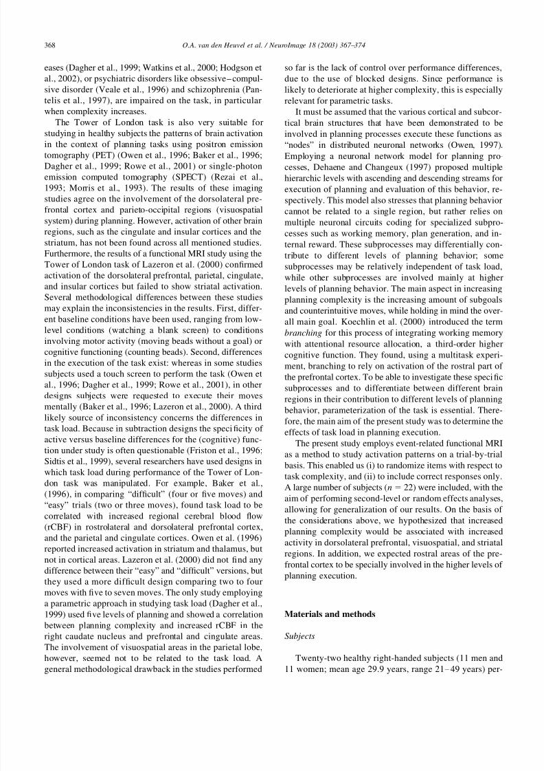
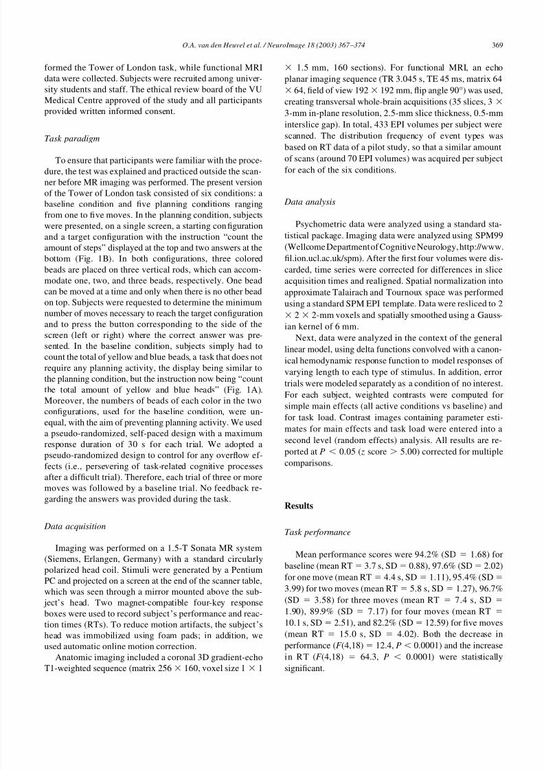
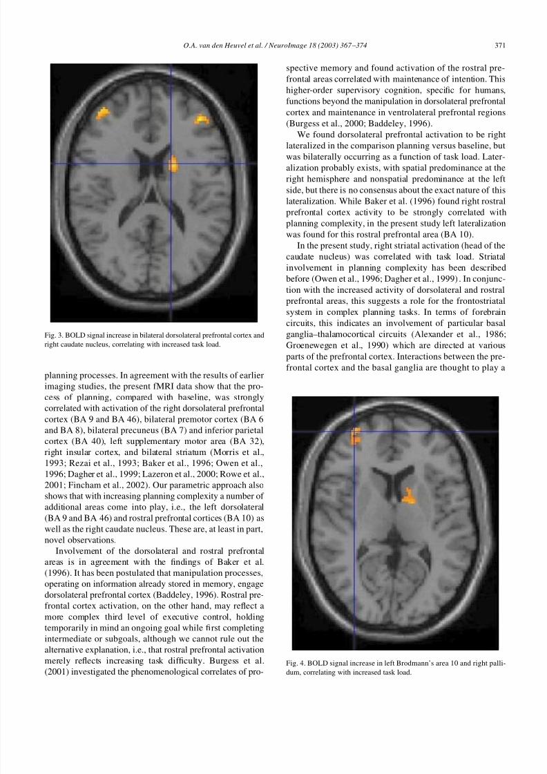
creased task load (Fig. 3 and 4), i.e., left dorsolateral pre-
frontal cortex, left insular cortex, right pallidum, and right
caudate nucleus. The left rostral prefrontal cortex (BA 10)
was also involved in the highest levels of planning com-
plexity only. The reverse contrast (decreased BOLD signal
associated with increased task load) did not show significant
activations except for bilateral primary visual cortex.
Discussion
In this article we present a parametric event-related func-
tional MRI version of the Tower of London task, suitable
for investigating the effects of increasing complexity in
Fig. 1. Display screen of the Tower of London task as used in the present
study: (A) baseline condition, (B) planning condition.
Fig. 2. BOLD signal increase in right dorsolateral prefrontal and bilateral
motor and visuospatial cortex during planning compared with baseline.
370 O.A. van den Heuvel et al. / NeuroImage 18 (2003) 367 – 374
8/12/2019 Frontostriatal System in Planning Complexity: A Parametric Functional Magnetic Resonance Version of Tower of London Task
http://slidepdf.com/reader/full/frontostriatal-system-in-planning-complexity-a-parametric-functional-magnetic 5/9
planning processes. In agreement with the results of earlier
imaging studies, the present fMRI data show that the pro-
cess of planning, compared with baseline, was strongly
correlated with activation of the right dorsolateral prefrontalcortex (BA 9 and BA 46), bilateral premotor cortex (BA 6
and BA 8), bilateral precuneus (BA 7) and inferior parietal
cortex (BA 40), left supplementary motor area (BA 32),
right insular cortex, and bilateral striatum (Morris et al.,
1993; Rezai et al., 1993; Baker et al., 1996; Owen et al.,
1996; Dagher et al., 1999; Lazeron et al., 2000; Rowe et al.,
2001; Fincham et al., 2002). Our parametric approach also
shows that with increasing planning complexity a number of
additional areas come into play, i.e., the left dorsolateral
(BA 9 and BA 46) and rostral prefrontal cortices (BA 10) as
well as the right caudate nucleus. These are, at least in part,
novel observations.Involvement of the dorsolateral and rostral prefrontal
areas is in agreement with the findings of Baker et al.
(1996). It has been postulated that manipulation processes,
operating on information already stored in memory, engage
dorsolateral prefrontal cortex (Baddeley, 1996). Rostral pre-
frontal cortex activation, on the other hand, may reflect a
more complex third level of executive control, holding
temporarily in mind an ongoing goal while first completing
intermediate or subgoals, although we cannot rule out the
alternative explanation, i.e., that rostral prefrontal activation
merely reflects increasing task dif ficulty. Burgess et al.
(2001) investigated the phenomenological correlates of pro-
spective memory and found activation of the rostral pre-
frontal areas correlated with maintenance of intention. This
higher-order supervisory cognition, specific for humans,
functions beyond the manipulation in dorsolateral prefrontal
cortex and maintenance in ventrolateral prefrontal regions
(Burgess et al., 2000; Baddeley, 1996).
We found dorsolateral prefrontal activation to be rightlateralized in the comparison planning versus baseline, but
was bilaterally occurring as a function of task load. Later-
alization probably exists, with spatial predominance at the
right hemisphere and nonspatial predominance at the left
side, but there is no consensus about the exact nature of this
lateralization. While Baker et al. (1996) found right rostral
prefrontal cortex activity to be strongly correlated with
planning complexity, in the present study left lateralization
was found for this rostral prefrontal area (BA 10).
In the present study, right striatal activation (head of the
caudate nucleus) was correlated with task load. Striatal
involvement in planning complexity has been describedbefore (Owen et al., 1996; Dagher et al., 1999). In conjunc-
tion with the increased activity of dorsolateral and rostral
prefrontal areas, this suggests a role for the frontostriatal
system in complex planning tasks. In terms of forebrain
circuits, this indicates an involvement of particular basal
ganglia–thalamocortical circuits (Alexander et al., 1986;
Groenewegen et al., 1990) which are directed at various
parts of the prefrontal cortex. Interactions between the pre-
frontal cortex and the basal ganglia are thought to play a
Fig. 3. BOLD signal increase in bilateral dorsolateral prefrontal cortex and
right caudate nucleus, correlating with increased task load.
Fig. 4. BOLD signal increase in left Brodmann’s area 10 and right palli-
dum, correlating with increased task load.
371O.A. van den Heuvel et al. / NeuroImage 18 (2003) 367 – 374

8/12/2019 Frontostriatal System in Planning Complexity: A Parametric Functional Magnetic Resonance Version of Tower of London Task
http://slidepdf.com/reader/full/frontostriatal-system-in-planning-complexity-a-parametric-functional-magnetic 6/9
role in the selection and switching of behavioral compo-
nents (Wise et al., 1996), which are crucially important for
the execution of a (complex) planning process. In the con-
text of the neuronal network model of Dehaene and Chan-
geux (1997), our finding of involvement of various cortical
and subcortical regions related to increased planning com-
plexity strengthens the idea of distributed corticocortical
and corticosubcortical networks as a neural basis for such
higher-order cognitive processes. The involvement of the
striatum might represent, at least in part, the predicted role
Table 1
Maxima of regions showing significant (P 0.05 ( z 5.00), corrected) BOLD signal increase during planning compared with baseline
Region of activation Cluster size
(voxels)
Left/
right
Brodmann’s
area
Talairach coordinate (mm) Z value
x y z
Dorsolateral PFC 5 R 46 46 36 28 5.57
7 R 9 40 30 30 5.17
Premotor cortex 12 L 6 24 20 50 5.17
31 L 6 26 6 52 5.17
7 L 6 26 4 54 5.08
90 R 6 26 4 54 6.05
SMA 3 L 32 6 20 46 5.02
Precuneus 277 L 7 4 56 48 6.42
R 7 6 58 50 5.47
Inferior parietal cortex 12 L 40 38 78 34 5.34
2 L 40 60 36 44 5.14
60 R 40 34 84 28 5.61
7 R 40 28 72 40 5.20
7 R 40 48 42 44 5.18
1 R 40 40 66 26 5.03
Caudate nucleus 8 L 8 8 0 5.01
Globus pallidus 41 R 12 6 6 5.53
Insular cortex 23 R 32 24 4 5.59
Table 2
Maxima of regions showing significant (P 0.05 ( z 5.00), corrected) BOLD signal increase correlating with increased task load
Region of activation Cluster size
(voxels)
Left/
right
Brodmann’s
area
Talairach coordinate (mm) Z value
x y z
Anterior PFC 2 L 10 36 60 8 5.02
Dorsolateral PFC 15 L 46 40 50 16 5.52
23 L 46 44 32 24 5.27
33 L 9 44 36 38 5.37
134 R 46 40 36 24 5.72
R 9 44 32 32 5.13
Premotor cortex 197 L 6 24 16 56 5.98
3 L 6 18 12 58 5.19
23 L 6 32 4 58 5.29
212 R 6 26 10 58 5.69
33 R 8 28 34 42 5.60
SMA 94 L 32 6 16 50 5.97
Precuneus 323 L 7 12 56 52 6.86
R 7 6 60 48 5.50
Inferior parietal cortex 51 L 40 42 40 36 5.89
87 L 40 48 42 46 5.73
7 L 40 40 78 34 5.47
15 L 40 40 50 16 5.52
5 L 40 24 76 50 5.37
52 R 40 38 72 34 5.83
20 R 40 50 42 42 5.64
3 R 40 48 42 50 5.00
3 R 40 34 64 44 5.06
Caudate nucleus, caput 23 R 18 6 18 5.43
Globus pallidus 23 R 12 4 4 5.58
Insular cortex 53 L 32 20 0 5.88
372 O.A. van den Heuvel et al. / NeuroImage 18 (2003) 367 – 374

8/12/2019 Frontostriatal System in Planning Complexity: A Parametric Functional Magnetic Resonance Version of Tower of London Task
http://slidepdf.com/reader/full/frontostriatal-system-in-planning-complexity-a-parametric-functional-magnetic 7/9
of a reward system in the cognitive processes (Dehaene and
Changeux, 2000).
As mentioned before, most previous studies used a block
design with an easy and a dif ficult planning condition,
thereby restricting the investigation of task load effects to a
subtraction analysis of both conditions. That no task load
effects were found in the functional MRI study of Lazeronet al. (2000) may be explained by this operationalization of
“easy” and “dif ficult” task conditions. The main aspect of
planning complexity is presumably the amount of subgoals
to be attained before reaching the main goal. The “easy”
condition of Lazeron et al. consisted of two- to four-move
items. Part of the three-move items and all four-move items
need the counterintuitive intermediate steps characteristic of
planning complexity. In the “dif ficult” condition subjects
had to solve five- to seven-move items, which are quite hard
to perform. Therefore, the difference between “easy” and
“dif ficult” may have been obscured by the presence of
complex items in the “easy” condition and the occurrence of ceiling effects in the “dif ficult” condition.
Our results are also at variance with Dagher et al. (1999),
who conducted a parametric analysis and divided areas
involved in planning behavior into those that did and those
that did not correlate with task complexity. Brain regions
where rCBF correlated with planning complexity included
bilateral dorsolateral prefrontal, premotor, and anterior cin-
gulate cortex and right caudate nucleus. In contrast with our
results, Dagher et al. (1999) found parietal activation not to
be correlated with task complexity. Since this PET study
consisted of only 6 subjects with 12 measurements per
subject, all data were analyzed using a fixed effects analysis,and results were presented uncorrected for multiple com-
parisons. Another important confounding factor may have
been that subjects had to perform the planning task by
touching the screen to move the beads. Motor execution is
not essential for planning and may produce motion artifacts,
confounding experimental effects. For this reason, we asked
our subjects to execute their plan by making the moves
mentally. Even without motor execution, massive activation
of motor and visuospatial areas was found. Parietal activa-
tion caused by motor execution, independent of specific
planning behavior, is not correlated with task complexity,
while visuospatial activity necessary for planning is indeed
correlated with task complexity.
Compared with previous studies, the present version of
the Tower of London task has two advantages. First, a
self-paced, parametric design allows flexible responding as
well as comparisons between subjects/groups at each task
level, providing us with the opportunity to investigate sub-
jects and groups with varying levels of performance. The
increased comparability across groups, using the same task,
is important for clinical studies, for instance, in patients
with decreased functioning of the frontostriatal system. Sec-
ond, in addition to variation in reaction time, performance
may also vary in hit percentage. Event-related analysis en-
ables a selective analysis of correct responses, by rejecting
the false responses. Moreover, adequate randomization of
trials is possible in event-related designs, in contrast to
block designs.
In summary, frontostriatal and visuospatial systems are
strongly involved in planning behavior and their level of
activity is related to task complexity as demonstrated by thedescribed self-paced, parametric event-related version of
the Tower of London task. By correcting for task perfor-
mance differences, it will be possible to investigate planning
behavior in subjects with decreased frontostriatal function.
Acknowledgments
This work was supported by the Dutch Organization for
Scientific Research (NWO), MW 940-37-018.
References
Alexander, G.E., DeLong, M.R., Strick, P.L., 1986. Parallel organization of
functionally segregated circuits linking basal ganglia and cortex. Annu.
Rev. Neurosci. 9, 357–381.
Baddeley, A., 1996. The fractionation of working memory. Proc. Natl.
Acad. Sci. USA 93, 13468 –13472.
Baker, S.C., Rogers, R.D., Owen, A.M., Frith, C.D., Dolan, R.J., Frack-
owiak, R.S., Robbins, T.W., 1996. Neural systems engaged by plan-
ning: a PET study of the Tower of London task. Neuropsychologia 34,
515–526.
Burgess, P.W., Quayle, A., Frith, C.D., 2001. Brain regions involved in
prospective memory as determined by positron emission tomography.
Neuropsychologia 39, 545–555.Burgess, P.W., Veitch, E., de Lacy, C.A., Shallice, T., 2000. The cognitive
and neuroanatomical correlates of multitasking. Neuropsychologia 38,
848 – 863.
Dagher, A., Owen, A.M., Boecker, H., Brooks, D.J., 1999. Mapping the
network for planning: a correlational PET activation study with the
Tower of London task. Brain 122, 1973–1987.
Dehaene, S., Changeux, J.P., 1997. A hierarchical neuronal network for
planning behavior. Proc. Natl. Acad. Sci. USA 94, 13293–13298.
Dehaene, S., Changeux, J.P., 2000. Reward-dependent learning in neuronal
networks for planning and decision making. Prog. Brain Res. 126,
217–229.
Fincham, J.M., Carter, C.S., Veen van, V., Stenger, A., Anderson, J.R.,
2002. Neural mechanisms of planning: a computational analysis using
event-related fMRI. Proc. Natl. Acad. Sci. USA 99, 3346 –3351.
Friston, K.J., Price, C.J., Fletcher, P., Moore, C., Frackowiak, R.S., Dolan,R.J., 1996. The trouble with cognitive subtraction. NeuroImage 4,
97–104.
Groenewegen, H.J., Berendse, H.W., Wolters, J.G., Lohman, A.H., 1990.
The anatomical relationship of the prefrontal cortex with the striato-
pallidal system, the thalamus and the amygdala: evidence for a parallel
organization. Prog. Brain Res. 85, 95–116.
Hodgson, T.L., Tiesman, B., Owen, A.M., Kennard, C., 2002. Abnormal
gaze strategies during problem solving in Parkinson’s disease. Neuro-
psychologia 40, 411– 422.
Koechlin, E., Corrado, G., Pietrini, P., Grafman, J., 2000. Dissociating the
role of the medial and lateral anterior prefrontal cortex in human
planning. Proc. Natl. Acad. Sci. USA 97, 7651–7656.
Lazeron, R.H., Rombouts, S.A., Machielsen, W.C., Scheltens, P., Witter,
M.P., Uylings, H.B., Barkhof, F., 2000. Visualizing brain activation
373O.A. van den Heuvel et al. / NeuroImage 18 (2003) 367 – 374

8/12/2019 Frontostriatal System in Planning Complexity: A Parametric Functional Magnetic Resonance Version of Tower of London Task
http://slidepdf.com/reader/full/frontostriatal-system-in-planning-complexity-a-parametric-functional-magnetic 8/9
during planning: the tower of London test adapted for functional MR
imaging. AJNR Am. J. Neuroradiol. 21, 1407–1414.
Morris, R.G., Ahmed, S., Syed, G.M., Toone, B.K., 1993. Neural correlates
of planning ability: frontal lobe activation during the Tower of London
test. Neuropsychologia 31, 1367–1378.
Owen, A.M., 1997. Cognitive planning in humans: neuropsychological,
neuroanatomical and neuropharmacological perspectives. Prog. Neuro-
biol. 53, 431– 450.
Owen, A.M., Doyon, J., Petrides, M., Evans, A.C., 1996. Planning and
spatial working memory: a positron emission tomography study in
humans. Eur. J. Neurosci. 8, 353–364.
Pantelis, C., Barnes, T.R., Nelson, H.E., Tanner, S., Weatherley, L., Owen,
A.M., Robbins, T.W., 1997. Frontal-striatal cognitive deficits in pa-
tients with chronic schizophrenia. Brain 120, 1823–1843.
Rezai, K., Andreasen, N.C., Alliger, R., Cohen, G., Swayze, V., O ’Leary,
D.S., 1993. The neuropsychology of the prefrontal cortex. Arch. Neu-
rol. 50, 636 – 642.
Robbins, T.W., 1998. Dissociating executive functions of the prefrontal
cortex, in: Roberts, A.C., Robbins, T.W., Weiskrantz, L. (Eds.), The
Prefrontal Cortex: Executive and Cognitive Functions, Oxford Univ.
Press, Oxford, pp. 117–142.
Rowe, J.B., Owen, A.M., Johnsrude, I.S., Passingham, R.E., 2001. Imaging
the mental components of a planning task. Neuropsychologia 39, 315–
327.
Shallice, T., 1982. Specific impaitments of planning. Philos. Trans. R. Soc.
London B Sci. 298, 199 –209.
Sidtis, J.J., Strother, S.C., Anderson, J.R., Rottenberg, D.A., 1999. Arebrain functions really additive? NeuroImage 9, 490 – 496.
Veale, D.M., Sahakian, B.J., Owen, A.M., Marks, I.M., 1996. Specific
cognitive deficits in tests sensitive to frontal lobe dysfunction in ob-
sessive– compulsive disorder. Psychol. Med. 26, 1261–1269.
Watkins, L.H., Rogers, R.D., Lawrence, A.D., Sahakian, B.J., Rosser,
A.E., Robbins, T.W., 2000. Impaired planning but intact decision
making in early Huntington’s disease: implications for specific fron-
tostriatal pathology. Neuropsychologia 38, 1112–1125.
Wise, S.P., Murray, E.A., Gerfen, C.R., 1996. The frontal cortex– basal
ganglia system in primates. Crit. Rev. Neurobiol. 10, 317–356.
374 O.A. van den Heuvel et al. / NeuroImage 18 (2003) 367 – 374

8/12/2019 Frontostriatal System in Planning Complexity: A Parametric Functional Magnetic Resonance Version of Tower of London Task
http://slidepdf.com/reader/full/frontostriatal-system-in-planning-complexity-a-parametric-functional-magnetic 9/9
Reproduced with permission of the copyright owner. Further reproduction prohibited without
permission.