From simulation to reciprocity: The case of complementary...
14
This article was downloaded by: [Universita di Padova] On: 22 July 2011, At: 00:59 Publisher: Psychology Press Informa Ltd Registered in England and Wales Registered Number: 1072954 Registered office: Mortimer House, 37-41 Mortimer Street, London W1T 3JH, UK Social Neuroscience Publication details, including instructions for authors and subscription information: http://www.tandfonline.com/loi/psns20 From simulation to reciprocity: The case of complementary actions Luisa Sartori a , Andrea Cavallo b , Giulia Bucchioni a & Umberto Castiello a a Department of General Psychology, University of Padova, Padova, Italy b Department of Psychology, Centre for Cognitive Science, University of Torino, Torino, Italy Available online: 21 Jul 2011 To cite this article: Luisa Sartori, Andrea Cavallo, Giulia Bucchioni & Umberto Castiello (2011): From simulation to reciprocity: The case of complementary actions, Social Neuroscience, DOI:10.1080/17470919.2011.586579 To link to this article: http://dx.doi.org/10.1080/17470919.2011.586579 PLEASE SCROLL DOWN FOR ARTICLE Full terms and conditions of use: http://www.tandfonline.com/page/terms-and-conditions This article may be used for research, teaching and private study purposes. Any substantial or systematic reproduction, re-distribution, re-selling, loan, sub-licensing, systematic supply or distribution in any form to anyone is expressly forbidden. The publisher does not give any warranty express or implied or make any representation that the contents will be complete or accurate or up to date. The accuracy of any instructions, formulae and drug doses should be independently verified with primary sources. The publisher shall not be liable for any loss, actions, claims, proceedings, demand or costs or damages whatsoever or howsoever caused arising directly or indirectly in connection with or arising out of the use of this material.
Transcript of From simulation to reciprocity: The case of complementary...

This article was downloaded by: [Universita di Padova]On: 22 July 2011, At: 00:59Publisher: Psychology PressInforma Ltd Registered in England and Wales Registered Number: 1072954 Registered office: Mortimer House,37-41 Mortimer Street, London W1T 3JH, UK
Social NeurosciencePublication details, including instructions for authors and subscription information:http://www.tandfonline.com/loi/psns20
From simulation to reciprocity: The case ofcomplementary actionsLuisa Sartori a , Andrea Cavallo b , Giulia Bucchioni a & Umberto Castiello aa Department of General Psychology, University of Padova, Padova, Italyb Department of Psychology, Centre for Cognitive Science, University of Torino, Torino, Italy
Available online: 21 Jul 2011
To cite this article: Luisa Sartori, Andrea Cavallo, Giulia Bucchioni & Umberto Castiello (2011): From simulation toreciprocity: The case of complementary actions, Social Neuroscience, DOI:10.1080/17470919.2011.586579
To link to this article: http://dx.doi.org/10.1080/17470919.2011.586579
PLEASE SCROLL DOWN FOR ARTICLE
Full terms and conditions of use: http://www.tandfonline.com/page/terms-and-conditions
This article may be used for research, teaching and private study purposes. Any substantial or systematicreproduction, re-distribution, re-selling, loan, sub-licensing, systematic supply or distribution in any form toanyone is expressly forbidden.
The publisher does not give any warranty express or implied or make any representation that the contentswill be complete or accurate or up to date. The accuracy of any instructions, formulae and drug doses shouldbe independently verified with primary sources. The publisher shall not be liable for any loss, actions, claims,proceedings, demand or costs or damages whatsoever or howsoever caused arising directly or indirectly inconnection with or arising out of the use of this material.

SOCIAL NEUROSCIENCE, 2011, iFirst, 1–13
From simulation to reciprocity: The caseof complementary actions
Luisa Sartori1, Andrea Cavallo2, Giulia Bucchioni1, and Umberto Castiello1
1Department of General Psychology, University of Padova, Padova, Italy2Department of Psychology, Centre for Cognitive Science, University of Torino, Torino, Italy
A large body of research reports that perceiving body movements of other people activates motor representationsin the observer’s brain. This automatic resonance mechanism appears to be imitative in nature. However, actionobservation does not inevitably lead to symmetrical motor facilitation: Mirroring the observed movement mightbe disadvantageous for successfully performing joint actions. What remains unknown is how we are to resolvethe possible conflict between the automatic tendency to “mirror” and the need to perform different context-relatedcomplementary actions. By using single-pulse transcranial magnetic stimulation, we found that observation ofa double-step action characterized by an implicit complementary request engendered a shift from symmetricalsimulation to reciprocity in the participants’ corticospinal activity. Accordingly, differential motor facilitation wasrevealed for the snapshots evoking imitative and complementary gestures despite the fact that the observed type ofgrasp was identical. Control conditions in which participants observed the same action sequence but in a contextnot implying a complementary request were included as well. The results provide compelling evidence that whenan observed action calls for a nonidentical complementary action, an interplay between the automatic tendencyto resonate with what is observed and to implicitly prepare for the complementary action does emerge. In otherwords, implicit complementary requests might have the ability to draw attention to specific features of the contextaffording nonidentical responses.
Keywords: Action observation; Transcranial magnetic stimulation; Complementary actions; Reach-to-grasp; Jointactions.
A large amount of evidence suggests that perceiv-ing body movements of other people activates motorrepresentations in the observer’s brain (for review,see Fadiga, Craighero, & Olivier, 2005; Rizzolatti,Fabbri-Destro, & Cattaneo, 2009; Rizzolatti, Fogassi,& Gallese, 2001). A tenet emerging from these stud-ies is that the activated motor representations appear tobe imitative in nature. This would reflect an automaticresonance mechanism of motor structures in line withthe observed movement.
Support for the idea of a basic neurophysio-logical system underlying such a motor resonancemechanism comes from different methodologicalapproaches. First and foremost, single-cell recordings
Correspondence should be addressed to: Umberto Castiello, Department of Psychology, University of Padova, via Venezia 8, 35131 Padova,Italy. E-mail: [email protected]
have demonstrated the existence of neurons, termed“mirror”, discharging both when a monkey grasps 3-D objects and when it observes the execution of asimilar action (di Pellegrino, Fadiga, Fogassi, Gallese,& Rizzolatti, 1992; Gallese, Fadiga, Fogassi, &Rizzolatti, 1996). Subsequently, functional magneticresonance imaging (fMRI) investigations (Buccinoet al., 2001; Decety et al., 1997; Gazzola & Keysers,2009; Gazzola, Rizzolatti, Wicker, & Keysers, 2007;Grafton, Arbib, Fadiga, & Rizzolatti, 1996; Grèzes,Costes, & Decety, 1999; Rizzolatti, Fadiga, Gallese, &Fogassi, 1996; Turella, Erb, Grodd, & Castiello, 2009)and magnetoencephalography (MEG) (Avikainen,Forss, & Hari, 2002; Hari et al., 1998; Nishitani &
© 2011 Psychology Press, an imprint of the Taylor & Francis Group, an Informa businesswww.psypress.com/socialneuroscience DOI: 10.1080/17470919.2011.586579
Dow
nloa
ded
by [
Uni
vers
ita d
i Pad
ova]
at 0
0:59
22
July
201
1

2 SARTORI ET AL.
Hari, 2000) uncovered the existence of similar neuralmechanisms within the human brain.
Furthermore, and of particular relevance forthe present study, transcranial magnetic stimulation(TMS) studies report that an observer’s motor sys-tem is facilitated by the mere viewing of motoractions (Aglioti, Cesari, Romani, & Urgesi, 2008;Avenanti, Bolognini, Malavita, & Aglioti, 2007;Baldissera, Cavallari, Craighero, & Fadiga, 2001;Borroni, Montagna, Cerri, & Baldissera, 2005; Fadiga,Fogassi, Pavesi, & Rizzolatti, 1995; Montagna, Cerri,Borroni, & Baldissera, 2005; Urgesi, Candidi, Fabbro,Romani, & Aglioti, 2006a). For instance, in the pio-neering study by Fadiga and colleagues (1995), single-pulse TMS was applied over the motor cortex ofparticipants observing a model reaching and graspingfor differently shaped objects. They demonstrated thatobserving an action induces an enhancement of themotor evoked potentials (MEPs) recorded from partic-ipants’ hand muscles corresponding to those involvedin the observed action.
Since then, similar paradigms have been use-fully applied to further investigate the nature ofthe corticospinal neural activity induced by peculiarvisual characteristics of an observed action (Alaerts,Heremans, Swinnen, & Wenderoth, 2009; Gangitano,Mottaghy, & Pascual-Leone, 2001; Maeda, Kleiner-Fisman, & Pascual-Leone, 2002; Urgesi, Moro,Candidi, & Aglioti, 2006b). For instance, it wasdemonstrated that the motor facilitation contingentupon action observation strictly reflects the tem-poral dynamics of the observed action kinematics(Gangitano et al., 2001), it is modulated by the lat-erality of the observed acting effector (Aziz-Zadeh,Maeda, Zaidel, Mazziotta, & Iacoboni, 2002), and itis not affected by the observer-model postural congru-ency (Urgesi et al., 2006a). Altogether, these findingssuggest that when we observe another individual act-ing, we “resonate” with her actions. Our motor systemsimulates under threshold the observed action in astrictly congruent fashion. The involved muscles arethe same as those used in the observed action, andtheir activation is temporally strictly coupled with thedynamics of the observed action (Fadiga et al., 2005).
A question that has yet to be fully investigated iswhether such automatic tendency to “mirror” is alwaysbeneficial in real-life situations. For instance, in avariety of circumstances in which two or more indi-viduals are involved in joint actions, complementaryrather than imitative actions appear to be more appro-priate. For example, if someone hands us a mug bythe handle, rather than imitate her action, we selecta grip which is complementary to hers. In such cir-cumstances, there would be a mismatch between the
types of grip adopted by the two agents. Mirroring theobserved movement might be detrimental to success-fully completing such joint action.
At this stage, the natural question is how are weto resolve the possible conflict between the automatictendency to “mirror” and the need to perform differentcontext-related complementary actions. Preliminaryanswers to this question come from recent studies inwhich this issue has been investigated by means ofTMS and fMRI techniques (Catmur, Walsh, & Heyes,2007; Catmur et al., 2008; Kokal, Gazzola, & Keysers,2009; Newman-Nordlund, van Schie, van Zuijlen, &Bekkering, 2007; see also Ocampo & Kritikos, 2010;van Schie, van Waterschoot, & Bekkering, 2008). Inthe first instance, a TMS study demonstrated that theproperties of the mirror system are not fixed but mayvary by a process of sensorimotor learning (Catmuret al., 2007, 2008; see also Heyes, Bird, Johnson, &Haggard, 2005). Training participants to perform indexfinger actions when they observe little finger actions,and vice versa, results in activation of primary motorcortical representations of the index finger when pas-sively observing little finger actions, and activationof representations of the little finger when observingindex finger actions. Therefore, nonidentical responseswere facilitated when associated with the observedstimuli. In the second instance, the role of the humanmirror neuron system for the coding of imitative andcomplementary actions has been recently investigatedby asking participants to prepare and execute imi-tative or complementary actions (Newman-Nordlundet al., 2007). Participants observed an actor grasp-ing a manipulandum, using either a precision or apower grip. In the imitative context, participants wererequested to perform the observed action, whereas inthe complementary context they were requested toexecute the other type of grasp. The data relative tothe behavioral part of this fMRI experiment indicatedthat, for the imitative context, reaction times (RTs)were faster for identical than nonidentical actions. Inthe complementary context, RTs were faster for non-identical than for identical actions. This suggests thatthe action context is critical for movement prepara-tion. In neural terms, results clearly indicate that themirror neuron system has the ability to link noniden-tical observed and executed actions as long as theyserve a common goal. Key areas of the mirror neu-ron system were more activated for the preparation ofcomplementary than imitative actions. This has beenexplained in terms of different kinds of mirror neurons.Strictly congruent mirror neurons, which respond toidentical observed and executed actions, may act in acontext-dependent manner. Broadly congruent mirrorneurons, which respond to nonidentical observed and
Dow
nloa
ded
by [
Uni
vers
ita d
i Pad
ova]
at 0
0:59
22
July
201
1

FROM SIMULATION TO RECIPROCITY 3
executed actions upon the same object, might be rele-vant to complementary actions. However, the questionof whether mirror neurons play a role in social interac-tions, implying different actions on different objects,has yet to be fully investigated.
In sum, these data indicate that action observa-tion does not inevitably lead to an imitative kind ofmotor facilitation. Merely changing the context inwhich an action is embedded can modulate the bias-ing effect of action observation (Catmur et al., 2007,2008; Newman-Nordlund et al., 2007). In this view,the potential conflict emerging between observed andnonidentical complementary actions might be resolvedflexibly in a double-step fashion by the same sys-tem; that is, a step in which the observed action hasto be experienced and understood in order to predictits goal, and a subsequent step in which associationsbetween observed and nonidentical movements areformed in order to eventually act in a complementarymanner. To date, no one has investigated whether sucha double-step process exists and how it unfolds.
Here we test the existence of a functional shift fromsymmetrical simulation to reciprocity in the arena ofcomplementary actions. We measured the effects ofsingle-pulse TMS on the muscle specificity of MEPsize during action observation at different times. MEPswere recorded from the abductor digiti minimi (ADM)and the first dorsal interosseus (FDI) muscles of theright hand during the observation of video clips rep-resenting a sequence of movements which might ormight not elicit a spontaneous complementary actionby the observer. As an example of the considered com-plementary actions, a model grasps a thermos filledwith coffee in a whole-hand grasp (WHG), that is,opposition of the thumb with the other fingers, andpours coffee into three cups located nearby. Then shestretches out her arm as if to pour coffee into a fourthcup which is out of her reach. Note that from theobserver’s point of view this cup was located on thebottom right corner of the image and would requirea precision grip (PG), that is, opposition between theindex finger and thumb by the handle, in order tobe reached. It is well known that the mere sight ofan object activates the representation of the actionthat can be performed on it, even in the absence ofexplicit intentions to act (Craighero, Fadiga, Rizzolatti,& Umiltà, 1998; Jeannerod, 1994; Tucker & Ellis,1998). Moreover, the results of a recent TMS studyshow that MEP facilitation was observed only whenthe handle of an object was located contralaterally withrespect to the site stimulated (Buccino, Sato, Cattaneo,Rodà, & Riggio, 2009).
Along these lines, we expect that MEPs recordedat the time the observer initially perceives the model
grasping the thermos might elicit both ADM and FDImuscle facilitation because such muscles are usuallyrecruited for a WHG. Conversely, the model’s moveto pour coffee into an out-of-reach cup might beable to elicit a nonidentical complementary gesture(i.e., PG) in the observer. At this stage, only MEPsrecorded from the FDI muscle should reveal a pro-nounced increase in activation. This is because the PGdoes not imply the recruitment of the ADM muscle.Furthermore, we predict that when the object possi-bly eliciting a complementary action is not present,symmetrical facilitation effects should emerge bothat the time the first manipulative action (i.e., WHG)is perceived and when observing the model sim-ply stretching her arm out holding the thermos (i.e.,WHG).
MATERIALS AND METHODS
Participants
Twenty healthy individuals (15 women and 5 men)aged 20–34 (mean 27 years) took part in the experi-ment. All were right-handed according to the StandardHandedness Inventory (Briggs & Nebes, 1975). Theyhad normal or corrected-to-normal visual acuity andwere free from any contraindication to TMS (Rossi,Hallett, Rossini, & Pascual-Leone, 2009; Wassermann,1998). All participants gave their written, informedconsent prior to their inclusion on the study andwere naive as to its purpose. Specific informationconcerning the study was provided after the experi-mental session was terminated. The experimental pro-cedures were approved by the Ethics Committee of theUniversity of Padova and were carried out in accor-dance with the principles of the 1964 Declaration ofHelsinki. None of the individuals taking part in theexperiment experienced discomfort or adverse effectsduring TMS.
Experimental stimuli
To create the stimulus material, we filmed a model per-forming four types of action sequences: (1) reachingand grasping a thermos, pouring coffee into three cupsand then stretching the arm out as to pour coffee into afourth cup which was beyond reach distance (Figure 1,panel a); (2) reaching and grasping a thermos, pouringcoffee into three cups, and then simply stretching thearm out holding the thermos (Figure 1, panel b); (3)reaching and grasping a sugar shaker, pouring sugarinto three cups, and then stretching the arm out to pour
Dow
nloa
ded
by [
Uni
vers
ita d
i Pad
ova]
at 0
0:59
22
July
201
1
4 SARTORI ET AL.
Figure 1. Frames extracted from the four video clips which served as stimuli for the present experiment. Specifically, for all video clips, theonset of the reach-to-grasp movement and the final phase of the action sequence are represented.
sugar into a fourth cup which was beyond reach dis-tance (Figure 1, panel c); (4) reaching and grasping asugar shaker, pouring sugar into three cups, and thensimply stretching the arm out holding the sugar shaker(Figure 1, panel d). The model naturally grasped the
thermos with a WHG, that is, the opposition of thethumb with the other fingers, and the sugar shaker witha PG, that is, the opposition of the thumb with theindex finger. As outlined in Figure 2, at the beginningof each video clip, the hand of the model was shown
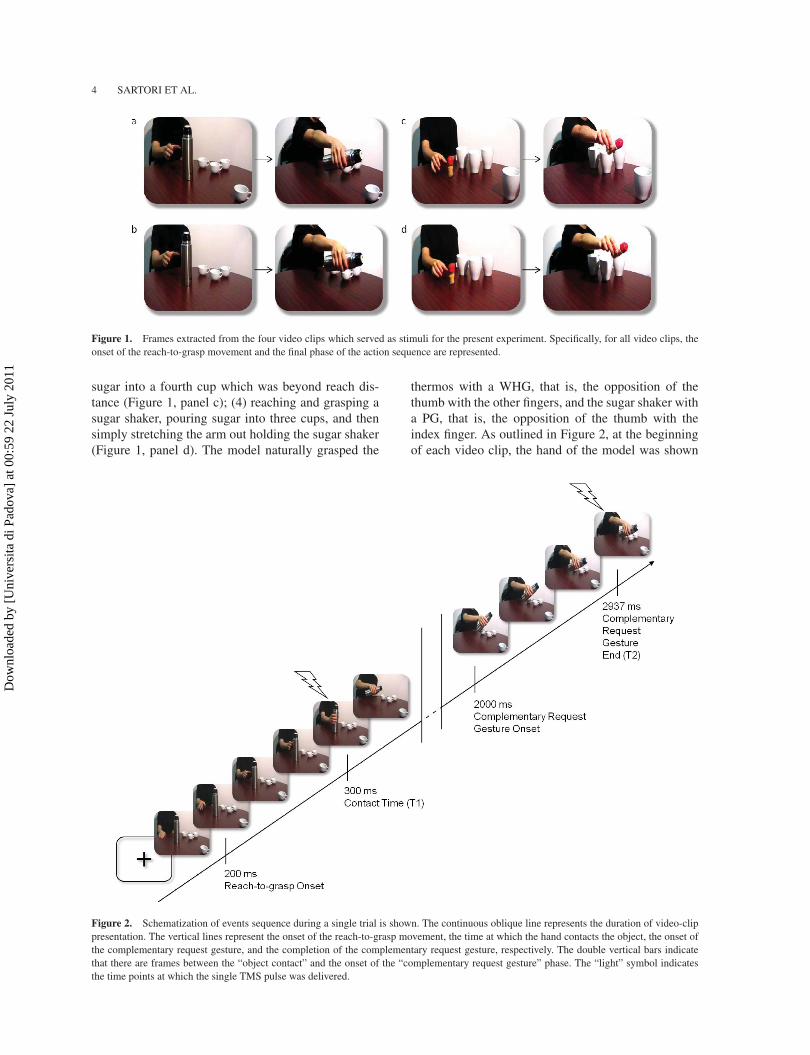
Figure 2. Schematization of events sequence during a single trial is shown. The continuous oblique line represents the duration of video-clippresentation. The vertical lines represent the onset of the reach-to-grasp movement, the time at which the hand contacts the object, the onset ofthe complementary request gesture, and the completion of the complementary request gesture, respectively. The double vertical bars indicatethat there are frames between the “object contact” and the onset of the “complementary request gesture” phase. The “light” symbol indicatesthe time points at which the single TMS pulse was delivered.
Dow
nloa
ded
by [
Uni
vers
ita d
i Pad
ova]
at 0
0:59
22
July
201
1

FROM SIMULATION TO RECIPROCITY 5
in a prone position resting on the table. After 200 ms,the model started her reach-to-grasp movement (i.e.,onset of the reach to grasp), and her fingers contactedthe first object at around 300 ms (i.e., contact time)(Figure 2). After 1700 ms, the model stretched out herarm as if to require a complementary action (i.e., onsetof the complementary request), which ended at 2937ms (Figure 2). The animation effect was obtained bypresenting a series of single frames, each lasting 33ms (resolution 720 × 576 pixels, with color depth of24 bits, and frame rate of 30 fps), plus the first and lastframes, which lasted 500 and 1000 ms, respectively.
TMS stimulation and MEP recording
TMS was delivered with a 70-mm, figure-of-eight coilconnected to a Magstim BiStim2 stimulator (Magstim,Whitlan, Dyfed, Wales, UK). The coil was angled 45◦relative to the interhemispheric fissure and perpendic-ularly to the central sulcus with the handle pointinglaterally and caudally (Brasil-Neto et al., 1992; Mills,Boniface, & Schubert, 1992). This orientation induceda posterior-anterior current in the brain, which tends toactivate corticospinal neurons indirectly via excitatorysynaptic inputs (Di Lazzaro et al., 1998). Pulses weredelivered over the left primary motor cortex (M1) cor-responding to the hand region. The coil was positionedin correspondence with the optimal scalp position(OSP), defined as the position at which the stimulationof a slightly suprathreshold intensity consistently pro-duced the largest MEP from both the ADM, the muscleserving little finger abduction, and the FDI, the muscleserving index finger flexion/extension muscles. Thecoil was held by a tripod, and its position was contin-uously checked by experimenters to keep it consistent.The resting motor threshold (rMT) was determinedfor each participant as the minimum intensity thatinduced reliable MEPs ( ≥50 µV peak-to-peak ampli-tude) in the relaxed muscle in 5 out of 10 consecutivetrials (Rossini et al., 1994). Stimulation intensity dur-ing the recording session was 110% of the rMT andranged from 38% to 59% (mean 48.5%) of the maxi-mum stimulator output intensity. MEPs were recordedsimultaneously from electrodes placed over the con-tralateral ADM and FDI muscles. Electromyographic(EMG) recording was performed through pairs of 9-mm diameter Ag-AgCl surface electrodes. The activeelectrodes were placed over the belly of the right ADMand FDI muscles and the reference electrodes overthe ipsilateral proximal interphalangeal joint (belly-tendon technique). Electrodes were connected to anisolated portable ExG input box linked to the main
EMG amplifier for signal transmission via twin fiber-optic cables (Professional BrainAmp ExG MR, BrainProducts, Munich, Germany). The ground was placedover the participants’ left wrist and connected to thecommon input of the ExG input box. The raw myo-graphic signals were band-pass filtered (20 Hz–1 kHz)and amplified prior to being digitized (5 kHz samplingrate), and stored on a computer for off-line analysis. Inorder to prevent contamination of MEP measurementsby background EMG activity, trials in which any EMGactivity greater than 100 μV was present in the 100-ms window preceding the TMS pulse were discarded.EMG data were collected for 200 ms after the TMSpulse.
Procedure
Each participant was tested in a single experimen-tal session lasting approximately 40 min. Testingwas carried out in a sound-attenuated Faraday room.Participants were seated in a comfortable armchairwith their head positioned on a fixed head rest so thatthe eye–screen distance was 80 cm. The right armwas positioned on a full-arm support, while the leftarm remained relaxed with the hand resting on thelegs. Participants were instructed to lay their handsin prone position as still and relaxed as possible. Thetask was to pay attention to the visual stimuli pre-sented on a 19-inch monitor (resolution 1280 x 1024pixels, refresh frequency 75 Hz, background lumi-nance of 0.5 cd/m2) set at eye level. Participantswere instructed to passively watch the video clipsand to avoid any movement. In order to maintain agood level of attention, participants were told that theywould be debriefed about what they had seen at theend of the experiment. For each of the four types ofvideo clips, 10 trials were presented for a total of40 trials. The order of presentation of the trials wasrandomized across participants. Prior to video presen-tation, baseline corticospinal excitability was assessedby acquiring 5 MEPs while the participants passivelywatched a white-colored fixation cross on black back-ground on the computer screen. Another series of5 MEPs was recorded at the end of the experimen-tal session. Comparisons of MEP amplitudes for thetwo series allowed us to check for any corticospinalexcitability change related to TMS per se. The aver-age amplitude of the two series allowed us to set theindividual baseline for data normalization procedures.TMS-induced MEPs from the right ADM and the rightFDI muscles were acquired once per video presenta-tion, at one of two counterbalanced time points: (1) onthe frame showing the contact of the fingers on the first
Dow
nloa
ded
by [
Uni
vers
ita d
i Pad
ova]
at 0
0:59
22
July
201
1

6 SARTORI ET AL.
object (T1, 300 ms) and (2) on the frame showing theend of the complementary request gesture (T2, 2937ms). Each video presentation was followed by a 10-srest interval. During the first 5 s of the rest period, amessage informing the participants to keep their handstill and fully relaxed was presented. This messagewas replaced by a fixation cross for the remaining 5 s.Five MEPs per muscle were acquired at every timepoint for each video, for a total of 80 MEPs per par-ticipant. The presentation of stimuli and the timingof TMS stimulation were managed by E-Prime V2.0software (Psychology Software Tools, Inc., Pittsburgh,PA, USA) running on a PC. Participants underwent thefollowing four experimental conditions.
Complementary PG action
In this condition, participants observed the videoclips representing a model performing a WHG as ifto handle a thermos filled with coffee and then pouringthe coffee into three cups located near her left side.After the coffee was poured into the third cup, themodel stretched out her arm as if to pour the coffee intoa fourth cup which was located out of reach. From anobserver’s point of view, this cup was situated on thebottom right corner of the image (Figure 1, panel a).Crucially, this cup afforded a PG movement in orderto be handled. Therefore, in this condition, there was amismatch between the observed model’s initial action(i.e., WHG) and the movement the observer wouldeventually perform as to complete the observed action(i.e., PG). A preliminary pilot investigation on a sam-ple of subjects with similar characteristics to those par-ticipating in the experiment indicated that the “fourth”cup strongly afforded a PG by the handle.
Control PG action
In this condition, participants observed the videoclips of the model performing the same actionsequence as for the “complementary PG action” con-dition except that the fourth cup was not present(Figure 1, panel b). Therefore, in this condition, thefinal part of the action sequence was not implyingthe performance of any complementary action by theobserver.
Complementary WHG action
In this condition, participants observed the videoclips of a model performing a PG as if to handle a
sugar shaker and then pouring its content into threecups located nearby to her left. After the sugar waspoured into the third cup, the model stretched out herarm as if to pour the sugar into a fourth cup whichwas located out of reach (Figure 1, panel c). Crucially,this cup afforded a WHG movement in order to behandled. Therefore, in this condition, there was a mis-match between the observed model’s action (i.e., PG)and the action the observer would eventually performso as to complete the observed movement (i.e., WHG).
Control WHG action
In this condition, participants observed the videoclips of the model performing the same actionsequence as for the “complementary WHG action”condition except that the fourth cup was not present(Figure 1, panel d). Therefore, in this condition, theaction sequence was not implying the performance ofany complementary action by the observer.
Data analysis
For each condition, peak-to-peak amplitudes of thecollected MEPs from both the ADM and FDI mus-cles were measured and averaged at each time point.MEP amplitudes deviating more than 2 SDs fromthe mean for each type of action and trials contam-inated by muscular preactivation were excluded asoutliers (<2%). A paired-sample t-test (two-tailed)was used to compare the amplitude of MEPs recordedfrom the ADM and FDI muscles in the two seriesof baseline trials presented at the beginning and atthe end of the experimental session. Ratios were thencomputed, using individual mean amplitude of MEPsrecorded in the two fixation-cross periods as base-line (MEP ratio = MEPobtained/MEPbaseline). For clar-ity, at analysis level, we will define the main fac-tors included within the analysis on the basis of thepresence/absence of the beyond-reach object possi-bly eliciting a complementary action. A repeated-measures analysis of variance (ANOVA) was con-ducted on the MEP ratios with “condition” (object,no-object), “type of observed grasp” (PG, WHG), and“time” (T1, T2) as within-subjects factors. Sphericityof the data was verified prior to performing statisticalanalysis (Mauchly’s test, p > .05). Post hoc, pairwisecomparisons were carried out by using t-tests, andthe Bonferroni correction for multiple comparisonswas applied. The comparisons between normalizedMEP amplitude and baseline were performed by usingone-sample t-tests.
Dow
nloa
ded
by [
Uni
vers
ita d
i Pad
ova]
at 0
0:59
22
July
201
1

FROM SIMULATION TO RECIPROCITY 7
RESULTS
Mean raw MEP amplitudes during the two base-line blocks administered at the beginning and theend of the experimental session were not signifi-cantly different for either the ADM muscle—-235vs. 297 μV, respectively—- t(19) = –0.87, p = .40, orthe FDI muscle—-692 vs. 585 μV, respectively—-t(19) = 0.91, p = .37. This suggests that TMS per sedid not induce any changes in corticospinal excitabilityin our experimental procedure. Mean MEP ratios fromthe ADM and the FDI muscles for each “type of con-dition” (object, no-object), “type of observed grasp”(PG, WHG), and “time” (T1, T2) are reported in Table1. Given that FDI is recruited for both PG and WHG,we did not expect any MEP modulation in terms oftype of observed grasp. Indeed, the repeated-measureANOVA on normalized MEP amplitude for the FDImuscle showed only a significant main effect of time,F(1, 19) = 4.65, p < .05, η2
p = .20. The repeated-measure ANOVA on normalized MEP amplitudes forthe ADM muscle yielded a statistically significantinteraction of “condition by type of grasp by time,”F(1, 19) = 28.81, p < .001, η2
p = .60. Normalizedmean amplitude for the FDI and the ADM muscles arereported in Table 1.
MEPs are modulated in terms ofcomplementary action
Post hoc comparisons indicated that MEP activationis modulated by the presence/absence of the objectcalling for a complementary action. Specifically, nor-malized MEP amplitude for the ADM muscle at T2
was smaller (p < .05) when participants observedthe model holding the thermos as if to approachthe beyond-reach cup affording a PG (i.e., objectcondition; Figure 3, panel a) than when participantsobserved the model simply holding the same thermoswith a WHG (i.e., no-object condition; Figure 3, panela). Conversely, MEP amplitude at T2 was greater (p <
.05) when participants observed the model holding thesugar shaker as if to approach the fourth cup affordinga WHG (i.e., object condition; Figure 3, panel b) thanwhen participants observed the model simply hold-ing the same sugar shaker with a PG (i.e., no-objectcondition; Figure 3, panel b). In contrasting MEPs atT2 against baseline, there was no ADM muscle acti-vation when the object calling for a complementaryaction required a PG action, t(19) = 0.7, p = .49. Sim-ilarly, there was no ADM activation when participantsobserved the model simply holding the sugar shakerwith a PG, t(19) = 0.64, p = .53. The very fact that wedid not find any statistically significant difference (ps
> .05) between the object and the no-object conditionsat T1 seems to suggest that the mere presence of thefourth cup affording either a PG or a WHG at the earlystage of the action sequence was not leading to anypriming effect. This should rule out the possibility thatdifferences across conditions may simply depend onthe presence/absence of the beyond-reach cup per se.
The time-course of complementaryactivations
In terms of normalized MEP amplitude for the ADMmuscle, no difference was noticed across delays (T1,T2) for the no-object conditions (ps > .05). The MEPamplitude evoked at T1 by the observation of a graspmovement (e.g., PG) was similar to that elicited bythe observation of a PG not implying any complemen-tary request at T2 (Table 1). As expected, statisticaldifferences arose for both the conditions in which thebeyond-reach object was present. Specifically, normal-ized MEP amplitude was larger (p < .05) when theTMS pulse was delivered at T1 during the observationof a WHG on the thermos than at T2 when participantsobserved the model holding the same thermos as if toapproach the cup affording a PG (Figure 4, panel a).One could argue that inferences about the motor facil-itation elicited by a PG relies on null results. However,the lack of differences from baseline was confined tothe ADM muscle. As revealed by the analysis on the
TABLE 1Normalized mean (±SEM) peak to peak amplitude of MEPs recorded from the FDI and ADM muscle during the two observation
conditions for each type of observed grasp at each trigger delay
Implicit complementary action sequences
WHG PG
Objectcondition (T1)
Objectcondition (T2)
No-objectcondition (T1)
No-objectcondition (T2)
Objectcondition (T1)
Objectcondition (T2)
No-objectcondition (T1)
No-objectcondition (T2)
FDI 1.31 ( ± 0.15) 1.54 ( ± 0.19) 1.15 ( ± 0.16) 1.4 ( ± 0.24) 1.13 ( ± 0.11) 1.34 ( ± 0.16) 1.15 ( ± 0.13) 1.25 ( ± 0.16)ADM 1.64 ( ± 0.28) 1.07 ( ± 0.10) 1.25 ( ± 0.19) 1.64 ( ± 0.23) 1.10 ( ± 0.15) 1.83 ( ± 0.29) 29 ( ± 0.19) 1.09( ± 0.14)
Dow
nloa
ded
by [
Uni
vers
ita d
i Pad
ova]
at 0
0:59
22
July
201
1

8 SARTORI ET AL.
Figure 3. The upper panels represent the means of the normalized MEP amplitudes across conditions (object, no-object) following the obser-vation of either a WHG (a) or a PG (b) at T2. Bars represent the SEM. The horizontal dotted line indicates MEP baseline. The lower panelsrepresent a typical MEP recording from the ADM muscle for one participant across conditions (object, no-object) following the observation ofeither a WHG (a) or a PG (b).
FDI muscle activity, the normalized MEP amplitudewas greater at T2 than at T1 (Table 1). In particular,when participants observed the model holding the ther-mos as if to approach the cup affording a PG, MEPamplitude for the FDI muscle was significantly greaterthan the baseline, t(19) = 2.75, p = .01. Coming backto ADM muscle activation, normalized MEP ampli-tude was smaller during the observation of a PG onthe sugar shaker (p < .05) at T1 than at T2 when par-ticipants observed the model holding the same sugarshaker as if to approach the beyond-reach cup afford-ing a WHG (Figure 4, panel b). Altogether, theseresults indicate a switch from a symmetrical motorresonance to a complementary activation of the ADMmuscle during the observation of an action sequence.
MEPs are modulated in terms of theobserved type of grasp
In terms of type of grasp, post-hoc comparisonsfor the ADM muscle revealed statistically significant
differences for both the object and the no-object con-ditions. In particular, MEP amplitude at T1 was greaterwhen observing a WHG on the thermos than a PGon the sugar shaker (Figure 5, panel a). This occurredfor both conditions at T1 (ps < .05) (Table 1) despitethe presence/absence of the fourth object. This mightsignify that at the early stage of the action sequenceparticipants were resonating with the model’s actionand ignoring the action-irrelevant fourth object. Andit corroborates the idea that the mere presence of thefourth object affording either a PG or a WHG at theearly stage of the action sequence (T1) was not lead-ing to any priming effect. Therefore, the possibilitythat the differences observed between the two objectconditions might simply depend on the mere pres-ence of the object can be rejected. Regarding T2, forthe no-object condition, MEP amplitude was greater(p < .05) when participants observed the model hold-ing a thermos (i.e., WHG) than when they observedthe model holding a sugar shaker (i.e., PG) (Table 1).Conversely, for the object condition, MEP amplitude atT2 was smaller (p < .05) when participants observed
Dow
nloa
ded
by [
Uni
vers
ita d
i Pad
ova]
at 0
0:59
22
July
201
1

FROM SIMULATION TO RECIPROCITY 9
Figure 4. The upper panels represent the means of the normalized MEP amplitudes across the time at which TMS was delivered (T1, T2)following the observation of either a WHG or a PG for the object condition. Bars represent the SEM. The horizontal dotted line indicates MEPbaseline. The lower panels represent a typical MEP recording from the ADM muscle for one participant across the time at which TMS wasdelivered (T1 and T2) following the observation of either a WHG or a PG for the object condition.
the model holding a thermos with a WHG while try-ing to pour coffee into the fourth cup eliciting a PGthan when participants observed the model performinga PG on the sugar shaker as if to approach the fourthcup affording a WHG (Figure 5, panel b).
DISCUSSION
The overarching aim of the present study was toinvestigate the effect of action observation in com-plementary contexts. The results suggest that whenan observed action calls for a nonidentical comple-mentary action, an interplay between the automatictendency to resonate with what is observed and toimplicitly prepare for the complementary action doesemerge. In other words, observed actions embeddingan implicit complementary request might have theability to prime nonidentical responses.
Little is known regarding how the inflexible ten-dency to match observed actions onto our motor
system can be reconciled with the request to preparenonidentical responses. In this respect, some investi-gations have focused on imitation and action obser-vation conditions. For instance, Heyes and colleagues(Catmur et al., 2007; Heyes et al., 2005) showed thatthe automatic effects of imitation can be abolished fol-lowing incompatible training. In the same vein, Gowenand colleagues (Gowen, Bradshaw, Galpin, Lawrence,& Poliakoff, 2010) have recently demonstrated thatautomatic imitation is not as “automatic” as previ-ously thought, but can be influenced by context. Inorder for visuomotor priming to occur, attention mustbe directed specifically to the action being performed.Other studies which have begun to consider task con-text, in the sense of the relation between model andobserver, have compared imitation and complementaryaction tasks (Newman-Nordlund et al., 2007; Ocampo& Kritikos, 2010; van Schie et al., 2008). In behavioralterms, these studies agree that there are differences inpreparing and executing complementary actions withrespect to imitative actions (Ocampo & Kritikos, 2010;
Dow
nloa
ded
by [
Uni
vers
ita d
i Pad
ova]
at 0
0:59
22
July
201
1

10 SARTORI ET AL.
Figure 5. The upper panels represent the means of the normalized MEP amplitudes across types of observed grasp (WHG, PG) recorded ateither T1 (a) or T2 (b) for the object conditions. Bars represent the SEM. The horizontal dotted line indicates MEP baseline. The lower panelsrepresent a typical MEP recording from the ADM muscle at either T1 (a) or T2 (b) for one participant for the object condition following theobservation of either a WHG or a PG.
van Schie et al., 2008). In neural terms, greater acti-vation during the preparation of complementary thanimitative actions has been found within key areas of themirror system, namely the inferior frontal gyrus andthe inferior parietal lobe (Newman-Nordlund et al.,2007). Our results extend this literature by demon-strating for the first time that corticospinal activationresulting from action observation does not necessar-ily introduce an imitative bias, but can as well primemotor activation for complementary actions depend-ing on contextual factors. They provide evidence offlexible stimulus-response adjustments, which are aprerequisite when people need to cooperate with andrespond to others in a different manner. Interestingly,in contrast to previous studies (e.g., Catmur et al.,2007), here we managed to reveal either symmetricalor complementary spontaneous corticospinal activa-tion by avoiding the use of instructions to participantsthat might have created a bias to mentally matchingor complementing the observed action. Our stimuli
had the ability to elicit a switch between the changesin MEP activity classically found following actionobservation and changes in MEP activity related tothe implicit complementary request embedded in theobserved stimulus.
Along these lines, a recent fMRI study has revealedthat the mirror neuron system is relevant to the plan-ning of both imitative and complementary actions(Newman-Nordlund et al., 2007). The basic idea is thatthe properties of a specific class of mirror neurons,namely the broadly congruent mirror neurons (Galleseet al., 1996), might have the ability to support the per-formance of complementary actions. This is becausebroadly congruent mirror neurons generalize the goalof an action across many types of instances, such asperforming a grasping movement with a PG or a WHG(Fogassi & Gallese, 2002; Gallese et al. 1996). Rather,in the present experiment, participants observed anobject-related movement which draw attention to anadditional object eliciting a different movement. This
Dow
nloa
ded
by [
Uni
vers
ita d
i Pad
ova]
at 0
0:59
22
July
201
1

FROM SIMULATION TO RECIPROCITY 11
might have determined an interplay between “mirror”and “canonical” neurons. This latter type of neuronresponds not only during the execution of behaviors,but also during the perception of the objects that arerelated to these behaviors (Rizzolatti & Craighero,2004). For instance, canonical motor neurons, whichbecome active during PG movements, also becomeactive upon presentation of a small object graspableby a PG. Conversely, canonical neurons that becomeactive during a WHG are selectively activated whena large object is shown (Murata et al., 1997). Inthis perspective, the need to perform a complemen-tary action involving a different object might implya combination of mirror and canonical neurons, cod-ing for different types of actions at different times.Indeed, we found MEP activity strongly indicativeof a pure “matching” mechanism at the start of theaction sequence and a “complementary” type of MEPactivity at the time the request for a complementaryaction, dictated by contextual factors, became evident.This points to a mechanism for recognizing objectaffordances (Gibson, 1979) and to the possible exis-tence of a specific type of intentional affordances;that is, “social affordances.” Intentional affordancesare produced by the establishment of a shared inten-tional space (Tomasello, 1999). Indeed, the presentresults suggest how social affordances might be criticalin order to automatically facilitate a complementarymotoric response. The crucial aspects of our exper-iment which favored a readiness to engage in jointaction are various. First, there is the presence of objectsthat are necessary for the action to occur (i.e., a cupthat can be held by the perceiver and a thermos/shakerthat can be poured by the actor). Second, there isthe ability of the observer to virtually take up theobject which is facing her. Third, we have the implicitrequest by the actor that opens up the affordance toengage in joint action. Finally, we have an appropri-ate relational orientation between actor and perceiverthat allows for joint action (i.e., facing rather thanbehind or to the side; within rather than outside thepersonal space). In light of this, the present resultsseem to suggest that automatic responses to anotherperson’s action have to do with the salient affordancesabout what one could do in this situation. In otherwords, making affordances salient evokes a readinessto enact them. This is in line with previous demonstra-tions that visual objects potentiate actions that mightbe performed on them, even in the absence of explicitintention to act (Buccino et al., 2009; Craighero et al.,1998; Jeannerod, 1994; Tucker & Ellis, 1998).
We are aware that our data cannot provide adetailed description of the time at which the proposed
functional shift occurs. This is because MEPs haveonly been recorded at two different stages of the actionsequence, namely during the first observed actionand during the unfolding of the implicit complemen-tary request. Nevertheless, such an approach might bevaluable for our understanding of how specific neu-ral networks flexibly adapt when contextual factorsdictate a mismatch between observed and performedactions.
Another aspect which particularly depicts the nov-elty of the present findings is concerned with theuse of stimuli which implicitly ask for a comple-mentary action. It might well be that the “comple-mentary” MEP activity recorded at the end of theaction sequence stemmed from inferring the inten-tions behind the observed action. That is, in a mannerwhich is congruent to the intentions of the observerrather than with what the model actually performed.Therefore, with a certain degree of caution, ourfindings indicate that different intentions might beassigned to a model’s action depending on context(i.e., object presence/absence). Specifically, the con-text calling for a complementary action induces anenhancement of MEPs, an idea which is in line withthe overarching intention to fulfill a specific outcomerather than with the tendency to resonate with themodel’s action. This result is not evident when the con-text within which the model’s action is performed doesnot subtend a “complementary” intention. This issuemight be particularly relevant to understanding howhumans coordinate their actions in social situationsin which the task at hand does not require simulat-ing the actions of another person (Sebanz, Bekkering,& Knoblich, 2006; Sebanz & Frith, 2004; Sebanz,Knoblich, & Prinz, 2003).
In conclusion, the present findings reconcile thenotion that action observation mechanisms inevitablyyield to the simulation of what is observed, andtherefore might not subserve the performance of non-identical complementary actions, with a more flexiblecontext-dependent view of action observation. Such aperspective entails an interplay between an initial sim-ulation process, which might allow one to experiencewhat is observed, and a process which elaborates theconsequences of the initially observed actions in termsof context and intentions.
Although recent studies have impressively extendedour view of the motor system and its cognitive func-tions, the role of the motor system in semanticsis largely unexplored and the study of complemen-tary actions is still in its infancy. Many questionsremain to be addressed, but future studies might ben-efit from the present findings for the determination
Dow
nloa
ded
by [
Uni
vers
ita d
i Pad
ova]
at 0
0:59
22
July
201
1

12 SARTORI ET AL.
of the neural mechanisms underlying complex socialsituations characterized by complementary behaviors.
Original manuscript received 23 November 2010Revised manuscript accepted 30 March 2011
First published online day/month/year
REFERENCES
Aglioti, S. M., Cesari, P., Romani, M., & Urgesi, C. (2008).Action anticipation and motor resonance in elite basket-ball players. Nature Neuroscience, 11, 1109–1116.
Alaerts, K., Heremans, E., Swinnen, S. P., & Wenderoth,N. (2009). How are observed actions mapped to theobserver’s motor system? Influence of posture and per-spective. Neuropsychologia, 47, 415–422.
Avenanti, A., Bolognini, N., Malavita, A., & Aglioti, S.M. (2007). Somatic and motor components of actionsimulation. Current Biology, 17, 2129–2135.
Avikainen, S., Forss, N., & Hari, R. (2002). Modulatedactivation of the human SI and SII cortices during obser-vation of hand actions. NeuroImage, 15, 640–646.
Aziz-Zadeh, L., Maeda, F., Zaidel, E., Mazziotta, J., &Iacoboni, M. (2002). Lateralization in motor facilitationduring action observation: A TMS study. ExperimentalBrain Research, 144, 127–131.
Baldissera, F., Cavallari, P., Craighero, L., & Fadiga, L.(2001). Modulation of spinal excitability during obser-vation of hand actions in humans. European Journal ofNeuroscience, 13, 190–194.
Borroni, P., Montagna, M., Cerri, G., & Baldissera, F. (2005).Cyclic time course of motor excitability modulation dur-ing observation of hand actions in humans. EuropeanJournal of Neuroscience, 13, 190–194.
Brasil-Neto, J. P., Cohen, L. G., Panizza, M., Nilsson, J.,Roth, B. J., & Hallett, M. (1992). Optimal focal tran-scranial magnetic activation of the human motor cortex:Effects of coil orientation, shape of the induced cur-rent pulse, and stimulus intensity. Journal of ClinicalNeurophysiolology, 9, 132–136.
Briggs, G. G., & Nebes, R. D. (1975). Patterns of handpreference in a student population. Cortex, 11, 230–238.
Buccino, G., Binkofski, F., Fink, G. R., Fadiga, L., Fogassi,L., Gallese, V., et al. (2001). Action observation activatespremotor and parietal areas in a somatotopic manner: AnfMRI study. European Journal of Neuroscience, 13, 400–404.
Buccino, G., Sato, M., Cattaneo, L., Rodà, F., & Riggio,L. (2009). Broken affordances, broken objects: A TMSstudy. Neuropsychologia, 47, 3074–3078.
Catmur, C., Gillmeister, H., Bird, G., Liepelt, R., Brass,M., & Heyes, C. (2008). Through the looking glass:Counter-mirror activation following incompatible senso-rimotor learning. European Journal of Neuroscience, 28,1208–1215.
Catmur, C., Walsh, V., & Heyes, C. (2007). Sensorimotorlearning configures the human mirror system. CurrentBiology, 17, 1527–1531.
Craighero, L., Fadiga, L., Rizzolatti, G., & Umiltà, C.(1998). Visuomotor priming. Visual Cognition, 5, 109–125.
Decety, J., Grèzes, J., Costes, N., Perani, D., Jeannerod, M.,Procyk, E., et al. (1997). Brain activity during observationof actions. Influence of action content and subject’sstrategy. Brain, 120, 1763–1777.
Di Lazzaro, V., Oliviero, A., Profice, P., Saturno, E.,Pilato, F., Insola, A., et al. (1998). Comparisonof descending volleys evoked by transcranial mag-netic and electric stimulation in conscious humans.Electroencephalography and Clinical Neurophysiology,109, 397–401.
di Pellegrino, G., Fadiga, L., Fogassi, L., Gallese, V., &Rizzolatti, G. (1992). Understanding motor events: Aneurophysiological study. Experimental Brain Research,91, 176–180.
Fadiga, L., Craighero, L., & Olivier, E. (2005). Human motorcortex excitability during the perception of others’ action.Current Opinion in Neurobiology, 15, 213–218.
Fadiga, L., Fogassi, L., Pavesi, G., & Rizzolatti, G. (1995).Motor facilitation during action observation: A magneticstimulation study. Journal of Neurophysiology, 73, 2608–2611.
Fogassi, L., & Gallese, V. (2002). The neural correlatesof action understanding in non-human primates. InM. I. Stamenov & V. Gallese (Eds.), Mirror neuronsand the evolution of brain and language (pp. 13–36).Philadelphia, PA: John Benjamins.
Gallese, V., Fadiga, L., Fogassi, L., & Rizzolatti, G. (1996).Action recognition in the premotor cortex. Brain, 119,593–609.
Gangitano, M., Mottaghy, F. M., & Pascual-Leone, A.(2001). Phase-specific modulation of cortical motoroutput during movement observation. Neuroreport, 12,1489–1492.
Gazzola, V., & Keysers, C. (2009). The observation andexecution of actions share motor and somatosensoryvoxels in all tested subjects: Single-subject analyses ofunsmoothed fMRI data. Cerebral Cortex, 19, 1239–1255.
Gazzola,V., Rizzolatti, G., Wicker, B., & Keysers, C. (2007).The anthropomorphic brain: The mirror neuron systemresponds to human and robotic actions. NeuroImage, 35,1674–1684.
Gibson, J. J. (1979). The ecological approach to visualperception. Boston, MA: Houghton Mifflin.
Gowen, E., Bradshaw, C., Galpin, A., Lawrence, A., &Poliakoff, E. (2010). Exploring visuomotor priming fol-lowing biological and non-biological stimuli. Brain andCognition, 74, 288–297.
Grafton, S. T., Arbib, M. A., Fadiga, L., & Rizzolatti, G.(1996). Localization of grasp representations in humansby positron emission tomography. 2. Observation com-pared with imagination. Experimental Brain Research,112, 103–111.
Grèzes, J., Costes, N., & Decety, J. (1999). The effects oflearning and intention on the neural network involved inthe perception of meaningless actions. Brain, 122, 1875–1887.
Hari, R., Forss, N., Avikainen, S., Kirveskari, E., Salenius,S., & Rizzolatti, G. (1998). Activation of human primarymotor cortex during action observation: A neuromagneticstudy. Proceedings of the National Academy of Sciencesof the United States of America, 95, 15061–15065.
Heyes, C., Bird, G., Johnson, H., & Haggard, P. (2005).Experience modulates automatic imitation. CognitiveBrain Research, 22, 233–240.
Dow
nloa
ded
by [
Uni
vers
ita d
i Pad
ova]
at 0
0:59
22
July
201
1

FROM SIMULATION TO RECIPROCITY 13
Jeannerod, M. (1994). The representing brain: Neuronal cor-relates of motor intention and imagery. Behavioral andBrain Sciences, 17, 187–202.
Kokal, I., Gazzola, V., & Keysers, C. (2009). Acting togetherin and beyond the mirror neuron system. NeuroImage, 47,2046–2056.
Maeda, F., Kleiner-Fisman, G., & Pascual-Leone, A.(2002). Motor facilitation while observing hand actions:Specificity of the effect and role of observer’s orientation.Journal of Neurophysiology, 87, 1329–1335.
Mills, K. R., Boniface, S. J., & Schubert, M. (1992).Magnetic brain stimulation with a double coil: Theimportance of coil orientation. Electroencephalographyand Clinical Neurophysiology, 85, 17–21.
Montagna, M., Cerri, G., Borroni, P., & Baldissera. F.(2005). Excitability changes in human corticospinal pro-jections to muscles moving hand and fingers while view-ing a reaching and grasping action. European Journal ofNeuroscience, 22, 1513–1520.
Murata, A., Fadiga, L., Fogassi, L., Gallese, V., Raos, V., &Rizzolatti, G. (1997). Object representation in the ven-tral premotor cortex (area F5) of the monkey. Journal ofNeurophysiology, 78, 2226–2230.
Newman-Nordlund, R. D., van Schie, H. T., van Zuijlen, A.M., & Bekkering, H. (2007). The mirror neuron systemis more activated during complementary compared withimitative action. Nature Neuroscience, 10, 817–818.
Nishitani, N., & Hari, R. (2000). Temporal dynamics ofcortical representation for action. Proceedings of theNational Academy of Sciences of the United States ofAmerica, 97, 913–918.
Ocampo, B., & Kritikos, A. (2010). Placing actions incontext: Motor facilitation following observation of iden-tical and non-identical manual acts. Experimental BrainResearch, 201, 743–751.
Rizzolatti, G., & Craighero, L. (2004). The mirror-neuronsystem. Annual Review of Neuroscience, 27, 169–192.
Rizzolatti, G., Fabbri-Destro, M., & Cattaneo, L. (2009).Mirror neurons and their clinical relevance. NatureClinical Practice: Neurology, 5, 24–34.
Rizzolatti, G., Fadiga, L., Gallese, V., & Fogassi, L. (1996).Premotor cortex and the recognition of motor actions.Cognitive Brain Research, 3, 131–141.
Rizzolatti, G., Fogassi, L., & Gallese, V. (2001).Neurophysiological mechanisms underlying the under-standing and imitation of action. Nature ReviewsNeuroscience, 2, 661–670.
Rossi, S., Hallett, M., Rossini, P. M., & Pascual-Leone,A. (2009). Safety, ethical considerations, and application
guidelines for the use of transcranial magnetic stim-ulation in clinical practice and research. ClinicalNeurophysiology, 120, 2008–2039.
Rossini, P. M., Barker, A. T., Berardelli, A., Caramia, M.D., Caruso, G., Cracco, R. Q., et al. (1994). Noninvasiveelectrical and magnetic stimulation of the brain, spinalcord and roots: Basic principles and procedures for rou-tine clinical application. Report of an IFCN Committee.Electroencephalography and Clinical Neurophysiology,91, 79–82.
Sebanz, N., Bekkering, H., & Knoblich, G. (2006). Jointaction: Bodies and minds moving together. Trends inCognitive Science, 10, 70–76.
Sebanz, N., & Frith, C. (2004). Beyond simulation? Neuralmechanisms for predicting the actions of others. NatureNeuroscience, 7, 5–6.
Sebanz, N., Knoblich, G., & Prinz, W. (2003). Representingother’s actions: Just like one’s own? Cognition, 88, B11–B12.
Tomasello, M. (1999). The cultural origins of human cogni-tion. Cambridge, MA: Harvard University Press.
Tucker, M., & Ellis, R. (1998). On the relations betweenseen objects and components of potential actions. Journalof Experimental Psychology: Human Perception andPerformance, 24, 830–846.
Turella, L., Erb, M., Grodd, W., & Castiello, U. (2009).Visual features of an observed agent do not modu-late human brain activity during action observation.NeuroImage, 46, 844–853.
Urgesi, C., Candidi, M., Fabbro, F., Romani, M., & Aglioti,S. M. (2006a). Motor facilitation during action obser-vation: Topographic mapping of the target muscle andinfluence of the onlooker posture. European Journal ofNeuroscience, 23, 2522–2530.
Urgesi, C., Moro, V., Candidi, M., & Aglioti, S. M. (2006b).Mapping implied body actions in the human motor sys-tem. Journal of Neuroscience, 26, 7942–7949.
van Schie, H. T., van Waterschoot, B. M., & Bekkering, H.(2008). Understanding action beyond imitation: Reversedcompatibility effects of action observation in imitationand joint action. Journal of Experimental Psychology:Human Perception and Performance, 34, 1493–1500.
Wassermann, E. M. (1998). Risk and safety of repeti-tive transcranial magnetic stimulation: Report and sug-gested guidelines from the International Workshop on theSafety of Repetitive Transcranial Magnetic Stimulation,June 5–7, 1996. Electroencephalography and ClinicalNeurophysiology, 108, 1–16.
Dow
nloa
ded
by [
Uni
vers
ita d
i Pad
ova]
at 0
0:59
22
July
201
1


![International Journal of Pavement Engineering An ... · International Journal of Pavement Engineering iFirst article, 2010, 1–14 Downloaded By: [Vandenbossche, Julie] At: 04:06](https://static.fdocuments.in/doc/165x107/60621336aa9f701a5d74f973/international-journal-of-pavement-engineering-an-international-journal-of-pavement.jpg)