Rodenticide Poisoning + Rat Killer paste poisoning management

Page 1/20
Dietary cottonseed consumption inducedsubfertility in male and female rats: a potent eco-friendly rodenticide compoundFariborz Nowzari
Shiraz UniversityFarhad Rahmanifar
Shiraz UniversityNader Tanideh
Shiraz Medical School: Shiraz University of Medical SciencesMohammad Reza Dorvash
Shiraz Medical School: Shiraz University of Medical SciencesArezoo Khoradmehr
Bushehr University of Medical SciencesNeda Baghban
Bushehr University of Medical SciencesAmin Tamadon ( [email protected] )
Bushehr University of Medical Sciences https://orcid.org/0000-0002-0222-3035
Research
Keywords: Cottonseed �our, Rat, Fertility, Rodenticide
Posted Date: May 21st, 2021
DOI: https://doi.org/10.21203/rs.3.rs-538459/v1
License: This work is licensed under a Creative Commons Attribution 4.0 International License. Read Full License
Page 2/20
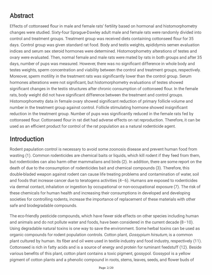
AbstractEffects of cottonseed �our in male and female rats’ fertility based on hormonal and histomorphometrychanges were studied. Sixty-four Sprague-Dawley adult male and female rats were randomly divided intocontrol and treatment groups. Treatment group was received diets containing cottonseed �our for 35days. Control group was given standard rat food. Body and testis weights, epididymis semen evaluationindices and serum sex steroid hormones were determined. Histomorphometry alterations of testes andovary were evaluated. Then, normal female and male rats were mated by rats in both groups and after 35days, number of pups was measured. However, there was no signi�cant difference in whole body andtestes weights, sperm concentration and viability between the control and treatment groups, respectively.Moreover, sperm motility in the treatment rats was signi�cantly lower than the control group. Serumhormones alterations were not signi�cant, but histomorphometry evaluations of testes showedsigni�cant changes in the testis structures after chronic consumption of cottonseed �our. In the femalerats, body weight did not have signi�cant difference between the treatment and control groups.Histomorphometry data in female ovary showed signi�cant reduction of primary follicle volume andnumber in the treatment group against control. Follicle stimulating hormone showed insigni�cantreduction in the treatment group. Number of pups was signi�cantly reduced in the female rats fed bycottonseed �our. Cottonseed �our in rat diet had adverse effects on rat reproduction. Therefore, it can beused as an e�cient product for control of the rat population as a natural rodenticide agent.
IntroductionRodent papulation control is necessary to avoid some zoonosis disease and prevent human food fromwasting (1). Common rodenticides are chemical baits or liquids, which kill rodent if they feed from them,but rodenticides can also harm other mammalians and birds (2). In addition, there are some report on thedeath of due to the consumption of rodenticides bait and chemical compounds (3). Therefore, thisdouble-bladed weapon against rodent can cause life treating problems and contamination of water, soiland foods that increase cancer due to teratogens activities (4–6). Humans are exposed to rodenticidesvia dermal contact, inhalation or ingestion by occupational or non-occupational exposure (7). The risk ofthese chemicals for human health and increasing their consumptions in developed and developingsocieties for controlling rodents, increase the importance of replacement of these materials with othersafe and biodegradable compounds.
The eco-friendly pesticide compounds, which have fewer side effects on other species including humanand animals and do not pollute water and foods, have been considered in the current decade (8–10).Using degradable natural toxins is one way to save the environment. Some herbal toxins can be used asorganic compounds for rodent population controls. Cotton plant, Gossypium hirsutum, is a commonplant cultured by human. Its �ber and oil were used in textile industry and food industry, respectively (11).Cottonseed is rich in fatty acids and is a source of energy and protein for ruminant feedstuff (12). Besidevarious bene�ts of this plant, cotton plant contains a toxic pigment, gossypol. Gossypol is a yellowpigment of cotton plants and a phenolic compound in roots, stems, leaves, seeds, and �ower buds of

Page 3/20
cotton (13). Gossypol can cause anorexia, a decrease in the growth rate, labored breathing and dyspnea,which also can cause lymphocyte cytotoxicity leading to immunode�ciency and toxic hepatitis (14, 15).On the other hand, gossypol as anti-viral agent has been used for therapeutic purposes in human (16).
The male contraceptive effect of gossypol has been approved in human (17). Gossypol has highpotential for crossing from gonadal barrier that increases its e�ciency of antifertility in mammalians(18). There are a lot of research about antifertility effect of gossypol on male animals but limited data areavailable about females (19). Gossypol consumption in human has more potential for inducing infertility(20). Gossypol affect male fertility by decreasing sperm motility, concentration and viability (21). Inaddition, decrease in serum LH, FSH and testosterone has been reported (22). Antifertility effects ofgossypol on males’ mammalians have been reported to be dose, time and strain dependent. For example,5–10 mg/kg daily gossypol acetic acid for 12 weeks in hamsters and rats induces complete infertility, butthis amount of gossypol has no signi�cant effect on rabbit fertility (23). Cui et al. (24) showed gossypolalong with methyltestosterone and ethinyl estradiol have infertility effect by apoptosis of rat’s germinalcells and can be used as contraceptive drugs for rodent’s papulation control. In addition, ultrastructureinvestigating with electron microscopy showed that gossypol affect epidydimal structure resulting indisruption of sperm maturation (25). In conclusion, gossypol induces infertility in male rats and otherrodents such as mice and hamsters and also on humans due to oxidative stress and damage tomitochondrial, germinal layers of seminiferous tube and Sertoli cells, but its effects are reversible (21, 26).
In female mammalian, reduction of follicles number and hormonal change that affect estrous cycle aswell as an increase in atretic follicle number that is responsible for infertility has been reported (22, 27). Inaddition, in pervious study on females, embryotoxic effect was investigated by in vitro and in vivo studyand the results showed interferes of embryonic development by gossypol administration (15). Its effecton estrous cycle has been reported in some case so that it caused to prolonged diestrus. However, there iscontroversies about cycling changes due to gossypol consumption (22).
Chemical based rodenticides are useful initially for immediate reduction of rat’s papulation but, as abovementioned, they are harmful. Because of low toxicity, cost effectiveness and speci�city of plant materials,herbal rodenticides have more potential for pest management (28, 29). In this study, we evaluate theeffect of diets containing gossypol on fertility histomorphometry and hormonal changes in male andfemale rats to investigate its potential for using as an eco-friendly rodenticide compound.
Materials And Methods
Animals and treatment16 male and 48 female adult Sprague-Dawley rats with approximately 220 g weight were purchased fromCenter of Comparative and Experimental Medicine of Shiraz University of Medical Sciences. Male ratswere randomly allocated into two groups (n = 8) and females were kept in two groups (control n = 32 andtreatment n = 16), each group was kept in a type III polypropylene cage. All rats were housed in a room

Page 4/20
with 12h light/dark condition cycle at temperature of 23 ± 1ºC. Water for all group was provided adlubitum. The control male and female rats had access to conventional rat pellet. The treatment male andfemale rats were fed by pellet containing 20% cottonseed �our. Body weight of all rats was measuredevery week for 35 days.
Preparation of plate and administrationCottonseeds were obtained from Fars province cotton farms and grinding in grinder machine, sieved with60 mesh sieves to separate coat of cottonseeds from the shell. The shell of cottonseeds was used inplate component. Conventional rodent chew was obtained from Center of Comparative and ExperimentalMedicine and was grinded to become powder. For producing one kg of pellet, rodent chew �our (800 g),cottonseed shell �our (200 g) and water were mixed for 10 min, converted to pellet using a pellet makingmachine and dried near fans for one day. All treatment rats were fed with the cottonseed pellet for 35days. Every week, the amount of food was measured to control each rat fed by 15 g pellet every day.
Pregnancy testAfter 35 days, control female rats (n = 16) were mated with treatment male rats and treatment females (n = 16) were mated with control male rats (ratio of 1 male per 2 females) for 4 days. Vaginal plug waschecked every day in morning and each female that had vaginal plug, was separated from the male. After22 days of pregnancy, the numbers of pups were recorded.
Serum sampling and sperm analysisAfter 35 days of cottonseed consumption, 8 male and 24 female rats were euthanized for samplecollecting. Blood samples were collected from heart and stored in tubes and allowed to become clotted,then centrifuged at 2000 rpm for 15 min. Serum obtained was freeze in -70 until hormone analysis. Lefttestis, epididymis and ovary were collected and �xed in 10% formalin-buffer. Right testis of each rat wascollected and epididymis was dissected from testis for sperm analysis. Right dissected testis wasweighed using a digital weighing scale (Acculab® ALC 210.4). Right epididymis was immediately slicedwith scalpel and threw into a medium size Eppendorf tube containing 1.5 ml warmed phosphate bufferedsaline (PBS), then incubated in 37 ºC for 30 min. Then, some drops of the sample were set on amicroscope slide and observed by a light microscope with 40× objective lens magni�cation to investigatesperm motility and estimate the percentage. For counting sperm, semen was diluted with PBS and set onNeubauer chamber. Five squares from central of the chamber were checked to count the number ofsperm. To investigate the sperm viability, semen was stained with eosin-nigrosin solution for 30 min andset on a microscopy slide. Dead sperm (violet head) and vital sperm (unstained head) in 10 random �eldsof slide were counted and the viability percentage was calculated.
Histological and histomorphometric analysisThe left testis, epididymis and ovary were �xed in formalin-buffer 10% for one month. Tissue processingwas done by tissue processor machine (model DS 2080/H, DID SABZ Co, Orumieh, Iran). After tissueprocessing, samples were embedded in para�n. Testis and epididymis samples were cut in 5 µm using

Page 5/20
microtome and set on microscope slides. After rehydration in decreasing dilution of ethanol, they werestained with hematoxylin and eosin.
A camera (Dino-Lite®, New Taipei City, Taiwan) was set up on a light microscope. In Dino capture 2.0(version 1.4.2.D) program the input magni�cation set on 725× and the objective lens was on 10× and unitset on 1 µm. For investigation of changes in spermatogenesis, some indices in seminiferous tubules wereevaluated.
For the �rst index, lumen diameter and total diameter of �ve tubules per each slide were measured. Thenaccording to the following formulas, cellular diameter, lumen area, cellular area and cross-section areawere calculated.
Cellular diameter = (total diameter – lumen diameter)/2
Lumen area = 3.142 × (lumen diameter)²/4
Cellular area = 3.142 × (cellular diameter)²
Cross section area = 3.142 × (total diameter)²/4
Evaluation of the spermatogenesis index was performed on the basis of a modi�ed scale of 0 to 7. Forthis purpose, the appearance of the spermatogenic cells throughout the seminiferous tubules wasevaluated. Number of cell layers, types of cells, and the presence of late spermatids in the seminiferoustubules were considered in this index. The indices were as follows: 0, presence of no spermatogenic cells;1, presence of only spermatogonia; 2, presence of spermatogonia and spermatocytes present; 3,spermatogonia, spermatocytes and round (early) spermatids with < 25 late spermatids per tubule; 4,presence of spermatogonia, spermatocytes, and round spermatids with up to 25–50 late spermatids pertubule; 5, presence of spermatogonia, spermatocytes, and round spermatids with up to 50–75 latespermatids per tubule; 6, presence of spermatogonia, spermatocytes, and round spermatids as well as upto 75–100 late spermatids per tubule; and 7, presence of all cell types present with > 100 late spermatidsper tubule.
The last index was the number of tubules per 5×5 mm² of transverse sections of seminiferous tubules.Based on this number, the numerical density of tubules was calculated as fallowing formula.
Numerical density = number of pro�les per unit area/(total diameter + average thickness of the section).
Left ovary was processed like testis with the difference that there are 10 slices (each 20 µm thickness)between 2 histology samples. Primary follicle was presented as one-layer cuboidal granulosa cells,secondary follicle was presented with two layer of cuboidal granulosa (with or without antrum), andtertiary follicle was presented as large antrum and several layers of granulosa cells. Number of primaryand secondary follicles was measured by 40x objective microscopy and every area was observed withcrinkle pattern. All data were obtained by a reproductive expert with blind observation. Tertiary and corpus

Page 6/20
luteum were measured by 10x microscopy. Area of follicle was measured by Dino capture 2.0 (version1.4.2.D) tools. For calculating volume, we calculated radius of these area and gain circle diameter. If wesuppose that 1 slice with its distance to another slice forms a cone frustum, we can use con volumeequation for calculating total volume of a follicle.
In this equation, d is circle diameter and h is distance between 2 slices (according to our slicing pattern is200 µm).
Hormone analysisLuteinizing hormone (LH), follicle-stimulating hormone (FSH) measured by radioimmunoassay (RIA) kit(Padyab Teb Company, Tehran, Iran). and testosterone were analysis by ELISA kit (PGI Company, Tehran,Iran). For testosterone analysis, all reagents, samples, control and standards were �rst prepared asinstructed. Standard and control samples were added to wells. Labaled HRP-conjugate was added toeach well and incubated at 37°C. Then, they were washed and TMB substrate was added to each welland incubated at room temperature. Finally, stop solution was added to wells and read immediately. ForFSH and LH hormonal analysis, brie�y, buffer bottle was warmed to melt and diluted with 100 mLdistilled water. Other components except separating reagent were added to assay buffer and replacedstopper. Working standards were prepared as company instruction. brie�y, 200 µL assay buffer waspipetted to non-speci�c binding and 100 µL was pipetted to zero standard tubes. 100 µL antiserum waspipetted into all tubes except non-speci�c binding (NSB) and total count (TC) tubes and all tubes werevortexed and stored for 4 h at room temperature. Next, 100 µL tracer was pipetted into all tubes. TC tubeswere stopped and put aside for counting. For second time, all tubes were vortexed and stored overnight atroom temperature. Next, 400 µL second antibody was added to each tube except TC tube, vortexed andincubated at room temperature for 10 min. Then, all tubes were centrifuged for 10 min at 1500 ×g and thesupernatant was discarded. The tubes were inverted on absorbent tissues to allow them drain for 5 min.The radioactivity present in each tube was determined by counting at last 60 sec in Gamma scintillationcounter (Dream GAMMA counter-5, LabLogic Company, USA) and compered with a standard curve fordetermining concentration of LH and FSH in samples. A standard curve for LH was drawn with Y-axis,present of Radio activity and X-axis, concentration of determinate LH. Sensitivity of LH and FSH kit wasdetermined as 0.22 mIU/mL and 0.09 mIU/mL, respectively.
Statistical analysisThe analysis was performed using GraphPad prism version 6 software (GraphPad software, La Jolla,USA). Means and standard error (SE) of the data of sperm analysis, weights, histomorphometry indicesof seminiferous tubules, and hormone assays were subjected to Kolmogorov-Smirnov test of normalityand analyzed by independent sample t-test. The post-hoc test was performed by LSD test. Thespermatogenesis index of seminiferous tubules and ovary structural change indices were comparedusing Mann-Whitney U test. P-value less than 0.05 was considered statistically signi�cant.
Page 7/20
Results
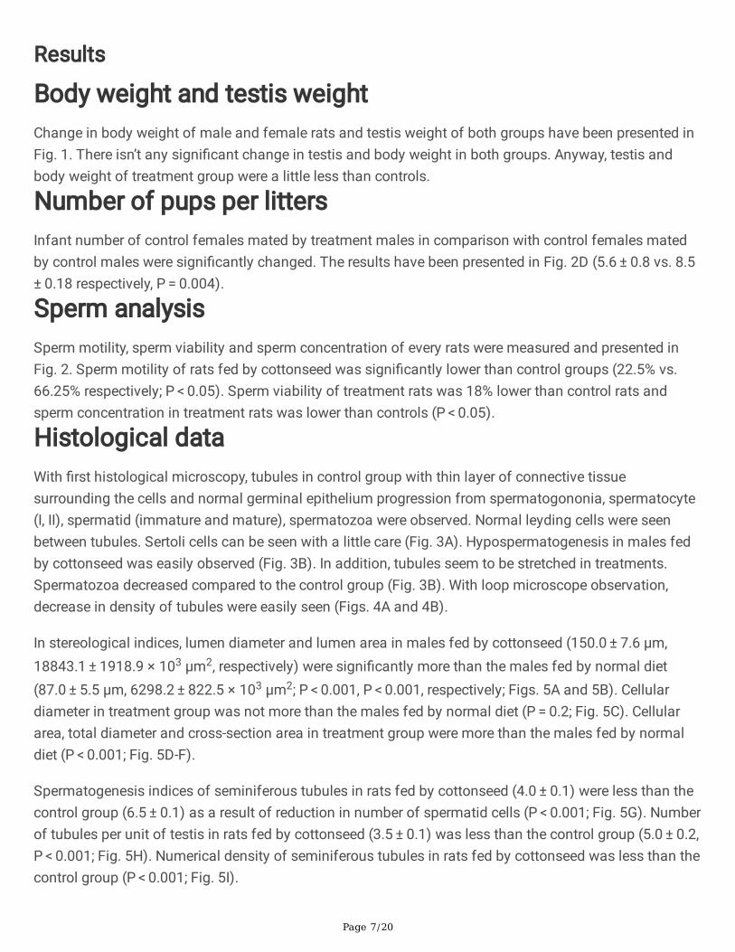
Body weight and testis weightChange in body weight of male and female rats and testis weight of both groups have been presented inFig. 1. There isn’t any signi�cant change in testis and body weight in both groups. Anyway, testis andbody weight of treatment group were a little less than controls.
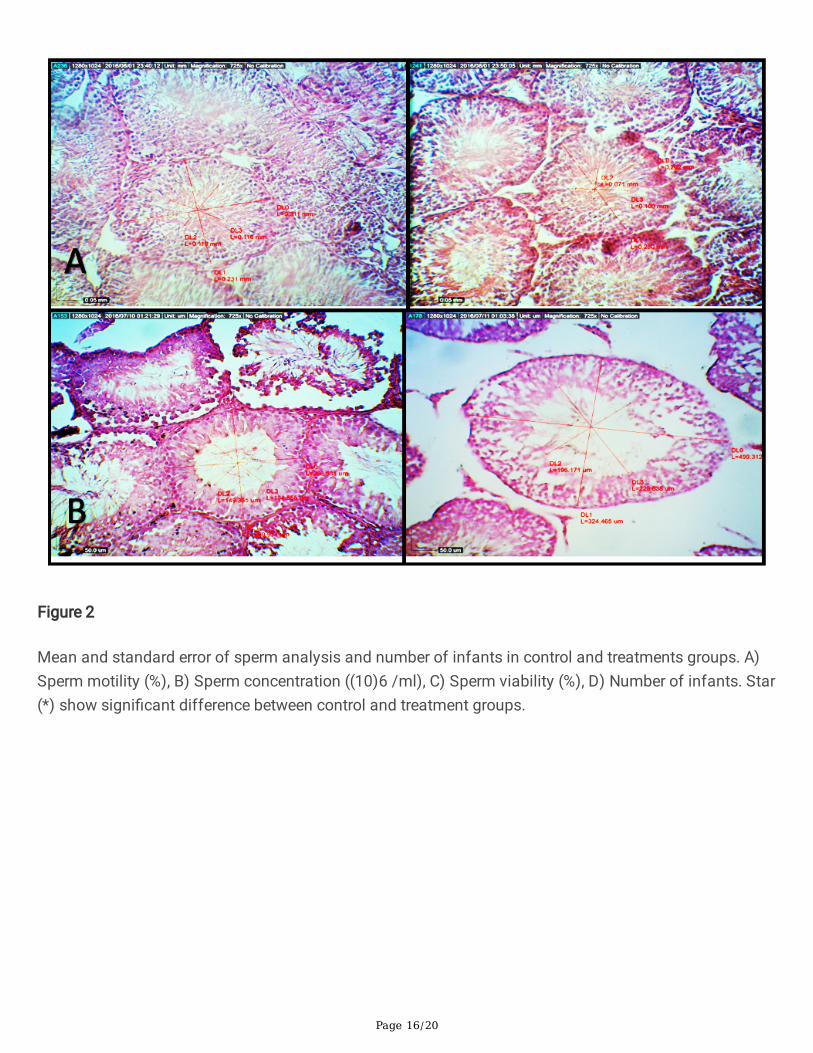
Number of pups per littersInfant number of control females mated by treatment males in comparison with control females matedby control males were signi�cantly changed. The results have been presented in Fig. 2D (5.6 ± 0.8 vs. 8.5 ± 0.18 respectively, P = 0.004).
Sperm analysisSperm motility, sperm viability and sperm concentration of every rats were measured and presented inFig. 2. Sperm motility of rats fed by cottonseed was signi�cantly lower than control groups (22.5% vs.66.25% respectively; P < 0.05). Sperm viability of treatment rats was 18% lower than control rats andsperm concentration in treatment rats was lower than controls (P < 0.05).
Histological dataWith �rst histological microscopy, tubules in control group with thin layer of connective tissuesurrounding the cells and normal germinal epithelium progression from spermatogononia, spermatocyte(I, II), spermatid (immature and mature), spermatozoa were observed. Normal leyding cells were seenbetween tubules. Sertoli cells can be seen with a little care (Fig. 3A). Hypospermatogenesis in males fedby cottonseed was easily observed (Fig. 3B). In addition, tubules seem to be stretched in treatments.Spermatozoa decreased compared to the control group (Fig. 3B). With loop microscope observation,decrease in density of tubules were easily seen (Figs. 4A and 4B).
In stereological indices, lumen diameter and lumen area in males fed by cottonseed (150.0 ± 7.6 µm,18843.1 ± 1918.9 × 103 µm2, respectively) were signi�cantly more than the males fed by normal diet(87.0 ± 5.5 µm, 6298.2 ± 822.5 × 103 µm2; P < 0.001, P < 0.001, respectively; Figs. 5A and 5B). Cellulardiameter in treatment group was not more than the males fed by normal diet (P = 0.2; Fig. 5C). Cellulararea, total diameter and cross-section area in treatment group were more than the males fed by normaldiet (P < 0.001; Fig. 5D-F).
Spermatogenesis indices of seminiferous tubules in rats fed by cottonseed (4.0 ± 0.1) were less than thecontrol group (6.5 ± 0.1) as a result of reduction in number of spermatid cells (P < 0.001; Fig. 5G). Numberof tubules per unit of testis in rats fed by cottonseed (3.5 ± 0.1) was less than the control group (5.0 ± 0.2,P < 0.001; Fig. 5H). Numerical density of seminiferous tubules in rats fed by cottonseed was less than thecontrol group (P < 0.001; Fig. 5I).

Page 8/20
On the other hand, total corpus luteum decreased in treatment group but not signi�cant. Ovarian cyst waspresence in treatment group. Number of primary follicles in rats fed by cottonseed were decreased incomparison with the control groups (3.8 vs 9.5, P = 0.03). There was no signi�cant decrease in number ofsecondary and tertiary follicles as well as corpus luteum numbers. In addition, total primary folliclevolume was decreased in cottonseed fed group (P = 0.02). Other follicle volumes did not signi�cantlychange. Ovary volume showed slightly increase in volume in group fed cottonseed. In addition, number ofatretic follicles increased in treatment group (P < 0.05; Fig. 7).
Hormone analysis dataTestosterone and gonadotropin hormone’s change in both groups were presented in Fig. 8. Level of serumtestosterone in rats fed by cottonseed showed a higher value than the control group (2.0 ± 0.4 vs. 1.3 ± 0.2, respectively; P = 0.2). Serum gonadotropin hormone and LH in treatment rats (2.35 ± 0.76) werehigher than the control (0.9 ± 0.3, P = 0.09). Treatment FSH (0.9 ± 0.2) was lower than the controls (1.3 ± 0.3, P = 0.2).
DiscussionIn this study, we evaluated consumption of cottonseed �our (16 g per day) on fertility changes in maleand female rats. Deleterious effect of gossypol on fertility of male mammalians has been widely reportedin the literature. In this study, testis weight and body weight in two group was similar and all rats havetheir normal appetite during the experiments that shown this amount of cottonseed haven’t enoughgossypol to have toxicity effect on growth. Singla et al. (19) used cottonseed oil for investigating itseffect on rats. Their results showed cottonseed oil consumption in appropriate dose had not any effecton body weight of animals (19). It seems that higher dose of this plant could affect body weight of ratsdue to changes on bait’s taste. In addition, there isn’t any clinical sign of gossypol toxicity like diarrhea,dehydration and distended abdomens in this research.
Signi�cant decrease in number of pups per litters is the result of subfertility in males and femalesbecause of decrease of number of primary follicle and alter estrus cycle, sperm concentration andmotility. Sperm motility is an important factor, which is affected by gossypol due to inhibition of theATPase activity in the spermatozoa (30) and reduction of mitochondrial activity (31). Serum testosteroneand LH level increased in cottonseed group but there was not statistically signi�cant. Other studiesshowed that intra peritoneal administration of gossypol reduced testosterone level (21, 22). It seems thatadministration route could affect content of gossypol reached to sexual organs. Correspondingly, in ourstudy, we used whole cottonseed �our, so there are some other ingredients in cottonseed �our that mayincrease testosterone and LH level in serum.
Anyway, despite of rise in testosterone level, subfertility effect was seen and also sperm concentrationwas decreased, so infertility effect of gossypol is more because of spermatogenesis dysfunction andsperm motility disorder. According to this, a decrease in the sperm concentration is not just because ofreduction of testosterone and it has some other reasons that can reduce it by gossypol. Due to

Page 9/20
suggestion of other studies (32–34), sperm concentration decreased by damage of germ cell andshedding immature spermatid in epididymis and seminiferous tubule or directly attaching of gossypol tomembrane of seminiferous tubule and following by facilitating the cell destruction through increasefragility of membrane in the testis. However, the other research stated that a decrease in the spermconcentration is due to a decrease in the serum testosterone (35). Depletion of spermatogenic cell inseminiferous tubule, histopathology investigation shows main toxicity effect of gossypol that previouslyreported (36). In a study conducted by Saleh et al. (37), gossypol acetate intra peritoneal administrationin male rats signi�cantly reduced the semen quality as evidenced by the decrease in the sperm count andmotility. Moreover, it has been reported that it changed the morphology of sperm and inhibited the α-glucosidase activity in gossypol group. Activity of α-glucosidase is widely used as an indicative markerfor sperm count, so low level of this enzyme indicates epididymal obstruction (37). In addition, it’s clearthat gossypol can affect spermatogonia cells and results in depletion of all cells in seminiferous tubules.
Gossypol inhibits spermatogenesis by impairing sperm motility with speci�c mitochondrial injury of thesperm tail (38) and decreasing concentration of semen (21). Gossypol can reduce cellular andmicrotubular 𝛽-tubular content in spermatocytes and spermatids (39). Another effect of gossypol isspermatozoid disturbance mechanism including the inhibition of release and utilization of ATP by thesperm cells (40). In a study conducted by Santana et al. (41), gossypol acetic acid was administrated bygastric gavage in male rats for investigation their infertility mechanism. They concluded that gavageadministration of 5 mg/kg body weight of gossypol acetic acid could induce subfertility in male rats (41).They reported that the major mechanism of inducing infertility by gossypol is inhibition of spermproduction and motility due to dramatic drop in the production of mitochondrial ATP (41). Furthermore,gossypol induces oxidative stress by increasing the formation of ROS and lipid peroxides, that affectplasma membrane permeability, ATPase activity, and glucose transport in sperms (42). In addition,gossypol inhibits Mg-ATPase and Ca-Mg-ATPase activity and calcium in�ux (31, 43) in spermatozoidplasmatic membranes (31). Infertility is the inability to conceive due to low semen quality that can causeby negative effect of cytotoxic component on spermatogenesis (44), so, it was resulted that cottonseed�our consumption could alter fertility in male rats.
Female rats fed cottonseed have body weight as same as females of the control group. The ovaryvolume was slightly higher than that of the control group. Hormonal analysis showed higher LH andlower FSH hormone. Other studies reported hormonal changes during gossypol administration. Lin et al.(45) reported that progesterone level was reduced in female rats that received 25 mg/kg gossypol viaintramuscular injection. In vitro studies reported that gossypol consumption could inhibit progesteronelevel secretion level in luteal cells (46).
Histomorphometry analysis showed reduction of primary follicle number and volume. FSH reductioncould alter the follicular maturation and reduce the follicular number. This can cause temporary infertility.The primordial follicle is �xed at the time of birth and stopping diets containing gossypol could returnfertility. The reduction of follicle has been reported in previous research on sheep, rats and mouse ovaries(22, 27). Furthermore, atretic follicle number was higher in treatment group, atretic follicle characterized
Page 10/20
as discontinuous basement membrane, retracted oocyte, pyknotic nucleus and disorganized granulosacells. Study conducted by Gadelha et al. (47), showed laying hens fed by cottonseed had more atreticfollicles at all stages of development. The mechanism of ovarian toxicity of gossypol has not been fullyunderstood, but may be due to cytotoxic and apoptotic activities. Embryonic changes have been reportedin few literatures. Louvandini et al. (48) and Jimenez et al. (49) reported gossypol can effect on malelambs of pregnant female sheep fed by cottonseed. They reported that male lambs showed signi�cantlower growth and lower testis weight as a proportion of the total body weight, and reduced lowertestosterone level than female rats (48, 49).
ConclusionsIn this study, it was showed that using cottonseed �our as a source of gossypol instead of gossypolacetic acid or other sources of gossypol such as cottonseed oil can induce infertility without toxic effectson body weight and has potential for using as a cheap and environment friendly contraception material.Gossypol could alter spermatogenesis and reduce sperm motility and viability in male rats. In addition, itcan reduce follicular number in female rat and alter hormonal level. Moreover, it was demonstrated thatcottonseed �our consumption in an appropriate daily dose could affect reproduction in male and femalerats and it could be used as an effective contraceptive diet for rodent and to replace chemical rodenticideproducts.
Declarations
Ethics approval and consent to participateThis investigation was performed in accordance with relevant guidelines and regulations of animalstudies of Ethical Committee of Shiraz University of Medical Sciences.
Consent for publicationNot applicable.
Availability of data and materialThe data used to support the �ndings of this study are included within the article.
FundingThis work was �nancially supported by grant of the Entrepreneurship Center of Shiraz University and vicechancellor for research and technology of Shiraz University of Medical Sciences.

Page 11/20
Competing interestsAuthor Fariborz Nowzari was employed by the company Arka Co. The remaining authors declare that theresearch was conducted in the absence of any commercial or �nancial relationships that could beconstrued as a potential con�ict of interest.
Authors' contributionsA.T., F.R., and N.T. conceived and designed the format of the manuscript. F.N., M.R.D., N.T. and F.R.collected the data, and drafted and edited the manuscript. A.T. drew the Figures. All the authors reviewedthe manuscript and all of them contributed to the critical reading and discussion of the manuscript. Allauthors have read and agreed to the published version of the manuscript.
AcknowledgmentsNot applicable.
References1. Panti-May JA, Sodá-Tamayo L, Gamboa-Tec N, Cetina-Franco R, Cigarroa-Toledo N, Machaín-
Williams C, et al. Perceptions of rodent-associated problems: an experience in urban and rural areasof Yucatan, Mexico. Urban Ecosystems. 2017;20(5):983-8.
2. Walther B, Geduhn A, Schenke D, Schlötelburg A, Jacob J. Baiting location affects anticoagulantrodenticide exposure of non‐target small mammals on farms. Pest Management Science.2021;77(2):611-9.
3. Sugunan S, Krishnan R, Kumar KS, Geetha S. Rodenticide poisoning in children: A study of clinicalpro�le and electrocardiographic changes. Indian Journal of Child Health. 2017;4(2):136-9.
4. Hill III T, Nelms MD, Edwards SW, Martin M, Judson R, Corton JC, et al. Editor’s highlight: NegativePredictors of carcinogenicity for environmental chemicals. Toxicological Sciences. 2017;155(1):157-69.
5. Jeephet K, Kamsa-Ard S, Bhudhisawasdi V, Kamsa-Ard S, Luvira V, Luvira V. Association betweenpesticide use and cholangiocarcinoma. Asian Paci�c Journal of Cancer Prevention. 2016;17(8):3979-82.
�. Ou Y, Mai J, Zhuang J, Liu X, Wu Y, Gao X, et al. Risk factors of different congenital heart defects inGuangdong, China. Pediatric research. 2016;79(4):549-58.
7. VoPham T, Bertrand KA, Hart JE, Laden F, Brooks MM, Yuan J-M, et al. Pesticide exposure and livercancer: a review. Cancer Causes & Control. 2017;28(3):177-90.

Page 12/20
�. Govindarajan M, Rajeswary M, Hoti S, Bhattacharyya A, Benelli G. Eugenol, α-pinene and β-caryophyllene from Plectranthus barbatus essential oil as eco-friendly larvicides against malaria,dengue and Japanese encephalitis mosquito vectors. Parasitology research. 2016;115(2):807-15.
9. Kedia A, Prakash B, Mishra PK, Singh P, Dubey NK. Botanicals as eco friendly biorational alternativesof synthetic pesticides against Callosobruchus spp.(Coleoptera: Bruchidae)—a review. Journal ofFood Science and Technology. 2015;52(3):1239-57.
10. Zhang C, Qu Y, Wu X, Song D, Ling Y, Yang X. Eco-friendly insecticide discovery via peptidomimetics:Design, synthesis, and aphicidal activity of novel insect kinin analogues. Journal of agricultural andfood chemistry. 2015;63(18):4527-32.
11. Soto-Blanco B. Gossipol e fatores antinutricionais da soja. Toxicologia Aplicada à VeterináriaManole, São Paulo. 2008:531-45.
12. Rico J, de Souza J, Allen M, Lock A. Nutrient digestibility and milk production responses to increasinglevels of palmitic acid supplementation vary in cows receiving diets with or without wholecottonseed. Journal of animal science. 2017;95(1):436-46.
13. Tian X, Ruan J, Huang J, Fang X, Mao Y, Wang L, et al. Gossypol: phytoalexin of cotton. ScienceChina Life Sciences. 2016;59(2):122-9.
14. Xu W-b, Xu L-h, Lu H-s, Ou-Yang D-y, Shi H-j, Di J-f, et al. The immunosuppressive effect of gossypolin mice is mediated by inhibition of lymphocyte proliferation and by induction of cell apoptosis. ActaPharmacologica Sinica. 2009;30(5):597-604.
15. Gadelha ICN, Fonseca NBS, Oloris SCS, Melo MM, Soto-Blanco B. Gossypol toxicity from cottonseedproducts. The Scienti�c World Journal. 2014;2014.
1�. Keshmiri-Neghab H, Goliaei B. Therapeutic potential of gossypol: an overview. Pharmaceuticalbiology. 2014;52(1):124-8.
17. Lim W, Ham J, Park S, Bae H, You S, Song G. Gossypol induces disruption of spermatogenesis andSteroidogenesis in male mice. Journal of agricultural and food chemistry. 2019;67(7):2075-85.
1�. Dabrowski K, Rinchard J, Lee K, Blom J, Ciereszko A, Ottobre J. Effects of diets containing gossypolon reproductive capacity of rainbow trout (Oncorhynchus mykiss). Biology of reproduction.2000;62(2):227-34.
19. Singla N, Garg M. Effect of crude cottonseed oil containing gossypol on fertility of male and estrouscycle of female Bandicota bengalensis Gray and Hardwicke. Journal of Applied Animal Research.2013;41(2):156-65.
20. Coutinho EM. Gossypol: a contraceptive for men. Contraception. 2002;65(4):259-63.
21. El-Sharaky A, Newairy A, Elguindy N, Elwafa A. Spermatotoxicity, biochemical changes andhistological alteration induced by gossypol in testicular and hepatic tissues of male rats. Food andChemical Toxicology. 2010;48(12):3354-61.
22. Gadelha ICN, Fernandes de Macedo M, Oloris SCS, Melo MM, Soto-Blanco B. Gossypol promotesdegeneration of ovarian follicles in rats. The Scienti�c World Journal. 2014;2014.

Page 13/20
23. Chang M, Gu Z, Saksena S. Effects of gossypol on the fertility of male rats, hamsters and rabbits.Contraception. 1980;21(5):461-9.
24. Cui G-H, Xu Z-L, Yang Z-J, Xu Y-Y, Xue S-P. A combined regimen of gossypol plus methyltestosteroneand ethinylestradiol as a contraceptive induces germ cell apoptosis and expression of its relatedgenes in rats. Contraception. 2004;70(4):335-42.
25. de Andrade SF, Oliva SU, Klinefelter GR, De Grava Kempinas W. Epididymis-speci�c pathologicdisorders in rats exposed to gossypol from weaning through puberty. Toxicologic pathology.2006;34(6):730-7.
2�. Hassan ME, Smith GW, Ott RS, Faulkner DB, Firkins LD, Ehrhart E, et al. Reversibility of thereproductive toxicity of gossypol in peripubertal bulls. Theriogenology. 2004;61(6):1171-9.
27. Câmara ACL, Gadelha ICN, Borges PAC, de Paiva SA, Melo MM, Soto-Blanco B. Toxicity of gossypolfrom cottonseed cake to sheep ovarian follicles. PLoS One. 2015;10(11):e0143708.
2�. Dubey N, Srivastava B, Kumar A. Current status of plant products as botanical pesticides in storagepest management. Journal of biopesticides. 2008;1(2):182-6.
29. Singh J, Mirza A. Natural Biological Products from Plants as Rodenticides. Natural BioactiveProducts in Sustainable Agriculture: Springer; 2020. p. 235-57.
30. Kalla NR, Vasudev M. Studies on the male antifertility agent‐Gossypol acetic acid. II. Effect ofgossypol acetic acid on the motility and ATPase activity of human spermatozoa. Andrologia.1981;13(2):95-8.
31. Breitbart H, Rubinstein S, Nass‐Arden L. Effect of gossypol‐acetic acid on calcium transport andATPase activity in plasma membranes from ram and bull spermatozoa. International journal ofandrology. 1984;7(5):439-47.
32. Ali IN, Fakhrildin MBM, Al-Obaidi SA. EFFECT OF AQUEOUS AND ALCOHOLIC EXTRACTS OFCOTTONSEED ON SOME PARAMETERS OF SPERMS AND REPRODUCTIVE PERFORMANCE OFALBINO MICE. Al-Nahrain Journal of Science. 2010;13(4):151-8.
33. Amini A, Kamkar F. The effects of gossypol on spermatogenesis in NMRI mice. Iranian Journal ofScience and Technology (Sciences). 2005;29(1):123-33.
34. Farrag AK, Ismail HI, Khalifa EA, El-Gamal MM. The possible synergistic effect of gossypol(cottonseed oil) and Schistosomiasis mansoni on the male reproductive organs in experimentinginfected mice. Tanta Medical Sci J. 2007;2(1):78-91.
35. Matsumoto AM. Effects of chronic testosterone administration in normal men: safety and e�cacy ofhigh dosage testosterone and parallel dose-dependent suppression of luteinizing hormone, follicle-stimulating hormone, and sperm production. The Journal of Clinical Endocrinology & Metabolism.1990;70(1):282-7.
3�. da Silva KM, Zart AL, Brum KB, dos Santos Fernandes CE. Histopathological and histomorphometrictesticular characteristics associated to reproductive condition in Bos indicus (Nellore) bulls. Semina:Ciências Agrárias. 2015;36(1):1935-44.

Page 14/20
37. Saleh SR, Attia R, Ghareeb DA. The ameliorating effect of berberine-rich fraction against gossypol-induced testicular in�ammation and oxidative stress. Oxidative medicine and cellular longevity.2018;2018.
3�. Akinola O, Dosunmu O, Dini L, Ajayi S. Proteinaceous diet inhibits gossypol-induced spermatotoxicity.European Journal of Histochemistry. 2006:205-8.
39. Teng O. Reversible changes in the content of cellular and microtubular tubulin in spermatogenic cellsafter gossypol treatment. Contraception. 1997;55(3):183-8.
40. Ueno H, Sahni MK, Segal SJ, Koide S. Interaction of gossypol with sperm macromolecules andenzymes. Contraception. 1988;37(3):333-41.
41. Santana AT, Guel� M, Medeiros HC, Tavares MA, Bizerra PF, Mingatto FE. Mechanisms involved inreproductive damage caused by gossypol in rats and protective effects of vitamin E. Biologicalresearch. 2015;48(1):1-8.
42. El-Mokadem M, Taha T, Samak M, Yassen A. Alleviation of reproductive toxicity of gossypol usingselenium supplementation in rams. Journal of Animal Science. 2012;90(9):3274-85.
43. Breitbart H, Mayevsky A, Nass-Arden L. Molecular mechanisms of gossypol action on sperm motility.The International journal of biochemistry. 1989;21(10):1097-102.
44. Panahi M, Karimaghai N, Rahmanifar F, Tamadon A, Vahdati A, Mehrabani D, et al. Stereologicalevaluation of testes in busulfan-induced infertility of hamster. Comparative Clinical Pathology.2015;24(5):1051-6.
45. Lin YC, Fukaya T, Rikihisa Y, Walton A. Gossypol in female fertility control: ovum implantation andearly pregnancy inhibited in rats. Life Sciences. 1985;37(1):39-47.
4�. Lin Y, Coskun S, Sanbuissho A. Effects of gossypol on in vitro bovine oocyte maturation andsteroidogenesis in bovine granulosa cells. Theriogenology. 1994;41(8):1601-11.
47. Gadelha I, Lima M, Melo M, Soto-Blanco B. Gossypol promotes the degeneration of chicken ovarianfollicles in vitro. Brazilian Journal of Poultry Science. 2016;18(3):505-10.
4�. Louvandini H, Corrêa PS, Amorín R, Liu L, Ieda EH, Jimenez CR, et al. Gestational and lactationalexposure to gossypol alters the testis transcriptome. BMC genomics. 2020;21(1):1-11.
49. Jimenez CR, Moretti DB, da Silva TP, Corrêa PS, da Costa RLD, Mui TS, et al. Cottonseed (gossypol)intake during gestation and lactation does affect the ovarian population in ewes and lambs?Research in Veterinary Science. 2020.
Figures

Page 15/20
Figure 1
Mean and standard error of Testis weight (A) and body weight (B) of male control and treatment groupsand female control and treatment groups (C).
Page 16/20
Figure 2
Mean and standard error of sperm analysis and number of infants in control and treatments groups. A)Sperm motility (%), B) Sperm concentration ((10)6 /ml), C) Sperm viability (%), D) Number of infants. Star(*) show signi�cant difference between control and treatment groups.

Page 17/20
Figure 3
Section of testes showing seminiferous tubule. H&E staining. Control (A) and treatments group (B) with100x magni�cation. Scale bars are 50 µm.

Page 18/20
Figure 4
Cross-section of testes showing whole seminiferous tubule with loop microscopy 30x magni�cation. H&Astaining. Control group (A) and treatment group (B).

Page 19/20
Figure 5
Mean and standard error of stereological indices And spermatogenesis index of control and treatmentgroup. A) lumen diameter (µm), B) lumen area (µm^2), C) cellular diameter (µm), D) cellular area (µm^2 ),E) total diameter (µm), F) cross-section area (µm^2), H) number of tubules per unit area of testis, I)numerical density of seminiferous tubules, G) spermatogenesis index Star (*) show signi�cant differencebetween control group and group fed by cottonseed.
Figure 6
Mean and standard error of ovary index of control and treatment group. A) primary follicle number, B)secondary follicle number, C) tertiary follicle number, D) corpus luteum number, E) total primary folliclevolume (µm3), F) total secondary follicle volume (µm3), G) total tertiary follicle volume (µm3), H) totalcorpus luteum volume (µm3), I) total ovary volume (µm3), J) total stroma volume (µm3). Star (*) showsigni�cant difference between control group and group fed by cottonseed.

Page 20/20
Figure 7
Cross section of ovary showing whole ovary with loop microscopy 30X magni�cation. H&A staining.Control group (A) and treatment group (B). notice to atretic follicle in both groups
Figure 8
Mean and standard error of hormone serum in control and treatment group. A) Luteinizing hormone(mlU/mL), B) Follicle stimulating hormone (mlU/mL), C) Testosterone (ng/ml).


![FEDERAL INSECTICIDE, FUNGICIDE, AND RODENTICIDE … · 1 FEDERAL INSECTICIDE, FUNGICIDE, AND RODENTICIDE ACT [As Amended Through P.L. 110–246, Effective May 22, 2008] TABLE OF CONTENTS](https://static.fdocuments.in/doc/165x107/5c8e4d4c09d3f216698bdefe/federal-insecticide-fungicide-and-rodenticide-1-federal-insecticide-fungicide.jpg)