
FRCS(Tr&Orth) VivasOrth)_-_Basic_Science...• ANATOMY – Synovial fibrosis – Anomolous tendon...
159
FRCS(Tr&Orth) Vivas Summer Term 2005
Transcript of FRCS(Tr&Orth) VivasOrth)_-_Basic_Science...• ANATOMY – Synovial fibrosis – Anomolous tendon...

FRCS(Tr&Orth) Vivas
Summer Term 2005
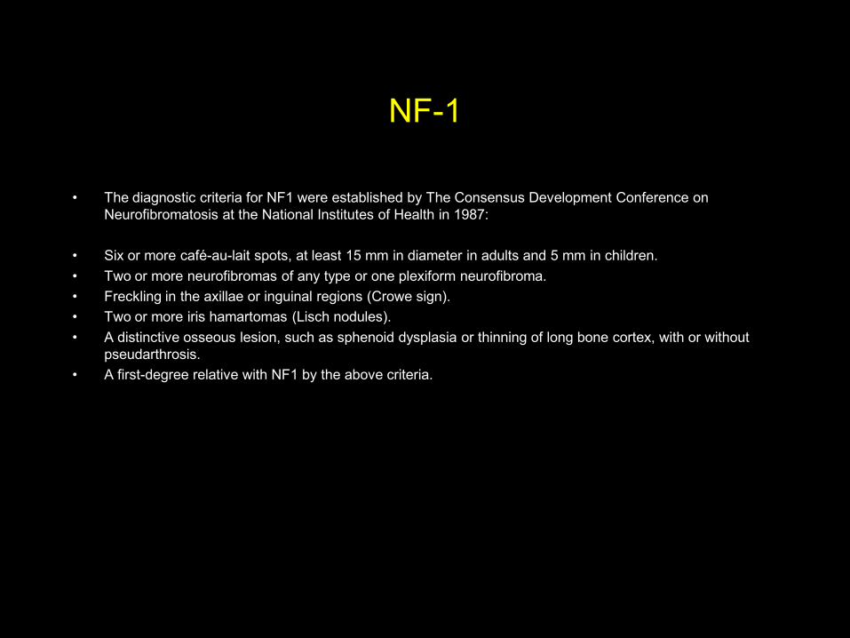
NF-1
• The diagnostic criteria for NF1 were established by The Consensus Development Conference on
Neurofibromatosis at the National Institutes of Health in 1987:
• Six or more café-au-lait spots, at least 15 mm in diameter in adults and 5 mm in children.
• Two or more neurofibromas of any type or one plexiform neurofibroma.
• Freckling in the axillae or inguinal regions (Crowe sign).
• Two or more iris hamartomas (Lisch nodules).
• A distinctive osseous lesion, such as sphenoid dysplasia or thinning of long bone cortex, with or without
pseudarthrosis.
• A first-degree relative with NF1 by the above criteria.

How do you check for a L4 level?
• Sensation on the medial side of the ankle, the
patellar tendon reflex, and plantar inversion are
associated with the L4 neurologic level.
• (Sensation on the lateral side of the ankle and the
Achilles tendon reflex are associated with the S1
neurologic level).

Which foot bones are ossified at birth?
• metatarsals are ossified at birth, along with the talus
and the calcaneus.
• The cuboid ossifies at 1 month, followed by the third,
second, and first cuneiforms.
• The navicular does not ossify until age 2 or 3 years.
• These facts are useful when interpreting radiographs
for congenital foot deformities such as clubfoot. The
location of the navicular must often be inferred from
the position of the first metatarsal

When to wt-bear after talar neck #’s
• Thordarson and associates used MRI to establish criteria for allowing a patient to begin
weight bearing. Their study suggested that patients with Hawkins types I and II fractures
should be allowed protective weight bearing when radiographic evidence of healing at the
fracture is present. None of the patients with a type I or II fracture in this study developed
late segmental collapse.
For patients with a Hawkins type III or IV fracture, the investigators recommended an MRI at
8 to 12 weeks postinjury to assess for osteonecrosis. The degree of osteonecrosis is then
classified based on the percent of talar body affected by osteonecrosis (type A,
homogenous bone throughout the body of the talus; type B, signal changes in up to 25 % of
the body of the talus; type C, signal changes in up to 25% to 50% of the body of the talus;
and type D, signal changes in more than 50 % of the body of the talus). The fracture
presented in this question is a type C. Type C fractures are kept non-weight bearing and the
MRI is repeated in 6 to 9 months after injury. If no progression of signal changes is noted,
protective weight bearing is allowed.

The oblique retinacular ligament connects with
what two structures?
• Landsmeer (oblique retinacular ligament) originates
from periosteum of prox phalynx, A2 and C1 pulleys.
• Inserts into the terminal tendon
• Function:To link PIPJ and DIPJ extension

Branches of axillary artery
1(Medial to pec minor)
– Supreme thoracic
2 (below pec minor)
Thoracoacromial artery (deltoid, acromial, pectoral and clavicular branches)
Lateral thoracic
3 (Lat to pec minor)
Subscapular
Anterior humeral circumflex
Posterior humeral circumflex
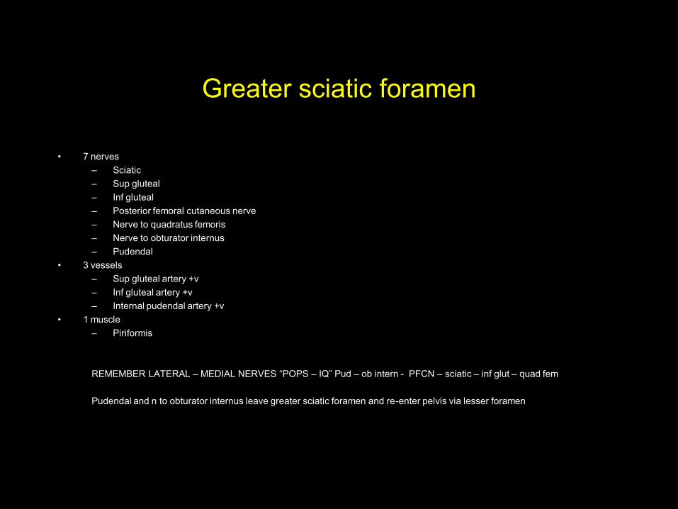
Greater sciatic foramen
• 7 nerves
– Sciatic
– Sup gluteal
– Inf gluteal
– Posterior femoral cutaneous nerve
– Nerve to quadratus femoris
– Nerve to obturator internus
– Pudendal
• 3 vessels
– Sup gluteal artery +v
– Inf gluteal artery +v
– Internal pudendal artery +v
• 1 muscle
– Piriformis
REMEMBER LATERAL – MEDIAL NERVES ―POPS – IQ‖ Pud – ob intern - PFCN – sciatic – inf glut – quad fem
Pudendal and n to obturator internus leave greater sciatic foramen and re-enter pelvis via lesser foramen
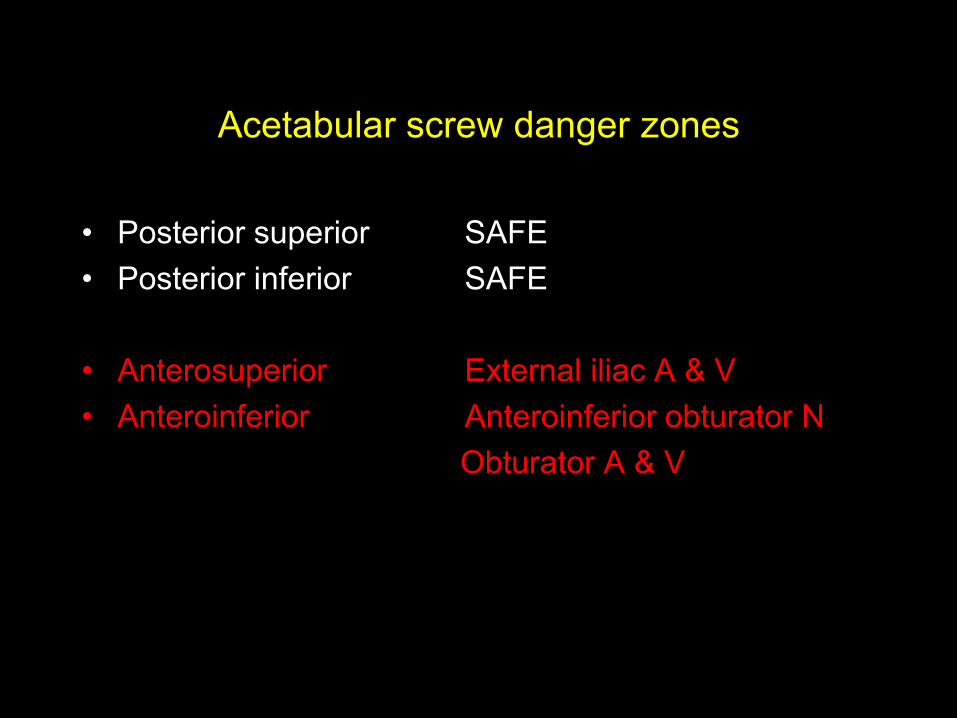
Acetabular screw danger zones
• Posterior superior SAFE
• Posterior inferior SAFE
• Anterosuperior External iliac A & V
• Anteroinferior Anteroinferior obturator N
Obturator A & V

Difference between UBC and ABC
• Unicameral bone cysts have a typical radiographic appearance:
• Metaphyseal
• Purely lytic
• Expand the bone in a symmetric fashion
• Often border the growth plate
• May have trabeculations in them once they have fractured
• ABC = Eccentric
• Unicameral bone cysts do not expand the bone beyond the width of the physis
• ABC may markedly expand the cortex.
• As ABCs expand into the soft tissues, there is generally a thin rim of periosteal
bone that outlines the expansion. With aneurysmal bone cyst, the lesion is
eccentric. Normal bone is present bordering the lesion. In contrast, in
unicameral bone cyst the entire medullary cavity is symmetrically involved.

Suprascapular nerve
• The suprascapular nerve is a branch of the upper trunk of the brachial plexus at
Erb’s point. The suprascapular nerve receives branches primarily from the fifth
cervical nerve root. The nerve follows the omohyoid muscle laterally and passes
beneath the anterior border of the trapezius muscle to the upper border of the
scapula where it joins the suprascapular artery. It passes through the
suprascapular notch deep to the transverse scapular ligament. The artery and
vein pass superficial to the ligament and join the nerve distally in the
suprascapular fossa. After innervating the supraspinatus muscle, the nerve
passes around the lateral free margin of the scapular spine (spinoglenoid notch)
to innervate the infraspinatus muscle.

Delbert classification
• Type I: Transphyseal fracture
• Type II: Transcervical fracture
• Type III: Basicervical fracture
• Type IV: Intertrochanteric fracture

What is this?
• CVT
• Note the equinus of the
hindfoot, calcaneus of
the forefoot, and the
crease in the sinus
tarsi. In patients with a
calcaneovalgus foot,
the hindfoot is in
calcaneus, not equinus.

What level myelodysplasia?
• This patient has active
quadriceps (which are
innervated through L2-L4)
and adductors (which are
innervated through L1-L3).
Because the patient’s knees
are slightly hyperextended
there is no hamstring
function. The patient’s right
foot has some dorsiflexion.
The lesion is rated as L4.
However, the patient’s right
side may be rated as L3

Boundaries of Hunter’s canal
• Hunter’s canal (adductor canal) is bound
anterolaterally by the vastus medialis, posterolaterally
by the adductor longus, and medially by the sartorius

• Syndrome Glycosaminoglycans
• Hurler syndrome Dermatan and heparan sulfate
• Hunter syndrome Dermatan and heparan sulfate
• Sanfilippo syndrome Heparan sulfate
• Morquio syndrome Keratin sulfate
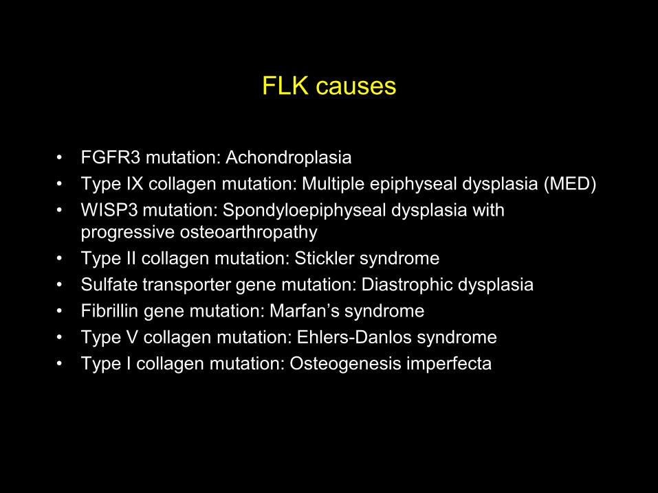
FLK causes
• FGFR3 mutation: Achondroplasia
• Type IX collagen mutation: Multiple epiphyseal dysplasia (MED)
• WISP3 mutation: Spondyloepiphyseal dysplasia with
progressive osteoarthropathy
• Type II collagen mutation: Stickler syndrome
• Sulfate transporter gene mutation: Diastrophic dysplasia
• Fibrillin gene mutation: Marfan’s syndrome
• Type V collagen mutation: Ehlers-Danlos syndrome
• Type I collagen mutation: Osteogenesis imperfecta
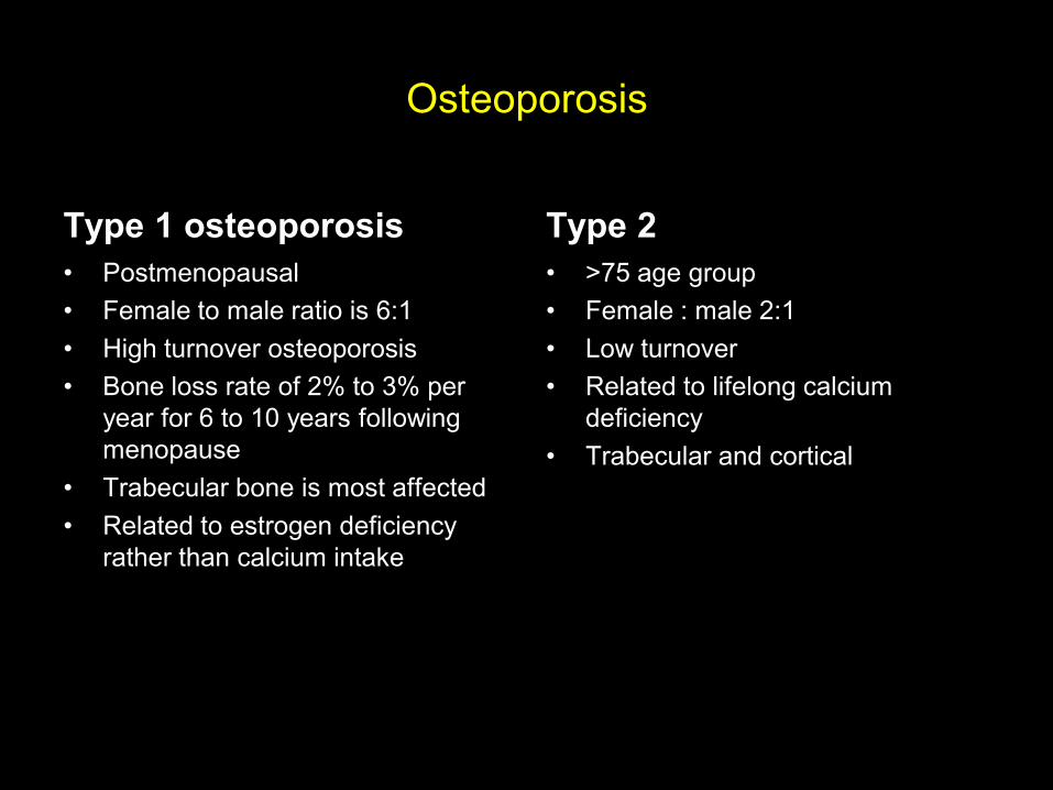
Osteoporosis
Type 1 osteoporosis
• Postmenopausal
• Female to male ratio is 6:1
• High turnover osteoporosis
• Bone loss rate of 2% to 3% per
year for 6 to 10 years following
menopause
• Trabecular bone is most affected
• Related to estrogen deficiency
rather than calcium intake
Type 2
• >75 age group
• Female : male 2:1
• Low turnover
• Related to lifelong calcium
deficiency
• Trabecular and cortical
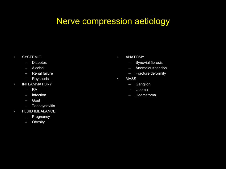
Nerve compression aetiology
• SYSTEMIC
– Diabetes
– Alcohol
– Renal failure
– Raynauds
• INFLAMMATORY
– RA
– Infection
– Gout
– Tenosynovitis
• FLUID IMBALANCE
– Pregnancy
– Obesity
• ANATOMY
– Synovial fibrosis
– Anomolous tendon
– Fracture deformity
• MASS
– Ganglion
– Lipoma
– Haematoma

Radial club hand
• Bilat in 50-72%
• Assoc VATER, Holt-Oram, TAR, Fanconis
• Stage I: Deficient distal radial epiphysis
• Stage II: Complete but short (hypoplasia)
• Stage III: Present proximally (partial aplasia)
• Stage IV: Total aplasia (most common)

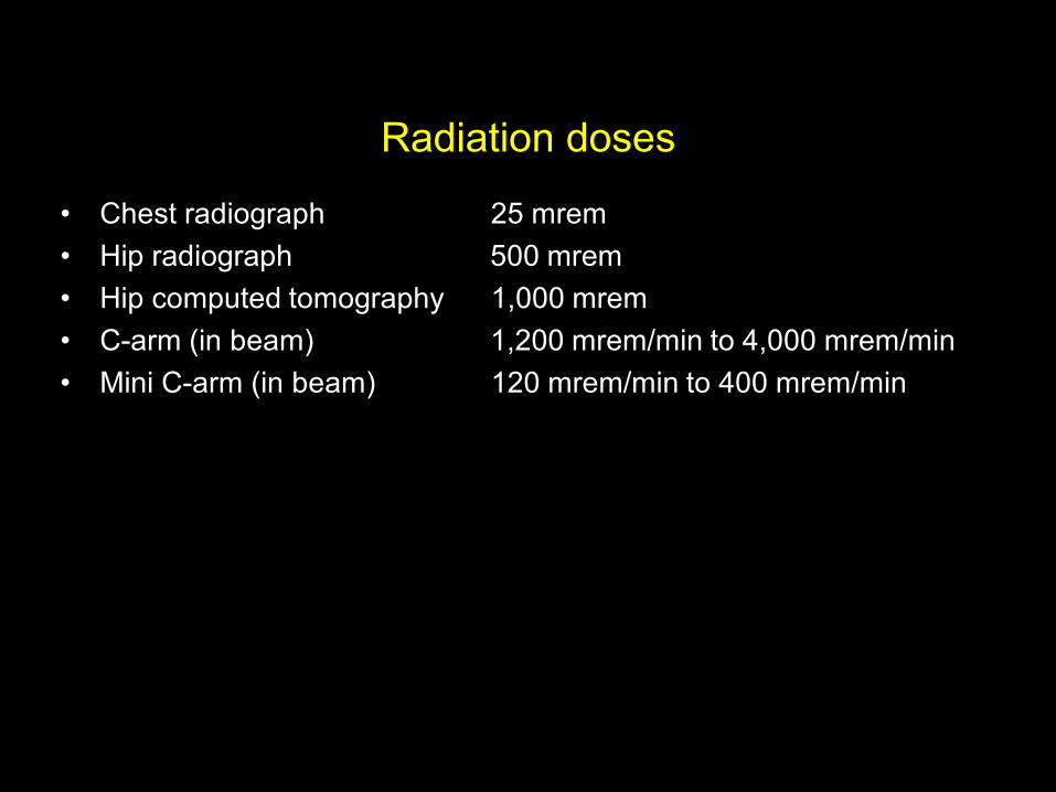
Radiation doses
• Chest radiograph 25 mrem
• Hip radiograph 500 mrem
• Hip computed tomography 1,000 mrem
• C-arm (in beam) 1,200 mrem/min to 4,000 mrem/min
• Mini C-arm (in beam) 120 mrem/min to 400 mrem/min
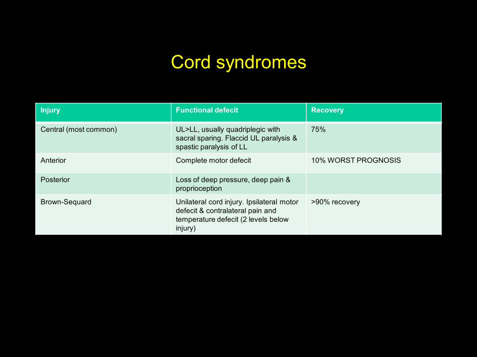
Cord syndromes
Injury Functional defecit Recovery
Central (most common) UL>LL, usually quadriplegic with
sacral sparing. Flaccid UL paralysis &
spastic paralysis of LL
75%
Anterior Complete motor defecit 10% WORST PROGNOSIS
Posterior Loss of deep pressure, deep pain &
proprioception
Brown-Sequard Unilateral cord injury. Ipsilateral motor
defecit & contralateral pain and
temperature defecit (2 levels below
injury)
>90% recovery

Key neurologic levels
• LEVEL MOTOR REFLEX
• C5 Deltoid Biceps
• C6 Wrist extension Brachioradialis
• C7 Wrist flex/Triceps Triceps
• C8 Finger flexion
• T1 Interossei
• L4 Tib ant Patellar
• L5 Toe extensors
• S1 Peroneal Achilles

Lumbar disc levels
LEVEL NERVE ROOT SENSORY LOSS MOTOR LOSS REFLEX LOSS
L1-L3 L2-L3 Anterior thigh Hip flexors None
L3-L4 L4 Medial calf Quads, tib ant Knee jerk
L4-l5 L5 Lat calf, dorsal foot EHL, EDL None
L5-S1 S1 Post calf, plantar foot Gastrosoleus Ankle jerk
S2-S4 S2,3,4 Perianal Bowel/bladder Cremasteric

What are the scenarios in a L4/L5 disc bulge?
• Normal bulge would hit the traversing L5 root causing
– + tension signs (decreased SLR)
– Decreased hip abductors
– Decreased EHL
– Pain and numbness lat leg and dorsum of foot
Far lateral disc could hit L4 as it exits
+ tension (FST)
Decreased tib ant
Loss of patella reflex

C-spine root compressions
• LEVEL ROOT MUSCLE SENS REFLEX• C3-C4 C4 Scapular Lat neck, shoulder None
• C4-C5 C5 Deltoid/biceps Lat arm Biceps
• C5-C6 C6 Wrist ext, biceps, triceps Radial forearm Brachioradialis
(supination)
C6-C7 C7 Triceps, wrist flexors Middle finger Triceps
C7-C8 C8 Finger flexors Ulnar hand None
C8-T1 T1 Interossei Ulnar forearm None

Tibial #’s – acceptable alignment
• Varus – valgus 5 degrees
• Sagittal plane 10 degrees
• Cortical apposition 50%
• Shortening 1 cm
• Rotation 10 degrees

What are these signs?
• Jeanne's sign identifies thumb metaphalangeal joint hyperextension of
10° to 15° with key pinch or gross grip.
• Froment's sign refers to the exaggeration of thumb interphalangeal joint
flexion during key pinch by the flexor pollicis longus in ulnar nerve
palsies.
• Wartenberg's sign is the inability to adduct the extended small finger
due to an ulnar nerve palsy.
• Duchenne's sign refers to clawing of the ring and small fingers.
• Pollock's sign is the inability to flex the distal interphalangeal joints of
the ring and small fingers in high palsies.

Blood supply to the physis
• Epiphyseal artery – terminates @ proliferative zone
• Perichondral artery
• Metaphyseal artery
• Nutrient arteries
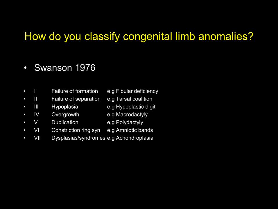
How do you classify congenital limb anomalies?
• Swanson 1976
• I Failure of formation e.g Fibular deficiency
• II Failure of separation e.g Tarsal coalition
• III Hypoplasia e.g Hypoplastic digit
• IV Overgrowth e.g Macrodactyly
• V Duplication e.g Polydactyly
• VI Constriction ring syn e.g Amniotic bands
• VII Dysplasias/syndromes e.g Achondroplasia

Indications for im nailing tibia
• Unacceptable alignment with closed reduction
• Significant soft tissue injury
• Segmental #
• Polytrauma/ipsilateral #’s
• Morbid obesity
• High energy/nstable

Tibial #’s – indications for amputation
• Warm ischaemia > 6hrs
• Absent plantar sensation
• Severe ipsilateral foot trauma
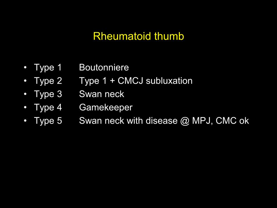
Rheumatoid thumb
• Type 1 Boutonniere
• Type 2 Type 1 + CMCJ subluxation
• Type 3 Swan neck
• Type 4 Gamekeeper
• Type 5 Swan neck with disease @ MPJ, CMC ok

Pseudohypoparathyroidism
• Pseudohypoparathyroidism (PHP) (Albright Hereditary Osteodystrophy [AHO]) -
end organ insensitivity; in AHO, germline mutation that leads to loss of function
of Galpha S (GNAS1); causes end-organ resistance to PTH.
• PHP - short stature, short metacarpals (4th and 5th), rounded facies
– Mental retardation, tetany
– Sex-linked dominant
• Laboratory features
– Hypocalcemia
– Hyperphopshatemia
– Normal PTH

Vitamin D-resistant rickets
• Vitamin D-resistant rickets may occur when there is an inability to convert 25
hydroxy vitamin D into 1,25 dihydroxy vitamin D.
• Patients develop secondary hyperparathyroidism. A low serum calcium level
causes an increased parathyroid hormone (PTH) level. Parathyroid hormone
causes the kidneys not to reabsorb phosphorus, and the serum phosphate is
low. The serum 1,25 dihydroxy vitamin D level is low.
• The metabolic profile is:
– Serum calcium Low
– Serum phosphate Low
– Serum PTH High
– 25 vitamin D Normal
– 1,25 vitamin D Very low
Treatment is by dietary 1,25 dihydroxy vitamin D.

DEFINITIONS

Define Spasticity
A motor disorder characterised by a velocity-dependent
in muscle tone

Spondylolisthesis
• I Dysplastic Child Congenital dysplasia of S1 sup facet
• II Isthmic 5 – 50 Predisposn with elongation/# of pars
(L5-S1)
• III Degenerative Old Facet arthroses leading to subluxation
(L4-L5)
• IV Traumatic Young Acute # other than pars
• V Pathologic Any Incompetence of bony elements
• VI Postsurgical Adult Xs resection of neural arches/facets
• Grade I= 0-25% slip, II = 25-50%, III = 50-75%, IV > 75%, V > 100%

Causes of Swan Neck
• ―Imbalance of forces @ PIPJ and a lax volar plate‖
• e.g MCPJ volar subluxation (Rheumatoids)
Mallet finger
Laceration
Transfer of FDS
Intrinsic contracture
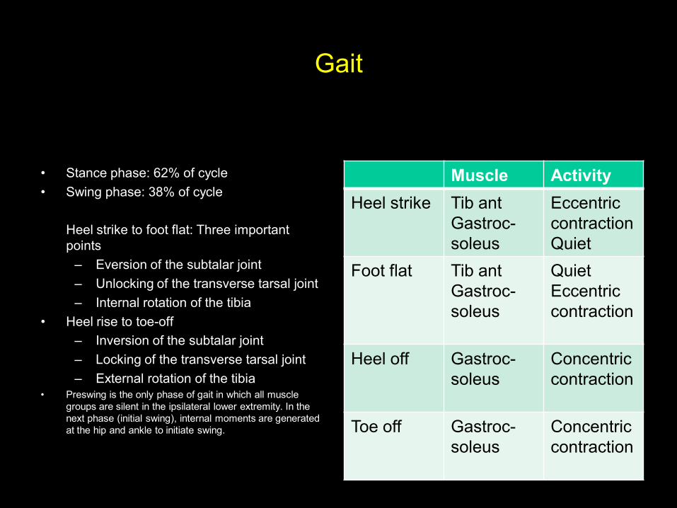
Gait
• Stance phase: 62% of cycle
• Swing phase: 38% of cycle
Heel strike to foot flat: Three important
points
– Eversion of the subtalar joint
– Unlocking of the transverse tarsal joint
– Internal rotation of the tibia
• Heel rise to toe-off
– Inversion of the subtalar joint
– Locking of the transverse tarsal joint
– External rotation of the tibia• Preswing is the only phase of gait in which all muscle
groups are silent in the ipsilateral lower extremity. In the
next phase (initial swing), internal moments are generated
at the hip and ankle to initiate swing.
Muscle Activity
Heel strike Tib ant
Gastroc-
soleus
Eccentric
contraction
Quiet
Foot flat Tib ant
Gastroc-
soleus
Quiet
Eccentric
contraction
Heel off Gastroc-
soleus
Concentric
contraction
Toe off Gastroc-
soleus
Concentric
contraction

Electrophysiology
• With electrodiagnostic testing, a clinician may find several characteristic features in different disorders:
• Denervation
– Fibrillation
– Positive sharp waves
– Fasciculations
• Neurogenic lesions
– Fasciculations
– Myokymic potentials
• Myopathies
– Complex repetitive discharges
– Myotonic discharges

Nerve injuries
Nerve Presentation Muscles lost
High radial Wrist drop EDC, EPL, APL, ECRB, ECRL, BR
Low radial Wrist drop EDC, EPL, APL
High ulnar Ulnar claw (intrinsic minus) Adductor poll, Interossei, FDP (ring &
little), FCU, Lumbricals (ring & little)
Low ulnar Severe ulnar claw Adductor poll, interossei, Lumbricals
(ring & little)
High median Ape hand PT, FCR, FDP (index & long), FPL,
APB, Lumbricals (index and long)
Low median Thenar wasting APB, Lumbricals (index and long)

What are osteocytes?
• Osteoblasts that become imbedded into the bone matrix
become osteocytes. Osteocytes are less metabolically active
because they do not produce large amounts of protein for
export. Thus, osteocytes have a higher nucleus to cytoplasm
ratio than osteoblasts. Osteocytes have fewer organelles as
they do not need extensive intracellular machinery to export
protein products.
• Osteocytes have extensive connections with other osteocytes
through the cell processes that travel through the canaliculi.
Strain generated signals such as cell deformation, streaming
potentials, or shear stress by fluid flow could be perceived by
the osteocytes and passed on to other cells.

Knee layers
• Medial
• I Sartorius
• II Sup MCL,
semimembranosus
• III Deep MCL, capsule
• Lateral
• I ITB, biceps, prepatella
bursa, peroneal nerve
• II Patella retinaculum,
patellofemoral lig
• III Arcuate lig, fabellofibular
lig, LCL, pop, pop-fib,
capsule

rank
• Activation of osteoclasts a complex.
• Surface receptors on osteoclast precursor cells are called RANK.
• Receptor activator of nuclear factor –kB ligand (RANKL) is expressed on the
surface of osteoblasts/stromal cells.
• RANKL proteins leave osteoblast and attach to the RANK receptor on the
osteoclast precursor.
• Macrophage colony stimulating factor (MCSF) then facilitates the production of
active osteoclasts from the osteoclast precursor.
Osteoprotegerin (OPG) is an inhibitor that is produced on the cell surface of
hematopoietic precursor cells and mature osteoclasts. OPG binds to RANK
receptor to inhibit the activation of osteoclasts.

Gustillo and Anderson
• Type I fractures have an open wound less than 1 cm in length.
• Type II wounds measure greater than 1 cm but less than 10 cm without
contamination, and the wounds can be closed without flap coverage.
• Type IIIA fractures have an open wound greater than 10 cm that can be
closed with delayed primary techniques. Segmental fractures and
gunshot injuries are also graded IIIA.
• Type IIIB fractures require rotational of free flap wound coverage.
• Type IIIC fractures are any open fractures with an associated vascular
injury that requires repair

#’s: im nails vs plates
• The periosteal blood supply cannot supply the inner two-thirds of the
cortex even if the endosteal blood supply has been interrupted, as in
intramedullary reaming.
• Blood flow markedly drops at the fracture site at the time of the fracture
and peaks at 2 weeks.
• Intramedullary reamed rods destroy the endosteal blood supply. In dog
experiments, the blood supply is reconstituted to normal in 120 days.
• In dog experiments, the blood supply is decreased in both plated and
rodded tibias at 42 and 90 days. The decrease is greater in the rodded
tibias.
• The oxygen tension is low in the fracture hematoma and in the newly
formed cartilage and bone. The oxygen tension is highest in the fibrous
tissue. The hypoxic state favors cartilage formation.
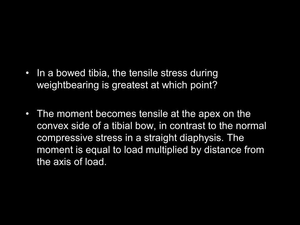
• In a bowed tibia, the tensile stress during
weightbearing is greatest at which point?
• The moment becomes tensile at the apex on the
convex side of a tibial bow, in contrast to the normal
compressive stress in a straight diaphysis. The
moment is equal to load multiplied by distance from
the axis of load.
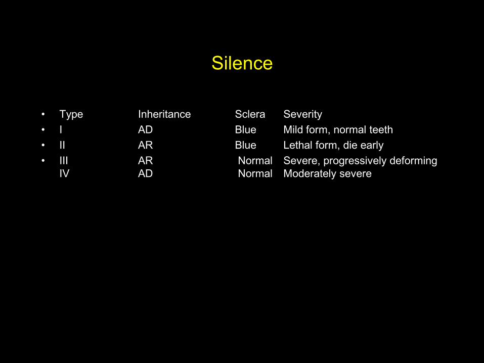
Silence
• Type Inheritance Sclera Severity
• I AD Blue Mild form, normal teeth
• II AR Blue Lethal form, die early
• III AR Normal Severe, progressively deforming
IV AD Normal Moderately severe

Layers of the foot
• First Layer
Abductor hallucis MPN (S2,S3) abducts and flexes Flexor digitorum brevis MPN
(S2,S3) flexes lateral four digits Abductor digit minimi LPN (S2,S3) abducts and
flexes fifth digit
• Second Layer
Quadratus plantae LPN (S2,S3) flexes lateral. four digits Lumbricals Medial one:
MPN flex MTPJ, ext PIP, DIP Lateral three: LPN
• Third Layer
Flexor hallucis brevis MPN (S2, S3) flex MTPJ Adductor hallucis LPN (S2, S3)
abducts first digit Flexor digiti minimi LPN (S2, S3) flex MTPJ
• Fourth Layer
Plantar interossei LPN (S2,S3) adducts digits
flex MTPJ Dorsal interossei LPN (S2,S3) adducts digits
flex MTPJ

OPG
• Soluble decoy receptor for RANKL
• Blocks osteoclast formation
• Reduces hypercalcemia
• Overexpression induces osteopetrosis
• Loss of expression induces osteoporosis
• Prevents calcification of large arteries

Define Neuropraxia
A physiological conduction block

Define Axonotomesis
Axonal disruption with an intact nerve sheath
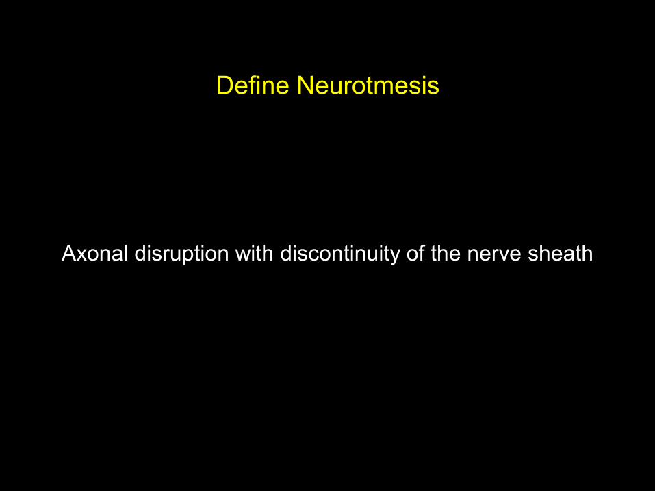
Define Neurotmesis
Axonal disruption with discontinuity of the nerve sheath

What is osteoporosis?
A skeletal disease characterised by a low bone mass
with a consequent increase in bone fragility

Describe Newton’s Laws
• FIRST (Inertia) = sum of all external applied forces is
zero.
• SECOND (Acceleration) = change in velocity is
proportional to the force.
• THIRD (Reaction) = for every action there is an equal
and opposite reaction.

What is stress?
Force per unit area (N/m2)
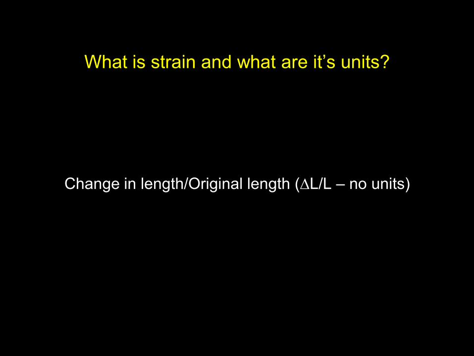
What is strain and what are it’s units?
Change in length/Original length (L/L – no units)

What properties does an elastic material
demonstrate?
Reversible deformation
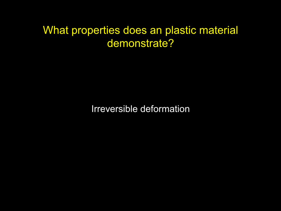
What properties does an plastic material
demonstrate?
Irreversible deformation

What is the yield point for a biomaterial?
Point at which there is a dramatic increase in strain with
little increase in stress (transition from elastic to
plastic phase)

What is the yield strength for a biomaterial?
Stress necessary to produce a specific amount of
permanent deformation (0.2%)

Define stiffness
Resistance to deformation - a product of stress/strain
(Young’s modulus)

What is the difference between stiffness and
rigidity in bio-engineering
Stiff = A material that is resistant to deformation
Rigid = A structure that is resistant to deformation

What is hardness?
Scratch resistance

What is fatigue?
Progressive failure due to application of cyclical
stresses below the ultimate tensile strength

What is prevalence in a population?
Total number of affected individuals at a single point in
time

What is incidence in a population?
Number of new cases per unit time

What is an odds ratio?
Likelihood of exposure causing disease

Define relative risk
Risk of developing disease without exposure

Define statistical power
The ability of a study to detect a TRUE difference

What types of statistical error do you know?
How might it be reduced?
• Type I () = study detects a difference that DOES
NOT exist = ―difference by chance alone‖ (related to
study design)
• - Type II () = study does not detect a difference
when one DOES exist (related to study power)

What is bias? Give an example?
A flaw in impartiality
(Blinding = study technique that minimises bias)

What is a confounding factor? Give an example
of how it may be reduced/removed
A variable that affects the validity of the conclusions of a
study
(Randomisation = study technique that minimises
confounding variables)

• Sens = TP/TP+FN
• Spec = TN/TN+FP
• PPV = TP/TP+FP
• NPV = TN/TN+FN
• Accuracy = TP+TN/All
Disease + Disease –
Test + TP 3 FP
Test - FN 4 TN
1 2

Define sensitivity
Proportion of true positives that are test positive

Define specificity
Proportion of true negatives that are test negative

What is a positive predictive value?
Proportion of test positives that are true positives

What is a negative predictive value?
Proportion of test negatives that are true negatives

What is an intercalated segment?
A segment whose stability depends on the compressive
load supplied by adjacent structures

What is a bone scan?
A radio-isotope imaging investigation

What is a DEXA scan? What are T-scores and
Z-scores?
Dual energy x-ray absorptiometry used to evaluate
bone mineral density
T-score = comparison with sex/race matched YOUNG
ADULT controls
Z-Score = comparison with sex/race and AGE matched
controls (not used in osteoporosis!!)
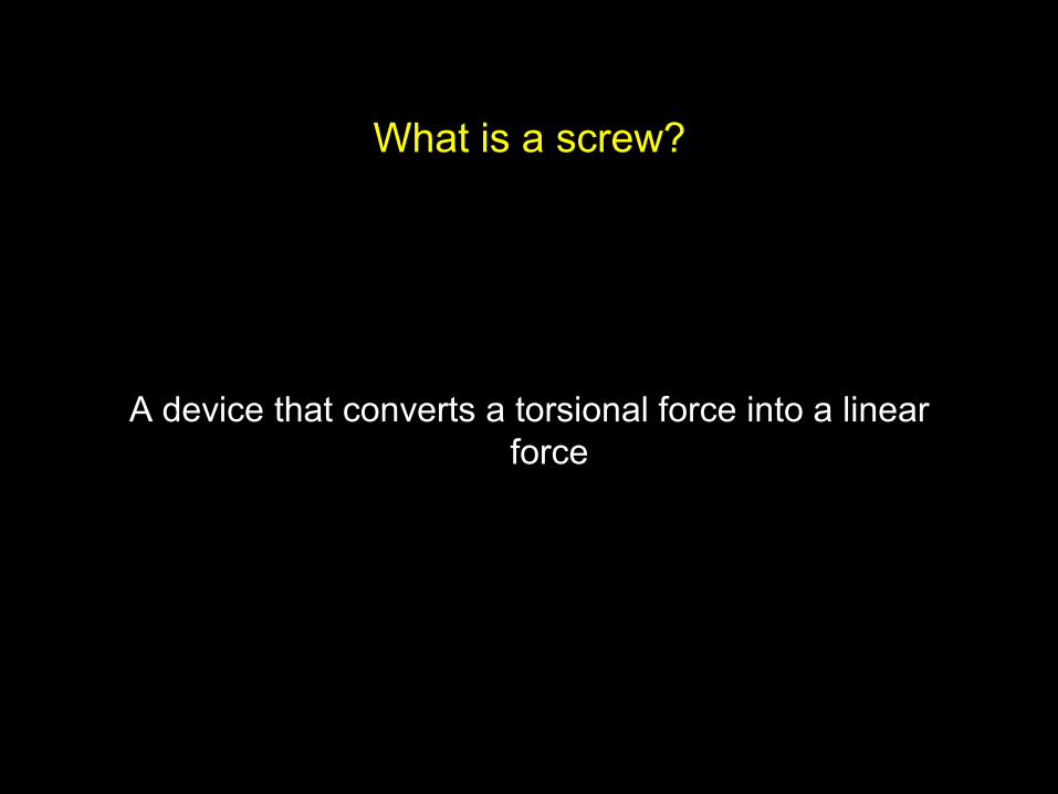
What is a screw?
A device that converts a torsional force into a linear
force

What are the drill/screw sizes that you
commonly use in fracture surgery?
Large Fragment
Small Fragment Mini Fragment
Outer Diameter (Cancellous) Outer Diameter (Cortical) Core Diameter Drill Size
6.5mm 4.5mm 3.0mm 3.2mm
4.0mm 3.5mm 2.4mm 2.5mm
N/A 2.7mm 1.9mm 2.0mm

What is the definition of a massive blood transfusion?
What are the indications for transfusion?
Massive blood transfusion = transfusion of TBV <24hrs
or 50% TBV <1hr
• Indications
– Emergency = urgent restoration of blood volume
– Elective = Hb <8 in fit patients & Hb <10 in
patients with C/P disease (NEJM 1999)

DRAWINGS

Draw the anatomy of the quadrilateral &
triangular spaces (in the shoulder!!)
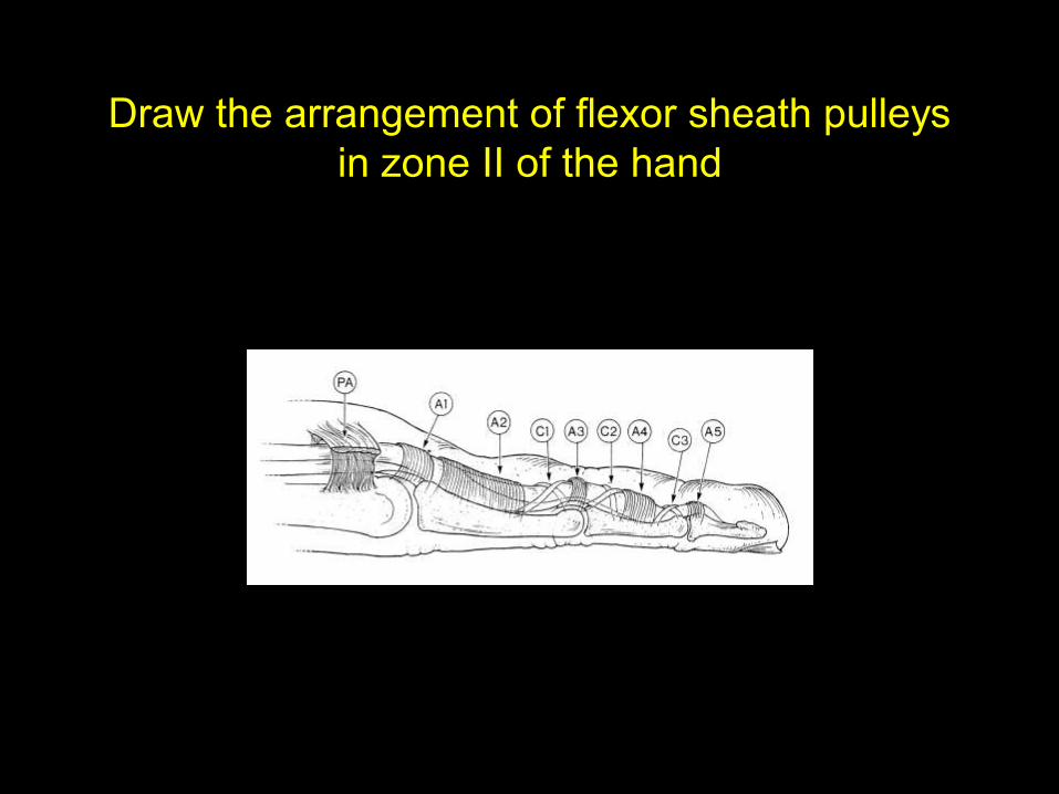
Draw the arrangement of flexor sheath pulleys
in zone II of the hand

Draw the extensor compartments at the wrist

Draw the fascial compartments of the hand.
How would you perform fasciotomies?

Draw the organisation of tracts in the spinal
cord

Meniscus
• 1. Only the peripheral 25% to 30% of the meniscus has a vascular
supply.
• 2. The medial meniscus functions as a secondary restraint to anterior
tibial translation (when the anterior cruciate ligament is cut).
• 3. Fifty percent of the compressive load of the knee is transmitted
through the meniscus when the knee is extended.
• 4. Eighty-five percent of the compressive load of the knee is borne by
the menisci when the knee is in 90° of flexion.
• 5. Meniscal fibrochondrocytes have the ability to proliferate and
synthesize matrix.
• 6. The medial meniscus is semicircular in shape; the lateral meniscus is
circular in shape.

• Timing
• Onset of injury site
•
• 7 to 10 days
• 2 to 5 weeks
(fibrillation, positive sharp
waves)
• 6 to 8 weeks
• Electrophysiologic
abnormality
• Conduction block across
nerve injury
• Reduced amplitudes on
distal stimulation
• Denervation changes on
electromyographic (EMG)
(fibrillation, positive sharp
waves)
Re-innervation on EMG

Foot compartments
• Calcaneal compartment
– Quadratus plantae
– Posterior tibial nerve, artery, and vein
– Lateral plantar nerve, artery, and vein
– Medial plantar nerve (variable)
• *Remember that the calcaneal compartment may communicate with the posterior tibial compartment.
• Interossei—(four separate compartments)
• Adductor muscle
• Medial
– Flexor hallucis
– Abductor hallucis
• Lateral
– Abductor digiti minimi
– Flexor digiti minimi
• Superficial
– Flexor digitorum brevis
– Lumbricals (four)
– Flexor digitorum longus
– Medial plantar nerve (variable)

Indications for retrograde i.m femoral nail
• Multisystem injury
• Trauma involving multiple extremity fractures
• Ipsilateral vascular injury
• Fracture in the morbidly obese
• Isolated fracture above a preexisting total knee prosthesis or below a hip
prosthesis
• Ipsilateral hip/acetabular fracture
• Spine injury or uncleared spine
• A relative indication is pregnancy as it reduces the amount of radiation directly to
the abdomen.
• Contraindications include skeletal immaturity and a
history of knee joint sepsis.

How do you do a discectomy
• The traditional surgery for the excision of a herniated
posterolateral lumbar disk is by means of a midline
incision. This procedure is then performed in a
stepwise fashion: The paraspinal musculature is
stripped from the lamina of the vertebra; the
ligamentum flavum is then excised; portions of the
superior and inferior lamina are removed; and the
nerve root and dural sac are identified and carefully
retracted. This is followed by excision of the
herniated disk material and wound closure.

Draw the fascial compartments of the foot. How
would you perform fasciotomies?

Draw a cross-section thorough a peripheral
nerve
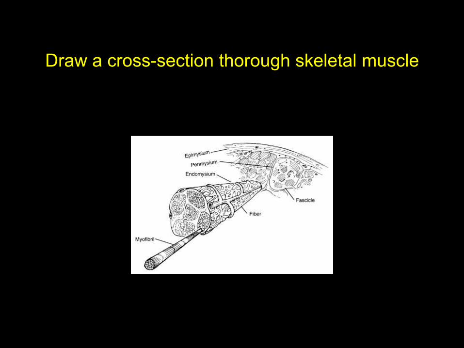
Draw a cross-section thorough skeletal muscle

Draw the microscopic arrangement of a skeletal
muscle fibre

Draw a FBD for forces around the shoulder

Draw a FBD for forces around the elbow
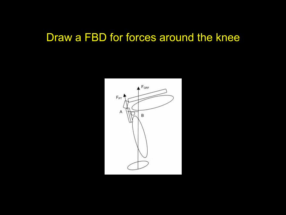
Draw a FBD for forces around the knee

Draw the anatomy of Femoral shaft fracture
displacement patterns

Draw a cross-section thorough an intervertebral
disc. How are the annular fibres organised?

Draw a cross-section of articular cartilage

Describe the molecular structure of the cartilage
layers
• Superficial tangential zone (gliding zone) Thin collagen fibrils are parallel to
the articular surface
• Chondrocytes are elongated with the axis parallel to the surface
• Proteoglycan content is at the lowest level
• Water content is at the highest level
• Middle zone Larger diameter collagen fibers/less organization
• Rounded chondrocytes
• Deep zone Collagen fibers are large and perpendicular to the articular surface
• Highest concentration of proteoglycans
• Lowest water content
• Chondrocytes are spherical and arranged in columnar fashion
• Calcified zone Small cells in cartilage matrix encrusted with apatitic salts

Draw a stress/strain curve for a biomaterial and
describe the key areas of the diagram
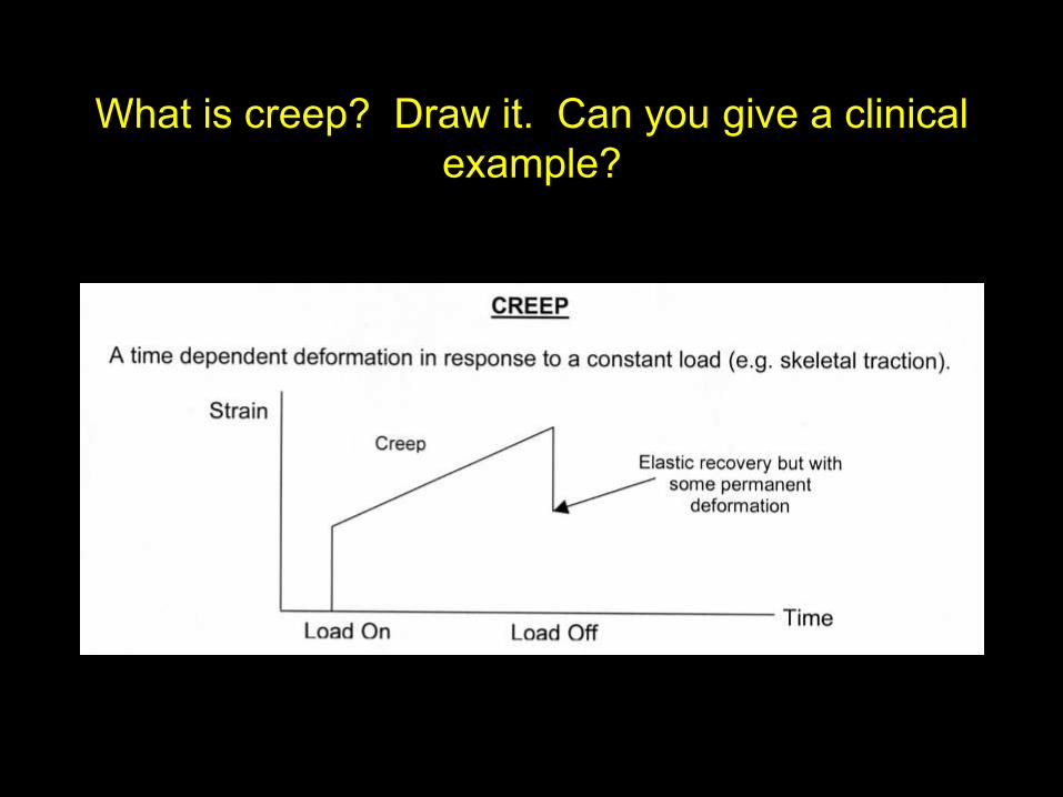
What is creep? Draw it. Can you give a clinical
example?

What is stress-relaxation? Draw it. Can you
give a clinical example?

What is hysteresis? Draw it

What is fatigue? What is a stress-riser? Draw
an S/n curve. How might we reduce fatigue?
• Fatigue – progressive failure due to application of cyclical stresses
below the ultimate tensile strength. It occurs through a process of
crack propogation.
• ―Stress riser‖ = a point where the concentration of stress exceeds the
mean stress for the material.
• Reduction strategies:
– - implant design (i.e. avoiding sudden changes in geometry).
– - surface treatments (e.g. polishing).
– - correct insertion of implants (to avoid load-bearing a device).
– - control of loading conditions (e.g. partial weightbearing)

What are the pre-requisites for normal gait?
• Gage
– Stability in stance
– Clearance in swing
– Foot pre-positioning
– Adequate stride length
– Energy conservation/efficiency

Pronator syn
• Sites of compression
– Supracondylar process
– Lig of Struthers (tip of supracondylar process – medial epicondyle)
– Deep head of p teres
– Origin of FDS
– Differentiate from CTS by sens disturbance over distributn of palmar cut
branch,ant prox forearm pain & tinels.NO night symptoms
– OE: Resisted elbow flexion with forearm supination (bicipital aponeurosis)
• Resisted forearm pronation with extended elbow (2 heads p teres)
• Isolated long finger PIPJ flexion (FDS origin)
Rx Non operative, if fails decompress everything!

Describe normal gait
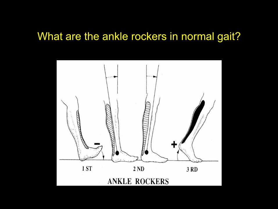
What are the ankle rockers in normal gait?

Describe Vitamin D Metabolism

What is the diagnosis?

Describe is the microscopic structure of bone
• Cellular Component (10% of cortical bone volume)
– osteoblasts (mesenchymal stem cells)
• osteocyte (90%) = osteoblast ―trapped‖ in mineralised osteoid – maintain bone; communicate via cellular processes.
• bone lining cell = cells lining quiescent bone with cellular processes penetrating matrix to osteocytes. PTH expose bone surface for o’clasts.
– osteoclasts (haematopoietic stem cells)
• produce H-ions = demineralisation.
• proteolytic enzymes = matrix removal)
• Matrix Component (composite material comprising 90% of cortical bone volume)
– Organic (40% dry weight) – resists tension forces
• collagen (type I ……..one = bone)
• proteoglycans (GAGs)
• matrix proteins (i.e. non-collagenous) = osteocalcin, -nectin, -pontin
– Inorganic (60% dry weight) – resists compression forces
• calcium hydroxyapatite
• osteocalcium phosphate (brushite)

Describe the blood supply of a long bone. How
does it vary between adult/child?
• Endosteal - Nutrient Artery (high pressure system) =
inner 2/3
• Periosteal - (low pressure system) = outer 1/3
• Metaphyseal/Epiphyseal – peri-articular vascular
plexus
(N.B. Direction)
– Adult (Normal) = Centrifugal (Fracture) =
Centripetal
– Child (Normal) = Centripetal – periosteum +++

What factors affect bone healing?
• Systemic = age; functional activity; nerve function;
nutrition; drugs(NSAIDs++); smoking
• Local = severity of local trauma; degree of bone loss;
vascular injury; type of bone fractured;
immobilisation; infection; local pathology
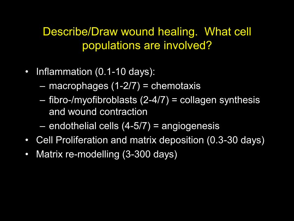
Describe/Draw wound healing. What cell
populations are involved?
• Inflammation (0.1-10 days):
– macrophages (1-2/7) = chemotaxis
– fibro-/myofibroblasts (2-4/7) = collagen synthesis
and wound contraction
– endothelial cells (4-5/7) = angiogenesis
• Cell Proliferation and matrix deposition (0.3-30 days)
• Matrix re-modelling (3-300 days)

Describe both the macroscopic & microscopic
structure of synovium
• Macroscopic
– Synovium (intimal layer) = avascular, aneural & arranged into microvilli to surface area
– Sunsynovium (fibrous/areolar/adipose) = cellular & vascular innervated with lymphatics
– Joint capsule/tendon sheath = thick fibrous tissue
• Microscopic
– Cellular (synoviocytes)
• A-cells (macrophages) = phagocytosis
• B-cells (fibroblast-like) = exocrone/synthetic cells
– Extra-cellular matrix
• Collagen (I, III, IV, V, VI)
• Proteins (fibronectin….)

Describe the composition of synovial fluid
• Synovial Fluid = blood ultra-filtrate containing:
– Hyaluronic acid
– Lubricin
– Proteinase
– Collagenase
– Prostaglandins

Blood supply to the spine
• Derived from segmental arteries
– Located @ vertebral midbodies via aorta
– Primary supply to dura & posterior elements from dorsal branches
– Ventral branches supply vertebral bodiesvia ascending and descending
branches which are delivered under PLL in 4 separate ostia
– Vertebral artery (from subclavian) ascends through transverse foramina C1-
C6 (anterior to and not through C7), posterior to longus coli muscle,
posterior to lateral masses, along top of C1, ventromedially aroundcord,
through foramen magnum before uniting at midline basilar artery.

Stages of Kienbocks (Lichtman)
Stage Description Treatment
1 Normal x-ray, signal changes on MRI Cast immobilization
If ulna- radial shortening
2 Sclerosis Ulna – radial shortening
Ulna + vascularized bone graft, distal radius osteotomy
3a Fragmentation & lunate collapse
Normal carpal alignment
Ulna – radial shortening
Ulna + vascularized bone graft, distal radius osteotomy
3b Fragmentation & lunate collapse
Loss of carpal height
Prox row carpectomy
Intercarpal arthrodesis
4 Lunate collapse with arthrosis Prox row carpectomy
Wrist arthrodesis

Hawkins classification
• Type 1 Undisplaced 13% AVN
• Type 2 Displaced neck # + Subluxation/dislocation subtalar joint 20-50%
• Type 3 Displaced neck # + Subluxation/dislocation subtalar + ankle joint ~100%
• Type 4 Displaced neck # + Dislocation subtalar + ankle + talonavicular joints ~100%

Blood supply to the talus
• Extraosseous
– Posterior tibial a
• Artery of tarsal canal, calcanael branches
– Dorsalis pedis
• Medial tarsal branches, artery to tarsal sinus
– Peroneal
• Also contributes to artery of tarsal sinus
• Intraosseous
– Talar head supplied by dorsalis pedis and tarsal sling (arteries of
tarsal canal and sinus)
– Talar body supplied by anastomosis of tarsal sling

Describe the microscopic structure of articular
cartilage
• Cellular (5%)
– Chondrocytes
• Extracellular (95%)
– Water (75%)
– Proteoglycans (20%) – function = trap and hold water
– Collagen (5%)
• Type II – predominantly
• Type IX – lies on surface of type II and acts as an interfibrillar glue
• Type X – calcified zone
– Adhesives
• Chondronectin
• Fibronectin
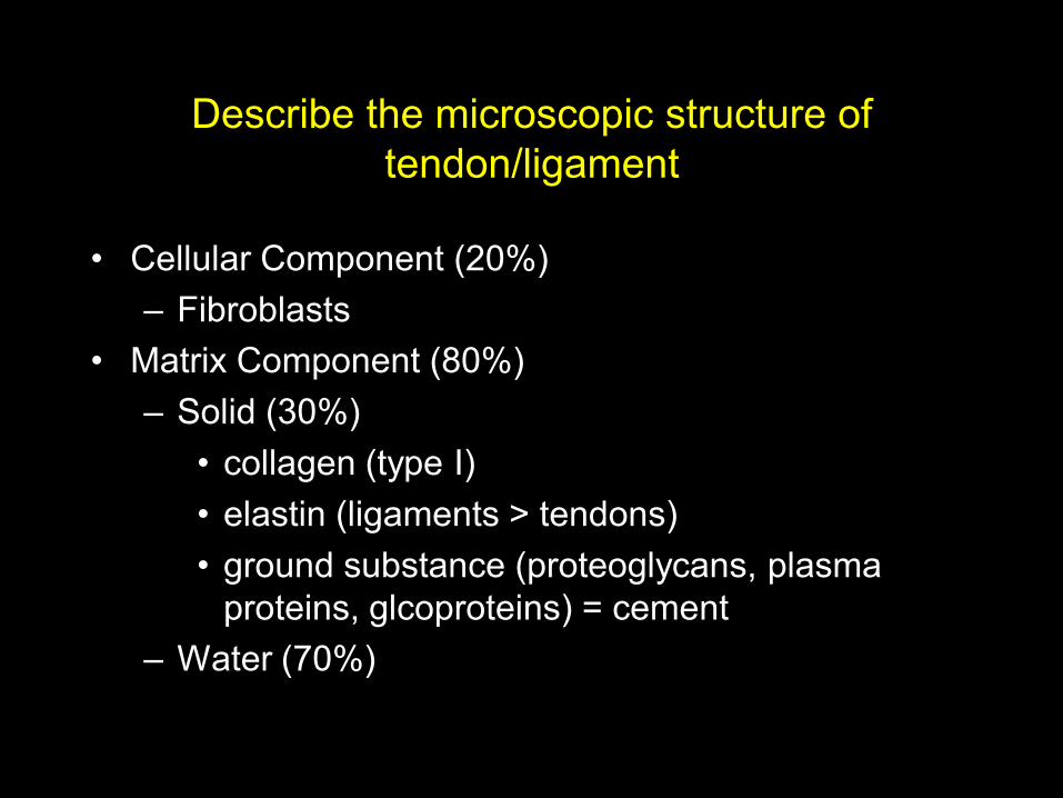
Describe the microscopic structure of
tendon/ligament
• Cellular Component (20%)
– Fibroblasts
• Matrix Component (80%)
– Solid (30%)
• collagen (type I)
• elastin (ligaments > tendons)
• ground substance (proteoglycans, plasma
proteins, glcoproteins) = cement
– Water (70%)
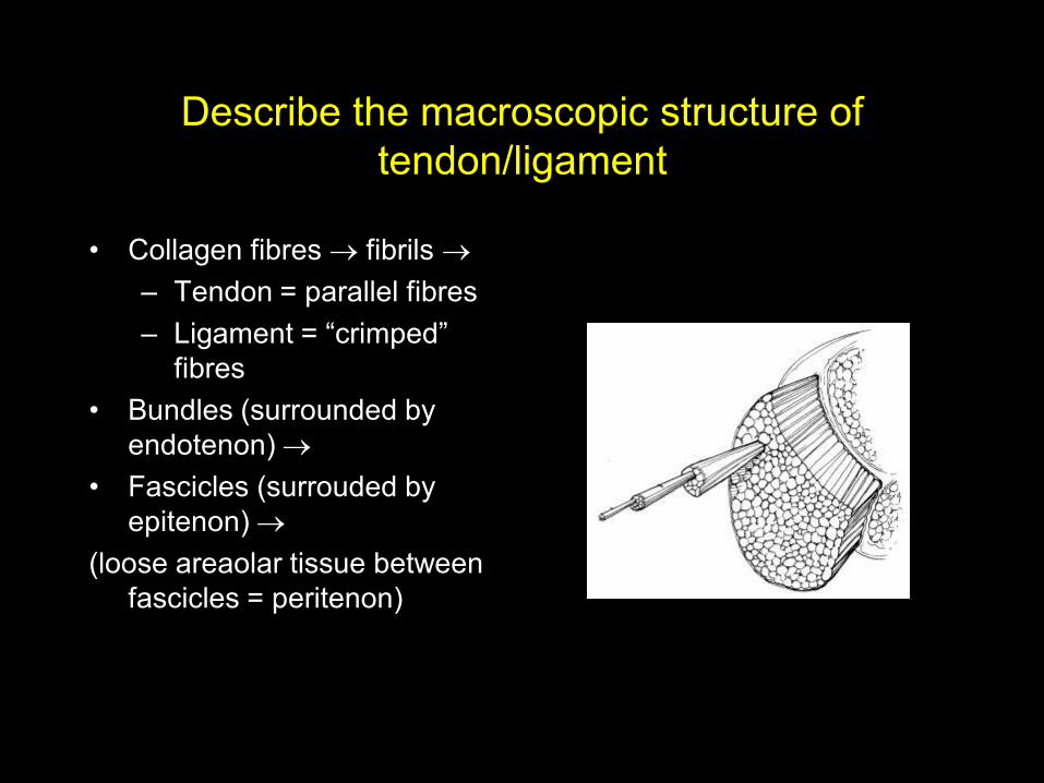
Describe the macroscopic structure of
tendon/ligament
• Collagen fibres fibrils
– Tendon = parallel fibres
– Ligament = ―crimped‖
fibres
• Bundles (surrounded by
endotenon)
• Fascicles (surrouded by
epitenon)
(loose areaolar tissue between
fascicles = peritenon)

Draw/Describe the biomechanics of
tendon/ligament failure
• 1 = ―toe‖ region = ―uncrimping‖ of relaxed collagen fibres
• 2 = elastic region
• 3 = yield point
• 4 = ultimate tensile strength failure (tendon)
• 5 = ultimate tensile strength failure (ligament -microfailure)
• 6 = macroscopically intact ligament with intra-substance failure (e.g. ACL)

What are the important features of a screening
programme? What problems may occur?
• The Disease
– documented natural history
– validated therapeutic protocol
– treatment benefit
• The Test
– accurate (product of sensitivity(TP’s) and specificity(TN’s))
– safe
– acceptable
– cheap
• Problems = ? population to be screened; compliance; interval
lesions; lead-time bias
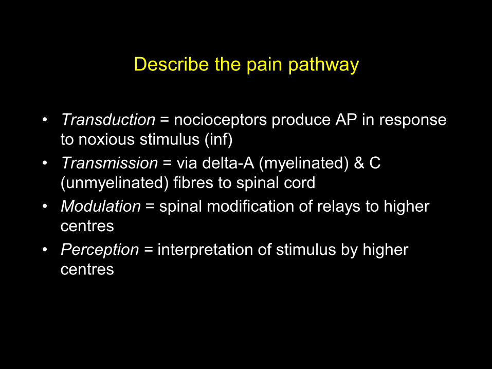
Describe the pain pathway
• Transduction = nocioceptors produce AP in response
to noxious stimulus (inf)
• Transmission = via delta-A (myelinated) & C
(unmyelinated) fibres to spinal cord
• Modulation = spinal modification of relays to higher
centres
• Perception = interpretation of stimulus by higher
centres

What are the general principles of managing a
soft-tissue defect?
• General principles = dirty wound clean wound.
Open wound closed wound using the
reconstruction ladder
• Reconstruction ladder = direct closure/granulation
skin graft (SSG/FTG) local flaps distant flaps
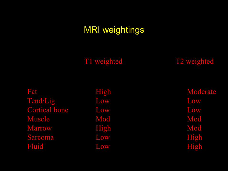
MRI weightings
T1 weighted T2 weighted
Fat High Moderate
Tend/Lig Low Low
Cortical bone Low Low
Muscle Mod Mod
Marrow High Mod
Sarcoma Low High
Fluid Low High
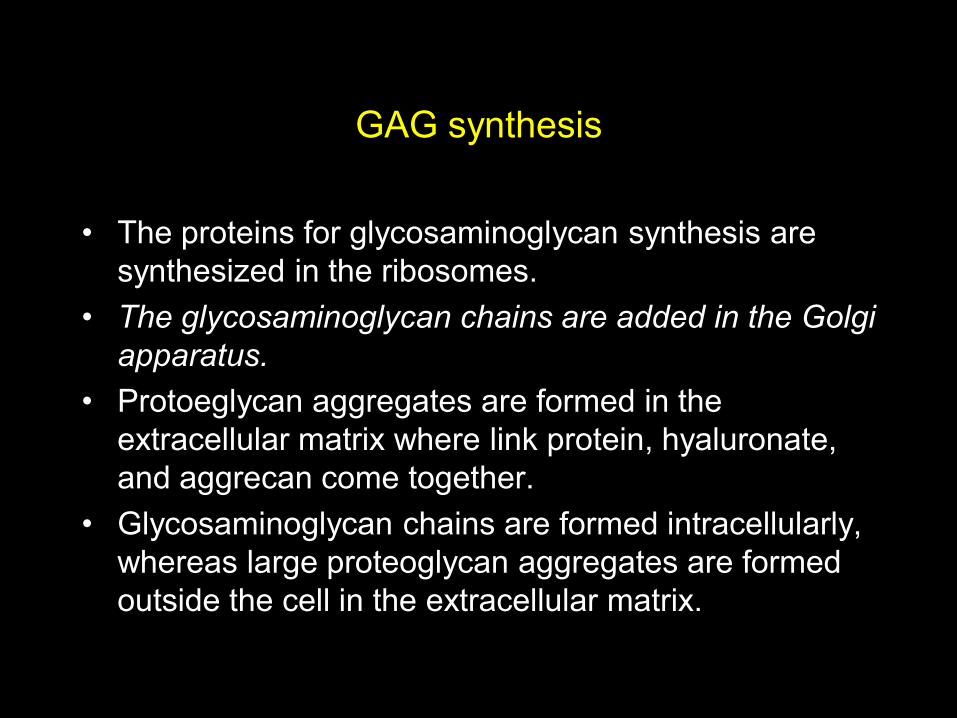
GAG synthesis
• The proteins for glycosaminoglycan synthesis are
synthesized in the ribosomes.
• The glycosaminoglycan chains are added in the Golgi
apparatus.
• Protoeglycan aggregates are formed in the
extracellular matrix where link protein, hyaluronate,
and aggrecan come together.
• Glycosaminoglycan chains are formed intracellularly,
whereas large proteoglycan aggregates are formed
outside the cell in the extracellular matrix.
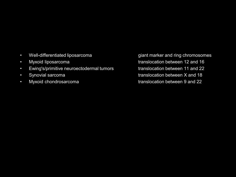
• Well-differentiated liposarcoma giant marker and ring chromosomes
• Myxoid liposarcoma translocation between 12 and 16
• Ewing's/primitive neuroectodermal tumors translocation between 11 and 22
• Synovial sarcoma translocation between X and 18
• Myxoid chondrosarcoma translocation between 9 and 22

How does diathermy work? What types are
there? List some safety issues
―..passage of high-frequency alternating current through body tissue with a point of concentration producing heat.‖ Frequency >50,000Hz removes neuromuscular response to current. Surgical diathermy = 400kHz – 10MHz.
• Types:
– Bipolar = generator both limbs of diathermy forceps
– Monopolar = generator active elctrode (point) plate electrode.
• Cutting = continuous current cell vaporisation
• Coagulation = pulsed current cell dessication & sealing of vessels
• Safety:
– properly checked / maintained equipment
– plate continuity alarm
– plate application (at least 70cm2)
– patient protection from contact with metal objects
– appropriate use of diathermy
– pacemaker – recently checked; avoid/reduce use; consider bipolar

Describe how you would design a study
• Identify clinical problem to be studied
• Literature search - ? work done already ?weaknesses in other studies
• Propose a hypothesis
• Study design
– Ethical approval
– Power calculation
– Recruiting study population
– Randomisation
– Observation/Measuremets
– Methods/Sources for data collection
• Data Collection
• Statistical analysis
• Interpretation of results & conclusions

What types of study do you know?
• Meta-analysis
• Observational studies
– Cross-sectional studies = prevalence
– Cohort studies (prospective)
– Case-control studies (retrospective)
• Interventional studies
– Randomised, controlled trials (prospective)
– Case reports/series

• In studying a newly recognized
disorder using a large population of
affected individuals, geneticists
discover that although the disorder
often affects siblings, it was rarely, if
ever, detected in their ancestors.
This disorder most closely follows
which pattern of inheritance: (A)
Autosomal dominant (B) Autosomal
recessive (C) Sex-linked (D)
Multifactorial (E) Anticipation
• Autosomal recessive conditions classically show
―horizontal‖ inheritance. Ancestors do not display the
gene because they would likely have only one copy
of the mutant allele. Only when two carriers
reproduce is the phenotype manifest in
approximately one-fourth of their offspring.
• Autosomal dominant inheritance is characterized by
vertical transmission. Many generations manifest the
trait because it takes only a single copy of a mutant
allele to display the phenotype.
• Sex-linked conditions are often traced back in a
family. Normally the males are affected and the
females are carriers.
• Multifactorial conditions are thought to result from the
combination of different genes. Although the risk of
recurrence in kindred is somewhat greater than the
population as a whole, it is still quite low (only a few
percent). It is rare for siblings to be affected.
• Anticipation refers to the phenomenon in which
successive generations are likely to display more
severe forms of a given disorder. Myotonic dystrophy
is a classic example of this phenomenon

Define Congenital Vertical Talus
• Irreducible dorsal dislocation of the navicular on the
talus with a fixed equinous hindfoot deformity
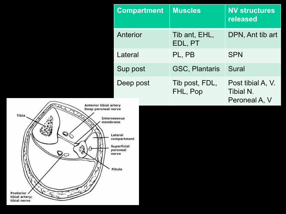
Compartment Muscles NV structures
released
Anterior Tib ant, EHL,
EDL, PT
DPN, Ant tib art
Lateral PL, PB SPN
Sup post GSC, Plantaris Sural
Deep post Tib post, FDL,
FHL, Pop
Post tibial A, V.
Tibial N.
Peroneal A, V


Modes of femoral stem loosening
Mode Mechanism Cause Findings
Ia Pistoning Subsidence of stem within cement RLL between stem & cement zones 1 & 2
Distal cement #
Stem displaced distally in cement
Ib Pistoning Subsidence of stem & mantle within bone RLL all 7 zones
II Medial stem pivot Lack of supermedial & inferolateral cement support Medial migration prox stem
Lat migration distal tip
Cement # zones 2 & 6
III Calcar pivot Medial & Lat toggle of distal stem
Hang up of stem collar on medial cortex
Windshield wiper rxn at distal stem
Sclerosis & thickening of bone @ stem tip
RLL zones 4 & 5
IV Cantilever bending Loss of prox cement support leaving distal stem still
fixed; allows for proximal cantilever bending
Stem crack or #
RLL zones 1 & 2, 6 & 7

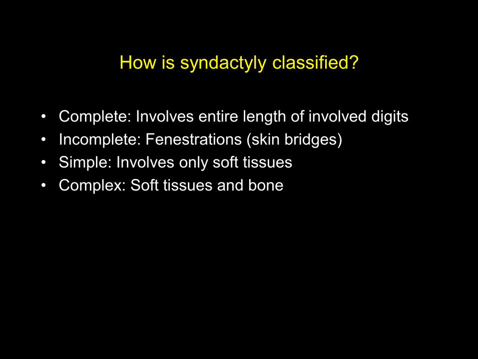
How is syndactyly classified?
• Complete: Involves entire length of involved digits
• Incomplete: Fenestrations (skin bridges)
• Simple: Involves only soft tissues
• Complex: Soft tissues and bone

What is camptodactyly & how’s it classified?
• Nontraumatic painless PIPJ flexion contracture
• Usually little finger
• Unknown aetiology
• Imbalance of forces between FDS and intrinsics (lumbricals and
interossei)
• Type 1 Most common, uni or bilat PIPJ contracture in healthy infant
• Type 2 Same as above but seen adolescence esp girls
• Type 3 Assoc with syndromes

Radial clinodactyly
• Congenital curvature of digit in radioulnar plane
• Type 1 Minor angulation, normal length v. common
• Type 2 Minor angulation, short (3% and 25% Downs)
• Type 3 Marked delta phalynx ( c shaped epiphysis)

What is the AIS?
• Head
• Face
• Neck
• Thorax
• Abdo, pelvis
• Spine
• UL
• LL
• External
The ISS is the sum of the squares for the highest AIS grades in the 3 most
severely injured ISS regions. >18 = polytrauma

What are the MESS variables?
• Skeletal and soft tissue injury
– Low, med, high, very high energy
Limb ischaemia (score doubled if >6hrs)
Shock
Age
Score >7 = Amputate

Antibiotics 1
• Penicillins: Bactericidal, inhibit peptidoglycan synth. Bind to membranes
(aka Beta lactams)
• Cephalosporins:Bactericidal. Inhibit cell-wall synthesis (also beta lactams)
• Aminoglycosides: e.g Gentamicin. Bactericidal. Inhibit protein synthesis by
binding to cytoplasmic rRNA 30s subunit
• Macrolides: e.g erythromycin: Inhibit dissociation of tRNA from ribosomes during
translacation by binding to 50s subunit
• Quinolones: Inhibit DNA gyrase, stops supercoiling
• Glycopeptides: e.g vanc/teic. Inhibit cell membrane synthesis (hit glycan
subunits in the cell wall
• Tetracyclines: Bacteriostatic. Identical action to aminoglycosides, just doesn’t kill
them!
• Rifampicin: Bactericidal. Prevents RNA transcription
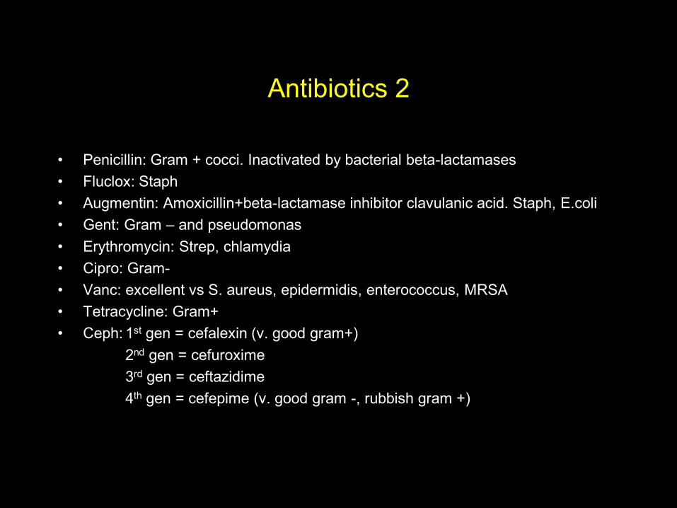
Antibiotics 2
• Penicillin: Gram + cocci. Inactivated by bacterial beta-lactamases
• Fluclox: Staph
• Augmentin: Amoxicillin+beta-lactamase inhibitor clavulanic acid. Staph, E.coli
• Gent: Gram – and pseudomonas
• Erythromycin: Strep, chlamydia
• Cipro: Gram-
• Vanc: excellent vs S. aureus, epidermidis, enterococcus, MRSA
• Tetracycline: Gram+
• Ceph: 1st gen = cefalexin (v. good gram+)
2nd gen = cefuroxime
3rd gen = ceftazidime
4th gen = cefepime (v. good gram -, rubbish gram +)

Common bacteria
Gram+ cocci Gram- cocci Gram+bacilli Gram-bacilli
S.aureus N. meningitidis C. perfringens Pseudomonas
S. epidermidis N. gonorrhoea C. tetani Eikenella
Strep E. coli
Salmonella

What do interossei do?
• 4 dorsal Abductors (DAB)
– Insert index, middle (both ways)
and ring
3 palmar Adductors (PAD)
– Adduct index, ring and little
fingers
These muscles flex the MCPJ
and extend the PIPJ via the
lateral bands

What do lumbricals do?
• Originate on FDP
• Insert on radial lateral band of
extensor expansion
• Pass volar to transverse metacarpal
ligament
• Median n supplies radial 2
lumbricals (unipennate)
• Ulnar n supplies ulnar 2 (bipennate)

Stages of Charcot arthropathy
Stage Description Treatment
0 Unilateral oedema, erythema
No break in skin
Negative x-ray
Cast immobilization, NWB
1 Unilateral oedema, erythema
X-ray = osseous destruction, joint
subluxation/dislocation
2 Decreased oedema, erythema and warmth
X-ray shows coalescence of fracture
fragments
WB immobilization until tissue
homeostasis is evident then AFO with
locked or limited motion ankle
3 No or limited oedema, erythema or warmth
X-ray = consolidation and remodelling
AFO used on limited basis


![Large buccal fat pad lipoma: A rare case report...gland lipoma in 2 cases, angiolipoma in 2 cases, and spindle cell lipoma in 3 cases [10]. The most common presentation of BFP lipoma](https://static.fdocuments.in/doc/165x107/5e610a1252021369db53e163/large-buccal-fat-pad-lipoma-a-rare-case-report-gland-lipoma-in-2-cases-angiolipoma.jpg)

![Kundalini - Evolution and Enlightenment (ed. John White) (459p) [Anomolous].pdf](https://static.fdocuments.in/doc/165x107/55cf8f31550346703b99d250/kundalini-evolution-and-enlightenment-ed-john-white-459p-anomolouspdf.jpg)


![Swami Narayanananda - The Secrets of Prana, Pranayama & Yoga Asanas (149p) [Anomolous]](https://static.fdocuments.in/doc/165x107/577c78221a28abe0548edebb/swami-narayanananda-the-secrets-of-prana-pranayama-yoga-asanas-149p.jpg)


