Frank Bernhard Institute of Biophysical Chemistry ... · Frank Bernhard Institute of Biophysical...
37
Frank Bernhard Institute of Biophysical Chemistry University of Frankfurt, Germany Cell-Free Expression of Membrane Proteins in Artificial Environments 4 th P4EU Meeting Porto 2013
Transcript of Frank Bernhard Institute of Biophysical Chemistry ... · Frank Bernhard Institute of Biophysical...

Frank Bernhard
Institute of Biophysical Chemistry
University of Frankfurt, Germany
Cell-Free Expression of Membrane Proteins in Artificial Environments
4th P4EU Meeting Porto 2013

Coupled Transcription-Translation
- Bacterial S30 extract
Removal of endogenous amino acids
Elimination of endogenous mRNA
- T7-RNA polymerase
- Template DNA (T7-regulatory sequences),
e.g. pET-vectors, pIVEX
- tRNA
- Amino acids
- RNase, protease inhibitors
- Energy system: acetyl/creatine phosphate,
pyruvate, kinases
- NTPs
- Suitable buffer system
- Mg2+-, K+-Ions
Optional additives:
I. Efficiency enhancer: PEG, spermidine, cAMP, oxalate, coenzyme A, NAD…
II. Chemical chaperones: Glycerol, polysaccharides, alcohols…
III. Protein chaperones: GroEL/ES…
IV. Target specific stabilizers: Ligands, cofactors, inhibitors, subunits…
V. Solubilization compounds: Detergents, lipids, surfactants…
VI. Redox systems: DTT, glutathione system, DsbA/C…
Applications of cell-free expression
I. High throughput, protein expression in artificial environments
Proteome expression screens by robotic devices, X-ray, NMR!, reducing inclusion body formation by addition of stabilizers
II. Expression of disulfide-bridged proteins
Venome peptides, antibodies…
Modulation of oxidizing/reducing conditions
III. Co-expression of multisubunit complexes
Identical vectors (no care about compatibility, copy number..)
Pre-expression, co-expression
IV. Excellent system for protein labelling
Fluorescence enhanced amino acids, seleno-methionine, stable isotopes
V. Expression of proteins that cannot be obtained in living hosts Toxins, unstable proteins, apo-enzymes, artificial cofactor incorporation
membrane proteins

Cell-Free Membrane Protein Expression
Continuous exchange cell-free system
(CECF)
Method Development
Reckel et al (2010) Methods Mol. Biol.
Kai et al (2011) Methods Mol. Biol.
Haberstock et al (2012) Prot. Expr. Purif.
Junge et al (2010) New Biotechnol.
Junge et al (2008) Cell. Mol. Life Sci.
Sobhanifar et al (2010) J. BioMol. NMR
Reckel et al (2008) PNAS
Schwarz et al (2008) Proteomics
Roos et al (2012) Mol. Membr. Biol.
Feeding Mix
Reaction Mix
Maxi-CECF Reactor
Reaction
Mix
Feeding Mix
Mini-CECF Reactor

Cloning/DNA preparation / linear PCR fragments
Transformation
Incubation (overnight – days)
Induction of expression
Harvest
Cell-disruption
Isolation of membrane fraction
Extraction of MPs out of membrane
Purification
Reaction set-up
Incubation (overnight)
(Solubilization 1-3 hrs)
Reaction/Fermentation set-up
Cellular - Cell-Free Membrane Protein Expression
Cellular Cell-free
Targeting
Translocation
Toxicity
Proteolysis Vector issues
Cell handling
Media
Sterility
Fusions
Promoter
Regulation/Induction
+ efficient labelling
+ individualized protocols
+ artificial hydrophobic
environments

I: → Yield = Basic expression protocol: P-CF
Tag variation, template design and quality, codon usage
Basic compounds (ions, extract source, energy system)
RM:FM ratio, re-feeding of substrates
Systematic 3-Level Protocol Development
Junge et al., (2011) New Biotechnology 28:262-71.
Schwarz et al., (2008) Proteomics 8:3933-46.
Junge et al., (2008) Cell. Mol. Life Sci. 65:1729-55.
II: → Solubility = Array of expression protocols: D-CF + L-CF
Type and concentration of hydrophobic compound(s), detergents, lipids,
D/L-mixtures, bicelles, liposomes, nanodiscs
III: → Quality = Selection of one/few individual protocols
Stability, homogeneity, activity
Detergent exchange; 1st and 2nd detergent, additives, stabilizer
Quality screening of sample arrays
Customized target specific production protocols

Roos et al. (2012) MolMemBiol.
Monitoring of Cell-Free Membrane Protein Expression

130 Targets
80 % success,
Size: > 100 kDa, 15 TMS
Schwarz et al. (2010) Proteomics 10: 1762-79

E. coli cell-free system: Purification
Single step purification with affinity chromatography
Reaction mix / resolubilized pellet can be loaded directly on the affinity matrix

G-Protein Coupled Receptors (GPCRs)
Junge et al. (2010) J. Struct. Biol. 172: 94-106
Junge et al. (2008) Cell. Mol. Life Sci. 65: 1729-55
Klammt et al. (2007) J. Struct. Biol. 158: 482-93
Klammt et al. (2007) FEBS J. 274: 3257-69
bovine
Rhodopsin
rat
Corticotropin
releasing
factor
human
Vasopressin
type 2

Cell-free Expression Modes for Membrane Protein Synthesis
► 3 basic ways to produce soluble membrane proteins
DL-CF

P-CF Expression Mode
● Spin down
● Wash precipitate
● Option: Selective solubilization
● Option: Complete Solubilization (1 h, RT)
● Affinity purification, e.g. Ni-NTA

Km(MPP+) = 35 µM (30 µM*)
turnover number = 19 sec-1 (20 sec-1*) MPP:1-methyl-4-phenyl-pyridinium
*rOCT1 from insect cells
180
130
100
73
54
48
35
24
1 2 3
rOCT1
180
130
100
73
54
48
35
24
1 2 3
rOAT1
Eukaryotic Organic Cation/Anion Transporters
Cooperation: T. Keller/H. Koepsell
Cation selectivity of rOCT1 IC50:
TEA+: 159 µM / 196 µM*
TPeA+: 2.9 µM / 1.8 µM*
TBuA+: 20 µM / 19 µM*
Keller et al. (2008) Biochemistry 47: 4552-4564
Keller et al., (2011) J. Biol. Chem. 286: 37874-86
P-CF

Presenilin 1(PS1): 53 kDa
Presenilin-NTF (NTF): 31 kDa
Presenilin-CTF (CTF): 20 kDa
Nicastrin: 78 kDa
Pen-2: 12 kDa
Aph-1: 27 kDa
Sobhanifar et al. (2010), PNAS 107, 9644-9649
Sobhanifar et al. (2010), J Biomol NMR 46, 33-43
Solution NMR structure of P-CF expressed presenilin-1 CTF

Positron Research Group B. Pucci / A. Polidori J. L. Popot
L-CF D-CF

Cell-free expression of GnRHR
Test different hydrophobic environments
Nanodiscs
DMPC
DMPG
DOPC
DPPC
DOPC+DPPC
POPC
Detergents Amphipol: A8-35
D-CF:
Brij-35
Brij-58
Brij-78
Solubilization of P-CF
precipitates:
LMPG
LMPC
DDM
DPC
Ligand binding studies: MST, Biacore, Pull-down

Soluble expression in D-CF mode
P-CF Brij35 Brij58 Brij78 DDM DPC Digitonin DHPC
M RM
PR from Digitonin in DHPC, Superdex200
-10
0
10
20
30
40
50
60
70
0 0.5 1 1.5 2 2.5 3
elution volume [ml]
ab
so
rpti
on
[m
AU
]
-10
-5
0
5
10
15
20
25
30
35
40ab
so
rpti
on
[m
AU
]280 nm
380 nm
535 nm
Sina Reckel

Throughput Cell-Free Yield + Solubility + Quality
Optimization Screening
Brij58 DPC P-CF DMPC Brij58 + DMPC Triton + DMPC
Digitonin : diC7PC 4:1
Reckel et al. (2011) Angew. Chem. Intl. Ed.

Quality Screening of Endothelin-A Receptor Samples
P-CF
P-CF
D-CF
Junge et al. (2010) J. Struct. Biol. 172: 94-106

T. Meier
D. Matthies
MPI of Biophysics
Frankfurt/Main
Cell-Free Expression of Multisubunit Complexes
F1F0 ATP Synthase
3 membrane protein subunits
5 soluble protein subunits
25 protomers, 542 kDa

Cell-Free Expression of Multisubunit Complexes
Matthies et al. (2011) J. Mol. Biol. 413: 593-603
In vivo expression Cell-free expression

Adapted from J. Popot et al. 2011
Nonionic APol
A8-35
Amphipols

Roos et al (2012) Mol Membr Biol
Proverbio et al (2013) BBA
L-CF Expression Mode

Adapted from Nath et al. 2006
Nanodiscs

Nanodiscs Liposomes Consistence between + - preparations Monodispersity + - Time and temperature + + stability of MP Access form both sides + - of the membrane Quantification of + (+) protein insertion Purificationof“loaded” + (+) bilayers only
Insertion of CF produced MPs into
Nanodiscs

Lipid MSP1 :: Lipid MSP1E3 :: Lipid
DMPC 1:80 1:115
POPC 1:55 1:85
DOPC 1:30 1:80
DMPG 1:70 1:110
DOPG 1:30 1:90
DOPE 1:30 1:80
PC Soybean 1:40 1:60
PL E. coli 1:40 1:60
TL E. coli 1:40 1:50
PL Brain 1:45 1:70
TL Brain 1:35 1:60
Co-translational insertion into preformed nanodiscs
No detergent contact of membrane protein Roos et al (2012) BBA
In Vitro Nanodisc Formation

DOPC nanodisc
0
50
100
150
200
250
0,0
0
0,1
10
,23
0,3
40
,46
0,5
70
,68
0,8
00
,91
1,0
2
1,1
41
,25
1,3
71
,48
1,5
91
,71
1,8
21
,93
2,0
52
,16
2,2
82
,39
2,5
02
,62
2,7
3
2,8
52
,96
3,0
73
,19
3,3
03
,41
3,5
33
,64
DOPC 1:60
DOPC 1:80
DOPC 1:100
Best: 1:60
0
50
100
150
200
250
0,0
0,1
0,3
0,4
0,5
0,7
0,8
0,9
1,0
1,2
1,3
1,4
1,6
1,7
1,8
2,0
2,1
2,2
2,4
2,5
2,6
2,7
2,9
3,0
3,1
3,3
3,4
3,5
3,7
Volume (ml)
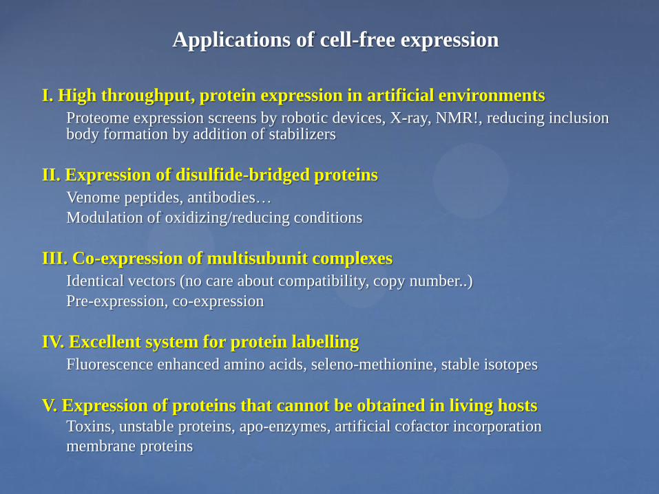
DPPC NDs
DPPC 1:100
DPPC 1:150
DPPC 1:170
DPPC 1:200
Best: 1:100

0
50
100
150
200
250
0,0
0,1
0,2
0,3
0,5
0,6
0,7
0,8
0,9
1,0
1,1
1,3
1,4
1,5
1,6
1,7
1,8
1,9
2,0
2,2
2,3
2,4
2,5
2,6
2,7
2,8
3,0
3,1
3,2
3,3
3,4
3,5
3,6
DOPC:DPPC 1:30:60
DOPC:DPPC 1:40:80
DOPC:DPPC 1:40:40
DOPC-DPPC NDs
Best: 1:40:40 (or 1:30:60)

SugE
Roos et al (2012) BBA
Proteorhodopsin
MraY Translocase
Optimization of Nanodisc Complex Formation

Zocher et al. (2011) ACS Nano
Single Molecule Force Microscopy of Bacteriorhodopsin
in Nanodiscs
Bacteriorhodopsin inserts as a
Trimer
Roos et al (2012) BBA

Laser flash induced time-dependent absorbance changes of green
proteorhodopsin reconstituted from DHPC into DMPC/DMPA
proteoliposomes and DMPC/DMPA nanodiscs. The PR-nanodisc sample
shows a spectral behavior similar to that of proteoliposomes.
Moers et al. (2013) BBA
Ranaghan et al., JACS 2011
Functional Analysis of PR/Nanodisc Complexes
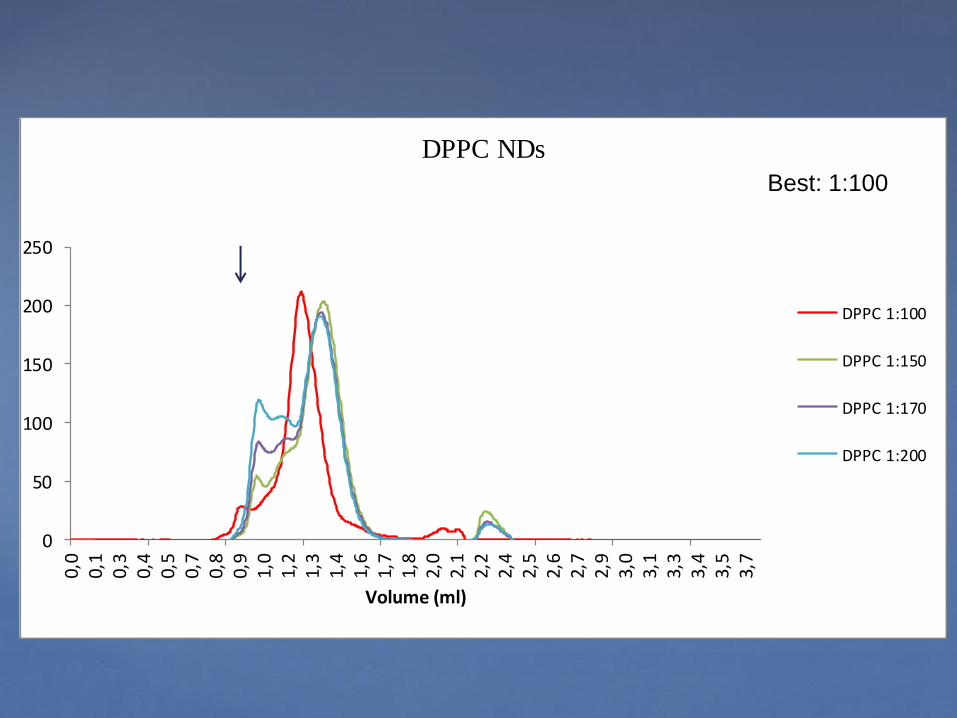
Ligand nM IC50 (IC50 literature)
ET-1 8 (0.3)
Ala-4-ET-1 2663 (> 2000)
BQ-123 38 (1.4)
IRL-1620 300 (> 500)
Ligand nM IC50 (IC50 literature)
ET-1 9 (0.2)
Ala-4-ET-1 4 (0.33)
BQ-123 1395 (> 1500)
IRL-1620 0.005 (0.1-56)
ETA: ET-1 = BQ-123 >> ET-3, 4-Ala-ET-1, IRL-1620
ETB: ET-1 = ET-3, 4-Ala-ET-1, IRL-1620 >> BQ-123
Kd ET-1: 1.7 (0.4-0.8)
Kd ET-3: 3400 (>2000)
Kd ET-1: 0.4-0.7 (0.1-0.8)
Kd ET-3: 0.2 (0.1-0-8)
Differential Ligand Binding of GPCR/Nanodisc Complexes
Proverbio et al. (2013) BBA

{
Biosynthesis pathway of Lipid II in E.coli
Cell Wall Biosynthesis in Bacteria

Biosynthesis of Peptidoglycan Precursor
Published in: Tetsuya Tanino; Bayan Al-Dabbagh; Dominique Mengin-Lecreulx; Ahmed Bouhss; Hiroshi Oyama; Satoshi Ichikawa; Akira Matsuda;
J. Med. Chem. 2011, 54, 8421-8439.
DOI: 10.1021/jm200906r
Copyright © 2011 American Chemical Society

Lipid Requirements of E. coli MraY Translocase
Roos et al (2012) BBA
Ma et al (2011) JBC

Goethe University, Biophysical Chemistry • Volker Dötsch
• Natascha Rogova (Expression screening, extract preparation)
• Davide Proverbio, Erika Orbán, Ralf Rues, Fang Dong (GPCRs)
• Erik Henrich (L-CF, nanodiscs, translocase)
• Christopher Hein (Site specific labelling)
• Former Members: Edith Buchinger, Stefan Haberstock, Frederike Junge,
Lei Kai, Yi Ma, Sina Reckel, Christian Roos, Solmaz Sobhanifar
Thomas Meier Klaus Fendler
Vladimir Shirokov Alexander Spirin
Clemens Glaubitz Rupert Abele Joseph Wachtveitl
Hermann Koepsell
Hans-Georg Sahl Tanja Schneider
Stefan Kubick
Martin Caffrey Coilin Boland
Daniel Müller
Alain Milon
Bernard Mouillac
Core Center for Cell-Free Expression
www.structuralbiology.eu