FoxG1 and TLE2 act cooperatively to regulate ventral ... · differentiation in the dorsal...
10
1553 RESEARCH ARTICLE INTRODUCTION The vertebrate forebrain is a complex neural network consisting of a wide variety of highly specified neurons and glia. The correct localisation and timed differentiation of a variety of neuron subtypes requires the coordinated action of a large number of signalling cascades and transcriptional regulators (Hebert and Fishell, 2008). One factor involved in the timing of neuronal differentiation and in the specification of subdomain identity is the transcriptional repressor FoxG1, a member of the Fox/Forkhead family of winged- helix transcription factors [previously known as BF-1 and renamed Foxg1 for mouse, FoxG1 for other chordates and FOXG1 for human (Kaestner et al., 2000)]. FoxG1 expression is conserved throughout vertebrates in dividing progenitors of the ventricular zone and early postmitotic neurons in the telencephalic neuroepithelium (Bourguignon et al., 1998; Murphy et al., 1994; Tao and Lai, 1992; Zhao et al., 2009). Loss- and gain-of-function experiments in different species have established FoxG1 as a maintenance factor for neural progenitors. Knockout of Foxg1 leads to premature differentiation in the dorsal telencephalon and to a drastic increase in early-fate CR neurons accompanied by a loss of late progenitors (Hanashima et al., 2004; Xuan et al., 1995). Conversely, in gain-of- function experiments in frogs, a high dose of FoxG1 expands the progenitor population (Bourguignon et al., 1998; Hardcastle and Papalopulu, 2000), whereas in chicken it causes overgrowth of the neural tube (Ahlgren et al., 2003). siRNA knockdown experiments have demonstrated a role for the level of FoxG1 in controlling the timing of cortical layer differentiation (Shen et al., 2006), suggesting a concentration-dependent activity. A second important role of FoxG1 is during the specification of the ventral telencephalon (subpallium) and its delineation against the dorsal telencephalon (pallium) and basal diencephalon (hypothalamus). In the mouse Foxg1 knockout, all markers for the ventral telencephalon are lost from an early stage, suggesting that the ventral telencephalon is never specified, while dorsal markers spread ventrally (Martynoga et al., 2005). In fish, morpholino (MO) knockdown experiments demonstrated a similar loss of the subpallium, accompanied by a ventral expansion of dorsal telencephalon markers and a ‘slippage’ of ventral telencephalic cells into the hypothalamic territory of the diencephalon (Danesin et al., 2009). Recently, it has been shown that Foxg1 is required cell- autonomously in the acquisition of ventral (subpallial) telencephalic identity in the mouse (Manuel et al., 2010). How does FoxG1 exert these functions? At the molecular level, FoxG1 acts mainly as a transcriptional repressor by direct and indirect mechanisms (Bourguignon et al., 1998; Dou et al., 2000; Li et al., 1995; Seoane et al., 2004; Yao et al., 2001). FoxG1 shows direct repression on reporter constructs (Li et al., 1995; Yao et al., 2001), and in vivo it directly represses p27Xic and wnt8b (Hardcastle and Papalopulu, 2000; Danesin et al., 2009). However, it can also negatively regulate TGF signalling by binding to Smad and FoxO transcription factors (Dou et al., 2000; Seoane et al., 2004). The proliferation-promoting effect of mouse Foxg1 is independent of its DNA-binding ability (Dou et al., 2000; Hanashima et al., 2002). Transcriptional repression by FoxG1 is partly mediated by recruiting transcriptional co-repressors of the Groucho/Transducin-like enhancer of split (TLE) and AT-rich interaction domain (ARID) families (Sonderegger and Vogt, 2003; Tan et al., 2003; Yao et al., 2001). These co-repressors, in turn, Development 137, 1553-1562 (2010) doi:10.1242/dev.044909 © 2010. Published by The Company of Biologists Ltd 1 Faculty of Life Sciences, Michael Smith Building, University of Manchester, Oxford Road, Manchester M13 9PT, UK. 2 Medical Research Council Centre for Developmental Neurobiology, King’s College London, London SE1 1UL, UK. *Present address: AstraZeneca, Tinsdaler Weg 183, 22880 Wedel, Germany † These authors contributed equally to this work ‡ Author for correspondence ([email protected]) Accepted 23 February 2010 SUMMARY FoxG1 is a conserved transcriptional repressor that plays a key role in the specification, proliferation and differentiation of the telencephalon, and is expressed from the earliest stages of telencephalic development through to the adult. How the interaction with co-factors might influence the multiplicity and diversity of FoxG1 function is not known. Here, we show that interaction of FoxG1 with TLE2, a Xenopus tropicalis co-repressor of the Groucho/TLE family, is crucial for regulating the early activity of FoxG1. We show that TLE2 is co-expressed with FoxG1 in the ventral telencephalon from the early neural plate stage and functionally cooperates with FoxG1 in an ectopic neurogenesis assay. FoxG1 has two potential TLE binding sites: an N-terminal eh1 motif and a C-terminal YWPMSPF motif. Although direct binding seems to be mediated by the N-terminal motif, both motifs appear important for functional synergism. In the neurogenesis assay, mutation of either motif abolishes functional cooperation of TLE2 with FoxG1, whereas in the forebrain deletion of both motifs renders FoxG1 unable to induce the ventral telencephalic marker Nkx2.1. Knocking down either FoxG1 or TLE2 disrupts the development of the ventral telencephalon, supporting the idea that endogenous TLE2 and FoxG1 work together to specify the ventral telencephalon. KEY WORDS: FoxG1, TLE, Xenopus, Telencephalon FoxG1 and TLE2 act cooperatively to regulate ventral telencephalon formation Martin Roth 1, *, Boyan Bonev 1,† , Jennefer Lindsay 1,† , Robert Lea 1,† , Niki Panagiotaki 1 , Corinne Houart 2 and Nancy Papalopulu 1,‡ DEVELOPMENT
Transcript of FoxG1 and TLE2 act cooperatively to regulate ventral ... · differentiation in the dorsal...

1553RESEARCH ARTICLE
INTRODUCTIONThe vertebrate forebrain is a complex neural network consisting ofa wide variety of highly specified neurons and glia. The correctlocalisation and timed differentiation of a variety of neuron subtypesrequires the coordinated action of a large number of signallingcascades and transcriptional regulators (Hebert and Fishell, 2008).One factor involved in the timing of neuronal differentiation and inthe specification of subdomain identity is the transcriptionalrepressor FoxG1, a member of the Fox/Forkhead family of winged-helix transcription factors [previously known as BF-1 and renamedFoxg1 for mouse, FoxG1 for other chordates and FOXG1 for human(Kaestner et al., 2000)]. FoxG1 expression is conserved throughoutvertebrates in dividing progenitors of the ventricular zone and earlypostmitotic neurons in the telencephalic neuroepithelium(Bourguignon et al., 1998; Murphy et al., 1994; Tao and Lai, 1992;Zhao et al., 2009). Loss- and gain-of-function experiments indifferent species have established FoxG1 as a maintenance factor forneural progenitors. Knockout of Foxg1 leads to prematuredifferentiation in the dorsal telencephalon and to a drastic increasein early-fate CR neurons accompanied by a loss of late progenitors(Hanashima et al., 2004; Xuan et al., 1995). Conversely, in gain-of-function experiments in frogs, a high dose of FoxG1 expands theprogenitor population (Bourguignon et al., 1998; Hardcastle andPapalopulu, 2000), whereas in chicken it causes overgrowth of the
neural tube (Ahlgren et al., 2003). siRNA knockdown experimentshave demonstrated a role for the level of FoxG1 in controlling thetiming of cortical layer differentiation (Shen et al., 2006), suggestinga concentration-dependent activity.
A second important role of FoxG1 is during the specification ofthe ventral telencephalon (subpallium) and its delineation againstthe dorsal telencephalon (pallium) and basal diencephalon(hypothalamus). In the mouse Foxg1 knockout, all markers for theventral telencephalon are lost from an early stage, suggesting thatthe ventral telencephalon is never specified, while dorsal markersspread ventrally (Martynoga et al., 2005). In fish, morpholino (MO)knockdown experiments demonstrated a similar loss of thesubpallium, accompanied by a ventral expansion of dorsaltelencephalon markers and a ‘slippage’ of ventral telencephalic cellsinto the hypothalamic territory of the diencephalon (Danesin et al.,2009). Recently, it has been shown that Foxg1 is required cell-autonomously in the acquisition of ventral (subpallial) telencephalicidentity in the mouse (Manuel et al., 2010).
How does FoxG1 exert these functions? At the molecular level,FoxG1 acts mainly as a transcriptional repressor by direct andindirect mechanisms (Bourguignon et al., 1998; Dou et al., 2000; Liet al., 1995; Seoane et al., 2004; Yao et al., 2001). FoxG1 showsdirect repression on reporter constructs (Li et al., 1995; Yao et al.,2001), and in vivo it directly represses p27Xic and wnt8b(Hardcastle and Papalopulu, 2000; Danesin et al., 2009). However,it can also negatively regulate TGFsignalling by binding to Smadand FoxO transcription factors (Dou et al., 2000; Seoane et al.,2004). The proliferation-promoting effect of mouse Foxg1 isindependent of its DNA-binding ability (Dou et al., 2000;Hanashima et al., 2002). Transcriptional repression by FoxG1 ispartly mediated by recruiting transcriptional co-repressors of theGroucho/Transducin-like enhancer of split (TLE) and AT-richinteraction domain (ARID) families (Sonderegger and Vogt, 2003;Tan et al., 2003; Yao et al., 2001). These co-repressors, in turn,
Development 137, 1553-1562 (2010) doi:10.1242/dev.044909© 2010. Published by The Company of Biologists Ltd
1Faculty of Life Sciences, Michael Smith Building, University of Manchester, OxfordRoad, Manchester M13 9PT, UK. 2Medical Research Council Centre forDevelopmental Neurobiology, King’s College London, London SE1 1UL, UK.
*Present address: AstraZeneca, Tinsdaler Weg 183, 22880 Wedel, Germany†These authors contributed equally to this work‡Author for correspondence ([email protected])
Accepted 23 February 2010
SUMMARYFoxG1 is a conserved transcriptional repressor that plays a key role in the specification, proliferation and differentiation of thetelencephalon, and is expressed from the earliest stages of telencephalic development through to the adult. How the interactionwith co-factors might influence the multiplicity and diversity of FoxG1 function is not known. Here, we show that interaction ofFoxG1 with TLE2, a Xenopus tropicalis co-repressor of the Groucho/TLE family, is crucial for regulating the early activity of FoxG1.We show that TLE2 is co-expressed with FoxG1 in the ventral telencephalon from the early neural plate stage and functionallycooperates with FoxG1 in an ectopic neurogenesis assay. FoxG1 has two potential TLE binding sites: an N-terminal eh1 motif and aC-terminal YWPMSPF motif. Although direct binding seems to be mediated by the N-terminal motif, both motifs appear importantfor functional synergism. In the neurogenesis assay, mutation of either motif abolishes functional cooperation of TLE2 with FoxG1,whereas in the forebrain deletion of both motifs renders FoxG1 unable to induce the ventral telencephalic marker Nkx2.1.Knocking down either FoxG1 or TLE2 disrupts the development of the ventral telencephalon, supporting the idea that endogenousTLE2 and FoxG1 work together to specify the ventral telencephalon.
KEY WORDS: FoxG1, TLE, Xenopus, Telencephalon
FoxG1 and TLE2 act cooperatively to regulate ventraltelencephalon formationMartin Roth1,*, Boyan Bonev1,†, Jennefer Lindsay1,†, Robert Lea1,†, Niki Panagiotaki1, Corinne Houart2 andNancy Papalopulu1,‡
DEVELO
PMENT

1554
recruit chromatin-modifying enzymes such as histone deacetylasesto the transcription factor complex (Chen et al., 1999) or possessdemethylation activity themselves (Yamane et al., 2007).
The Groucho/TLE family of transcriptional co-repressors isutilised by a large number of transcription factors to confer repressoractivity. TLEs can enhance the activity of active repressors, turninactive transcription factors into repressors, or even converttranscriptional activators into repressors. Groucho/TLEs areinvolved in the regulation of a variety of signalling pathways,including Notch, Wnt, TGF superfamily and EGF signalling, andshow partially overlapping expression in various developing tissues(Buscarlet and Stifani, 2007; Chen and Courey, 2000; Gasperowiczand Otto, 2005; Hasson and Paroush, 2006; Zamparini et al., 2006).Expression of TLE1 and TLE3 has been reported in the ventricularzone of the murine telencephalon (Dehni et al., 1995; Leon andLobe, 1997) and overexpression of TLE1 causes a delay orinhibition of neurogenesis in vivo and in cultured cortical neurons(Nuthall et al., 2004; Yao et al., 2000). Even though the fullmechanism of TLE inhibition of neurogenesis in the developingforebrain still has to be resolved, TLE1 has been shown to bind agroup of transcription factors involved in telencephalon patterningand differentiation, including Hes1, FoxG1 and members of the Sixfamily (Kobayashi et al., 2001; Nuthall et al., 2002; Yao et al., 2001).Transfected FoxG1 acts as a repressor of cortical neurogenesis andthis effect can be enhanced by TLE1 or reversed by the distantfamily member TLE6 (Marcal et al., 2005). Since FoxG1 is co-expressed with members of the Groucho/TLE family (Yao et al.,2001), we hypothesised that there might be a specific requirementfor individual TLEs for the activity of FoxG1 in forebraindevelopment.
Here, we describe the cloning and characterisation of Xenopustropicalis TLE2, about which little was known. We show that aconserved N-terminal eh1 motif is necessary for the physicalinteraction of FoxG1 with TLE2, whereas the C-terminal domain,which has previously been suggested to contain a TLE binding motif,is unnecessary. Nevertheless, we find that the C-terminal domain isnecessary for the functional synergism of FoxG1 with TLE2, eitheralone or in combination with the N-terminal domain. We also showthat FoxG1 is co-expressed with TLE2 in the ventral telencephalon(subpallium), and either FoxG1 or TLE2 knockdown abolishes orreduces the development of this region. Our findings suggest that aspatially restricted member of the Groucho/TLE family, TLE2,interacts with FoxG1 to specify the ventral telencephalon.
MATERIALS AND METHODSSequences and constructsBLAST searches with human TLE sequences were performed in an ESTdatabase (http://informatics.gurdon.cam.ac.uk/online/xt-fl-db.html) andEST clusters were retrieved.
Clusters Xt7.1-ANBT192.5.5, Xt7.1-TEgg056g07.3 and Xt7.1-TGas107e13.3 contain the homologue of TLE1, TLE2 and TLE4,respectively; Xt7.1-CABD14417.5 contains the homologue of the shortform AES. X. tropicalis sequence IDs: TLE1 (GU014558), TLE2(GU014559), TLE4 (GU014560) and AES (GU014561). Human sequenceIDs: TLE1 (NP_005068.2), TLE2 (AAH17364), TLE3 (AAH43247) andTLE4 (NP_008936). Additional BLAST search with the JGI X. tropicalisgenome server (http://genome.jgi-psf.org/Xentr4/Xentr4.home.html)confirmed these results and did not identify any other sequences. Sequencealignments were performed by CLUSTALW (Larkin et al., 2007) onhttp://align.genome.jp/ using standard settings.
To generate antisense RNA probes, we used the following single ESTclones: TGas001p14 for TLE1, TEgg002e09 for TLE2, TGas107e13 forTLE4 and TGas097a10 for AES.
Full-length constructs and Flag-tagged constructs were cloned from ESTsequences by PCR and inserted into pCS2 using the following restrictionsites: TLE1 was cloned via XbaI, TLE2 via XbaI, TLE4 via EcoRI/XbaI andAES via EcoRI/XbaI. Since there was no full-length clone available forTLE4, the sequence was first amplified from X. tropicalis stage (st.) 19cDNA using 5�-GCATTAGGCTTACTATTAACACAAGGAGTC-3� and5�-TACAAGTATAATACAAATTCAGAGAATCACAA-3�. Flag epitopes(DYKDDDDK) were introduced by linker-PCR at the N-terminus directlyafter the start methionine. X. tropicalis FoxG1 was not found in the ESTdatabase and was PCR cloned from genomic sequence in pSC2. X. tropicalisFoxG1 has no introns in the coding sequence.
Xenopus laevis FoxG1-HA has been described elsewhere (Regad et al.,2007) and was used as a template for all mutants. FoxG1-N3A-HA wasgenerated by mutating I20, L23 and V24 into alanines using the StratageneQuikChange II Site-Directed Mutagenesis Kit and the QuikChange PrimerDesign Program (www.stratagene.com) to design oligos. Similarly, FoxG1-C4A-HA was generated by mutating L309, V311, L314 and V315 intoalanines. FoxG1-HA constructs to mutate F233, F246, W255 and F260 intoalanines were generated the same way. For FoxG1-C-HA, sequencesencoding amino acids 1-253 and 261-436 were amplified by recombinantPCR using 5�-TGGTGCAAGGAGAGCAGGGAGCCCGCTCTATC -CATAAAGGT-3� and 5�-GCGGGCTCCCTGCTCTCCTTGCACCAC -CCAAGGGC CAGC-3� as inner primers and SP6 and T7 as outer primers.The final fragment was ligated, following EcoRI/XbaI digestion, into pCS2.
mRNA injections and embryo processingXenopus embryos were injected in one cell at the two-cell stage andprocessed as described (Bourguignon et al., 1998). lacZ mRNA (200 pg) wasco-injected as a linage tracer. All mRNA injections were performed in X.laevis, using X. laevis FoxG1 and X. tropicalis TLE2 mRNA. Antisenseprobes for X. laevis N-tubulin and X. tropicalis elrC have been describedpreviously (Bourguignon et al., 1998; Carruthers et al., 2003). For other X.tropicalis antisense probes we used the following ESTs: for Dlx5, cloneTNeu071c08; for TLE1, TGas001p14; for TLE2, TEgg002e09; for AES,TGAs097a10; and for emx1, TNeu056k18. emx1 was cloned by PCR intopSC2.
X-Gal staining and in situ hybridisation were carried out as described(Bourguignon et al., 1998).
MO design and injectionAll MO experiments were performed in X. tropicalis with MOs designedagainst the X. tropicalis genes. MOs were designed and produced by GeneTools. All MOs used were FITC labelled. The Control MO is the standardGene Tools control 25-mer (5�-CCTCTTACCTCAGTTACAATTTATA-3�). The upstream/ATG FoxG1 MO is 5�-TCACAAGGATTGGAGCCG-GACACTC-3� (–26 to –2); the TLE2 exon 1-intron 1 splice MO is 5�-ACGTCACACAACACACTTACCGGAG-3�. To detect the efficiency ofthe TLE2 exon 1-intron 1 splice MO, we performed semi-quantitative RT-PCR using primers that anneal in exon 1 (5�-CAAACCGAGACAGTCC-CACT-3�) and exon 3 (5�-TTTCTGTTGCCAGCTTTTCA-3�) of TLE2.The predicted product size is 220 bp. Interfering with the splicing eventwould lead to larger products or to premature degradation of the mRNA.
Cell culture transfectionsHEK 293T cells from ATCC (30-2002) were maintained in DMEMsupplemented with 10% FBS (DE14-801FH, Bio-Whittaker) andpenicillin/streptomycin. Individual plasmid DNA constructs weretransfected into cells plated in 6-well plates with Lipofectamine 2000(Invitrogen) following the manufacturer’s protocol. The following amountswere used: GFP, 1 g; FoxG1-HA and FoxG1 mutants, 0.5 g; TLE1-Flagand TLE2-Flag, 1.5 g. Each transfection was supplemented with pCS2 toa total of 2 g DNA.
Co-immunoprecipitation and western blottingCo-immunoprecipitation (co-IP) and western blotting of HEK 293T cellswere performed by standard methods. Forty-eight hours post-transfection,PBS-washed HEK 293T cells were scraped using 0.5 ml IP-lysis buffer (50
RESEARCH ARTICLE Development 137 (9)
DEVELO
PMENT

mM Tris-acetate pH 7.5, 300 mM NaCl, 1 mg/ml BSA, 2% IGEPAL orNP40, protease inhibitors) and rotated for 20 minutes at 4°C followed by 10minutes centrifugation at 16,000 g.
Supernatant was collected as cell lysate and 10 l kept for expressionanalysis. Cell lysates were supplemented with another 0.5 ml IP-lysis bufferand 1.5 g anti-Flag antibody (F1804, Sigma) and rotated for 3 hours at 4°C.Following the addition of 30 l preincubated Protein A/G Sepharose (sc-2003, Santa Cruz), samples were rotated overnight at 4°C. After washingthree times with IP-washing buffer (50 mM Tris-acetate pH 7.5, 300 mMNaCl, 0.5% IGEPAL, 0.1% SDS), Sepharose beads were incubated with 30l SDS-loading buffer and, after heating for 10 minutes at 95°C andcentrifugation for 3 minutes at 16,000 g, the supernatant was loaded on a10% SDS-PAGE gel.
Western blotting was performed in a semi-dry system according to themanufacturer’s instructions (Hoefer). PVDF membrane (Millipore) wasblocked with 5% dry milk powder in PBS containing 0.1% Tween 20 for 30minutes followed by incubation with anti-Flag-HRP (1:1000; A8592,Sigma) or anti-HA-HRP (1:1000; 12013819001, Roche) antibodies inblocking buffer overnight. Following three washing steps in PBS containing0.1% Tween 20, blots were developed using ECL reagents (GE Healthcare).
Cryosectioning and immunohistochemistryFor X. tropicalis FoxG1 immunohistochemistry, embryos were fixed for 24hours at 4°C in Dent’s Fixative (80% methanol, 20% DMSO), washed threetimes for 10 minutes in PBS, transferred into 15% fish gelatine/15% sucrosefor 16 hours, sectioned on a cryostat and stained following standardprotocols (Regad et al., 2007).
For tissue culture immunohistochemistry, HeLa cells grown on coverslipswere fixed with PBS containing 4% paraformaldehyde for 20 minutes andprocessed as described (Regad et al., 2007).
The following primary antibodies were used: rabbit anti-FoxG1 (1:50)(Regad et al., 2007), mouse anti-phospho-histone H3 (anti-pH3; 1:500,Upstate) and rat anti-HA (1:200, Roche).
For sectioning after whole-mount in situ hybridisation, embryos wereprocessed as above and dried sections (2 hours at room temperature) weretreated for 2 minutes with acetone followed by 2 minutes in PBS containing0.1% Triton X-100 and then embedded in 90% glycerol.
TUNEL stainingX. tropicalis embryos were injected in one cell at the two-cell stage with 7.5ng of the respective FITC-conjugated MO (Control, TLE2 or FoxG1 MO).At st. 28, embryos were fixed and sectioned at 12 m using a Leica CM3050S cryostat. TUNEL staining was performed using the TMR Red In Situ CellDeath Detection Kit according to the manufacturer’s instructions (Roche).The injected side was identified using mouse anti-FITC primary (1:250,Roche) and Alexa Fluor 488 goat anti-mouse secondary (1:500, MolecularProbes) antibodies.
ElectroporationX. tropicalis embryos were injected at the one-cell stage with 15 ng X.tropicalis FoxG1 MO. At st. 25, embryos were injected in the brain ventriclewith 30 l of a mixture of 1 g/l CMV--gal and 100 ng/l CMV-FoxG1DNA (X. laevis) or mutated versions thereof (note that the X. tropicalis MOdoes not recognise X. laevis FoxG1 as it is targeted to an upstream regionthat is not present in the X. laevis construct). Immediately after injection,embryos were electroporated using home-made platinum electrodes and anSD9 stimulator (Grass Technologies) at the following settings: 20V, 50-millisecond pulse duration, 1-second interpulse space, six pulses. Embryoswere fixed and analysed for -gal and Nkx2.1 expression at st. 31-32.
RESULTSIsolation of X. tropicalis TLEsWe scanned the largest available X. tropicalis EST collection(http://informatics.gurdon.cam.ac.uk/online/xt-fl-db.html) forhomologues of the six human members of the Groucho/TLE family(Gasperowicz and Otto, 2005). Three of the resulting four hits couldeasily be assigned as the homologues of human TLE1, TLE4 andAES based on sequence homology. The last sequence had only 64%
and 67% identity to human TLE2 and TLE3, respectively. However,synteny analysis revealed that the sequence has the sameneighbouring chromosomal genes as human and mouse TLE2/Tle2(Fig. 1). We therefore decided to name this gene X. tropicalis TLE2,a decision supported by phylogenetic tree analysis (see Fig. S1 in thesupplementary material). The adjacent gene to TLE2 in the human,murine and Xenopus genome is AES/Grg5, a truncated member ofthe TLE family that has been generated by tandem gene duplication(Bajoghli, 2007) and shows dominant-negative activity (Roose etal., 1998). Also, TLE1 and TLE4 are arranged adjacent to each otherin both the human and Xenopus genome. X. tropicalis TLE1, TLE2,TLE4 and AES were fully sequenced and TLE1, TLE2 and TLE4were shown to have the typical Groucho family domain structure(Fig. 2). Xenopus laevis TLE2, TLE4 and AES have been clonedbefore and named ESG1, Xgrg-4 and AES/Xgrg-5, respectively(Choudhury et al., 1997; Roose et al., 1998).
Expression of TLEs and FoxG1 in X. tropicalisAt the neural plate stage (st. 16), all three TLEs (TLE1, TLE2 andAES) were expressed throughout the anterior neural plate (ANP).Expression of TLE1 appeared to be stronger at the anterior-mostneural fold, similar to FoxG1 expression in the presumptivetelencephalic area (Fig. 3, left column). For TLE2 and AES, thestrongest expression was in a small horseshoe-shaped area in a moremedial part of the ANP that is fated to give rise to the ventraldiencephalon (Eagleson and Harris, 1990). At late neurula stage (st.22-23) TLE1, TLE2 and AES were partially co-expressed, showingareas of stronger and weaker expression along the neural tube aswell as in the eye primordium. In their anterior-most expression theyall extended into the ventral forebrain region and possiblyoverlapped with FoxG1 (Fig. 3, middle column). At early tadpolestage (st. 28), the expression of the individual TLEs became morespecific (Fig. 3, right column). TLE1 showed strong expression inthe telencephalon, the eye, mid- and hindbrain and the branchial
1555RESEARCH ARTICLEFoxG1 and TLE2 specify the subpallium
Fig. 1. Assignment of X. tropicalis TLE homologues by sequencehomology and synteny. (A)X. tropicalis and human TLE1 and TLE4are clearly homologous but TLE2 is difficult to assign to a human TLEbased on sequence similarity. (B)However, synteny analysis reveals thatX. tropicalis and human TLE2 share the same chromosomal neighbours,including AES. Similarly, TLE1 and TLE4 show the same inverted tandemchromosomal arrangement in both species.
DEVELO
PMENT

1556
arches and weaker expression along the neural tube. TLE2 appearedto be expressed in two stripes in the midbrain/diencephalon area andin the eye, weakly along the neural tube and diffusely in thebranchial arch region. However, in whole-mounts, TLE2 appearedto be absent from the telencephalon (Fig. 3, red arrowhead). AES
showed expression in the forebrain, eye, branchial arches, oticvesicle and along the neural tube, similar to the expression of AESin X. laevis (Molenaar et al., 2000). The expression of TLE4 was lowand diffuse (data not shown), unlike X. laevis TLE4 (Xgrg-4), whichis widely expressed but is higher in neural tissues (Molenaar et al.,2000). FoxG1 was expressed throughout the telencephalon, theolfactory placodes and the cranial nerves as well as in the anteriorretina. This expression pattern is identical to that of X. laevis FoxG1and the two gene products are 96% identical at the amino acid level(see Fig. S2 in the supplementary material).
In cleared or sectioned whole-mount embryos at st. 22 (Fig. 4D),X. tropicalis TLE1 and TLE2 were expressed in the forebrain andalso in more posterior parts of the CNS. Within the telencephalon,TLE1 showed a substantial overlap with FoxG1 (Fig. 4B,G),whereas TLE2 overlapped with FoxG1 only in the ventral-mosttelencephalon (Fig. 4B,K,E). FoxG1 and TLE2, but not TLE1, werealso co-expressed in the anterior optic chiasm, which is the ventralboundary of the telencephalon with the diencephalon (Fig. 4C,H,L)(Bachy et al., 2002).
FoxG1 synergises with TLE2FoxG1 affects neuronal differentiation in a dose-dependentmanner when it is ectopically overexpressed in Xenopus embryos(Bourguignon et al., 1998). A low dose of FoxG1 cell-autonomously enhances neurogenesis, whereas a higher dosecell-autonomously suppresses endogenous neurogenesis. Thus,FoxG1 misexpression forms a convenient assay to test forsynergism of a factor with FoxG1. To examine whether FoxG1and TLE2 work together, we injected TLE2 mRNA together witha low dose of FoxG1. Low-dose injection of FoxG1 mRNA alone
RESEARCH ARTICLE Development 137 (9)
Fig. 2. Amino acid sequence alignment of X. tropicalis TLE1, TLE2,TLE4 and AES. The domain structure of a typical Groucho protein isshown diagrammatically at the bottom (reviewed by Jennings and Ish-Horowicz, 2008). The numbers on the diagram correspond to theamino acid sequence of X. tropicalis TLE2. The Q domain is responsiblefor homo- and heterodimerisation with other TLEs (Grbavec et al.,1998), the adjacent GP (glycine/proline-rich) domain recruits histonedeacetylases (HDACs), the CcN domain contains phosphorylation sitesand a nuclear localisation signal and SP is a serine/proline-rich domain.The WDR (WD Repeat) domain is highly conserved and mosttranscription factors interact with TLEs via this domain. To demarcatethe different domains, the GP and SP domains are boxed in thesequence alignment. Definitions of conservation features: asterisk,positions that have a single, fully conserved residue; colon, one of thefollowing ‘strong’ groups is fully conserved: STA, NEQK, NHQK, NDEQ,QHRK, MILV, MILF, HY, FYW; full-stop, one of the following ‘weaker’groups is fully conserved: CSA, ATV, SAG, STNK, STPA, SGND, SNDEQK,NDEQHK, NEQHRK, FVLIM, HFY.
Fig. 3. Expression of X. tropicalis TLEs. Expression patterns of TLE1,TLE2, AES and FoxG1 as shown by whole-mount in situ hybridisation ofX. tropicalis embryos at neural plate stage (st. 16; frontal view), earlytailbud (st. 22-23; frontal view), and late tailbud (st. 28; lateral view).TLE1 and TLE2 show expression throughout the anterior neural plate(st. 16), but TLE1 is strongest in the anterior neural ridge (black arrow),whereas TLE2 is strongest in a more medial ridge (black arrow). Notethe expression of TLE1, but not TLE2, in the forebrain of st. 28embryos, which largely overlaps with the telencephalic expression ofFoxG1 (telencephalon indicated by red arrowheads). Ey, eye; m,midbrain; h, hindbrain; ba, brancial arches; di, diencephalon; ov, oticvesicle.
DEVELO
PMENT
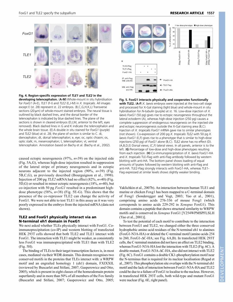
caused ectopic neurogenesis (97%, n39) on the injected side(Fig. 5A,G), whereas high-dose injection resulted in suppressionof the lateral stripe of primary neurogenesis and in ectopicneurons adjacent to the injected region (90%, n39) (Fig.5B,C,G), as previously described (Bourguignon et al., 1998).Injection of 200 pg TLE2 mRNA had no effect (82%, n40) (Fig.5D,G) or resulted in minor ectopic neurogenesis (18%, n40), butco-injection with 50 pg FoxG1 resulted in a predominant high-dose phenotype (58%, n38) (Fig. 5E-G). This shows that thepresence of the co-repressor TLE2 can change the activity ofFoxG1. We were not able to test TLE1 in this assay as it was verypoorly expressed in the embryo from the injected mRNA (data notshown).
TLE2 and FoxG1 physically interact via anN-terminal eh1 domain in FoxG1We next asked whether TLEs physically interact with FoxG1. Co-immunoprecipitation (co-IP) and western blotting of transfectedHEK 293T cells showed that both TLE2 and TLE1 interact withFoxG1. The interaction with TLE1 might be weaker, as consistentlyless FoxG1 was immunoprecipitated with TLE1 than with TLE2(Fig. 5H).
The binding of TLEs to their target transcription factors is, in mostcases, mediated via their WDR domain. This domain recognises twoconserved motifs in the proteins that TLEs interact with: a WRPWmotif and an engrailed homology 1 (eh1) domain, FXIXXIL(reviewed by Buscarlet and Stifani, 2007; Gasperowicz and Otto,2005), which is present in eight classes of the homeodomain proteinsuperfamily and in more than 50% of all members of the Fox family(Buscarlet and Stifani, 2007; Gasperowicz and Otto, 2005;
Yaklichkin et al., 2007b). An interaction between human TLE1 andmurine or chicken Foxg1 has been mapped to a C-terminal domainof Foxg1 (Sonderegger and Vogt, 2003; Yao et al., 2001),comprising amino acids 276-336 of mouse Foxg1 (whichcorresponds to amino acids 229-292 in Xenopus FoxG1). Thisdomain contains a peptide that shows structural similarity to WRPWmotifs and is conserved in Xenopus FoxG1 [Y254WPMSPFLSLH(Yao et al., 2001)].
To test the potential of each motif to contribute to the interactionbetween FoxG1 and TLE2, we changed either the three conservedhydrophobic amino acid residues of the N-terminal eh1 to alanines(FoxG1-N3A-HA) or deleted the C-terminal motif (amino acids 254to 260, FoxG1-C-HA; see Fig. 6A,B). In transfected HEK 293Tcells, the C-terminal mutation did not have an effect on TLE2 binding,whereas FoxG1-N3A-HA lost the interaction with TLE2 (Fig. 6C). Adouble mutant, FoxG1-N3A-C-HA, also did not interact with TLE2(Fig. 6C). FoxG1 contains a double CK1-phosphorylation motif nearthe N-terminus that is required for its nuclear localisation (Regad etal., 2007). This phosphorylation site overlaps with the N-terminal eh1motif and the lack of interaction between FoxG1-N3A-HA and TLE2could be due to a failure of FoxG1 to localise to the nucleus. However,in transfected HEK 293T cells, both wild-type and mutant FoxG1were nuclear (Fig. 6E, right panel).
1557RESEARCH ARTICLEFoxG1 and TLE2 specify the subpallium
Fig. 4. Region-specific expression of TLE1 and TLE2 in thedeveloping telencephalon. (A-M)Whole-mount in situ hybridisationfor FoxG1 (A-E), TLE1 (F-I) and TLE2 (E,J-M) in X. tropicalis. All imagesexcept E (st. 28) represent st. 22 embryos. (B,C,G,H,K,L) Transversesections (20m) of whole-mount stained embryos. The neural tissue isoutlined by black dashed lines, and the dorsal border of thetelencephalon is indicated by blue dashed lines. The plane of thesections is shown in cleared embryos (D,I,M, anterior to the left, eyesremoved). Black dashed lines in G and K indicate the telencephalon andthe whole brain tissue. (E)A double in situ stained for FoxG1 (purple)and TLE2 (blue) at st. 28; the plane of section is similar to C. di,diencephalon; dt, dorsal telencephalon; e, eye; oc, optic chiasm; os,optic stalk; m, mesencephalon; t, telencephalon; vt, ventraltelencephalon. Annotation based on Bachy et al. (Bachy et al., 2002).
Fig. 5. FoxG1 interacts physically and cooperates functionallywith TLE2. (A-F)X. laevis embryos were injected at the two-cell stageand processed for X-Gal staining (light blue) and whole-mount in situhybridisation for N-tubulin (purple) at st. 16. Low-dose injection of X.laevis FoxG1 (50 pg) gives rise to ectopic neurogenesis throughout thelateral ectoderm (A), whereas high-dose injection (250 pg) causes acomplete suppression of endogenous neurogenesis on the injected siteand ectopic neuronogenesis outside the X-Gal staining area (B,C).Injection of X. tropicalis FoxG1 mRNA gave rise to similar phenotypes(not shown). Co-expression of 200 pg X. tropicalis TLE2 with 50 pg X.laevis FoxG1 (E,F) gives rise to a phenotype that is similar to high-doseinjections (250 pg) of FoxG1 alone (B,C). TLE2 alone has no effect (D).(A,B,D,E) Dorsal views; (C,F) lateral views. In all panels, anterior is to theleft. (G)Percentage of low-dose and high-dose phenotypes resultingfrom each injection. (H)Co-immunoprecipitation of X. laevis FoxG1-HAand X. tropicalis TLE-Flag with anti-Flag antibody followed by westernblotting with anti-HA. The bottom panel shows loading of equalamounts of lysates followed by western blotting with anti-Flag andanti-HA. TLE2-Flag strongly interacts with FoxG1-HA, whereas TLE1-Flag expressed at similar levels shows slightly weaker binding.
DEVELO
PMENT

1558
A computational analysis of eh1 motifs within the Fox family oftranscription factors revealed that in addition to the fully conservedeh1 motif at the N-terminus (F18SINSLV), there is a ‘remnant’ eh1motif in the C-terminal region of FoxG1 that lacks the highlyconserved phenylalanine [L309SVDRLV (Yaklichkin et al.,2007b)]. Mutation of the four hydrophobic amino acid residues ofthe remnant C-terminal eh1 motif to alanines (FoxG1-C4A-HA) didnot affect TLE binding (Fig. 6C) and was not investigated further.In parallel, we tested the ability of all FoxG1 mutants to bind TLE1,and the results were very similar to those obtained with TLE2 (datanot shown).
Both the N-terminal and C-terminal domains areresponsible for functional cooperation with TLE2To address the functional activity of the FoxG1 mutants, we assessedthe potential of the mutants to convert a low-dose FoxG1 mRNAinjection into a high-dose phenotype. Injection of 400 pg TLE2mRNA alone had only a minor effect on neurogenesis, showing amild low-dose phenotype (30%, n61) (Fig. 6D). Injection of wild-type FoxG1 together with TLE2 showed a strong conversion to ahigh-dose phenotype (74%, n46); however, the FoxG1-N3A-HAand the double mutant FoxG1-N3A-C-HA produced a very low
percentage of the high-dose phenotype when co-injected with TLE2(8%, n50 and 11%, n45, respectively). Surprisingly, the deletionmutant FoxG1-C-HA was unable to efficiently convert a low-doseinjection into a high-dose phenotype in the presence of TLE2 (5%,n66) (Fig. 6D). All three FoxG1 mutants were able to induce a low-dose or high-dose phenotype when overexpressed alone, althoughthe double mutant showed a reduced low-dose phenotype, in thesense that the number of ectopic neurons observed was lower (datanot shown). This suggest that all mutants are expressed and arefunctionally active and, in addition, that the double mutant is moresevere that the single mutants. Taken together, these results suggestthat the N-terminal eh1 motif within FoxG1 plays a crucial role inthe physical and functional interaction with TLEs. The C-terminalmotif does not appear necessary for interaction with TLE2, but it isrequired for functional synergism, which could be indirect.
Knockdown of FoxG1 or TLE2 leads to reductionof the ventral telencephalonIf FoxG1 and TLE2 interact in vivo in the developing forebrain wewould expect common aspects in their respective knockdownphenotypes. FoxG1 is encoded by an intronless gene and wasknocked down by an ATG/upstream MO, whereas TLE2 was
RESEARCH ARTICLE Development 137 (9)
Fig. 6. Motifs in FoxG1 that are responsible for the binding and functional cooperativity of FoxG1 and TLE2. (A)Amino acid sequence ofX. laevis FoxG1 and potential TLE binding motifs. The DNA-binding domain is shown in blue, the N-terminal eh1 motif is highlighted in red, the C-terminal WRPW-like motif in green and the cryptic eh1 motif in purple. (B)Schematic representation of the FoxG1 mutants. The N-terminal domainis shown in dark grey, the DNA-binding domain in blue, the potential TLE1 interaction domain in orange and the HA epitope in red. Beneath isshown the sequence of mutated motifs (same colour coding as in A). (C)Immunoprecipitation (IP) of HEK 293T cells transfected with FoxG1-HA orFoxG1 mutants together with TLE2-Flag, showing that mutation of the N-terminal eh1 motif (FoxG1-N3A-HA), either alone or simultaneously withthe C-terminal motif (FoxG1-N3A-C-HA), leads to loss of binding to TLE2. The top two panels show IP with anti-Flag followed by western blottingwith anti-HA and anti-Flag. Lower panels show the input (pre-IP lysates) analysed by western blotting with anti-Flag and anti-HA. (D)Thephenotypes of embryos injected with the indicated amounts of X. tropicalis TLE2 and X. laevis FoxG1 or FoxG1 mutant mRNA were scored as low-dose or high-dose as in Fig. 5. TLE2 does not cooperate with any of the three mutants to convert a low-dose FoxG1 injection into a high-dosephenotype. (E)HEK 293T cells transfected with either FoxG1-HA or FoxG1-N3A-HA. Both proteins are nuclear.
DEVELO
PMENT

knocked down by injection of a splice MO targeted to the exon 1-intron 1 boundary. The knockdown of FoxG1 protein wasdetermined by immunohistochemistry with anti-FoxG1 antibody(Fig. 7A), and the effectiveness of the TLE2 splice MO was assessedby aberrant splicing as detected by PCR (Fig. 7B,C).
To assess possible similarities in the loss-of-function phenotypewe injected Control, FoxG1 or TLE2 MO into X. tropicalis embryosand performed whole-mount in situ hybridisation for the ventraltelencephalon marker Nkx2.1. Nkx2.1 is also expressed in thediencephalon, but within the telencephalon it is good marker for theventral-most part of the subpallium: the pallidum or medialganglionic eminence (Bachy et al., 2002). In FoxG1 MO-injectedembryos, the subpallial expression of Nkx2.1 was entirely missing(100%, n11), consistent with previous data from mouse and fish(Martynoga et al., 2005; Manuel et al., 2010; Danesin et al., 2009),and in TLE2 MO embryos it was greatly reduced (70%, n10) (Fig.7G).
To assess whether FoxG1 knockdown also affects neurogenesisin the Xenopus forebrain, we studied the effect of FoxG1 and TLE2MO knockdown on elrC expression, which is one of the earliestmarkers of neuronal differentiation, being expressed in cellsundergoing the transition from proliferation to differentiation(Carruthers et al., 2003; Good, 1995; Perron et al., 1999). Weobserved a reduction in the expression domain of elrC in the
forebrain of st. 19 embryos after either FoxG1 or TLE2 MOinjection (Fig. 7H-K). Loss of elrC expression could indicate afailure of early neural specification, increased cell death or a failureto exit the progenitor compartment. We did not observe a statisticallysignificant increase in cell death with either MO (Fig. 7L,M,N; seeFig. S3E in the supplementary material). Failure to exit theprogenitor compartment should result in an increase in proliferation.However, we found a significant reduction (by 40%) in pH3-positivecells in the FoxG1 MO-injected side as compared with the controlside in both st. 19 and st. 28 embryos (n12, P0.003) (Fig. 7O; seealso anti-pH3-stained green nuclei in Fig. 7A), suggesting that theloss of elrC is due to a loss of neural specification in the ventralforebrain, rather than to any prolonged maintenance of cells in theprogenitor state.
To address whether the loss of ventral telencephalic fate isaccompanied by an increase in dorsal markers, we assessed theexpression of emx1, a dorsal telencephalic (pallial) marker (Bachyet al., 2002). emx1 expression was expanded ventrally in the FoxG1MO embryos, but not in the TLE2 MO embryos (see Fig. S3 in thesupplementary material), suggesting that FoxG1 knockdown, butnot TLE2 knockdown, causes a widespread respecification.
Finally, to test for functional synergism of FoxG1 with TLE2 invivo, we tested the ability of wild-type and mutated FoxG1 to induceNkx2.1 expression in a FoxG1 MO background (Fig. 7P-T). We
1559RESEARCH ARTICLEFoxG1 and TLE2 specify the subpallium
Fig. 7. The effect of FoxG1 and TLE2 MO on forebraindevelopment. (A)Transverse sections of forebrain of FoxG1MO-injected (7.5 ng) embryos (st. 28) stained with DAPI(blue), anti-pH3 (green) and anti-FoxG1 (red). Note thereduction of FoxG1 staining and in pH3-positive cells on theMO-injected (left) side in the presumptive FoxG1 expressionregion. Dashed white line indicates the midline. (B)Schematicrepresentation of the TLE2 splice MO (red) and location ofPCR primers in relation to the exon-intron arrangement ofTLE2. (C)Semi-quantitative PCR from embryos injected with10 ng Control MO (lanes 1, 4), 10 ng TLE2 MO (lanes 2, 5) or20 ng TLE2 MO (lanes 3, 6). Primers used were ornithinedecarboxylase (ODC) as control (lanes 4-6) and TLE2 exon 1and TLE2 exon 3 (lanes 1-3) (see B). (D-G)Nkx2.1 expression inthe forebrain of st. 34 control embryos and embryos injectedat the one-cell stage with 10 ng Control MO (E), 15 ng FoxG1MO (F) and 15 ng TLE2 MO (G). The expression in the ventraltelencephalon (red arrowhead) is reduced or missing in TLE2and FoxG1 morphant embryos (F,G). di, diencephalon, dt,dorsal telencephalon, vt, ventral telencephalon. (H-N)Embryosinjected in one cell at the two-cell stage with FoxG1 MO (7.5ng) and TLE2 MO (7.5 ng) processed for in situ hybridisationfor elrC (H-K) and TUNEL staining (red nuclei; L-N). Injectedside is to the right, as marked by X-Gal staining (H-K) or FITC(L-N). (H,J)Frontal views; (I,K-N) transverse sections throughthe forebrain of st. 19 (H-K) or st. 32 (L-N) embryos. Reducedforebrain elrC expression on the MO-injected side is indicatedby red arrows. There was no statistically significant increase inTUNEL staining. (O)In FoxG1 MO-injected forebrains therewas a 40% decrease (P0.003, paired t-test, n12 embryos) inpH3-positive cells. (P-T)Transverse sections through theforebrain of FoxG1 MO-injected embryos, subsequentlyelectroporated with wild-type FoxG1 and various FoxG1mutated versions, as indicated, together with lacZ DNA andanalysed for Nkx2.1 expression. Light blue (X-Gal staining)indicates cells that have taken up the electroporated DNA (redarrowheads indicate examples of electroporated cells); darkpurple indicates cells expressing Nkx2.1. The experiment wasrepeated four times with similar results. See Materials andmethods for details. Scale bars: 100m.
DEVELO
PMENT

1560
found that wild-type FoxG1 and the N-terminal and C-terminalmutants (FoxG1-N3A and FoxG1-C) were able to induce Nkx2.1,suggesting that in the forebrain each motif is able to compensate forthe absence of the other. However, the double mutant, in which bothN-terminal and C-terminal motifs are affected (FoxG1-N3A-C),was unable to induce Nkx2.1. This finding supports the notion thatcooperation between the TLE2-binding eh1 motif and the C-terminal WRPW-like motif is necessary for ventral telencephalonspecification.
DISCUSSIONCloning and expression of X. tropicalis TLEsIn this study, we have cloned and characterised the expression offour X. tropicalis TLEs: TLE1, TLE2, TLE4 and AES. We haveshown that TLE1, TLE2 and AES are highly expressed in thenervous system; however, they did not show identical expressionpatterns. We focused on the expression of TLE1 and TLE2 in thedeveloping forebrain, specifically the telencephalon. Expression ofTLE2 has previously been described only by northern blotting(Choudhury et al., 1997).
We have shown that these genes are co-expressed at the neuralplate stage, but show a bias in the strength of expression towardsdifferent future subdomains of the forebrain. Later on, thesedifferences are amplified, as TLE1 is expressed throughout thetelencephalon whereas TLE2 is restricted to the ventral-most part,i.e. the subpallium and the optic chiasm, which forms the borderbetween telencephalon and diencephalon and where TLE1 is notexpressed. TLE1 and TLE2 are expressed in more-posterior regionsin distinct patterns, which we have not detailed here. The mousehomologue of TLE1 is expressed in the embryonic telencephalon(Dehni et al., 1995; Xuan et al., 1995; Yao et al., 1998). TLE2exhibits very limited expression in the mouse forebrain (Grbavec etal., 1998) and is also expressed in the developing mammalianpancreas and in chicken feather buds (Hoffman et al., 2008;Houghton et al., 2003).
Whether TLEs have distinct or overlapping (redundant) functionsis an open question and there are data to support either possibility(Gasperowicz and Otto, 2005; Yao et al., 1998). Our expressionanalysis of Xenopus TLE1 and TLE2 supports the view that thesetranscriptional co-factors have distinct functions; their neuralexpression profiles only partially overlap, providing the potential tointeract with different transcription factors.
The N-terminal eh1 motif is important for physicalassociation of TLEs with FoxG1Previous studies in chicken and mouse that analysed the interactionbetween TLE1 and Foxg1 used large deletions to identify potentialbinding sites (Sonderegger and Vogt, 2003; Yao et al., 2001). Bothgroups identified a polypeptide of ~65-70 amino acids C-terminalof the Foxg1 DNA-binding domain as crucial for the interactionwith TLE1 in in-vitro assays. Within the binding region, the shortpeptide motif YWPMSPFLSLH was reported to show similarity toWRPW motifs (Yao et al., 2001). Another study has shown that theanti-neurogenic activity of TLE1 is due to binding of TLE1 to Hes1and other WRPW motif-containing proteins, rather than to eh1motif-containing binding partners, such as FoxG1 (Buscarlet et al.,2008). Therefore, it was unclear whether the co-repressor TLE1, orthe eh1 motif of Foxg1, is important for the activity of Foxg1 in thedeveloping telencephalon.
However, in co-IP assays performed in cell culture betweenFoxG1 and TLE2, we could not detect any effect on TLE2 bindingto FoxG1 when the C-terminal motif is deleted. The mutation of
single aromatic amino acids (crucial components of both knownmotifs) in this region also had no effect on binding (data not shown).By contrast, we identified an N-terminal eh1 motif in FoxG1 as themain binding site for TLE2. Although the co-IP assay does not allowus to conclude with certainty that the interaction between FoxG1 andGroucho/TLE is direct, the eh1 motif shows the strongestconservation with known TLE binding sites (Yaklichkin et al.,2007a; Yaklichkin et al., 2007b).
How can the differences to previous reports be explained? Onepossibility is that FoxG1 interacts with TLE1 and TLE2 via differentdomains. However, this is not supported by our data, as we have notfound any differences between TLE1 and TLE2 in co-IP assays(except for a lower amount of immunoprecipitated FoxG1 withTLE1). Another possibility is that the differences are due to theassays used. For example, the work on TLE1 used in vitro assayswith bacterially derived proteins, which might lack importantmodifications necessary for binding to the eh1 motif. Indeed, aprevious study has shown that the phosphorylation state of S/Tresidues next to the initial phenylalanine in an eh1 motif has aninfluence on the interaction with Groucho (Goldstein et al., 2005)and we have previously described a double casein kinase Iphosphorylation site that overlaps with the N-terminal eh1 motif(Regad et al., 2007). TLEs can also be post-translationally modified,for example by hyperphosphorylation, which is also induced by thepresence of FoxG1 (Buscarlet et al., 2008; Nuthall et al., 2002;Nuthall et al., 2004), or by sumoylation (Ahn et al., 2009). Ouranalysis bears the advantage of having co-factors and proteinmodifications present and, under these conditions, which betterresemble those in vivo, we detect the N-terminal eh1 motif of FoxG1as the main binding motif for TLE2.
Functional cooperation of TLE2 with FoxG1 in vivoWe have used a Xenopus neurogenesis assay (Bourguignon et al.,1998) to assess the cooperation of FoxG1 with TLE2 in the embryo.Since the expression of FoxG1 in the posterior neural plate andlateral ectoderm is ectopic, this assay can only be used to showfunctional synergism, rather than to draw any conclusions on thespecific activity of FoxG1 in the developing forebrain. With thisassay, we have shown that TLE2 modulates the activity of FoxG1.When expressed together with TLE2, a low dose of FoxG1suppresses endogenous neurogenesis, which is otherwise onlyobserved when FoxG1 is overexpressed at a high dose. We interpretthis finding to mean that the presence of TLE2 allows FoxG1 tofunction more efficiently as a transcriptional repressor. This isanalogous to the observation that co-expression of TLE1 enhancesthe repressor activity of FoxG1 on a FoxG1-sensitive reporterconstruct (Yao et al., 2001).
Mutating the N-terminal eh1 motif (FoxG1-N3A-HA) leads toa loss of functional cooperation with TLE2, consistent with theloss of TLE2 binding by this mutant in co-IP experiments. TheFoxG1-C-HA mutant, which represents a deletion of the C-terminal YWPMSPF motif, also shows a lack of functionalcooperation with TLE2 in the neurogenesis assay. This isintriguing because deletion of this site does not abolish interactionwith TLE2 in co-IP assays. Therefore, we suggest that eventhough this site is not necessary for TLE2 binding, it is importantfor the functional cooperation of FoxG1 with TLE2. Oneexplanation is that a third factor exists, which binds FoxG1 at thismotif and has an influence on FoxG1/TLE activity. As the motifYWPMSPF also contains a MAP kinase consensus site (P-X-S/T-P), it could serve as a possible protein-protein interaction site thatis dependent on phosphorylation.
RESEARCH ARTICLE Development 137 (9)
DEVELO
PMENT

Our own work has also uncovered differences that depend on theassay used. In the neurogenesis assay, mutation of either the N-terminal or C-terminal motif is sufficient to interfere with thefunctional cooperation of exogenous FoxG1 and TLE2. However,within the forebrain, each motif seems to be able to compensate forthe absence of the other. In both assays, simultaneous interferencewith both motifs renders FoxG1 inactive. One possible explanationis that both mutants are compromised in recruiting co-repressors, butdifferent target genes have different threshold requirements fortranscriptional repression. The presence of different additional co-factors or FoxG1/TLE modifications in the different assays mightalso play a role. Although we do not yet fully understand the detailsof FoxG1-TLE interaction, the common denominator in all thefunctional assays used in this work is that the double mutant is moreseverely affected that any single mutant. These findings suggest thatboth the N-terminal eh1 motif and the C-terminal WRPW-like motifcontribute cooperatively to FoxG1 activity, either by interaction withTLE alone or in combination with other co-factors.
FoxG1 and TLE2 knockdown affect ventralforebrain specificationIf FoxG1 and TLE2 interact in vivo one would expect that thephenotype of knocking down FoxG1 would be similar to that ofknocking down TLE2. Indeed, both FoxG1 and TLE2 knockdownaffected Nkx2.1 expression in the ventral telencephalon. Theexpression of Nkx2.1 in this area was eliminated with the FoxG1MO and reduced with the TLE2 MO. This is consistent with theexpression profile of FoxG1 and TLE2 and, particularly, with theirco-expression in the ventral telencephalon (subpallium). The factthat FoxG1 knockdown has more severe effects than TLE2knockdown in the telencephalon can be explained by the observationthat FoxG1 is expressed throughout the entire telencephalon,whereas TLE2 is restricted to the ventral-most part.
The requirement for FoxG1 in the specification of the ventraltelencephalon is consistent with previous findings in mouse andzebrafish (Danesin et al., 2009; Martynoga et al., 2005). Here, wesuggest that this event is mediated by the specific interaction ofFoxG1 with the transcriptional co-repressor TLE2, which mightprovide a mechanism for the cell-autonomous role of FoxG1 in thisregion (Manuel et al., 2010). Although we have focused on TLE2,we cannot exclude a contribution by TLE1 and finer assays will beneeded to distinguish the contribution of each factor.
One of the hallmarks of the FoxG1 knockout and knockdownphenotype in mice and zebrafish is premature neuronaldifferentiation, particularly in the dorsal forebrain (pallium) (Danesinet al., 2009; Xuan et al., 1995), which transiently results in a greaternumber of neurons. In Xenopus, we have not seen this increase butinstead observed a reduction in newly postmitotic neurons in thetelencephalon of st. 19 tadpoles, marked by the expression of elrC.This is unlikely to reflect a delay in neurogenesis accompanied byincreased proliferation, as we detected this reduction over a longperiod of time (st. 19 to st. 25) and we observed a reduction in mitoticcells. Therefore, we suggest that the reduction of elrC expressionsignifies a failure in the specification of telencephalic tissue thatmight precede the specification of neurogenesis. Indeed, this wouldbe consistent with our earlier observation that FoxG1 is sufficient toinduce the neuroepithelial marker Sox3 in isolated naïve ectoderm(animal caps) (Hardcastle and Papalopulu, 2000). The differencebetween the Xenopus phenotype and that seen in mouse andzebrafish, in which an increased number of neurons is transientlydetected, could be due to phylogenetic differences in the developmentof the forebrain. Alternatively, partial compensation by other genes,
as originally suggested for the mouse (Xuan et al., 1995), might resultin different phenotypes. In this respect, it might be relevant that inzebrafish there are three Foxg1 genes: foxg1a is the main Foxg1paralogue expressed in the telencephalon, but foxg1b, which wouldnot be targeted by the foxg1a MO, is transiently expressed in theventral telencephalon (Zhao et al., 2009). At present, we cannotexclude the possibility that the reduction in elrC is due to prematureneurogenesis that depletes the overall number of progenitors andhence reduces the overall number of neurons generated.
In conclusion, based on the specific overlap of expressionbetween FoxG1 and TLE2 in the ventral telencephalon, theirphysical and functional interaction as shown by co-IP and aneurogenesis assay, the induction of Nkx2.1 in the forebrain and thesimilar phenotypes resulting from MO knockdown, we suggest thatFoxG1 and TLE2 synergise to specify the ventral telencephalon. Itremains to be seen whether other regionally expressed co-factors,such as JARID1B (PLU-1; KDM5B) (Tan et al., 2003), mediatesome of the other activities of FoxG1.
AcknowledgementsWe thank Nitin Sabherwal for help with the manuscript. This work was fundedby a Wellcome Trust Senior Fellowship grant to N. Papalopulu (057819/Z/05).N. Panagiotaki and B.B. are Wellcome Trust four-year PhD students. Depositedin PMC for release after 6 months.
Competing interests statementThe authors declare no competing financial interests.
Supplementary materialSupplementary material for this article is available athttp://dev.biologists.org/lookup/suppl/doi:10.1242/dev.044909/-/DC1
ReferencesAhlgren, S., Vogt, P. and Bronner-Fraser, M. (2003). Excess FoxG1 causes
overgrowth of the neural tube. J. Neurobiol. 57, 337-349.Ahn, J. W., Lee, Y. A., Ahn, J. H. and Choi, C. Y. (2009). Covalent conjugation of
Groucho with SUMO-1 modulates its corepressor activity. Biochem. Biophys. Res.Commun. 379, 160-165.
Bachy, I., Berthon, J. and Retaux, S. (2002). Defining pallial and subpallialdivisions in the developing Xenopus forebrain. Mech. Dev. 117, 163-172.
Bajoghli, B. (2007). Evolution of the Groucho/Tle gene family: gene organizationand duplication events. Dev. Genes Evol. 217, 613-618.
Bourguignon, C., Li, J. and Papalopulu, N. (1998). XBF-1, a winged helixtranscription factor with dual activity, has a role in positioning neurogenesis inXenopus competent ectoderm. Development 125, 4889-4900.
Buscarlet, M. and Stifani, S. (2007). The ‘Marx’ of Groucho on development anddisease. Trends Cell Biol. 17, 353-361.
Buscarlet, M., Perin, A., Laing, A., Brickman, J. M. and Stifani, S. (2008).Inhibition of cortical neuron differentiation by Groucho/TLE1 requires interactionwith WRPW, but not Eh1, repressor peptides. J. Biol. Chem. 283, 24881-24888.
Carruthers, S., Mason, J. and Papalopulu, N. (2003). Depletion of the cell-cycleinhibitor p27(Xic1) impairs neuronal differentiation and increases the number ofElrC(+) progenitor cells in Xenopus tropicalis. Mech. Dev. 120, 607-616.
Chen, G. and Courey, A. J. (2000). Groucho/TLE family proteins andtranscriptional repression. Gene 249, 1-16.
Chen, G., Fernandez, J., Mische, S. and Courey, A. J. (1999). A functionalinteraction between the histone deacetylase Rpd3 and the corepressor grouchoin Drosophila development. Genes Dev. 13, 2218-2230.
Choudhury, B. K., Kim, J., Kung, H. F. and Li, S. S. (1997). Cloning anddevelopmental expression of Xenopus cDNAs encoding the Enhancer of splitgroucho and related proteins. Gene 195, 41-48.
Danesin, C., Peres, J. N., Johansson, M., Snowden, V., Cording, A.,Papalopulu, N. and Houart, C. (2009). Integration of telencephalic Wnt andhedgehog signaling center activities by Foxg1. Dev. Cell 16, 576-587.
Dehni, G., Liu, Y., Husain, J. and Stifani, S. (1995). TLE expression correlateswith mouse embryonic segmentation, neurogenesis, and epithelialdetermination. Mech. Dev. 53, 369-381.
Dou, C., Lee, J., Liu, B., Liu, F., Massague, J., Xuan, S. and Lai, E. (2000). BF-1interferes with transforming growth factor beta signaling by associating withSmad partners. Mol. Cell. Biol. 20, 6201-6211.
Eagleson, G. W. and Harris, W. A. (1990). Mapping of the presumptive brainregions in the neural plate of Xenopus laevis. J. Neurobiol. 21, 427-440.
Gasperowicz, M. and Otto, F. (2005). Mammalian Groucho homologs:redundancy or specificity? J. Cell. Biochem. 95, 670-687.
1561RESEARCH ARTICLEFoxG1 and TLE2 specify the subpallium
DEVELO
PMENT

1562
Goldstein, R. E., Cook, O., Dinur, T., Pisante, A., Karandikar, U. C., Bidwai, A.and Paroush, Z. (2005). An eh1-like motif in odd-skipped mediates recruitmentof Groucho and repression in vivo. Mol. Cell. Biol. 25, 10711-10720.
Good, P. J. (1995). A conserved family of elav-like genes in vertebrates. Proc. Natl.Acad. Sci. USA 92, 4557-4561.
Grbavec, D., Lo, R., Liu, Y. and Stifani, S. (1998). Transducin-like Enhancer ofsplit 2, a mammalian homologue of Drosophila Groucho, acts as atranscriptional repressor, interacts with Hairy/Enhancer of split proteins, and isexpressed during neuronal development. Eur. J. Biochem. 258, 339-349.
Hanashima, C., Shen, L., Li, S. C. and Lai, E. (2002). Brain factor-1 controls theproliferation and differentiation of neocortical progenitor cells throughindependent mechanisms. J. Neurosci. 22, 6526-6536.
Hanashima, C., Li, S. C., Shen, L., Lai, E. and Fishell, G. (2004). Foxg1suppresses early cortical cell fate. Science 303, 56-59.
Hardcastle, Z. and Papalopulu, N. (2000). Distinct effects of XBF-1 in regulatingthe cell cycle inhibitor p27(XIC1) and imparting a neural fate. Development 127,1303-1314.
Hasson, P. and Paroush, Z. (2006). Crosstalk between the EGFR and othersignalling pathways at the level of the global transcriptional corepressorGroucho/TLE. Br. J. Cancer 94, 771-775.
Hebert, J. M. and Fishell, G. (2008). The genetics of early telencephalonpatterning: some assembly required. Nat. Rev. Neurosci. 9, 678-685.
Hoffman, B. G., Zavaglia, B., Beach, M. and Helgason, C. D. (2008). Expressionof Groucho/TLE proteins during pancreas development. BMC Dev. Biol. 8, 81.
Houghton, L., Freeman, A. and Morgan, B. A. (2003). Expression andregulation of Groucho-related genes in the embryonic chicken feather bud. Dev.Dyn. 226, 587-595.
Jennings, B. H. and Ish-Horowicz. (2008). The Groucho/TLE/Grg family oftranscriptional co-repressors. Genome Biol. 9, 205.1-205.7.
Kaestner, K. H., Knochel, W. and Martinez, D. E. (2000). Unified nomenclaturefor the winged helix/forkhead transcription factors. Genes Dev. 14, 142-146.
Kobayashi, M., Nishikawa, K., Suzuki, T. and Yamamoto, M. (2001). Thehomeobox protein Six3 interacts with the Groucho corepressor and acts as atranscriptional repressor in eye and forebrain formation. Dev. Biol. 232, 315-326.
Larkin, M. A., Blackshields, G., Brown, N. P., Chenna, R., McGettigan, P. A.,McWilliam, H., Valentin, F., Wallace, I. M., Wilm, A., Lopez, R. et al. (2007).Clustal W and Clustal X version 2.0. Bioinformatics 23, 2947-2948.
Leon, C. and Lobe, C. G. (1997). Grg3, a murine Groucho-related gene, isexpressed in the developing nervous system and in mesenchyme-inducedepithelial structures. Dev. Dyn. 208, 11-24.
Li, J., Chang, H. W., Lai, E., Parker, E. J. and Vogt, P. K. (1995). The oncogeneqin codes for a transcriptional repressor. Cancer Res. 55, 5540-5544.
Manuel, M., Martynoga, B., Yu, T., West, J. D., Mason, J. O. and Price, D. J.(2010). The transcription factor Foxg1 regulates the competence oftelencephalic cells to adopt subpallial fates in mice. Development 137, 487-497.
Marcal, N., Patel, H., Dong, Z., Belanger-Jasmin, S., Hoffman, B., Helgason,C. D., Dang, J. and Stifani, S. (2005). Antagonistic effects of Grg6 andGroucho/TLE on the transcription repression activity of brain factor 1/FoxG1 andcortical neuron differentiation. Mol. Cell. Biol. 25, 10916-10929.
Martynoga, B., Morrison, H., Price, D. J. and Mason, J. O. (2005). Foxg1 isrequired for specification of ventral telencephalon and region-specific regulation ofdorsal telencephalic precursor proliferation and apoptosis. Dev. Biol. 283, 113-127.
Molenaar, M., Brian, E., Roose, J., Clevers, H. and Destree, O. (2000).Differential expression of the Groucho-related genes 4 and 5 during earlydevelopment of Xenopus laevis. Mech. Dev. 91, 311-315.
Murphy, D. B., Wiese, S., Burfeind, P., Schmundt, D., Mattei, M. G., Schulz-Schaeffer, W. and Thies, U. (1994). Human brain factor 1, a new member ofthe fork head gene family. Genomics 21, 551-557.
Nuthall, H. N., Husain, J., McLarren, K. W. and Stifani, S. (2002). Role forHes1-induced phosphorylation in Groucho-mediated transcriptional repression.Mol. Cell. Biol. 22, 389-399.
Nuthall, H. N., Joachim, K. and Stifani, S. (2004). Phosphorylation of serine 239of Groucho/TLE1 by protein kinase CK2 is important for inhibition of neuronaldifferentiation. Mol. Cell. Biol. 24, 8395-8407.
Perron, M., Furrer, M. P., Wegnez, M. and Theodore, L. (1999). Xenopus elav-like genes are differentially expressed during neurogenesis. Mech. Dev. 84, 139-142.
Regad, T., Roth, M., Bredenkamp, N., Illing, N. and Papalopulu, N. (2007).The neural progenitor-specifying activity of FoxG1 is antagonistically regulatedby CKI and FGF. Nat. Cell Biol. 9, 531-540.
Roose, J., Molenaar, M., Peterson, J., Hurenkamp, J., Brantjes, H., Moerer, P.,van de Wetering, M., Destree, O. and Clevers, H. (1998). The Xenopus Wnteffector XTcf-3 interacts with Groucho-related transcriptional repressors. Nature395, 608-612.
Seoane, J., Le H. V., Shen, L., Anderson, S. A. and Massague, J. (2004).Integration of Smad and forkhead pathways in the control of neuroepithelialand glioblastoma cell proliferation. Cell 117, 211-223.
Shen, Q., Wang, Y., Dimos, J. T., Fasano, C. A., Phoenix, T. N., Lemischka, I.R., Ivanova, N. B., Stifani, S., Morrisey, E. E. and Temple, S. (2006). Thetiming of cortical neurogenesis is encoded within lineages of individualprogenitor cells. Nat. Neurosci. 9, 743-751.
Sonderegger, C. K. and Vogt, P. K. (2003). Binding of the corepressor TLE1 toQin enhances Qin-mediated transformation of chicken embryo fibroblasts.Oncogene 22, 1749-1757.
Tan, K., Shaw, A. L., Madsen, B., Jensen, K., Taylor-Papadimitriou, J. andFreemont, P. S. (2003). Human PLU-1 has transcriptional repression propertiesand interacts with the developmental transcription factors BF-1 and PAX9. J.Biol. Chem. 278, 20507-20513.
Tao, W. and Lai, E. (1992). Telencephalon-restricted expression of BF-1, a newmember of the HNF-3/fork head gene family, in the developing rat brain.Neuron 8, 957-966.
Xuan, S., Baptista, C. A., Balas, G., Tao, W., Soares, V. C. and Lai, E. (1995).Winged helix transcription factor BF-1 is essential for the development of thecerebral hemispheres. Neuron 14, 1141-1152.
Yaklichkin, S., Steiner, A. B., Lu, Q. and Kessler, D. S. (2007a). FoxD3 and Grg4physically interact to repress transcription and induce mesoderm in Xenopus. J.Biol. Chem. 282, 2548-2557.
Yaklichkin, S., Vekker, A., Stayrook, S., Lewis, M. and Kessler, D. S. (2007b).Prevalence of the EH1 Groucho interaction motif in the metazoan Fox family oftranscriptional regulators. BMC Genomics 8, 201.
Yamane, K., Tateishi, K., Klose, R. J., Fang, J., Fabrizio, L. A., Erdjument-Bromage, H., Taylor-Papadimitriou, J., Tempst, P. and Zhang, Y. (2007).PLU-1 is an H3K4 demethylase involved in transcriptional repression and breastcancer cell proliferation. Mol. Cell 25, 801-812.
Yao, J., Liu, Y., Husain, J., Lo, R., Palaparti, A., Henderson, J. and Stifani, S.(1998). Combinatorial expression patterns of individual TLE proteins during celldetermination and differentiation suggest non-redundant functions formammalian homologs of Drosophila Groucho. Dev. Growth Differ. 40, 133-146.
Yao, J., Liu, Y., Lo, R., Tretjakoff, I., Peterson, A. and Stifani, S. (2000).Disrupted development of the cerebral hemispheres in transgenic miceexpressing the mammalian Groucho homologue transducin-like-enhancer ofsplit 1 in postmitotic neurons. Mech. Dev. 93, 105-115.
Yao, J., Lai, E. and Stifani, S. (2001). The winged-helix protein brain factor 1interacts with groucho and hes proteins to repress transcription. Mol. Cell. Biol.21, 1962-1972.
Zamparini, A. L., Watts, T., Gardner, C. E., Tomlinson, S. R., Johnston, G. I.and Brickman, J. M. (2006). Hex acts with beta-catenin to regulateanteroposterior patterning via a Groucho-related co-repressor and Nodal.Development 133, 3709-3722.
Zhao, X. F., Suh, C. S., Prat, C. R., Ellingsen, S. and Fjose, A. (2009). Distinctexpression of two foxg1 paralogues in zebrafish. Gene Expr. Patterns 9, 266-272.
RESEARCH ARTICLE Development 137 (9)
DEVELO
PMENT