Foxe et al (in press)
48
Foxe, McCourt & Javitt “Parietal Control of Spatial Attention” 1 Right hemisphere control of visuospatial attention: 'Line-bisection' judgments evaluated with high-density electrical mapping and source analysis. John J. Foxe a,b,c,* , Mark E. McCourt d , Daniel C. Javitt a,e a The Cognitive Neurophysiology Laboratory, Nathan S. Kline Institute for Psychiatric Research, Program in Cognitive Neuroscience and Schizophrenia, 140 Old Orangeburg Road, Orangeburg, New York 10962, USA b Department of Neuroscience c Department of Psychiatry and Behavioural Science, Albert Einstein College of Medicine, 1300 Morris Park Avenue, Bronx, New York 10461, USA d Department of Psychology, North Dakota State University Fargo, North Dakota 58105, USA e Department of Psychiatry, New York University School of Medicine, 550 1 st Avenue, New York, New York 10016, USA NeuroImage (in press). Please do not cite without prior permission. *Correspondence can be addressed to: Dr. John J. Foxe The Cognitive Neurophysiology Laboratory, Nathan S. Kline Institute for Psychiatric Research, Program in Cognitive Neuroscience and Schizophrenia, 140 Old Orangeburg Road, Orangeburg, NY 10962, USA Vox: (845) 398-6547; Fax: (845) 398-6545 e-mail: [email protected]
Transcript of Foxe et al (in press)

Foxe, McCourt & Javitt “Parietal Control of Spatial Attention” 1
Right hemisphere control of visuospatial attention: 'Line-bisection' judgments evaluated with
high-density electrical mapping and source analysis.
John J. Foxe a,b,c,* , Mark E. McCourt d, Daniel C. Javitt a,e
a The Cognitive Neurophysiology Laboratory,Nathan S. Kline Institute for Psychiatric Research,
Program in Cognitive Neuroscience and Schizophrenia,140 Old Orangeburg Road,
Orangeburg, New York 10962, USA
b Department of Neurosciencec Department of Psychiatry and Behavioural Science,
Albert Einstein College of Medicine,1300 Morris Park Avenue,
Bronx, New York 10461, USA
d Department of Psychology,North Dakota State University
Fargo, North Dakota 58105, USA
e Department of Psychiatry,New York University School of Medicine,
550 1st Avenue,New York, New York 10016, USA
NeuroImage (in press). Please do not cite without prior permission.
*Correspondence can be addressed to:
Dr. John J. FoxeThe Cognitive Neurophysiology Laboratory,Nathan S. Kline Institute for Psychiatric Research, Program in Cognitive Neuroscience and Schizophrenia, 140 Old Orangeburg Road, Orangeburg, NY 10962, USAVox: (845) 398-6547; Fax: (845) 398-6545e-mail: [email protected]

Foxe, McCourt & Javitt “Parietal Control of Spatial Attention” 2
ABSTRACT
The ‘line-bisection’ task has proven an especially useful clinical tool for assessment of spatial
neglect syndrome in neurological patients. Here, we investigated the neural processes involved
in performing this task by recording high-density event-related potentials (ERP) from 128 scalp-
electrodes in normal observers. We characterize a robust net negative potential from 170-400 ms
post stimulus presentation that correlates with line-bisection judgments. Topographic mapping
shows three distinct phases to this negativity. The first phase (~170-190 ms) has a scalp
distribution exclusively over the right parieto-occipital and lateral-occipital scalp, consistent with
generators in the region of the right temporo-parietal junction and right lateral occipital cortices.
The second phase (~190-240 ms) sees the emergence of a second negative focus over the right
central-parietal scalp, consistent with subsequent involvement of right superior parietal cortices.
In the third phase (~240-400 ms), the topography becomes dominated by this right central-
parietal negativity. Inverse source-modeling confirmed that right hemisphere lateral occipital,
inferior parietal and superior parietal regions were the likeliest generators of the bulk of the
activity associated with this effect. The line stimuli were also presented at three contrast levels
(3%, 25% and 100%) in order to manipulate both the latency of stimulus processing and the
relative contributions from magnocellular and parvocellular inputs. Through this manipulation,
we show that the line-bisection effect systematically tracks/follows the latency of the N1
component, which is considered a temporal marker for object processing in the ventral visual
stream. This pattern of effects suggests that this task invokes an allocentric (object-based) form
of visuo-spatial attention. Further, at 3% contrast, the line-bisection effect was equivalent to the
effects seen at higher contrast levels, suggesting that parvocellular inputs are not necessary for
successful performance of this task.
Foxe, McCourt & Javitt “Parietal Control of Spatial Attention” 3
INTRODUCTION
Line-bisection tasks are a commonly used metric for the clinical assessment of visuo-spatial
neglect syndrome, a condition that predominantly results from vascular lesions to the right
inferior parietal or temporo-parietal cortex (e.g. Vallar and Perani, 1987; Cappa et al., 1991;
Mesulam, 2000; Na et al., 2000; Kerkhoff, 2001; see Karnath, 2001 for a comprehensive
treatment). Patients with visuospatial neglect syndrome generally bisect lines significantly to the
right of veridical center (Robertson and Halligan, 1999), due either to a decrement in the ability
to allocate attention to left visual space, or to hyper-attention to rightward space. Clinical
findings from neglect patients allied to the scores of neuroimaging studies showing a
predomination of right parietal activity during tasks requiring visuo-spatial attention (e.g.
Corbetta et al., 2000; Coull et al., 2001) have led to the formulation that while both left and right
parietal areas are involved in attention to the right visual field, right parietal areas alone may
control attention to the left visual hemifield (e.g. Heilman and van den Abell, 1980; Weintraub
and Mesulam, 1987). A measure of support for this conjecture is derived from the finding that
neurologically normal subjects demonstrate a phenomenon known as ‘pseudoneglect’ (Bowers &
Heilman, 1980), in which a significant and systematic misbisection of lines (or space) occurs that
is leftward of veridical center. This leftward tendency in normal observers has been theorized to
result from a profound parietal asymmetry in attentional control, which gives rise to some degree
of hyper-attentiveness to the left visual field (in space-based attention), or to the left-hand side of
individual objects (in object-based attention) (see Jewell and McCourt, 2000; McCourt, 2001a;
McCourt & Jewell, 1999; McCourt et al., 2000).
Functional imaging (fMRI) studies have confirmed a central role for right parietal cortices in
performance of both line-bisection tasks (Weiss et al., 2000; Fink et al., 2000a, 2000b, 2001,
Foxe, McCourt & Javitt “Parietal Control of Spatial Attention” 4
2002; Galati et al., 2000) as well as judgments of object location relative to the midsagittal plane
(Vallar et al., 1999; Galati et al., 2000). There have, to date, been no electrophysiological
investigations of line-bisection; consequently the timing of right parietal involvement relative to
the timing of ongoing stimulus processing is as yet unknown. In the current study, we performed
high-density electrical mapping (from 128 scalp electrodes) of the visual event-related potential
(ERP) while subjects engaged in either a tachistoscopic forced-choice line-bisection task
(McCourt & Olafson, 1997) or in a control task in which they simply judged whether lines were
transected or not. Our main objectives were to both confirm the involvement of and define the
timecourse of right parietal activity in performance of the line-bisection task. In particular, we
wished to assess the relationship of parietal attentional processes in the dorsal visual processing
stream to the processing of the line stimuli by object recognition areas of the ventral visual
stream. An open question is whether line midpoint judgments can only be made upon
completion of object recognition processes for the object that is to be bisected. In the present
study, we systematically varied the timing of object-recognition processes by varying the
contrast level at which lines were displayed. We used the well-characterized N1 component of
the VEP as an index of the timing of object recognition processes, since this component has
repeatedly been shown to be correlated with the processes involved in object recognition (e.g.
Allison et al., 1999; Bentin et al., 1999; Doniger et al., 2000, 2001). Systematically varying the
latency of this component allowed us to assess whether bisection processes tracked this latency
manipulation.
Foxe, McCourt & Javitt “Parietal Control of Spatial Attention” 5
MATERIALS AND METHODS
Subjects
Nine (4 male) neurologically normal, paid volunteers (ages 19-45 years, mean = 29.2 years)
participated. All subjects provided written informed consent, and the Institutional Review Boards
of the Nathan Kline Research Institute and North Dakota State University approved all
procedures. All subjects possessed normal or corrected-to-normal vision and were right-handed,
as measured using the Oldfield (1971) laterality inventory (mean score = 58.3, sd=22.7).
Instrumentation and Stimuli
Subject responses were sensed and collected, and stimuli were presented as 640x480 pixel
images on a 21" flat-screen monitor; frame refresh rate was 60 Hz and mean display luminance
was 22 cd/m2. The generation and sequencing of stimuli and the collection of subject responses
were accomplished using the ERTS software package (Beringer, 1995). Luminance and contrast
calibrations were made using a photometer.
Stimuli were line segments (29 cm wide by 0.5 cm high) presented for 150 ms. At the
viewing distance of 108 cm, lines subtended 15.3o (width) by 0.27o (height) of visual angle. All
lines were horizontally and vertically centered within the display. Seventy-five percent of the
lines were transected at one of 25 different locations ranging from ±0.6o of visual angle relative
to veridical line midpoint. This range of transector locations is sufficient to determine perceived
line midpoint in most observers (McCourt, 2001a). The remaining 25% of lines were non-
transected. Lines were presented at three levels of Michelson contrast: 3%, 25%, and 100%. This
range of stimulus contrast was employed for both methodological and theoretical reasons.
Methodologically, the presence of low contrast lines ensured that the visual discriminations in
the feature detection task (transected versus non transected) would not be trivially easy, and that

Foxe, McCourt & Javitt “Parietal Control of Spatial Attention” 6
subjects would remain alert and vigilant throughout the blocks of trials. Theoretically, it was of
interest to determine whether there would be any unique behavioral or electrophysiological
signature regarding the lowest contrast stimulus that, at 3% contrast, is well below the operating
range of the parvocellular division of the geniculostriate projection, and thus preferentially
engages the magnocellular stream (Shapley and Perry, 1986).
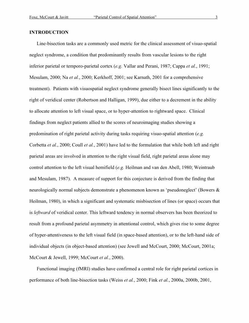
Figure 1 illustrates examples of the line stimuli. Both members of the upper pair of lines (A,
B) possess 100% contrast, and are transected to the left of center, by −0.33o (−2.17%) and −0.60o
(−3.91%), respectively. Both members of the lower pair of lines (E, F) possess 3% contrast and
are transected to the right of center, by +0.47o (3.04%) and +0.17o (1.09%), respectively. Lines C
and D possess 25% contrast; C is veridically transected and line D lacks a transector. Pairs (A,
B) and (E, F) differ in contrast polarity. Lines of varying contrast and contrast polarity appeared
with equal frequency. The order of appearance of all line types was quasi-randomized within
blocks of trials.
Insert Figure 1 Here
Procedures and Tasks
Subjects were seated in a comfortable chair with their midsagittal plane aligned with the
display monitor. Stimuli were viewed binocularly through natural pupils.
Each block of trials consisted of 200 line presentations. On alternate blocks subjects
performed either one of two tasks. In the Control Task subjects performed an oddball (feature
discrimination) task in which they judged whether lines were transected (75% of trials) or not
(25% of trials). The non-bisected lines thus served as "target" stimuli. Use of the oddball task as
the control condition ensured that subjects were actively engaged in an attentionally demanding

Foxe, McCourt & Javitt “Parietal Control of Spatial Attention” 7
task in both conditions and that equivalent button push responses were made during both tasks. If
a simple passive viewing task were to be used, changes in general arousal level might have
accounted for any effects seen.
In the Line-bisection task subjects made judgments regarding transector location (left versus
right) relative to perceived line midpoint by depressing the appropriate mouse button "left" or
"right", respectively. Subjects were instructed to respond to non-transected lines by depressing
either mouse button.
Subjects used their dominant (right) hand to depress mouse buttons. Subjects were instructed
to delay their responses for at least 1 second following stimulus presentation to obviate motor
artifacts in the EEG signal. Inter-trial intervals were approximately 2 sec., since subsequent trials
were initiated 750 ms following the previous response. Each subject completed 3 or 4 blocks of
both Line-bisection and Control Task trials. At each level of line contrast subjects made either
six or eight "left-right" judgments at each transector location, such that estimates of perceived
line midpoint in each line contrast condition were determined based on 150-200 bisection trials.
Each block of trials was completed in approximately 9 minutes.
Measurements and Analyses
Behavioral Measures
For behavioral analysis the dependent measure was the proportion of trials on which subjects
indicated that transectors appeared to the "left" of perceived line midpoint. The method of
constant stimuli was used to derive psychometric functions, and nonlinear least-squares
regression was used to fit a cumulative Gaussian distribution to the psychometric functions (see
McCourt & Jewell, 1999). Based on these fits, transector locations corresponding to a 50% "left"

Foxe, McCourt & Javitt “Parietal Control of Spatial Attention” 8
response rate were determined. The transector location for which "left" and "right" responses
occur with equal frequency is called the "point of subjective equality" (pse) and is an objective
measure of perceived line midpoint. Inferential statistical analyses were performed on the pse
values.
Electrophysiological Measures
Continuous EEG was acquired from 128 scalp electrodes (impedances < 5kΩ), referenced to
nose, band-pass filtered from 0.05 to 100Hz, and digitized at 500Hz. Data were epoched off-line
from −100 pre-stimulus to 500 ms post-stimulus and baseline-corrected from −100 to 0 ms.
Thereafter, trials with blinks and eye movements were rejected on the basis of horizontal and
vertical electro-oculogram. An artifact rejection criterion of ±60µV was used at all other scalp
sites to reject trials with excessive EMG or other noise transients. The average number of
accepted trials per condition across subjects was 476 (S.D. = 84.2). Responses to the non-
transected lines, which served as "target" stimuli in the control task, were not included in the
averages nor analyzed further. This latter point is important, as during the control task the N2/P3
component complex associated with infrequent target stimuli was generated for these stimuli (see
e.g. Ritter and Vaughan, 1969; He et al., 2001). Thus, use of these responses as constituents of
the "control" averaged response would have been problematic. Also, false alarms (which were
very few) were also excluded from this analysis.
The latencies and scalp topographies of the standard ERP componentry over posterior scalp
were identified from group-averaged waveforms collapsed across the Control and Line-bisection
conditions. Repeated measures analyses of variance (ANOVA) was used to test for significant
differences between conditions for four pre-selected time-windows, centered at the peak

Foxe, McCourt & Javitt “Parietal Control of Spatial Attention” 9
amplitude of the P1 (92-100 ms), the N1 (174-182 ms), the P2 (230-240 ms) and the following
negative deflection over occipito-temporal regions, N280ot, (270-280 ms). For these time-
windows, a measure of integrated amplitude was derived between the response and the 0 µV
baseline. ANOVAs possessed a repeated-measures 2 X 2 X 4 design with the following factors:
condition (control, line-bisection), hemisphere (left, right) and electrode (4 electrode pairs over
lateral occipito-parietal scalp).
Topographic Mapping
Scalp topographic maps in the present study represent interpolated potential distributions,
derived from the 128-scalp measurements and based on the computation of a common average
reference. These interpolated potential maps are displayed on the 3-D reconstruction of an
average rendered scalp surface (derived from anatomical MRIs), using the boundary element
method (BEM; e.g. Fuchs et al., 1998) and as implemented in the CURRY multimodal
neuroimaging analysis software package (Version 4.0 − Philips Research, Hamburg, Germany).
Exact electrode locations were assessed for each subject on the day of testing by 3D-
digitization of the locations of the scalp electrodes with respect to fiduciary landmarks (i.e. the
nasion and pre-auricular notches) using a magnetic digitization device (Polhemus Fastrak and
3DspaceDX software, Neuroscan, Inc.). Electrode placement was highly consistent across
subjects due to the use of a custom-designed electrode cap that constrained inter-electrode
spacing and placement. An averaged version of these electrode locations was projected onto the
averaged rendered head for computation of the group topographic data.
Lastly, one obvious constraint of the printed page is that only a limited number of
discrete maps can be shown to represent a given topographic distribution, and such static maps

Foxe, McCourt & Javitt “Parietal Control of Spatial Attention” 10
fail to depict the full spatio-temporal dimensionality of the data. This can make it particularly
difficult for the reader to assess the extent of contribution to the maps of background noise. In
determining the display-gain to be used for the maps in the current study, we followed the
topography over its entire timecourse (through observing animated time-series). Observation of
these maps in the baseline period (from -100 ms up to the onset of significant activity at about 50
ms) allowed us to determine the relative contribution of noise to the topographic maps. The gain
was then set so that background noise during this baseline period accounted for less than 1-2
topographic lines of potential in the maps.
This is more readily seen in the animation appendix to this paper, which can be viewed or
downloaded at (http//academicpress.com/ni/linebisection.avi ???)
Dipole source analyses
Information about the intracranial generators contributing to effects seen in the data was
also obtained through dipole source analysis using electro-magnetic source estimation (EMSE)
as implemented in CURRY version 4.0 software. This method assumes that there are a limited
and distinct number of active brain regions over the evoked potential epoch, each of which can
be approximated by an equivalent dipole. Dipole generators are placed within a three-shell
spherical volume conductor model and overlaid on and adjusted to a BEM-segmented structural
MRI (in this case, an averaged brain). The forward solution to this dipole configuration is tested
against the observed experimental data. When not fixed, the positions and orientations of the
dipoles are iteratively adjusted to minimize the residual variance between the forward solution
and the observed data. The upper bound of the number of modeled dipole sources is determined
using a test dipole (Scherg & Picton, 1991). If the number of modeled sources, m, is adequate,
Foxe, McCourt & Javitt “Parietal Control of Spatial Attention” 11
then addition of another source (test dipole) and solving for m+1 sources would not be expected
to further reduce the residual variance, above that attributable to noise. The reader is referred to
the following papers for comprehensive treatments of source localization procedures (Simpson,
1995a,b; Scherg and Berg, 1997; Michel et al., 2001).
RESULTS
Behavioural Results
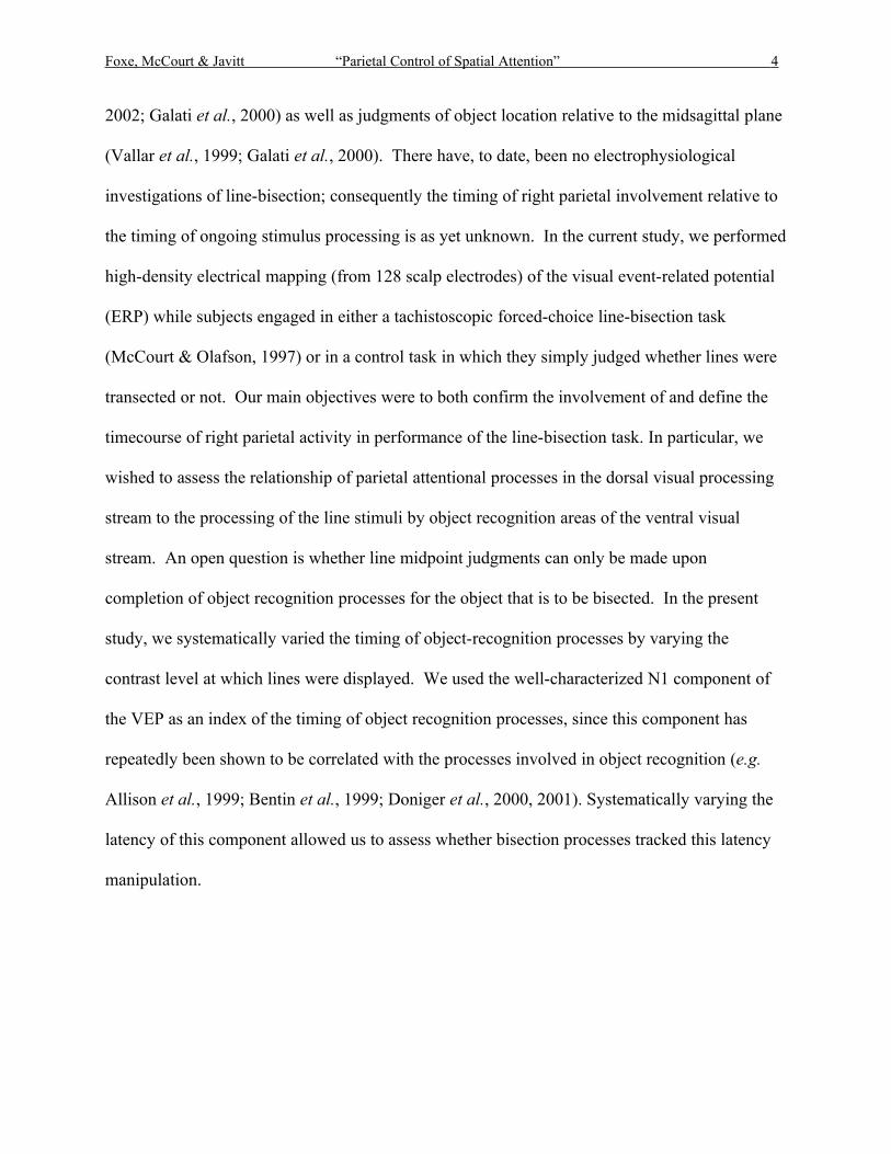
Figure 2 presents group-averaged psychometric functions obtained in the Line-bisection
condition at each level of line contrast. Open symbols plot mean percent “left” responses (±1
s.e.m.) against transector location (in degrees relative to veridical line midpoint). The data are
Insert Figure 2 Here
well behaved, and the range of transector locations sampled is observed to encompass perceived
line midpoint in this sample of subjects. Solid lines depict cumulative Gaussian distributions
fitted to the data by nonlinear least-squares regression. The cumulative Gaussian function is
described by the equation:
f(x,α,β,σ)=α(50+50(erf((x−β)/20.5 σ)))
where x is transector location (in degrees relative to veridical line midpoint), α is an overall gain
parameter, β is the x-axis location corresponding to the mean of the underlying Gaussian density
function (i.e., pse − the transector location at which left-right responses occur with equal frequency),
and σ is its standard deviation. The error function (erf) is an approximation to the cumulative
Gaussian distribution, for which there is no closed-form analytical expression. The horizontal
dashed line in each panel indicates the 50% “left” response rate; the transector location for which

Foxe, McCourt & Javitt “Parietal Control of Spatial Attention” 12
the cumulative Gaussian intersects the dashed line is one measure of perceived line midpoint
(pse).
The solid symbols (and vertical dotted lines) in each panel plot mean pse (±1 s.e.m.) based
on the analysis of psychometric functions from individual observers. The agreement with pse
estimates from the group-averaged fits is excellent, and reveals the systematic leftward error
(pseudoneglect) in perceived line midpoint that typifies the performance of neurologically normal
right-handed observers (Jewell & McCourt, 2000; McCourt, 2001a). A one-way repeated-measures
ANOVA conducted on the pse values in each line contrast condition revealed no significant effect
of line contrast, F(2, 16) = 0.74, p = 0.49. While single-sample t-tests comparing mean pse values in
each line contrast condition against veridical fail to achieve significance: 100%, t(8) = −1.63, p =
0.14; 25%, t(8) = −1.73, p = 0.12; 3%, t(8) = −1.68, p = 0.13, increasing statistical power by
collapsing across levels of line contrast revealed, however, that grand mean pse is shifted
significantly leftward of veridical by approximately 0.2o, or 1.4%, t(26) = −2.98, p = 0.006.
Electrophysiological Results
The analysis of behavioural results revealed that line contrast had no significant effect on
bisection performance, and we therefore collapsed across the three levels of line contrast in the
initial statistical analysis of the electrophysiological data. Inspection of group-averaged visual
evoked potentials for both the Line-bisection and Control Task conditions revealed the
traditional series of ERP components, including P1, N1 and P2 (Figure 3.). These components
were maximal over visual cortices.
Insert Figure 3 Here

Foxe, McCourt & Javitt “Parietal Control of Spatial Attention” 13
The earliest robust component, the P1, showed no amplitude or latency difference between
conditions. However, over right hemisphere occipito-parietal sites, responses in the line-
bisection condition showed a markedly increased negativity relative to those in the control task;
this difference began during the peak of the N1 component and then continued for some 300 ms.
At left hemisphere electrode sites, this negative shift was seen to onset later during the N1/P2
transition. This negativity can be seen in Figure 3, where the right hemisphere electrode site of
maximal difference is plotted along with the equivalently positioned left hemisphere site.
As expected, the ANOVA for the P1 latency bin (92-100 ms) yielded no significant main
effect of condition (p = 0.62). The ANOVA for the N1 latency range yielded a significant
interaction of Condition X Hemisphere (F (1,8) = 5.19, p = 0.05). Follow-up planned-
comparisons showed that this was due to a robust main effect of condition over the right
hemisphere electrode sites (F (1,8) = 8.8, p < 0.02), whereas no main effect of condition was
seen over the equivalent left hemisphere sites in this latency range (p = 0.44). The ANOVA for
the P2 latency range revealed a significant main effect of condition (F(1,8) = 31.33, p < 0.001)
but no interaction of Condition X Hemisphere (p = 0.68). Similarly, the ANOVA for the N280ot
latency range revealed only a significant main effect of condition (F(1,8) = 29.27, p < 0.001).
As the ANOVA for N1 showed that the effect of Condition onset earlier over the right
hemisphere, we performed a post-hoc analysis to determine rough onset times for the effect in
both hemispheres. We used a series of paired two-tailed t-tests between the Control and Line-
bisection conditions at the four representative pairs of electrode sites used in the above analyses.
Tests were conducted at latencies preceding the P2 peak to mark the earliest timepoint that
conformed to a 0.05 criterion. Onset latencies across the four left and across the four right
hemisphere electrode sites were averaged to provide a best estimate of onset in a given
Foxe, McCourt & Javitt “Parietal Control of Spatial Attention” 14
hemisphere. A point was only accepted as the earliest divergence if at least 11 subsequent
consecutive time-points (>20ms at 500 Hz digitization rate) met the 0.05 criterion (see also
Guthrie and Buchwald, 1991; Foxe and Simpson, 2002). The criterion was met at 172 ms for the
right hemisphere and 208 ms for the left, indicating that bisection-related attentional processes
onset in the right hemisphere some 30-40 ms earlier than they do in the left hemisphere.
Topographic Analysis
Figure 4 illustrates that the net negative activity associated with line-bisection judgments
(hereafter referred to as the “line-bisection effect”) consists of three distinct phases that can be
discerned through topographic mapping.
- Dominating the earliest phase of the response (~170-190 ms) is a negative focus
concentrated over the right lateral parieto-occipital scalp. There is little or no
concurrent effect over the equivalent left scalp.
- In the second phase (190-240 ms) an additional robust negative focus is evident over
right central parietal scalp, during which the right parieto-occipital focus persists.
Negativity is also evident over left lateral-occipital cortex during this phase.
- In the final phase, as the point of maximal effect is approached (~310 ms), the
topographic pattern becomes dominated by the right central-parietal negativity.
While Figure 4 shows these three distinct phases, the reader is referred to the accompanying
web appendix (http//academicpress.com/ni/linebisection.avi ???). Readers can download an
animation of the scalp topography of the line-bisection effect that better illustrates the spatio-
temporal dynamics of this negative potential.
Insert Figure 4 Here

Foxe, McCourt & Javitt “Parietal Control of Spatial Attention” 15
Analysis by Contrast Level
We next investigated the timing of the line-bisection effect at the three different contrast
levels that were employed (100%, 25% and 3%). We wished to assess the relationship of the
timing of the effect to ongoing stimulus processing as indexed by the N1 component. First we
established the peak latency of N1 for each of the 3 contrast conditions by the following method.
We identified the scalp-site of maximum N1 amplitude by inspecting the group-averaged voltage
waveforms over the right temporo-occipital scalp for the control condition. We then measured
the latency of N1 for each individual subject at this scalp site, where N1 latency was defined as
the point of maximum negative deflection in the latency window between 120 and 240 ms (i.e.
following the P1 component up until P2). Clearly identifiable N1’s were obtained for 8 of the 9
subjects in this study. We excluded from this phase of the analysis the one subject for whom a
clear N1 could not be identified. By this method we obtained the following N1 latency measures:
100%: 156.5 ms (± 17.3); 25%: 163.1 ms (± 21.5); 3%: 192.1 ms (± 19.2). Paired t-tests showed
that the latency difference between the 100% and 25% conditions reached significance (p<0.05),
and in turn, that the difference between 25% and 3% was also significant (p<0.002). Thus, the
contrast manipulation was successful in its aim of systematically manipulating N1 latency.
We now used the running t-test method described above to register the approximate onset of
the line-bisection effect at each of the three contrast conditions. Recall that a given time-point is
only accepted as the earliest divergence if at least 11 subsequent consecutive time-points (>20ms
at 500 Hz digitization rate) meet the 0.05 criterion. The 11-point criterion was met at the
following latencies for the respective contrast levels: 100%: 164 ms; 25%: 172 ms; 3%: 210 ms.
Thus, in all three cases, the onset of significant difference in the line-bisection effect lags the
peak latency of N1 and appears to systematically track the latency of N1. This pattern can be

Foxe, McCourt & Javitt “Parietal Control of Spatial Attention” 16
clearly seen in Figure 5, which illustrates the responses obtained at the three different line
contrasts.
Insert Figure 5 Here
Dipole Source-Analysis
A strong set of a-priori hypotheses regarding the likely intracranial generators of the line-
bisection effect was possible due to the series of detailed functional imaging studies of this task
that have been previously conducted, as discussed above (see also Fig 6). We used the following
strategy to guide the present source analysis. We performed a meta-analysis of three previous
fMRI reports on the line-bisection task (Fink et al, 2000a; Fink et al., 2001; Fink et al., 2002).
We made a list of regions that were consistently activated in the task, that is, regions that were
activated in at least two of the three studies. For example, the right lateral inferior posterior
parietal region was activated in all three of these studies with some minor variation in the exact
locus of the center of activation. As would be expected, slight variations in the exact coordinates
of a given activation were found across these studies, so we tabulated and averaged the Talairach
co-ordinates of common activations in order to derive a single average set of coordinates for a
given area.
This strategy led to the following list of five areas: Right Lateral Occipital Cortex
(RLOC- X=34, Y=-89, Z=03), Right Lateral Inferior Posterior Parietal Cortex (RIPP- 41, -40,
50), Right Lateral Superior Posterior Parietal Cortex (RSPP- 23, -58, 61), Medial
Striate/Extrastriate Cortex (mXST- 00, -80, 14) and Left Cerebellum (LCer- -30, -60, -30).
We used these five sets of co-ordinates as a guide in our spatio-temporal source-analysis,
seeding dipoles in these regions when fitting data over time epochs during which we had
Foxe, McCourt & Javitt “Parietal Control of Spatial Attention” 17
hypothesized that activity was being generated in these areas. The stability of a given dipole
location was tested by also allowing each dipole in turn to freely fit the data. Where one of these
“seeded” dipoles did not sufficiently explain the data, we employed additional dipoles that were
freely fit to the data. By this combination of approaches (both solving the forward and inverse
problem), we arrived at a solution that employed seven dipoles, including the five from the list
and 2 additional dipoles.
The source solution is shown in Figure 6 and the dipole locations and orientations are listed
in Table 1. The following section details the exact strategy, step by step, that was taken to arrive
at this solution:
1) A dipole was fixed in the coordinates of RLOC and allowed to fit the data in the initial 10
msec period of the effect (172-182 msec). This dipole accounted for 57.9% of the
variance in the data for this epoch. Note that allowing the dipole to freely fit the data in
this period resulted in a dipole that went more medially and superiorly into the medial
occipital cortex (-7, -87, 24). This freely fit dipole resulted in no real improvement in fit
with essentially the identical percentage of the variance explained (58.1%). The dipole
was therefore fixed in the RLOC and its orientation was also fixed (see Table 1).
2) A second dipole was fixed in the RIPP location. The fitting period was extended for
another 10 msec (172-192) and the dipole orientation was allowed to freely fit the data.
This resulted in an explained variance of 63.1% over the 20 msec epoch. Again, we
allowed this dipole to freely fit as a test. This caused the dipole to move to a more
inferior and medial location in the region of the right superior occipital gyrus (32, -84,
29) with an improvement in explained variance to 69.1%. This was a clear improvement
in the proportion of explained variance, suggesting that there was significant activity in

Foxe, McCourt & Javitt “Parietal Control of Spatial Attention” 18
the region of the right superior occipital / occipito-temporal region that needed to be
explained in this early period. We therefore fixed this second dipole in the freely fit
location in the right superior occipital gyrus (RSOG).
3) We added a third dipole, again fixing its location in the RIPP, and allowed it to fit for
orientation over the 172-192 msec epoch. Explained variance improved to 73.8%.
Allowing this dipole to freely fit for location and orientation resulted in only a very slight
change in location (<1 cm) and no improvement in explained variance. We therefore
fixed the dipole in the RIPP cords defined above. We tested the stability of the fit so far
by allowing dipole #2 to freely fit this epoch again. This resulted in no change in the
dipole’s position or orientation, indicative of a relatively stable fit.
4) We added a fourth freely fitting dipole and extended the analysis window out to 202
msec (a 30 msec epoch). This resulted in a second dipole in the region of the superior
occipital gyrus / middle temporal gyrus (49, -82, 22) that was more lateral than dipole #2
above. Explained variance for the 30 msec epoch was 71.0%.
5) The proximity of this dipole to dipole #2 suggested that these two dipoles might be trying
to explain the same data. We tested this by turning off dipole #2 and allowing #4 to freely
fit over this 30 msec epoch. This resulted in a dipole that moved slightly more medially
between the two previous dipole locations (45, -85, 25). The explained variance was
71.5%. Clearly, only a single dipole in the RSOG was required. Therefore, we fixed the
location and orientation of this latter dipole.
6) We then added a dipole in the RSPP and again extended the epoch by 10 msec to 212
msec. The reader will note that during this latter 10 msec period, the focus over superior
parietal scalp has begun to emerge strongly, as detailed in the topographic analysis above.

Foxe, McCourt & Javitt “Parietal Control of Spatial Attention” 19
Addition of this dipole resulted in an explained variance of 76.8% over the 40 msec
epoch (172-212). It is noteworthy that at the end of the epoch, explained variance was
83.5% (212 msec). Allowing this dipole to freely fit resulted in only a very slight change
of location, indicating that its location in RSPP was a relatively stable fit, so the dipole
was fixed in the coordinates defined above.
7) We next added both a medial striate and left cerebellar dipole and extended the fitting
window to 50 msec (172-222 msec), allowing the orientations of these two dipoles to fit
the data. These six dipoles resulted in an explained variance of 81.7% over the epoch
with a best fit at the last point (222 msec) of 93.0%. We fixed these latter two dipoles
orientations.
8) We then opened up the epoch across the duration of the entire effect (172 – 400 msec)
and allowed these six dipoles to fit the data. This resulted in an average explained
variance of 92.0% over this 228 msec epoch (see Fig 7B). It is of note that during the
period when the effect was of largest amplitude 220-320 msec (i.e. highest signal-to-
noise in our data), the averaged explained variance was ~97.5%.
9) Finally, it is clear from the topographic data that there is activity over the left hemisphere
that is unlikely to be simply a result of volume conduction from right hemisphere
activations. Left hemisphere activations are also seen in the fMRI data although the exact
loci across studies are not consistent (except for the left cerebellum). We added a left
hemisphere inferior posterior parietal dipole (-38, -42, 40) and fit it to a window from
200 to 260 msec. These coordinates were taken from Fink et al. (2001). It was during this
epoch that the strongest activity over left scalp was observed electrophysiologically. We
then fixed its orientation and opened the epoch up to the entire window (172-400 msec)
Foxe, McCourt & Javitt “Parietal Control of Spatial Attention” 20
and refit the data with these seven dipoles. Explained variance across the entire epoch
was only marginally improved to 92.4%.
Insert Figure 6 Here and Table 1 about here
It is noteworthy that Fink and colleagues also find consistent activation of right frontal
regions including orbito-frontal cortex and the right dorsolateral prefrontal cortex. In our study,
we also see foci of electrical activity over frontal regions but these activations occur relatively
late in time, mostly after the posterior parieto-occipital line-bisection effect is over. These frontal
effects are not treated further in the present data analysis.
It is important to emphasize that the fMRI activations seen in previous studies, and which
we have used to guide this source analysis, cannot be though of as discrete activations of
individual brain regions. Rather, as can be seen in Figure 7, many of these activation clusters are
much larger than a single cortical area and are likely to represent the activity of a cluster of
functional areas. The same is certainly the case for the electrophysiological results reported in
the present manuscript, where a given scalp topography almost certainly represents coordinated
activity within a cluster of functionally related areas. For instance the lateral occipital cortex
contains a cluster of subregions and it would be incorrect to think of the initial phase of the line-
bisection effect as representing activity in just a single one of these. As such, fitting each cluster
or time-epoch with a single equivalent current dipole clearly represents a highly over simplified
model of activity within a given cluster of areas. As such, it is important that the reader should
consider these dipoles to represent a “center-of gravity” rather than a discrete neural locus or a
single neural event.

Foxe, McCourt & Javitt “Parietal Control of Spatial Attention” 21
DISCUSSION
The current findings explicate the brain mechanisms underlying performance of the visual
line-bisection task, a perceptual version of the perceptual-motor task that is frequently employed
in the clinic to disclose the presence and severity of visuospatial neglect syndrome. We define an
electrophysiological correlate of line-bisection judgments, which manifests as a right parieto-
occipital negative potential that is significantly earlier in latency and larger in magnitude than
that over left scalp. This net negative potential change is seen to persist over a latency window
from approximately 170-400 ms post stimulus, and consists of three distinct phases that can be
discerned through topographic mapping. Dominating the earliest phase of the response
(~165-190 ms) is a negative focus concentrated over the right lateral parieto-occipital scalp. In
this early phase, the effect appears to be largely if not entirely unilateral with no differences seen
over the left hemisphere. In the second phase (190-240 ms), a distinct additional negative focus
develops over more superior right central-parietal scalp, during which the right parieto-occipital
focus persists. In the final phase, as the point of maximal amplitude effect is approached (~310
ms), the topographic pattern becomes dominated by this second focus over right central-parietal
scalp. We refer to this net negative difference as the line-bisection effect.
The Intracranial Generators of the Line-Bisection Effect:
Recent fMRI investigations using very similar experimental paradigms to ours have detailed
a network of brain areas that are activated during line-bisection judgments (see e.g. Fink et al.,
2000a, 2000b, 2001). Figure 7, which is adapted from work by Fink and colleagues (Fink et al.,
2001), illustrates the regional distribution of the activated regions during a line-bisection task,
and clearly shows the involvement of right inferior and superior parietal cortices, in addition to
earlier visual areas, frontal regions and regions in the cerebellum. Our topographic results

Foxe, McCourt & Javitt “Parietal Control of Spatial Attention” 22
(Figure 3) appear to be in good agreement with these findings and provide the critical temporal
activation pattern across these regions. Although topographic mapping of the ERP alone permits
only relatively crude spatial localization in terms of the intracranial generators responsible for a
given scalp topography, observation of the correspondence between the fMRI results of Fink et
al. (2001) and the present data allows for a degree of added confidence in such interpretations.
Clearly, the scalp topography of the earliest phase of the line-bisection effect suggests that
regions in the right lateral occipital cortex and the right temporo-parietal-occipital junction (TPJ)
are the likeliest generators, rather than parietal areas. Indeed, previous fMRI results show an
extended region of robust activation in these more inferior right occipital regions (e.g. Fink et al.,
2001). In turn, the second phase of the line-bisection effect, during which a strong focus develops
over the right central-parietal scalp, is likely generated in regions of the right superior parietal
cortex, where again, fMRI shows a very strong regional activation.
To further assess the putative intracranial sources of the effect, we conducted a source
analysis to augment our topographic mapping data. A stable fit that accounted for more than 90%
of the variance contained in the data was found, which included generators in right lateral
occipital cortex, right superior occipital gyrus, right lateral inferior posterior parietal cortex and
right lateral superior posterior parietal cortex. Additional generators were modeled in medial
occipital cortex, the left cerebellum and left lateral inferior parietal cortex. The latter dipole in
left inferior parietal cortex contributed very little to the source solution. To arrive at this
solution, we took advantage of the previous fMRI studies of line-bisection to guide our source
investigation (e.g. Fink et al., 2001). The model shows that dipoles seeded in the major activation
centers previously defined, provide a very stable fit of our data.
Insert Figure 7 Here
Foxe, McCourt & Javitt “Parietal Control of Spatial Attention” 23
It is noteworthy that the earliest phase of the line-bisection effect we describe here has a
somewhat more inferior distribution than might be predicted for a generator in the inferior
parietal lobule and may be more consistent with a primary generator in superior occipito-
temporal or lateral occipital regions of cortex. Source analysis suggested that two dipoles were
needed to account for his early phase, one in right lateral occipital cortex and a second in right
lateral superior occipital gyrus, directly adjacent to the middle temporal gyrus. Karnath (2001)
has recently suggested that the rostral portion of the superior temporal cortex, just inferior to the
temporo-parietal junction, is the principal region involved in spatial awareness and the primary
site of injury in cases of neglect (see also Karnath et al., 2001). Karnath argues that this region
represents the interface between the ventral (object: what) and dorsal (spatial: where) visual
processing streams. Our data appear to accord well with this interpretation, particularly when the
temporal sequence of activations is taken into account. First, we find that there is extensive
visual processing prior to the occurrence of any spatial attention effects, as indexed by the N1
component of the VEP which has been repeatedly implicated as a marker for ventral stream
object processing (e.g., Allison et al., 1999; Doniger et al., 2000, 2001; Murray et al., 2002).
The onset of the initial phase of the line-bisection effect is seen to follow the peak of the N1 and
appears to be generated in and around the right lateral occipital region and the right superior
occipital gyrus, which is relatively close to the TPJ. As Karnath notes, the TPJ is ideally situated
at the junction between the two visual processing streams. Subsequently, the effect appears to be
transmitted into inferior parietal and then more superior parietal areas of the dorsal stream,
potentially for further spatial processing. A strong prediction based on the present results is that
object recognition processes, as indexed by the N1 component, should be relatively intact in
neglect patients but that the essential link or relay between initial object-based processing
Foxe, McCourt & Javitt “Parietal Control of Spatial Attention” 24
(accomplished within the ventral stream) and subsequent visuo-spatial processing (subserved by
the dorsal stream) is selectively impaired.
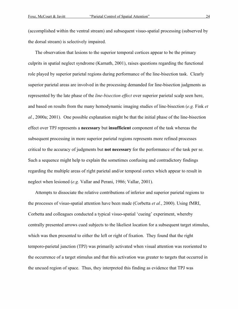
The observation that lesions to the superior temporal cortices appear to be the primary
culprits in spatial neglect syndrome (Karnath, 2001), raises questions regarding the functional
role played by superior parietal regions during performance of the line-bisection task. Clearly
superior parietal areas are involved in the processing demanded for line-bisection judgments as
represented by the late phase of the line-bisection effect over superior parietal scalp seen here,
and based on results from the many hemodynamic imaging studies of line-bisection (e.g. Fink et
al., 2000a; 2001). One possible explanation might be that the initial phase of the line-bisection
effect over TPJ represents a necessary but insufficient component of the task whereas the
subsequent processing in more superior parietal regions represents more refined processes
critical to the accuracy of judgments but not necessary for the performance of the task per se.
Such a sequence might help to explain the sometimes confusing and contradictory findings
regarding the multiple areas of right parietal and/or temporal cortex which appear to result in
neglect when lesioned (e.g. Vallar and Perani, 1986; Vallar, 2001).
Attempts to dissociate the relative contributions of inferior and superior parietal regions to
the processes of visuo-spatial attention have been made (Corbetta et al., 2000). Using fMRI,
Corbetta and colleagues conducted a typical visuo-spatial ‘cueing’ experiment, whereby
centrally presented arrows cued subjects to the likeliest location for a subsequent target stimulus,
which was then presented to either the left or right of fixation. They found that the right
temporo-parietal junction (TPJ) was primarily activated when visual attention was reoriented to
the occurrence of a target stimulus and that this activation was greater to targets that occurred in
the uncued region of space. Thus, they interpreted this finding as evidence that TPJ was
Foxe, McCourt & Javitt “Parietal Control of Spatial Attention” 25
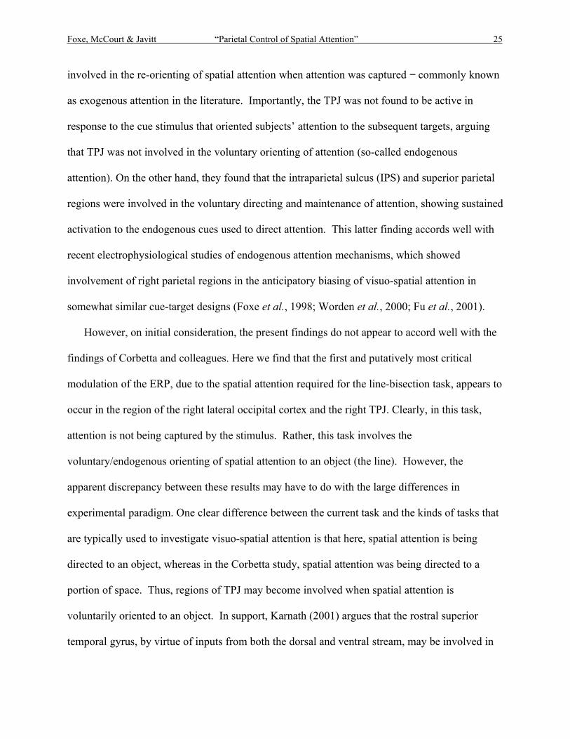
involved in the re-orienting of spatial attention when attention was captured − commonly known
as exogenous attention in the literature. Importantly, the TPJ was not found to be active in
response to the cue stimulus that oriented subjects’ attention to the subsequent targets, arguing
that TPJ was not involved in the voluntary orienting of attention (so-called endogenous
attention). On the other hand, they found that the intraparietal sulcus (IPS) and superior parietal
regions were involved in the voluntary directing and maintenance of attention, showing sustained
activation to the endogenous cues used to direct attention. This latter finding accords well with
recent electrophysiological studies of endogenous attention mechanisms, which showed
involvement of right parietal regions in the anticipatory biasing of visuo-spatial attention in
somewhat similar cue-target designs (Foxe et al., 1998; Worden et al., 2000; Fu et al., 2001).
However, on initial consideration, the present findings do not appear to accord well with the
findings of Corbetta and colleagues. Here we find that the first and putatively most critical
modulation of the ERP, due to the spatial attention required for the line-bisection task, appears to
occur in the region of the right lateral occipital cortex and the right TPJ. Clearly, in this task,
attention is not being captured by the stimulus. Rather, this task involves the
voluntary/endogenous orienting of spatial attention to an object (the line). However, the
apparent discrepancy between these results may have to do with the large differences in
experimental paradigm. One clear difference between the current task and the kinds of tasks that
are typically used to investigate visuo-spatial attention is that here, spatial attention is being
directed to an object, whereas in the Corbetta study, spatial attention was being directed to a
portion of space. Thus, regions of TPJ may become involved when spatial attention is
voluntarily oriented to an object. In support, Karnath (2001) argues that the rostral superior
temporal gyrus, by virtue of inputs from both the dorsal and ventral stream, may be involved in

Foxe, McCourt & Javitt “Parietal Control of Spatial Attention” 26
both spatial orienting and in the analysis of objects in space. By this reasoning, both the findings
of Corbetta et al. (2000) and the present findings are consonant with activity in the TPJ. Of
course, given the spatial uncertainty regarding the exact generator locus of the early line-
bisection effect from ERP topographic mapping alone, an alternate explanation may be that sub-
regions in and around the temporo-parietal junction serve different roles and that the region in
TPJ that is active during exogenous attention in the Corbetta et al. (2000) study is not the same
region that is activated in the present study.
Magnocellular and parvocellular involvement in spatial attention
Arising in the retina, and projecting centrally into temporal and parietal cortical regions, the
primate visual system is comprised of two principal neural pathways, the parvocellular (P) and
magnocellular (M) processing streams (Shapley & Perry, 1986). The M stream has a predominant
dorsal projection to areas V2, V3, V4, MT, MST and area 7a in posterior parietal cortex. The P
stream courses ventrally to areas V2, VP, V4 and to regions of inferotemporal cortex (DeYoe &
Van Essen, 1988; Webster & Ungerleider, 1998). Cells in the M pathway possess high contrast
sensitivity, and demonstrate early response saturation; cells of the P pathway are far less sensitive,
and respond in a graded fashion even to high contrast stimuli (Kaplan & Shapley, 1982; 1986).
Neuronal contrast response functions are well described by the Michaelis-Menten equation, where
C50 represents the stimulus contrast producing half-maximal response (the semi-saturation constant).
Primate P and M LGN neurons possess median values of C50 of 0.50 and 0.11, respectively; for cells
in primary visual cortex (V1), which receive both M and P input, this value is 0.33, and for area MT
it is 0.07 (Sclar et al., 1990).
It is presently unknown to what extent visuospatial attentional mechanisms depend
differentially upon M and P pathway input. Relevant is evidence that VEP's to luminance

Foxe, McCourt & Javitt “Parietal Control of Spatial Attention” 27
contrast in the neglected hemifield of neglect patients are delayed compared to the non-neglected
hemifield. The delay is exacerbated at lower luminance contrasts, and no latency differences are
observed for isoluminant chromatic stimuli (Spinelli et al., 1994; 1996). These findings have
been interpreted to suggest that neglect may, at least in part, result from a selective impairment
of M stream input to parietal cortical regions. Anatomical evidence also indicates that the
parietal cortex receives rich input from the M stream, supported by recent functional
neuroimaging evidence in humans (Tootell et al., 1995a;b) that neurons in dorsal V3 are
activated at low stimulus contrasts. Area V3, in turn, receives M stream input from layer 4B of
V1 and from the "thick stripes" of V2, and has reciprocal connections with other regions
innervated by the M stream, such as parietal cortex and V5 (Shipp et al., 1994; Zeki & Shipp,
1988; Webster & Ungerleider, 1998).
The present behavioral results, as well as those of a more extensive study involving 59
observers (McCourt, unpublished results), reveal that leftward error (pseudoneglect) on the line-
bisection task is remarkably stable as a function of stimulus contrast over the range of 1.5-100%,
which readily encompasses the transition from M to P stream function. It is clear from these data
that parvocellular inputs do not appear to be a necessary component for successful performance
of the line-bisection task, as at contrast levels below about 8% there is little or no parvocellular
input (Tootell et al., 1988). The current electrophysiological data provide similar evidence
against a major role for parvocellular inputs as at the 3% contrast level, a robust line-bisection
effect is observed and this effect has highly similar morphology to the effects seen for the 25%
and 100% contrast conditions. The line-bisection effect at 3% contrast persists despite large
effects upon P1 amplitude and N1 latency due to the contrast manipulation. Rather, the primary

Foxe, McCourt & Javitt “Parietal Control of Spatial Attention” 28
effect of the contrast manipulation upon the line-bisection effect was simply to delay its onset,
which will be discussed in the next section.
Confluence of object-based and space-based attentional processing
While the most prominent omnibus symptom of neglect syndrome is simply left inattention,
the existence of numerous subtypes of neglect has become increasingly clear, dissociated along
the dimensions of near (peripersonal) versus far (extrapersonal) space (Halligan and Marshall,
1991; Vuilleumier et al., 1998; Cowey et al., 1994; Tegner & Levander, 1991b), perceptual
versus motor origin (Bisiach et al., 1990; Tegner & Levander, 1991a), and referenced to
egocentric (self-centered) versus allocentric (object-based) spatial coordinate systems
(Behrmann, 1999; Driver & Pouget, 2000; Walker, 1995). Similar distinctions have been made
with respect to pseudoneglect, where the degree of leftward error on line-bisection tasks is
influenced by motor activity (McCourt et al., 2001a), viewing distance (McCourt et al., 2001b)
and object geometry (McCourt & Jewell, 1999; McCourt et al., 2001c). Our results are
particularly interesting with respect to the distinction between egocentric and allocentric
attention. Note that spatial judgments of line midpoint in a bisection task necessarily involve
computing the spatial location of a specific feature (the transector) with respect to features of an
object (the line itself), and thus explicitly invokes object-based attention. Our
electrophysiological results speak to this issue.
Perhaps the best-known effect of selective visuo-spatial attention upon the ERP is the oft-
reported modulation of the P1 component, which is observed when attention is directed to a
specific portion of the visual field while other parts of visual space are ignored (e.g. Van Hoorhis
and Hillyard, 1977; Martinez et al., 1999; 2001). This spatial attention effect occurs relatively
early in visual processing (typically in the 70-130 ms latency range) and is an example of the
Foxe, McCourt & Javitt “Parietal Control of Spatial Attention” 29
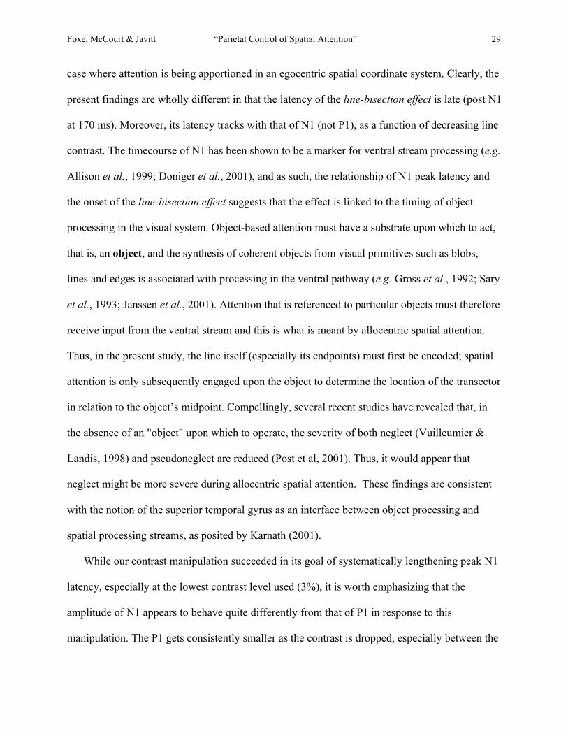
case where attention is being apportioned in an egocentric spatial coordinate system. Clearly, the
present findings are wholly different in that the latency of the line-bisection effect is late (post N1
at 170 ms). Moreover, its latency tracks with that of N1 (not P1), as a function of decreasing line
contrast. The timecourse of N1 has been shown to be a marker for ventral stream processing (e.g.
Allison et al., 1999; Doniger et al., 2001), and as such, the relationship of N1 peak latency and
the onset of the line-bisection effect suggests that the effect is linked to the timing of object
processing in the visual system. Object-based attention must have a substrate upon which to act,
that is, an object, and the synthesis of coherent objects from visual primitives such as blobs,
lines and edges is associated with processing in the ventral pathway (e.g. Gross et al., 1992; Sary
et al., 1993; Janssen et al., 2001). Attention that is referenced to particular objects must therefore
receive input from the ventral stream and this is what is meant by allocentric spatial attention.
Thus, in the present study, the line itself (especially its endpoints) must first be encoded; spatial
attention is only subsequently engaged upon the object to determine the location of the transector
in relation to the object’s midpoint. Compellingly, several recent studies have revealed that, in
the absence of an "object" upon which to operate, the severity of both neglect (Vuilleumier &
Landis, 1998) and pseudoneglect are reduced (Post et al, 2001). Thus, it would appear that
neglect might be more severe during allocentric spatial attention. These findings are consistent
with the notion of the superior temporal gyrus as an interface between object processing and
spatial processing streams, as posited by Karnath (2001).
While our contrast manipulation succeeded in its goal of systematically lengthening peak N1
latency, especially at the lowest contrast level used (3%), it is worth emphasizing that the
amplitude of N1 appears to behave quite differently from that of P1 in response to this
manipulation. The P1 gets consistently smaller as the contrast is dropped, especially between the
Foxe, McCourt & Javitt “Parietal Control of Spatial Attention” 30
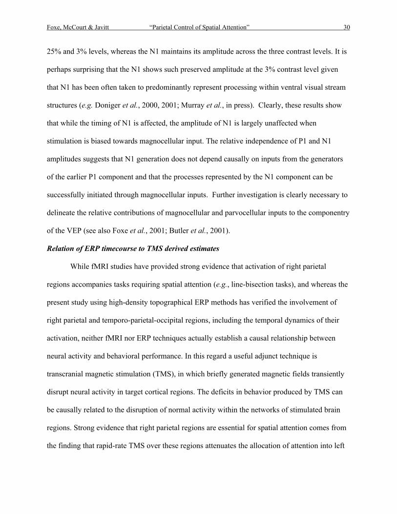
25% and 3% levels, whereas the N1 maintains its amplitude across the three contrast levels. It is
perhaps surprising that the N1 shows such preserved amplitude at the 3% contrast level given
that N1 has been often taken to predominantly represent processing within ventral visual stream
structures (e.g. Doniger et al., 2000, 2001; Murray et al., in press). Clearly, these results show
that while the timing of N1 is affected, the amplitude of N1 is largely unaffected when
stimulation is biased towards magnocellular input. The relative independence of P1 and N1
amplitudes suggests that N1 generation does not depend causally on inputs from the generators
of the earlier P1 component and that the processes represented by the N1 component can be
successfully initiated through magnocellular inputs. Further investigation is clearly necessary to
delineate the relative contributions of magnocellular and parvocellular inputs to the componentry
of the VEP (see also Foxe et al., 2001; Butler et al., 2001).
Relation of ERP timecourse to TMS derived estimates
While fMRI studies have provided strong evidence that activation of right parietal
regions accompanies tasks requiring spatial attention (e.g., line-bisection tasks), and whereas the
present study using high-density topographical ERP methods has verified the involvement of
right parietal and temporo-parietal-occipital regions, including the temporal dynamics of their
activation, neither fMRI nor ERP techniques actually establish a causal relationship between
neural activity and behavioral performance. In this regard a useful adjunct technique is
transcranial magnetic stimulation (TMS), in which briefly generated magnetic fields transiently
disrupt neural activity in target cortical regions. The deficits in behavior produced by TMS can
be causally related to the disruption of normal activity within the networks of stimulated brain
regions. Strong evidence that right parietal regions are essential for spatial attention comes from
the finding that rapid-rate TMS over these regions attenuates the allocation of attention into left
Foxe, McCourt & Javitt “Parietal Control of Spatial Attention” 31
hemispace, producing visual extinction similar to that in neglect patients (Pascual-Leone et al.,
1994; Fierro et al., 2000). More recently a study using single-pulse TMS over right posterior
parietal cortex (electrode site P6) reported significant rightward deviation of line midpoint
judgments in a line-bisection task (i.e., neglect-like behavior) when pulses were delivered 150
ms post stimulus (Fierro et al., 2001). This value agrees very well with the observed timecourse
of initial right parietal activation as revealed in the present ERP study, and the optimal
stimulation location over site P6 is highly similar to the observed center of the initial negative
focus of the line-bisection effect.
One potentially informative direction for a future TMS study would be to selectively
interrupt the early (parieto-occipital) and later (superior-parietal) phases of the line-bisection
effect to attempt to dissociate and identify the separate contributions of these two distinct regions
to line-bisection judgments. Mapping both the exact timing and topography of these information
processing phases on a subject-by-subject basis could further enhance the specificity of this
method, as some degree of temporal and topographic variation was observed between subjects in
the current sample.
Clinical utility of ERP measures of the ‘Line-Bisection Effect’
One problematic issue surrounding the use of standard line-bisection tasks to define spatial
neglect syndrome is that motor responses are often required from the patient, such as asking
them to draw bisectors with a pencil through lines on a page, or to point to the longer/shorter end
of a displayed line. Critically, there are often significant motor deficits in patients with lesions
that involve frontal cortex and also visual cortices, and this can be a confounding factor in results
of the line-bisection task that involve such overt responses (see e.g. Darling et al., 2001). That
is, systematic bisection errors by a motor-compromised patient could reflect an inability or

Foxe, McCourt & Javitt “Parietal Control of Spatial Attention” 32
reluctance to move the drawing or pointing hand in the necessary direction for correct
judgments. The presence of a highly robust ERP effect over right occipito-parietal areas may
provide a simple means for assessing integrity of function in patients without the necessity for
overt motor or even verbal responses.
Summary
In conclusion, the current study reveals a robust negative potential over the right lateral
occipito-parietal and right central-parietal scalp that indexes the neural processing involved in
performance of the ‘line-bisection’ task. Through high-density topographic mapping, we detail
the spatio-temporal dynamics of this processing on the scalp surface. The observed topographies
are consistent with recent hemodynamic imaging studies that have suggested a prominent role
for regions in and around the right temporo-parietal junction and regions of the right superior
parietal cortices in this task. Source analysis confirmed the involvement of these areas as well as
early involvement of right lateral occipital cortices. The present data provide the critical temporal
activation pattern across these regions and underscore the importance of defining the temporal
patterns of attentional modulation, which will be critical to our understanding of the mechanisms
of attentional control in the human brain (e.g. Schroeder et al., 2001).

Foxe, McCourt & Javitt “Parietal Control of Spatial Attention” 33
ACKNOWLEDGEMENTS
We express our sincere appreciation to Deirdre Foxe, Beth Higgins and Dr. Micah Murray
for their technical help with this study. We are most grateful to Dr. Gereon Fink and his
colleagues for generously allowing us to reproduce their fMRI data in the current report. Dr.
Antigona Martinez provided valuable comments on an earlier version of the manuscript for
which we are indebted to her. Our thanks also go to two anonymous reviewers for their careful
and constructive comments. This work was supported in part by grants from the National
Institute of Mental Health (MH63434 – JJF; MH49334 – DCJ), the National Eye Institute
(EY12267 – MEM), North Dakota EPSCoR (MEM), the Neuropsychiatric Research Institute,
Fargo, ND (MEM), the North Dakota State University Development Foundation (MEM), and by
generous support from the Burroughs Wellcome Fund.

Foxe, McCourt & Javitt “Parietal Control of Spatial Attention” 34
REFERENCES
Allison, T., Puce, A., Spencer, D. and McCarthy, G. 1999. Electrophysiological studies of
human face perception I: Potentials generated in occipitotemporal cortex by face and
non-face stimuli. Cereb. Cortex 9: 415-430.
Behrmann, M. 1999. Spatial reference frames and hemispatial neglect. In The New Cognitive
Neurosciences, 2nd Edition, (M.S. Gazzaniga, Ed.), pp. 651-666. MIT Press, Cambridge,
MA.
Bentin, S., Mouchetant-Rostaing, Y., Giard, M.H., Echallier, J.F. and Pernier, J. 1999. ERP
manifestations of processing printed words at different psycholinguistic levels: time
course and scalp distribution. J. Cogn. Neurosci. 11: 235-260.
Beringer J. 1995. Experimental Run Time System (Version 3.13), Berisoft Corporation,
Frankfurt, Germany.
Bisiach, E., Geminiani, G., Berti, A. and Rusconi, M.L. 1990. Perceptual and premotor factors of
unilateral neglect. Neurology 40: 1278-1281.
Bowers, D. and Heilman, K.M.1980. Pseudoneglect: effects of hemispace on a tactile line
bisection task. Neuropsychologia 18: 491-498.
Butler, P.D., Schechter, I., Zemon, V., Schwartz, S.G., Greenstein, V.C., Gordon, J., Schroeder,
C.E. and Javitt, D.C. 2001. Dysfunction of early-stage visual processing in schizophrenia.
Am. J. Psychiatry 158: 1126-1133.
Cappa, S. F., Guariglia, C., Messa, C., Pizzamiglio L. and Zoccolotti P. 1991. Computed
tomography correlates of chronic unilateral neglect. Neuropsychology 5: 195-204.
Foxe, McCourt & Javitt “Parietal Control of Spatial Attention” 35
Corbetta, M., Kincade, J.M., Ollinger, J.M., McAvoy, M.P. and Shulman, G.L. 2000. Voluntary
orienting is dissociated from target detection in human posterior parietal cortex. Nat.
Neurosci. 3: 292-297.
Coull, J.T., Nobre, A.C. and Frith, C.D. 2001. The noradrenergic α2 agonist clonidine modulates
behavioural and neuranotomical correlates of human attentional orienting and alerting.
Cereb. Cortex 11: 73-84.
Cowey, A., Small, M. and Ellis, S. 1994. Left visuo-spatial neglect can be worse in far than in
near space. Neuropsychologia 32: 1059-1066.
Darling, W.G., Rizzo, M. and Butler, A.J. 2001 Disordered sensorimotor transformations for
reaching following posterior cortical lesions. Neuropsychologia 39: 237-254.
DeYoe, E.A. and Van Essen, D.C. 1988. Concurrent processing streams in monkey visual cortex.
Trends Neurosci. 11: 219-226.
Doniger, G.M., Foxe, J.J., Murray, M.M., Higgins, B.A., Snodgrass, J.G., Schroeder, C.E. and
Javitt, D.C. 2000. Activation time-course of ventral visual stream object-recognition
areas: High density electrical mapping of perceptual closure processes. J. Cogn.
Neurosci. 12: 615-621.
Doniger, G.M., Foxe, J.J., Murray, M.M., Higgins, B.A., Schroeder, C.E. and Javitt, D.C.
2001.Visual perceptual learning in human object recognition areas: A repetition priming
study using high-density electrical mapping. NeuroImage 13: 305-313.
Driver, J. and Pouget, A. 2000. Object centered visual neglect, or relative egocentric neglect? J.
Cogn. Neurosci. 12: 542-545.
Foxe, McCourt & Javitt “Parietal Control of Spatial Attention” 36
Fierro, B., Brighina, F., Oliveri, M., Piazza, A., La Bua, V., Buffa, D. and Bisiach, E. 2000.
Contralateral neglect induced by right posterior parietal rTMS in healthy subjects.
Neuroreport 11: 1519-1521.
Fierro, B., Brighina, F., Piazza, A., Oliveri, M. and Bisiach, E. 2001. Timing of right parietal and
frontal cortex activity in visuo-spatial perception: A TMS study in normal individuals.
Neuroreport 12: 2605-2607.
Fink, G.R., Marshall, J.C., Shah, N.J., Weiss, P.H., Halligan, P.W., Grosse-Ruyken, M.,
Ziemons, K., Zilles, K. and Freund, H.J. 2000a. Line bisection judgments implicate right
parietal cortex and cerebellum as assessed by fMRI. Neurology 28: 1324-31.
Fink, G.R., Marshall, J.C., Weiss, P.H., Shah, N.J., Toni, I., Halligan, P.W. and Zilles, K. 2000b.
'Where' depends on 'what': A differential functional anatomy for position discrimination
in one- versus two-dimensions. Neuropsychologia 38: 1741-1748.
Fink, G.R., Marshall, J.C., Weiss, P.H. and Zilles, K. 2001. The neural basis of vertical and
horizontal line bisection judgments: An fMRI study of normal volunteers. Neuroimage
14: S59-67.
Fink, G.R., Marshall, J.C., Weiss, P.H., Toni, I. and Zilles, K. 2002. Task instructions influence
the cognitive strategies involved in line bisection judgments: Evidence from modulated
neural mechanisms revealed by fMRI. Neuropsychologia 40: 119-130.
Foxe, J.J., Simpson, G.V. and Ahlfors, S.P. 1998. Parieto-occipital ~10 Hz activity reflects
anticipatory state of visual attention mechanisms. Neuroreport 9: 3929-3933.
Foxe, J.J., Doniger, G.M., and Javitt, D.C. 2001. Early visual processing deficits in
schizophrenia: impaired P1 generation revealed by high-density electrical mapping.
Neuroreport 12: 3815-3820.
Foxe, McCourt & Javitt “Parietal Control of Spatial Attention” 37
Foxe, J.J. and Simpson, G.V. 2002. Timecourse of activation flow from V1 to frontal cortex in
humans: A framework for defining "early" visual processing. Exp. Brain Res. 142:
139-150.
Fu, K.G., Foxe, J.J., Murray, M.M., Higgins, B.A., Javitt, D.C. and Schroeder, C.E. 2001.
Attention-dependent suppression of distracter visual input can be cross-modally cued as
indexed by anticipatory parieto-occipital alpha-band oscillations. Cogn. Brain Res. 12:
145-512.
Fuchs, M., Drenckhahn, R., Wischmann, H.A. and Wagner, M. 1998. An improved boundary
element method for realistic volume-conductor modeling. IEEE Trans. Biomed. Eng. 45:
980-997.
Galati, G., Lobel, E., Vallar, G., Berthoz, A., Pizzamiglio, L. and Le Bihan, D. 2000. The neural
basis of egocentric and allocentric coding of space in humans: A functional magnetic
resonance study. Exp. Brain Res. 133: 156-164.
Gross, C.G., Rocha-Miranda, C.E. and Bender, D.B. 1972. Visual properties of neurons in
inferotemporal cortex of the Macaque. J. Neurophysiol. 35: 96-111.
Guthrie, D. and Buchwald, J.S. 1991. Significance testing of difference potentials.
Psychophysiology 28: 240-244.
Halligan, P.W., and Marshall, J.C. 1991. Left neglect for near but not far space in man. Nature
350: 498-500.
He, B., Lian, J., Spencer, K.M., Dien, J., and Donchin, E. 2001. A cortical potential imaging
analysis of the P300 and novelty P3 components. Hum. Brain Mapp. 12: 120-130.

Foxe, McCourt & Javitt “Parietal Control of Spatial Attention” 38
Heilman, K.M. and Van Den Abell, T. 1980. Right hemisphere dominance for attention: the
mechanism underlying hemispheric asymmetries of inattention (neglect). Neurology 30:
327-330.
Janssen, P., Vogels, R., Liu, Y. and Orban, G.A. 2001. Macaque inferior temporal neurons are
selective for three-dimensional boundaries and surfaces. J. Neurosci. 21: 9419-9429.
Kaplan, E. and Shapley, R. 1982. X and Y cells in the lateral geniculate nucleus of macaque
monkeys. J. Physiol. 330: 125-143.
Kaplan, E. and Shapley, R.M. 1986. The primate retina contains two types of ganglion cells, with
high and low contrast sensitivity. Proc. Natl. Acad. Sci. U.S.A. 83: 2755-2757.
Karnath, H.O. 2001. New insights into the functions of the superior temporal cortex. Nat. Rev.
Neurosci. 2: 568-576.
Karnath, H.O., Ferber, S. and Himmelbach, M. 2001. Spatial awareness is a function of the
temporal not the posterior parietal lobe. Nature 411: 950-953.
Kerkhoff, G. 2001. Spatial hemineglect in humans. Prog. Neurobiol. 63: 1-27.
Jewell, G. and McCourt, M.E. 2000. Pseudoneglect: a review and meta-analysis of performance
factors in line bisection tasks. Neuropsychologia 38: 93-110.
Martinez, A., Anllo-Vento, L., Sereno, M.I., Frank, L.R., Buxton, R.B., Dubowitz, D.J., Wong,
E.C., Hinrichs, H., Heinze, H.J. and Hillyard, S.A. 1999. Involvement of striate and
extrastriate visual cortical areas in spatial attention. Nat. Neurosci. 2: 364-369.
Martinez, A., DiRusso, F., Anllo-Vento, L., Sereno, M.I., Buxton, R.B. and Hillyard, S.A. 2001.
Putting spatial attention on the map: timing and localization of stimulus selection
processes in striate and extrastriate visual areas. Vision Res. 41: 1437-1457.
Foxe, McCourt & Javitt “Parietal Control of Spatial Attention” 39
McCourt, M.E. 2001a. Performance consistency of normal observers in forced-choice
tachistoscopic visual line bisection. Neuropsychologia 39: 1065-1076.
McCourt, M.E., Freeman, P., Tahmahkera-Stevens, C. and Chaussee, M. 2001a. The influence of
unimanual response on pseudoneglect magnitude. Brain Cogn. 45: 52-63.
McCourt, M.E. and Garlinghouse, M. 2000b. Asymmetries of visuospatial attention are
modulated by viewing distance and visual field elevation: Pseudoneglect in peripersonal
and extrapersonal space. Cortex 36: 715-732.
McCourt, M.E. and Garlinghouse, M. 2000c. Stimulus modulation of pseudoneglect: Effect of
line geometry. Neuropsychologia 38: 520-524.
McCourt, M.E., Garlinghouse, M. and Slater, J. 2000. Centripetal versus centrifugal bias in
visual line bisection: focusing attention on two hypotheses. Front. Biosci. 5: D58-71.
McCourt, M.E. and Jewell, G. 1999. Visuospatial attention in line bisection: Stimulus
modulation of pseudoneglect. Neuropsychologia 37: 843-855.
McCourt, M.E. and Olafson, C. 1997. Cognitive and perceptual influences on visual line
bisection: psychophysical and chronometric analyses of pseudoneglect.
Neuropsychologia 35: 369-380.
Mesulam, M.M. 2000. Attentional networks, confusional states, and neglect syndromes. In
Principles of Behavioral and Cognitive Neurology, 2nd Edition (M.M. Mesulam, Ed.), pp.
174-256. Oxford University Press, Oxford.
Michel, C.M., Thut, G., Morand, S., Khateb, A., Pegna, A.J., Grave de Peralta, R., Gonzalez, S.,
Seeck, M., Landis, T. 2001. Electric source imaging of human brain functions. Brain
Res. Brain Res. Rev. 36: 108-118.

Foxe, McCourt & Javitt “Parietal Control of Spatial Attention” 40
Murray, M.M., Wylie, G.R., Higgins, B.A., Javitt, D.C., Schroeder, C.E. and Foxe, J.J. The
spatio-temporal dynamics of illusory contour processing: Combined high-density
electrical mapping, source analysis, and functional magnetic resonance imaging. J.
Neurosci. 22: 5055-5073.
Na, D.L., Adair, J.C., Hye Choi, S., Won Seo, D., Kang, Y. and Heilman, K.M. 2000.
Ipsilesional versus contralesional neglect depends on attentional demands. Cortex 36:
455-467.
Oldfield, R.C. 1971. The assessment and analysis of handedness: The Edinburgh Inventory.
Neuropsychologia 9: 97-113.
Pascual-Leone, A., Gomez-Tortosa, E., Grafman, J., Alway, D., Nichelli, P. and Hallett, M.
1994. Induction of visual extinction by rapid-rate transcranial magnetic stimulation of
parietal lobe. Neurology 44: 494-498.
Post, R.B., Caufield, K.J. and Welch, R.B. 2001. Contributions of object- and space-based
mechanisms to line bisection errors. Neuropsychologia 39: 856-864.
Robertson, I.H. and Halligan, P.W. 1999. Spatial Neglect: A Clinical Handbook for Diagnosis
and Treatment. Psychology Press.
Ritter W, Vaughan HG Jr. 1969. Averaged evoked responses in vigilance and discrimination: a
reassessment. Science 164: 326-328.
Sary, G., Vogels, R. and Orban, G.A. 1993. Cue-invariant shape selectivity of macaque inferior
temporal neurons. Science 260: 995-997.
Scherg, M., and Picton, T.W. 1991. Separation and identification of event-related potential
components by brain electric source analysis. Electroencephalogr. Clin. Neurophysiol.
Suppl. 42: 24-37.
Foxe, McCourt & Javitt “Parietal Control of Spatial Attention” 41
Scherg, M., and Berg, P. 1996. New concepts of brain source imaging and localization.
Electroencephalogr. Clin. Neurophysiol. Suppl. 46: 127-137.
Schroeder, C.E., Mehta, A.D. and Foxe, J.J. 2001. Determinants and mechanisms of attentional
modulation of neural processing. Front. Biosci. 6: D672-684.
Sclar, G., Maunsell, J.H.R. and Lennie, P. 1990. Coding of image contrast in central visual
pathways of the macaque monkey. Vision Res. 30: 1-10.
Shapley, R. and Perry, V.H. 1986. Cat and monkey retinal ganglion cells and their functional
roles. Trends Neurosci. 9: 229-235.
Shipp, S., de Jong, B.M., Zihl, J., Frackowiak, R.S.J. and Zeki, S. 1994. The brain activity
related to residual motion vision in a patient with a bilateral lesion of V5. Brain 117:
1023-1038.
Simpson, G.V., Pfleiger, M.E., Foxe, J.J., Ahlfors, S.P., Vaughan, H.G.Jnr., Hrabe, J., Ilmoniemi,
R.J., and Lantos, G. 1995a. Dynamic neuroimaging of brain function. J. Clin. Neurophysiol.
12: 1-18.
Simpson, G.V., Foxe, J.J., Vaughan, H.G., Jr. Mehta, A.D., and Schroeder, C.E. 1995b. Integration
of electrophysiological source analyses, MRI and animal models in the study of visual
processing and attention. Electroencephalogr. Clin. Neurophysiol. Suppl. 44: 76-92.
Spinelli, D., Angelina, P., De Luca, M. and Burr, D.C. 1996. VEP in neglect patients have longer
latencies for luminance but not for chromatic patterns. Neuroreport 7: 815-819.
Spinelli, D., Burr, D.C. and Morrone, M.C. 1994. Spatial neglect is associated with increased
latencies of visual evoked potentials. Vis. Neurosci. 11: 909-918.
Talairach J, and Tournoux P. 1988. Co-Planar Stereotaxic Atlas of the Human Brain. New York,
NY: Thieme.
Tegner, R. and Levander, M. 1991a. Through a looking glass. A new technique to demonstrate
Foxe, McCourt & Javitt “Parietal Control of Spatial Attention” 42
directional hypokinesia in unilateral neglect. Brain 114: 1943-1951.
Tegner, R. and Levander, M. 1991b. The influence of stimulus properties on visual neglect. J.
Neurol. Neurosurg. Psychiatry 54: 882-887.
Tootell, R.B., Hamilton, S.L. and Switkes, E. 1988. Functional anatomy of macaque striate
cortex. IV. Contrast and magno-parvo streams. J. Neurosci. 8: 1594-1609.
Tootell, R.B., Reppas, J.B., Dale, A.M., Look, R.B., Sereno, M.I., Malach, R., Brady, T.J. and
Rosen, B.R. 1995a. Visual motion aftereffect in human cortical area MT revealed by
functional magnetic resonance imaging. Nature 375: 139-141.
Tootell, R.B.H., Reppas, J.B., Kwong, K.K., Malach, R., Born, R.T., Brady, T.J., Rosen, B.R.
and Belliveau, J.W. 1995b. Functional analysis of human MT and related visual cortical
areas using functional magnetic resonance imaging. J. Neurosci. 15: 3215-3230.
Vallar, G., Lobel, E., Galati, G., Berthoz, A., Pizzamiglio, L. and Le Bihan, D. 1999. A fronto-
parietal system for computing the egocentric spatial frame of reference in humans. Exp.
Brain Res. 124: 281-286.
Vallar, G. and Perani, D. 1986. The anatomy of unilateral neglect after right-hemisphere stroke
lesions. A clinical/CT-scan correlation study in man. Neuropsychologia 24: 609-622.
Vallar, G. and Perani, D. 1987. The anatomy of spatial neglect in humans. In Neurophysiological
and Neuropsychological Aspects of Spatial Neglect (M. Jennerod, Ed.), pp. 235-258.
North Holland, Amsterdam.
Vallar, G. 2001. Extrapersonal visual unilateral spatial neglect and its neuroanatomy.
Neuroimage 14: S52-58.
Van Hoorhis, S.T. and Hillyard, S.A. 1977. Visual evoked potentials and selective attention to
points in space. Percept. Psychophys. 22: 54-62.
Foxe, McCourt & Javitt “Parietal Control of Spatial Attention” 43
Vuilleumier, P. and Landis, T. 1998. Illusory contours and spatial neglect. Neuroreport 9:
2481-2484.
Vuilleumier, P., Valenza, N., Mayer, E., Reverdin, A. and Landis, T. 1998. Near and far visual
space in unilateral neglect. Ann. Neurol. 43: 406-410.
Walker, R. 1995. Spatial and object-based neglect. Neurocase 1: 371-383.
Webster, M.J. and Ungerleider, L.G. 1998. Neuroanatomy of visual attention. In The attentive
brain (R. Parasuraman, Ed.), pp. 19-34. MIT Press: Cambridge.
Weintraub, S. and Mesulam, M.M. 1987. Right cerebral dominance in spatial attention. Further
evidence based on ipsilateral neglect. Arch. Neurol. 44: 621-625.
Weiss, P.H., Marshall, J.C., Wunderlich, G., Tellmann, L., Halligan, P.W., Freund, H.J., Zilles,
K. and Fink, G.R. 2000. Neural consequences of acting in near versus far space: a
physiological basis for clinical dissociations. Brain 123: 2531-2541.
Worden, M.S., Foxe, J.J., Wang, N. and Simpson, G.V. 2000. Anticipatory biasing of
visuospatial attention indexed by retinotopically specific alpha-band
electroencephalography increases over occipital cortex. J. Neurosci. 20: RC63 (1-6).
Zeki, S. and Shipp, S. 1988. The functional logic of cortical connections. Nature 335: 311-317.

Foxe, McCourt & Javitt “Parietal Control of Spatial Attention” 44
FIGURE CAPTIONS
Figure 1. Line Stimuli Used in the Experiment. Both members of the upper pair of lines (A,
B) possess 100% contrast, and are transected to the left of center, by −0.33o (−2.17%) and −0.60o
(−3.91%), respectively. Both members of the lower pair of lines (E, F) possess 3% contrast and
are transected to the right of center, by +0.47o (3.04%) and +0.17o (1.09%), respectively. Lines C
and D possess 25% contrast; C is veridically transected and line D lacks a transector. Pairs (A,
B) and (E, F) differ in contrast polarity. Lines of varying contrast and contrast polarity appeared
with equal frequency. The order of appearance of all line types was quasi-randomized within
blocks of trials.
Figure 2. Behavioral Data. Group-averaged psychometric functions obtained in the Line
Bisection condition at each level of line contrast. Open symbols plot mean percent “left”
responses (±1 s.e.m.) against transector location (in degrees relative to veridical line midpoint).
Solid lines depict cumulative Gaussian distributions fitted to the data by nonlinear least-squares
regression. The horizontal dashed line in each panel indicates the 50% “left” response rate; the
transector location for which the cumulative Gaussian intersects the dashed line is one measure
of perceived line midpoint (pse).
The solid symbols (and vertical dotted lines) in each panel plot mean pse (±1 s.e.m.) based
on the analysis of psychometric functions from individual observers. The agreement with pse
estimates from the group-averaged fits is excellent, and reveals the systematic leftward error
(pseudoneglect) in perceived line midpoint that typifies the performance of neurologically normal
right-handed observers.
Figure 3. Sustained Negative Modulation during Line-Bisection Task. The 128-channel
electrode montage is shown here projected onto a three-dimensional scalp-reconstruction (from
Foxe, McCourt & Javitt “Parietal Control of Spatial Attention” 45
structural MRI data) of one of the subjects (BH). Group-averaged (n=9) ERP voltage waveforms
are plotted for two electrode sites (darkened in Montage display), showing the marked negative
deflection when subjects performed the line-bisection task (solid trace) relative to the control
task (dashed trace). The bottom two panels show the p-values (pointwise 2-tailed t-test
comparisons between conditions), illustrating the timecourse of significant differences at these
electrode sites.
Figure 4. Topographic Mapping of the Line-Bisection Effect. Group-averaged (n=9)
potential maps are shown for three timepoints during the three phases of the 'line-bisection'
effect. These topographies are derived from the difference waves, obtained by subtracting the
control-condition from the line-bisection condition. Surface negativity is depicted in blue/cyan
and positivity in red/yellow. The first phase of the effect (top maps ~176 ms) shows a highly
lateralized negative focus over right temporo-parietal scalp. During the second phase (middle
maps ~220 ms), at least two distinct negative foci are evident with the emergence of a strong
central parietal negative focus. During the third phase, the time-period of the largest effect
(bottom maps ~310 ms), the topography is dominated by a right central parietal negativity.
Figure 5. Three levels of contrast. Group-averaged (n=9) voltage waveforms are shown for the
three levels of display contrast used (same parieto-occipital scalp site as shown in figure 3). The
line-bisection effect can be seen at each level. As N1 latency increases with decreasing contrast,
the line-bisection effect moves systematically with this N1 latency shift.
Figure 6. Source analysis of the Line-Bisection Effect. A). The locations of the seven dipoles
used to model the line-bisection effect are shown projected into a 3D rendering of an averaged
human brain. Each dipole location is color-coded. B). The amount of variance explained in the
data by these seven dipoles across the 172-400 msec epoch of the effect. C). Waveforms
Foxe, McCourt & Javitt “Parietal Control of Spatial Attention” 46
depicting the source strength of each dipole are plotted for the 172-400 msec epoch (same color
convention as used in A).
Figure 7. fMRI Activation During the Line-Bisection Task. Comparison of the fMRI
activations seen here with the scalp-topography of the line-bisection effect shown in figure 4
shows strong correspondence between the electrophysiological and hemodynamic data sets.
(adapted with permission from Fink et al. 2001. The neural basis of vertical and horizontal line
bisection judgments: An fMRI study of normal volunteers. Neuroimage 14: S59-67.) Areas of
significant activation are shown projected into standard stereotactic space in a sagittal, coronal
and axial view (from left to right).
TABLE CAPTION
Table 1. Talairach coordinates and orientations are given for each dipole.

Foxe, McCourt & Javitt “Parietal Control of Spatial Attention” 47
Appendix:
Readers are encouraged to visit the NeuroImage website at the following URL to
download the animation file which accompanies the present report
http//academicpress.com/ni/linebisection.avi ???
This animation shows the evolution of the "line-bisection effect" at a rate approximately 100
times slower than real time. It begins with a display of the 128-channel montage as the 3D rendered head
rotates before settling with a view of the scalp from directly behind the head. The electrodes then
disappear before the topographic maps begin. The counter at the bottom right corner of the panel shows
time in milliseconds and the animation of topography begins at 0 ms which is when the stimulus
appeared. The group-averaged data have been projected onto a 3D-rendered scalp surface. Recall that the
line-bisection effect is derived by subtracting the control condition from the experimental condition and
therefore, all activity associated with the common sensory processing of these stimuli has been eliminated
from the data. Therefore, it can be seen that for the first 150 ms, there is no activity that exceeds noise.
The following is a description of the evolution of the effect, which may help to orient the reader
as they observe the animation.
- At about 160 ms, a negative focus over right parieto-occipital scalp is seen to emerge and build
rapidly in amplitude. By 172 ms (the point at which the effect reaches statistical significance)
the negativity is clearly discernable. This right parieto-occipital negativity continues to build
in amplitude up until ~200-210 ms.
- At approximately 188 ms, the initial right parieto-occipital negativity appears to extend
dorsally and centrally over mid-parietal scalp and this extension of the negativity quickly
develops into a second distinct focus by approximately 206-212 ms.
- By 210 ms, a third negative limb begins to be apparent over the left parieto-occipital
scalp although this left scalp negativity never becomes fully distinct from the two right
foci.

Foxe, McCourt & Javitt “Parietal Control of Spatial Attention” 48
- At 224 ms, the right parieto-occipital focus begins to decrease in amplitude and the
topography becomes dominated by the central parietal negativity. This continues until
approximately 260 ms when the right parieto-occipital focus begins to increase its
amplitude for a second time.
- Between 260 and ~340 ms, this pattern is essentially maintained. However, the reader
will notice that both the right parieto-occipital and central parietal foci appear to wax
and wane somewhat independently during this period. While it is impossible to
determine at this stage what these dynamics represent functionally, we would speculate
that these modulations are likely to represent successive rounds of activation within
functionally distinct modules of the spatial attention network, and perhaps
communication between two distinct centers of the network.
- By 340 ms, the negativity becomes concentrated over central parietal scalp and the
bilateral parieto-occipital foci are diminishing in amplitude. By 400 ms, the effect is
essentially over.
Note that the reader may find it useful to use the manual advance option on their media player to
control the progression of the animation in order to get a better look at the topographic transition
points detailed above.