A functional chitosan-based hydrogel as a wound dressing ...

i
FORMULATION OF WOUND HEALING HYDROGEL FROM
KERATIN PROTEIN
RABIATUL ADAWIYAH BINTI ZAUKIFLI
Thesis submitted in fulfilment of the requirements
for the award of the degree of
Bachelor of Chemical Engineering
Faculty of Chemical & Natural Resources Engineering
UNIVERSITI MALAYSIA PAHANG
JUNE 2012

vi
ABSTRACT
Keratin is a natural protein extracted from the chicken feather. In the developed country,
the usage of keratin in the personal care products is widely used. The personal care
product produced from the keratin protein is conditioning shampoo, anti aging cream,
facial cleanser and others. There are some differences between the personal care
products already produced by the other developed country because the raw materials to
extract the protein is sheep wool while in this research, the extraction of keratin protein
is from chicken feathers. Keratin solution, polyvinyl alcohol. Polyvinyl pyrrolidone,
glutaraldehyde and ammonium thioglycollate was mixed intimately. The combination
of the solution was then cast and hardened through a freezing-thawing process for 1
hour and this process was repeated up to 7 times to obtain a hydrogel. The related test
was conducted to test the effectiveness of the wound healing product.
vii
ABSTRAK
Keratin ialah protein semulajadi yang di ekstrak daripada bulu ayam. Di negara
membangun, penggunaan keratin dalam produk pengajaan diri sangat meluas. Produk
pengajaan diri yang di buat menggunakan keratin ialah shampoo, krim anti menuaan,
pencucui muka, ubat menyembuh luka dan sebagainya. Terdapat beberapa perbezaandi
antara produt penjagaan diri yang di buat di negara menbangun kerana mereka
menggunakan ekstrak keratin daripada bulu kambing biri-biri manakal di dalam kajian
ini mneggunakan ekstrak keratin daripada bulu ayam. Keratin, polyvinyl alcohol,
polyvinyl pyrrolidone glutaraldehyde dan ammonium thioglycollate di campurkan.
Campuran tersebut melalui proses beku dan rendamselama satu jam. Proses ini di
lakukan sebanyak tujuh kali untuk mendapatkn gel. Ujian terhadapgel tersebut di
lakukanbagi menguji keberkesanan gel tersebut.

viii
TABLE OF CONTENTS
DECLARATION ii
DEDICATION iv
ACKNOWLEDGEMENTS v
ABSTRACT vi
ABSTRAK vii
TABLE OF CONTENTS viii
LIST OF TABLE x
LIST OF FIGURES xi
LIST OF ABBREVIATIONS & SYMBOLS xii
CHAPTER 1 INTRODUCTION
1.1 Background of Studies 1
1.2 Problem Statement 2
1.3 Research Objective 2
1.4 Scope of the research 2
1.5 Rationale & Significance of Study 3
CHAPTER 2 LITERATURE REVIEW
2.1 Feather 4
2.2 Protein 5
2.3 Keratin 6
2.4 Biomedical Product 8
2.5 Wound 9
2.6 Wound Healing Process 10
2.7 Hydrogel by Freezing-Thawing Process 11

ix
CHAPTER 3 METHDOLOGY
3.1 Introduction 12
3.2 Material and Apparatus 12
3.2.1 Material and Reagent 12
3.2.1.1 Material and Reagent for Protein Production 12
3.2.1.2 Material and Reagent for Wound Healing Hydrogel 12
3.2.2 Apparatus 13
3.2.3 Equipment 13
3.3 Research Methodolgy 14
3.3.1 Research Methodology for Protein Production 14
3.3.1.1 Feather Treatment 14
3.3.1.2 Dissolving of Chicken Feather 14
3.3.1.3 Protein Precipitation 14
3.3.1.4 Protein Purification 14
3.3.1.5 Biuret Test 15
3.3.1.6 Analysis Sample 15
3.4 Research Methodology for Wound Healing Hydrogel Production 15
CHAPTER 4 RESULTS AND DISCUSSION
4.1 Introduction 17
4.2 Formulation of Hydrogel 17
4.3 Toxicology Testing 19
4.4 Animal Testing 19
CHAPTER 5 CONCLUSION
5.1 Conclusion 22
5.2 Recommendation 23
REFERENCE 24

x
LIST OF TABLE
Table No. Title Page
2.1 The acid amino composition of chicken feather 7
3.1 Formulation of hydrogel 16

xi
LIST OF FIGURE
Figure No. Title Page
1.1 The anatomy of chicken feather 1
2.1 Structure of chicken feather 4
2.2 The protein structure 5
2.3 The reduction of disulfide bond 8
2.4 Stages of wound repair 10
4.1 Appearance of Hydrogel 18
4.2 Toxicology test on animal 19
4.3 General Appearance of the wound site area 20
xii
LIST OF ABBREVIATIONS & SYMBOLS
FTIR Fourier-Transform Infra-Red
PVA Polyvinyl Alcohol
PVP Polyvinyl Pyrrolidone
SIFP S-Sulfonated Keratin Intermediate Filament Protein
SHSP S-Sulfonated Keratin High Sulfur Protein

CHAPTER 1
INTRODUCTION
1.1 Background of Study
Chicken feather wastage is made up approximately eleven million pound from
the commercial poultry processing plant annually. The disposal process for chicken
feather is expensive. It is also can be difficult because the chicken feather is burning up
with the incinerator plant, buried in the soils and also recycled as a low quality of
poultry foods. These processes mostly give the bad effects to the environment,
especially the burning of chicken feather which will release the green house gasses in
the air. There are several alternatives invented based on the chicken feather application,
but the wastage of chicken feather is still does not change as much as possible because
of its low requirement.
Keratin is a natural protein extracted from the chicken feather as shown in
Figure 1.1. In the developed country, the usage of keratin in the personal care products
Figure 1.1: The anatomy of a chicken feather
2
is widely used. The personal care product produced from the keratin protein is
conditioning shampoo, anti aging cream, facial cleanser and others. There are some
differences between the personal care products already produced by the other developed
country because the raw materials to extract the protein is sheep wool while in this
research, the extraction of keratin protein is from chicken feathers. These two materials
will produce two different sequences of amino acids.
1.2 Problem Statement
Nowadays, the demand of the chicken is increasing throughout the year from the
consumer. Poultry slaughterhouses produce large amount of feather. Further, burning
feather in special installations is very economically ineffective. The uncontrolled
disposal of feather leads to pollution in our environment. Five percent of the body
weight of poultry is feather.
Demand from consumer for keratin based product such as wound healing is
increasing. There are variety of events that wounds and lesions can be caused such as
surgery, traumatic injury, burn, abrasion and also skin grafts. The different result and
problem will occur in healing process (Kelly et al. 2010).
1.3 Research Objectives
1.3.1 To extract the keratin protein from the chicken feather.
1.3.2 To prepare the formulation of biomedical product from keratin protein.
1.3.3 To produce the wound care product containing keratin from chicken feather.
1.3.4 To analyse the wound care product.
1.4 Scope of Research
To achieve the objective of this research, the scopes have been identified in this
research. The scopes of this research are listed as below:-
3
1.4.1 Study the extraction of keratin protein from chicken feather
1.4.2 Freezing-thawing method will be used to produce hydrogel wound care product.
1.4.3 Testing on the animal will be conducted to analyse the wound care product
effectiveness.
1.5 Significance of Study
The research is to be done to develop new product which is wound care product.
The research try to develop new formulation of wound care product that containing
natural keratin protein extracted from chicken feather. Natural keratin protein extracted
from chicken feather has a wide range of use in biomedical product such as wound care
product that contains beta keratin and acid amino composition.
4
CHAPTER 2
LITERATURE REVIEW
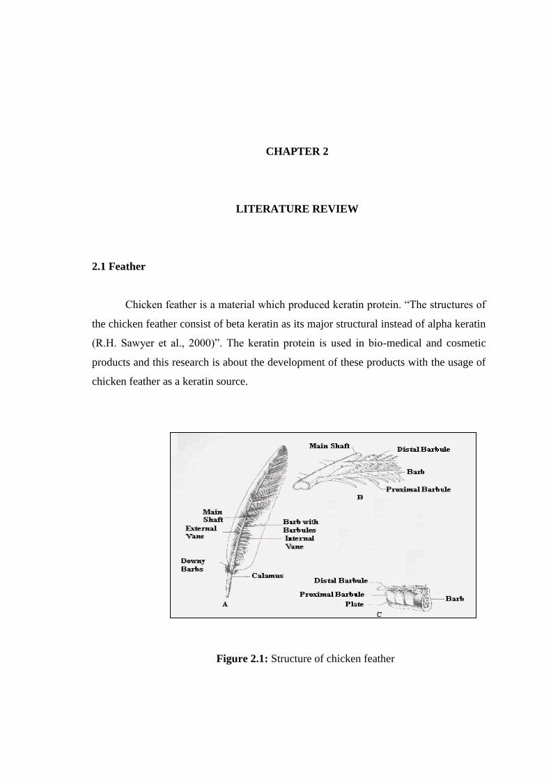
2.1 Feather
Chicken feather is a material which produced keratin protein. “The structures of
the chicken feather consist of beta keratin as its major structural instead of alpha keratin
(R.H. Sawyer et al., 2000)”. The keratin protein is used in bio-medical and cosmetic
products and this research is about the development of these products with the usage of
chicken feather as a keratin source.
Figure 2.1: Structure of chicken feather

5
2.2 Protein
Proteins play a big role in the human system daily life which provides the
structure to the human body system and also transporting oxygen in the human blood
regulating system. Human bodies depending on proteins to perform the life better and
protein also have its composition and structure. The differences of each protein
classification play its different role in the human body.
Figure 2.2: The protein structure
6
2.3 Keratin
The term ‘keratin’ originally referred to the broad category of in soluble proteins
that associate as intermediate filaments (Ifs) and form the bulk of cytoplasmic epithelia
and epidermal appendage structures ( hair, wool, horns, hooves and nails) (Jillian G.
Rouse, Mark E. Van Dyke, 2009).
As per Claudio et.al (2010) keratin represent a group of fibrous protein with
high sulphur content produces in some epithelial cell of vertebrate such as reptiles, birds
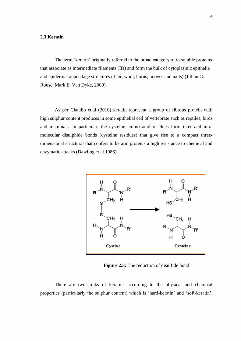
and mammals. In particular, the cysteine amino acid residues form inter and intra
molecular disulphide bonds (cysteine residues) that give rise to a compact three-
dimensional structural that confers to keratin proteins a high resistance to chemical and
enzymatic attacks (Dawling et.al 1986).
There are two kinks of keratins according to the physical and chemical
properties (particularly the sulphur content) which is ‘hard-keratin’ and ‘soft-keratin’.
Figure 2.3: The reduction of disulfide bond
7
The content of sulphur in ‘hard-keratin’ found in hair, wool, feathers, nails and horns is
> 3% whereas in ‘soft-keratin’ is < 3% (Fraser et.al 1972).
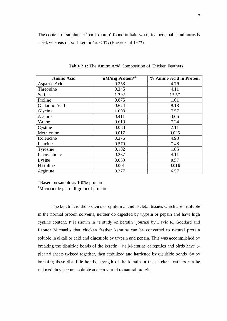
Table 2.1: The Amino Acid Composition of Chicken Feathers
Amino Acid uM/mg Protein*1
% Amino Acid in Protein
Aspartic Acid 0.358 4.76
Threonine 0.345 4.11
Serine 1.292 13.57
Proline 0.875 1.01
Glutamic Acid 0.624 9.18
Glycine 1.008 7.57
Alanine 0.411 3.66
Valine 0.618 7.24
Cystine 0.088 2.11
Methionine 0.017 0.025
Isoleucine 0.376 4.93
Leucine 0.570 7.48
Tyrosine 0.102 1.85
Phenylalnine 0.267 4.11
Lysine 0.039 0.57
Histidine 0.001 0.016
Arginine 0.377 6.57
*Based on sample as 100% protein 1Micro mole per milligram of protein
The keratin are the proteins of epidermal and skeletal tissues which are insoluble
in the normal protein solvents, neither do digested by trypsin or pepsin and have high
cystine content. It is shown in “a study on keratin” journal by David R. Goddard and
Leonor Michaelis that chicken feather keratins can be converted to natural protein
soluble in alkali or acid and digestible by trypsin and pepsin. This was accomplished by
breaking the disulfide bonds of the keratin. The β-keratins of reptiles and birds have β-
pleated sheets twisted together, then stabilized and hardened by disulfide bonds. So by
breaking these disulfide bonds, strength of the keratin in the chicken feathers can be
reduced thus become soluble and converted to natural protein.

8
Keratin is insoluble in water, weak acids and bases as well as in organic
solvents. The amino acid content of keratin is characterised by a high cyctine content(
and at the same time sulphur), which may change within 2% wt and 18%wt, a
significant amount of hydroxyaminoacids, especially serine (about 15 % wt), and a lack
of hydroxyproline and hydroxylisine, among other substances. The chemical activity of
keratin is connected in a significant degree to the crytine content. The disulphide bonds
which is formed between two cysteine molecules is responsible for the high strength of
keratin and its resistance against the action of proteolitic enzymes. On the other hand,
keratin is very reactive, as cystine can easily be reduced, oxidised, and hydrolysed. In
order to precisely determine the possible future applications of keratin, it is necessary to
learn in detail the structure the potential possibilities of this valuable protein. (Krystyna
Wrzesnieska-Tosik, 2007).
2.4 Biomedical Product
Robert James Kelly et. all., (2010) studied the composite materials containing
keratin. In their findings, the application represents materials derived from keratin
proteins in combination with polymers, either as intimate mixtures with water soluble
polymer, or as chemically bound copolymers. An amino acid composition of keratin
fractions: S-sulfonated keratin intermediate filament protein (SIFP), pepetides derived
from S-sulfonated keratin intermediate filament protein (SIFP-pep), S-sulfonated
keratin high sulfur protein (SHSP), peptides derived from S-sulfonated keratin high
sulphur protein (SHSP-pep), S-sulfonated keratin peptide (SPEP) as used in the
invention. The conversion is confirmed using Fourier-Transform Infra-Red (FTIR)
spectroscopic studied as the S-sulfonated group gives rise to a strong and sharp
absorbance at 1022 cm-1 which is observed to disappear on exposure of the S-
sulfonated to the reagents described. Using a freezing-thawing process during the
constructing composite films or membranes can improve of the physical and mechanical
properties of the keratin-co-polymer composites.
9
Alisa Dawn Roddick-Lanzilotta et. all. (2010) studied the wound care products
containing keratin. This invention relates to would care product that provides a
biochemical environment around a wound to promote wound healing. In their
inventions to provide a keratin protein fraction that is intact and S-sulfonated for use in
wound care. This invention also provides a material for treating a wound including a
keratin protein fraction in which the protein fraction is fraction is from the among
filament protein family. A material for treating a wound includes a keratin protein
fraction in which the protein fraction is s-sulfonated. The protein fraction may be
hydrolysed, preferably s-sulfonated, from high sulphur protein family and an among
filament protein.
Sierpinski et al and Apel et al demonstrated that keratin-based hydrogels were
neuroinductive and capable of facilitating regeneration in a peripheral nerve injury
model. Keratin-filled hydrogels were shown to accelerate nerve regeneration as
evidenced by improved electrophysiological recovery and increased axon density at
early time points. The keratin gel used in these experiments acted on the injury site by
instigating thrombus formation and by forming a physical seal of the wound site that
acted as a porous scaffold to allow for cellular infiltration and granulose tissues
formation. The ability for keratin-based biometerials to be translated into the human
clinical setting is dependent on further research to elucidate the mechanism by which
these materials regulate hemostasis and nerve regeneration.
2.5 Wound
A wound is a type of injury in which skin is torn, cut or punctured or where
blunt force trauma causes a contusion and also can be classified into chronic wounds
and acute wounds. In pathology, it specially refers to a sharp injury which damages the
dermis of the skin. Wounds may be grouped according to the cause, the environment in
which they occur, their extent and whether they are clean or contaminated. The
10
microorganisms that typically infect wounds and the skin depend on what is present in
the environment, the state of the person’s immune system and the depth of the wound.
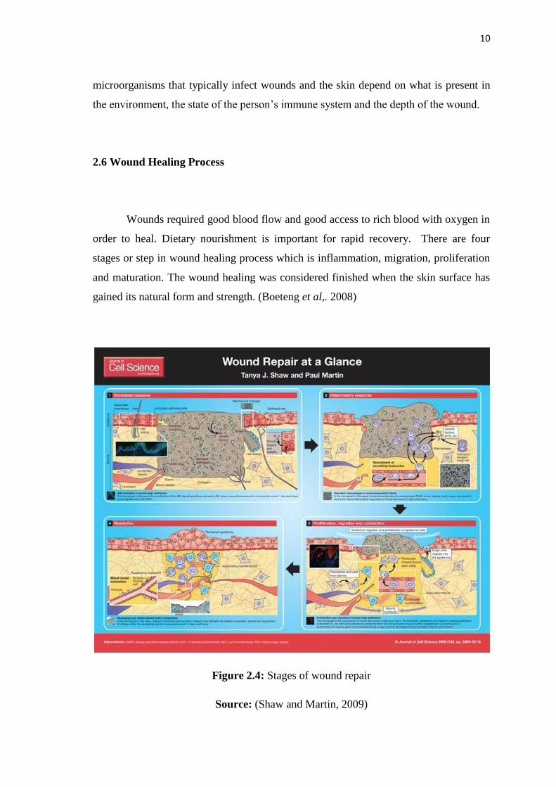
2.6 Wound Healing Process
Wounds required good blood flow and good access to rich blood with oxygen in
order to heal. Dietary nourishment is important for rapid recovery. There are four
stages or step in wound healing process which is inflammation, migration, proliferation
and maturation. The wound healing was considered finished when the skin surface has
gained its natural form and strength. (Boeteng et al,. 2008)
Figure 2.4: Stages of wound repair
Source: (Shaw and Martin, 2009)

11
2.7 Hydrogel by Freezing-Thawing Process
Freezing-thawing method was developing to avoid the cross linking processes
that potentially lead to the release of some polymers. (Christie M. Hassan, 2002). Result
from this process shows the freezing thawing process with chemical of PVA are high
tensile strength, high water content and high light transmittance.
12
CHAPTER 3
RESEARCH METHODOLOGY
3.1 Introduction
The method to extract the chicken feather and production of biomedical product
will be discussed in this chapter. The methods of this research will be start on the
production of the keratin protein from chicken feathers. Keratin protein extracted from
the chicken feather then will be use for developing biomedical product which is wound
care product. These products are to be analysed.
3.2 Material and Apparatus
3.2.1 Material and Reagents
3.2.1.1 Material and Reagent for Protein Production
i. Chicken feather
ii. Ether
iii. Sodium Sulfide
iv. Sodium Hydroxide
v. Ammonium Sulfate
vi. Potassium Hydroxide
vii. Copper Sulfate
3.2.1.2 Material and Reagent for Wound Care Product
i. Polyvinyalcohol
ii. Polyvinylpyrrolidone

13
iii. Glutaraldehyhe
iv. Ammonium Thioglycollate
v. Keratin Protein
vi. Water
3.2.2 Apparatus
i. Beaker
ii. Conical falask
iii. Volumetric flask
iv. Centrifuge tube
v. pH meter
vi. Thermometer
vii. Magnetic stirrer
viii. Schott bottle
ix. Forcep
x. Sample bottle
xi. Spatula
xii. Bottle reagent
xiii. Micropipette
xiv. Wash bottle
xv. Peri dish
3.2.3 Equipment
i. Centrifuge
ii. Hot and stirrer plate
iii. Water bath
iv. Ultrasonic water bath
v. Freezer
vi. Rheometer
vii. Mechanical stirrer

14
3.3 Research Methodology
3.3.1 Research methodology of Protein Production
3.3.1.1 Feathers Treatment
a) Soak the chicken feather in ether for 24 hours.
b) Wash the feathers with the soap water and dry the wet feathers under the
sunlight.
c) Collect all the dried feathers and blend the feather then keep the blend
feathers in the sealed plastic bag carefully.
3.3.1.2 Dissolving of Chicken Feather
a) Prepare the sodium sulphide solution in the conical flask.
b) Weight the blend feathers and add in into the sodium sulphide solution
c) Stir the solution for 6hours and maintain the condition of solution at 30°C
and pH range of 10-13.
d) Filter the solution and centrifuge the solution at 10000 rpm for 15 minutes.
e) Filter the solution to get the supernatant liquid.
f) Place the supernatant liquid in a beaker and stir the solution.
3.3.1.3 Protein Precipitation
a) Add an ammonium sulphate solution into the solution and centrifuge the
solution at 10000 rpm for 5 minutes.
b) Filter the solution to get supernatant liquid and solid particles.
c) Repeat step a) and b).
3.3.1.4 Protein Purification
a) Pour the deionized water into the solid particles and stir the solution
b) Centrifuge the solution at 10000 rpm for 5minutes and filter the solution to
get supernatant liquid and solid particles.
c) Use sodium hydroxide solution to dissolve the solid particles.
d) Centrifuge again the solution at 10000 rpm at 5 minutes. Collect the liquid
and discard the solids.
e) Repeat steps 1 to 4 for three times.
15
3.3.1.5 Biuret Test
a) Prepare the copper sulphate solution and potassium hydroxide solution.
b) Mix the protein solution with potassium hydroxide solution by 1:1 ratio.
c) Add three drops cooper sulphate solution in the mixture solution.
d) Observe and record the change in the solution.
e) Analyze the solution under UV-Visual to obtain its absorbance.
3.3.1.6 Analysis of the Sample
a) Analyze the protein solution in FTIR to obtain the wavelength graph.
b) Mix the ammonium sulfate with protein solution, collect and weight the
solid particles.
3.4 Research Methodology of Wound Healing Hydrogel Production
A 10% keratin solution was prepared using keratin powder dissolved in distilled
water with gradual addition of 1M NaOH over 2 hours under mechanical stirring. The
pH was maintained in the range 8.0-9.5 and finally adjusted to 8.5. The keratin protein
solution was centrifuged at 10000 rpm for 10 minutes in order to remove any air
bubbles and undissolved material. The resulting keratin protein solution was cast into a
peri dish and the solvents evaporated wider ambient conditions to leaves keratin
membrane. The solvent can also include some percentage of organic based aqueous can
also include some percentage of organic based aqueous miscible solvent, such as an
alcohol.
A 10% keratin solution that prepared as describe above intimately mixed with
water soluble polymer which is polyvinyl alcohol (PVA) comprising 20% solid content
and polyvinyl pyrrolidone (PVP) comprising 10% solid content to achieved a optimum
rheology and optimal composition (keratin: PVA: PVP = 100: 60: 40 (w/w, %) ) for
creating hydrogel. The cross-linking agent which is 0.05% to 0.1% of glutaraldehyde
was added into the blended solution. The solution then mixed with the final component
of chemical which is 1% of 0.25M ammonium thioglycollate solution. The combination

16
of the solution was then cast and hardened through a freezing-thawing for 1 hour. This
freeze-thaw cycle was repeated up to 7 times to obtain a hydrogel. This resulting
hydrogel was washed with distilled water multiple times to remove any unreacted
keratin and polymers.
The hydrogel get from freezing-thawing process then proceed with the rheology
test, toxicity test and testing on animal and the rate of healing of wound was observed.
Table 3.1: Formulation of Hydogel
Formulation,% w/w
Compound F1 F2
Polyvinylalcohol 20 15
Polyvinylpyrrolidone 15 15
Ammonium Thioglycollate 8 8
SIFP 10 10
Water 30 30
Glularaldehyde - 0.05