FORMULATION, DEVELOPMENT AND EVALUATION OF … · FORMULATION, DEVELOPMENT AND EVALUATION OF...
18
FORMULATION, DEVELOPMENT AND EVALUATION OF FILM-FORMING GEL FOR PROLONGED DERMAL DELIVERY OF TERBINAFINE HYDROCHLORIDE Vij N.N. * , Dr. Saudagar R.B. Kalyani Charitable Trust’s R.G. Sapkal College of Pharmacy, Anjaneri, Tal. Trimbakeshwar, Dist. Nasik-422213 Email: [email protected] Mobile no.: 9096373656 ABSTRACT: Purpose: The localized treatment of diseases of body tissues requires that the pharmaceutical active be maintained at the site of treatment for an effective period of time. Sweat, clothing, movements and getting washed away easily on contact with water are some of the problems that have limited the effectiveness and residence time of conventional topical formulations for treatment of fungal infections of skin. This necessitates longer treatment duration. Hence, a composition that adheres to skin surface afflicted and provides localized delivery of an antifungal agent is needed. Methods: The present work aims at designing a dosage form of TH referred to as a ‘film-forming gel’ which on application forms a thin, transparent film on skin surface. Eudragit RS PO and hydroxypropyl cellulose were used in combination to provide a matrix film that would permit the release of the antifungal agent for a prolonged time. The formulations were prepared using 3 2 full factorial design. They were tested for drying time, drug release, antifungal activity, skin irritation and stability studies. The gel was characterised for pH, viscosity, drug content, effective dosage volume and mechanical properties of the film formed after application; bioadhesion and water vapour permeability were also tested. Results: All the formulations showed results within acceptable range for various tests. The optimized formulation showed drug release of 99.84% and antifungal activity in terms of efficacy as 99.44%. Conclusion: Such a formulation can be claimed to decrease duration of therapy, will be more accepted by the patients and be a breakthrough in treating fungal infections of the skin. KEYWORDS: Terbinafine hydrochloride (TH); Fungal skin infections; Film-forming gel; Eudragit RS PO; Hydroxypropyl cellulose INTRODUCTION: Fungal diseases can be classified into 3 groups: the superficial, subcutaneous, and deep or systemic mycoses. Superficial infections are confined to skin, hair, nails or mucous membranes. The most common fungal skin infections are the dermatophytoses, pityriasis versicolor, and candidiasis. Approximately 90% of fungal skin infections are caused by ‘dermatophytes’, which are parasitic fungi affecting the skin, hair, nails. [1] One of the leading antifungal agents for topical treatment of fungal infections is terbinafine. It has been approved by the US Food and Drug Administration in cream, gel, solution and spray dosage forms. [2] Terbinafine is an allylamine antifungal agent widely utilized in the treatment of infections caused by dermatophytes. It is also reported to have good activity in vitro against Cryptococcus, some species of Candida, Penicillium marneffei, Aspergillus, and other filamentous fungi. [3] The mode of action for terbinafine involves inhibition of enzyme squalene epoxidase in fungal ergosterol biosynthesis, which induces accumulation of intracellular squalene and cell’s death. [1] Topical therapy is an attractive choice for the treatment of the cutaneous infections due to its advantages such as targeting of drugs to the site of infection and reduction of the risk of systemic side effects. [4] Systemic treatment is usually reserved for infections of the nails, extensive cutaneous infections or those which have not responded to topical therapy. Conventional topical formulations are unable to retain the drug over the skin for a prolonged period and hence necessitate longer treatment duration or have to be supplemented by oral therapy. [5] For effective local delivery of an antifungal that is applied to the surface of the skin, the agent must be partitioned firstly from the vehicle into the stratum corneum, and then partitioned to the local tissues including Vij N.N et al./ International Journal of Pharma Sciences and Research (IJPSR) ISSN : 0975-9492 Vol 5 No 09 Sep 2014 537
Transcript of FORMULATION, DEVELOPMENT AND EVALUATION OF … · FORMULATION, DEVELOPMENT AND EVALUATION OF...

FORMULATION, DEVELOPMENT AND EVALUATION OF FILM-FORMING GEL FOR PROLONGED DERMAL
DELIVERY OF TERBINAFINE HYDROCHLORIDE
Vij N.N.*, Dr. Saudagar R.B. Kalyani Charitable Trust’s R.G. Sapkal College of Pharmacy,
Anjaneri, Tal. Trimbakeshwar, Dist. Nasik-422213 Email: [email protected]
Mobile no.: 9096373656
ABSTRACT:
Purpose: The localized treatment of diseases of body tissues requires that the pharmaceutical active be maintained at the site of treatment for an effective period of time. Sweat, clothing, movements and getting washed away easily on contact with water are some of the problems that have limited the effectiveness and residence time of conventional topical formulations for treatment of fungal infections of skin. This necessitates longer treatment duration. Hence, a composition that adheres to skin surface afflicted and provides localized delivery of an antifungal agent is needed. Methods: The present work aims at designing a dosage form of TH referred to as a ‘film-forming gel’ which on application forms a thin, transparent film on skin surface. Eudragit RS PO and hydroxypropyl cellulose were used in combination to provide a matrix film that would permit the release of the antifungal agent for a prolonged time. The formulations were prepared using 32 full factorial design. They were tested for drying time, drug release, antifungal activity, skin irritation and stability studies. The gel was characterised for pH, viscosity, drug content, effective dosage volume and mechanical properties of the film formed after application; bioadhesion and water vapour permeability were also tested. Results: All the formulations showed results within acceptable range for various tests. The optimized formulation showed drug release of 99.84% and antifungal activity in terms of efficacy as 99.44%. Conclusion: Such a formulation can be claimed to decrease duration of therapy, will be more accepted by the patients and be a breakthrough in treating fungal infections of the skin. KEYWORDS: Terbinafine hydrochloride (TH); Fungal skin infections; Film-forming gel; Eudragit RS PO; Hydroxypropyl cellulose
INTRODUCTION: Fungal diseases can be classified into 3 groups: the superficial, subcutaneous, and deep or systemic mycoses. Superficial infections are confined to skin, hair, nails or mucous membranes. The most common fungal skin infections are the dermatophytoses, pityriasis versicolor, and candidiasis. Approximately 90% of fungal skin infections are caused by ‘dermatophytes’, which are parasitic fungi affecting the skin, hair, nails. [1] One of the leading antifungal agents for topical treatment of fungal infections is terbinafine. It has been approved by the US Food and Drug Administration in cream, gel, solution and spray dosage forms. [2] Terbinafine is an allylamine antifungal agent widely utilized in the treatment of infections caused by dermatophytes. It is also reported to have good activity in vitro against Cryptococcus, some species of Candida, Penicillium marneffei, Aspergillus, and other filamentous fungi. [3] The mode of action for terbinafine involves inhibition of enzyme squalene epoxidase in fungal ergosterol biosynthesis, which induces accumulation of intracellular squalene and cell’s death. [1]
Topical therapy is an attractive choice for the treatment of the cutaneous infections due to its advantages such as targeting of drugs to the site of infection and reduction of the risk of systemic side effects. [4] Systemic treatment is usually reserved for infections of the nails, extensive cutaneous infections or those which have not responded to topical therapy. Conventional topical formulations are unable to retain the drug over the skin for a prolonged period and hence necessitate longer treatment duration or have to be supplemented by oral therapy. [5]
For effective local delivery of an antifungal that is applied to the surface of the skin, the agent must be partitioned firstly from the vehicle into the stratum corneum, and then partitioned to the local tissues including
Vij N.N et al./ International Journal of Pharma Sciences and Research (IJPSR)
ISSN : 0975-9492 Vol 5 No 09 Sep 2014 537

the viable epidermis, dermis, subcutaneous tissue and appendages. This is problematic since antifungal compounds are generally hydrophobic and a means is needed to perform this partitioning to deliver therapeutically effective concentrations of active agent in situ. For effective delivery of drugs via dermal route, much effort has been invested in providing chemical enhancers for drug penetration, such as DMSO and azones. Many of these substances cause irritation and are not desirable due to their toxicity. Hence, there is a need for improved compositions for topical delivery of antifungal agents that would minimize the systemic exposure of the therapeutic agent. [4,6]
The need for multiple applications a day is frequently associated with poor compliance of patients. Thus, prolonging the contact time of active substances to the skin and thereby reducing the application frequency is subject of intensive research. [7] Sustained release delivery systems with features of both semisolid formulations and patches may be employed here. The concept of film forming formulations is very recent. Film forming formulations may be solutions, gels or emulsions. Film forming formulations are defined as non-solid dosage forms that produce a substantial film in situ after application on the skin or any other body surface. Such compositions can either be liquids or semisolids with a film forming polymer as basic material for the matrix. The formed film is sufficiently substantial to provide a sustained drug release to the skin. [8,9] Very few examples of film forming gel formulations have been reported in literature. BeeGentleTM and GELNIQUE are commercially available film forming gel formulations. [10,11]
We hypothesized that incorporation of the drug in a film forming gel would facilitate prolonged contact of the drug on the skin and the film formed on drying would improve its skin retention ability, thereby improving the topical treatment of fungal skin infections. This approach does not only sustain the release of drug and enhance percutaneous absorption, but may even allow for drug targeting to the skin or even its substructure, thereby enhancing drug efficacy and improving patient compliance by reducing application frequency. In this study a dermal gel containing terbinafine hydrochloride (TH) was prepared using the film forming polymer, Eudragit RS PO (Eudragit) and gelling agent, Hydroxypropyl cellulose (HPC). HPC also played the role of a secondary film forming polymer. Triethyl citrate (TEC) was used as a plasticizer. It is more efficient to use a multi-factorial design than one-factor-at-a-time experimentation since it can give a combination of variables that give better results for the optimization study. [9] Using the response surface analysis technique, we evaluated the effects of two factors: the amount of Eudragit and the amount of HPC on the drug release rate and antifungal activity by utilizing 32 full factorial design. The optimized formulation was also evaluated for skin irritation in rats.
MATERIALS AND METHODS: Materials:
TH was received as gift sample from FDC Ltd., Mumbai, India. Eudragit was received as gift sample from Evonik Degussa India Pvt. Ltd., Mumbai. HPC HF and TEC were purchased from SD Fine Chemicals, Mumbai. Wistar albino rats and were purchased from National Institute of Biosciences, Pune. All other chemicals were of analytical grade and were obtained commercially. Preparation of dermal gel:
The polymeric solutions of Eudragit RS PO and Hydroxypropyl cellulose were prepared in ethanol using dispersion method. Eudragit RS PO was sprinkled over 10 mL of ethanol containing triethyl citrate (7.5 % w/w of Eudragit RS PO). Hydroxypropyl cellulose was sprinkled over 10 mL of ethanol separately. Both solutions were allowed to swell for 24 hours to produce clear solutions. The polymeric solutions were mixed properly with continuous stirring. Accurately weighed quantity (0.25g) of the TH was dissolved in 5 mL ethanol. The drug solution and polymeric dispersion were mixed properly with continuous stirring and volume was made up to the mark using ethanol. [8,12-15]
32 factorial design was followed for the development of the formulations. In this design, 2 factors were evaluated each at 3 levels and experimental trials were performed at all 9 possible combinations as reflected in table I.
Table I: Composition of formulations as per 32 factorial design
Formulation code F1 F2 F3 F4 F5 F6 F7 F8 F9
Ingredient %
Terbinafine hydrochloride (w/v)
1 1 1 1 1 1 1 1 1
Eudragit RS PO (w/v) 5 5 5 12.5 12.5 12.5 20 20 20 HPC (w/v) 2 6 10 2 6 10 2 6 10 Triethyl citrate (w/w) 0.37 0.37 0.37 0.93 0.93 0.93 1.5 1.5 1.5 Ethanol (v/v) 100 100 100 100 100 100 100 100 100
Vij N.N et al./ International Journal of Pharma Sciences and Research (IJPSR)
ISSN : 0975-9492 Vol 5 No 09 Sep 2014 538

Evaluation of formulations: The formulations were tested clarity, pH, and viscosity. Clarity was checked visually and pH of the formulations was checked using digital pH meter 335. [5,13] The rheological properties of gels were determined by the Brookfield viscometer; type DV-II + PRO using spindle SC4-18.Viscosity values of the formulations were recorded at varying shear rates. [13]
Bioadhesion/Peel test:
The bioadhesive properties of the various formulations were evaluated using laboratory-assembled apparatus. The apparatus consisted of a modified double beam physical balance in which the left pan was replaced with a brass wire, to which a polypropylene disc of 2 cm height, 3.8 cm diameter and 2 cm thickness was hanged. Another polypropylene disc of 2 cm height and 1.5 cm diameter was placed right below the suspended disc upon the base of the balance. The right pan was replaced by the lighter pan so that, left pan weighs 5.25 g more than right pan. The lower polypropylene block was intended to hold the rat skin and placed in a beaker containing phosphate buffer solution pH 5.8 at 37 ± 2 0 C and enough of the film forming gel to make a thin layer over the skin. 1 mL gel was applied to the lower surface of the upper polypropylene cylinder. Then the gel was allowed to dry for up to 5 minutes. The weights on the right hand side were slowly added in increments of 0.5g till the film gets separated from the rat skin surface. The weight required for complete detachment is noted. (W1) (W1-5.25g) gives force required for detachment expressed in weight (g). The procedure was repeated 3 times and an average was computed. Using this technique, bioadhesion was evaluated through a measurement of the maximum force required to separate the film formed from the biological surface. This force could be correlated to the force required to detach the attached film from the skin in vivo. [15,16] Drug content:
To determine drug content 1 g of gel was taken in a 100 mL volumetric flask containing 10 mL phosphate buffer solution pH 5.8 and volume was made up to the mark with phosphate buffer solution pH 5.8 to get a concentration of 100μg/mL. An aliquot of 0.5 mL was transferred to a 10 mL volumetric flask and volume was made up with phosphate buffer solution pH 5.8. The absorbance of prepared solution was measured at λ max of 221 nm by using UV visible spectrophotometer. [13]
Drying time:
For the assessment of the drying time the formulation was applied to the inner sides of the forearm of a volunteer, who participated in the study on informed consent basis. After 2 minutes a glass slide was placed on the film without pressure. If no remains of liquid were visible on the glass slide after removal, the film was considered dry. If remains of liquid were visible on the glass slide the experiment was repeated until the film was found to be completely dry. [8]
Effective dosage volume:
The calculation of an effective dosage volume of the formulation to be applied per cm2 area of skin is necessary for ensuring that the drug is available at the site of fungal infection in concentrations above its minimum inhibitory concentration (MIC). [17] Pre-experiments were carried out to determine dose of drug to be delivered per cm2. For the calculation of an effective dosage volume, the formulation was applied to the inner sides of the forearm of a volunteer, who participated in the study on informed consent basis. Varying volumes of formulation (0.1 to 2 mL) were applied to an area of 2 cm2 on the inner sides of the forearm of the volunteer. The volume that did not flow away from the application site was noted. The amount of drug in this volume was calculated. Integrity of formulation on skin:
The formulation was applied to the forearm of a volunteer as described for the assessment of the drying time. The dry film was then worn overnight by the test subject. After 24 hours the test area was examined visually for completeness of the film, appearance of cracks or flaking. [8] Properties of film:
For the assessment of properties of the film, films were produced with a solvent evaporation technique by pouring 1 mL of the preparations into a stainless steel mould lined by Teflon (6 cm x 10 cm). The films were left to dry for 72 hours at room temperature (three hours ventilated in the open air to allow the evaporation of ethanol. The stickiness of the outer surface was tested by pressing cotton wool on the dry film under low pressure. Depending on the quantity of cotton fibres that were retained by the film the stickiness was rated high (dense accumulation of fibres on the film), medium (thin fibre layer on the film) or low (occasional or no adherence of fibres). The cosmetic attractiveness of the film was assessed by visual examination of the dry films. Transparent films with a low skin fixation had a high attractiveness as they were almost invisible. Opaque films and films with a medium skin fixation were considered less attractive as they exhibited an increased visibility and a slight
Vij N.N et al./ International Journal of Pharma Sciences and Research (IJPSR)
ISSN : 0975-9492 Vol 5 No 09 Sep 2014 539

wrinkling of the skin. Whitish films and films causing heavy wrinkling of the skin due to strong skin fixation displayed only a low attractiveness. The mechanical properties of the film were tested. The films were cut into size of 10 x 40 mm and the thickness of the film using a digital vernier calliper. Each film was measured at five positions (central and the four corners) and the mean thickness was calculated. Folding endurance was measured manually for the prepared films. A strip of film (10 x 40 mm) was cut and repeatedly folded at the same place till it broke. The number of times the film could be folded at the same place without breaking/cracking gave the value of folding endurance.18 Films were evaluated for tensile strength and % elongation using an apparatus assembled in the laboratory. Films of dimension 10 x 40 mm were attached to a support that was inextensible but flexible and this support was in turn held between two clamps separated by a distance of 3 cm. Clamps were designed to secure the patch without crushing it during the test. These were supported on a metal base. One of the clamps was fixed; the other one was movable and weights could be added to the movable clamp. During measurement, the films were pulled by the movable clamp with the addition of weights. The strength and elongation were measured when the films broke and tensile strength and % elongation were calculated using the following formulae. [19-22] Tensile strength = Tensile load at break Cross sectional area % Elongation = Maximum length recorded at break-Original length x 100 Original length Weight variation test:
For each formulation, three film samples (10 x 40 mm) were used. Each film sample was weighed individually and the average weight was calculated. [23] Drug content of films: [23,24]
Prepared film was put into 100 mL phosphate buffer solution pH 5.8 and stirred vigorously for 2 hours. Then the whole solution was sonicated for 15 minutes. The above solution was filtered and drug was estimated spectrophotometrically at λ max
Water vapour permeability: [8]
The water vapour permeability (WVP) was investigated according to a method modified from the British Pharmacopoeia. Films were produced with a solvent evaporation technique as described earlier. Circular samples with a diameter of 2.0 cm were cut from the dry film sheets with the help of a scalpel. For the sample
preparation 10 ml glass vials with an opening of 1.2 cm diameter (A = 1.13 cm2) were filled with approximately
8 g of distilled water, covered with the circular film samples and the vial was sealed tightly with an aluminium foil. To start the experiment, the top of the vial cap was opened and the weight of the vial was determined with an analytical scale. The vials (three replicates per formulation) were then placed into a desiccator containing a desiccant to create a climate of low relative humidity (approximately 0%). They were kept at a determined temperature (37°C) for 72 hours and weighed. From the weight loss of the vials W (g) the WVP was calculated
as the amount of water that had permeated through the film in relation to the surface area (A cm2) and the time
(t, 24 hours) using the following formula: WVP = W/ (A*t) (g cm-2 24 hrs-1) In-vitro Drug Release Study (Diffusion study): [5,23,25]
Laboratory-assembled apparatus resembling a Franz diffusion cell was used to determine the release profile of drug from film forming gel. The cell consisted of two chambers, the donor and the receptor compartment between which a diffusion membrane (egg membrane) was mounted. The donor compartment, with inner diameter 24 mm, was open i.e. exposed to the atmosphere at one end and the receptor compartment was such that it permitted sampling. The diffusion medium used was phosphate buffer solution pH 5.8 (PBS). 1 mL of the drug containing film forming gel was placed in the donor compartment over the drug release membrane and was separated from the receptor compartment by the egg membrane. The egg membrane was previously soaked for 24 hr. in PBS. The donor and receptor compartments were held together using a clamp. The position of the donor compartment was adjusted so that egg membrane just touches the diffusion medium. The whole assembly was fixed on a magnetic stirrer. The receptor compartment with 100 mL of PBS was placed on a thermostatically controlled magnetic stirrer. It was maintained at 37 ± 0.5 oC and stirred constantly at 50 rpm. Samples of 1 mL were collected at predetermined time intervals and analysed for drug content by UV Spectrophotometer at λ max against blank. The receptor phase was replenished with an equal volume of phosphate buffer at each time of sample withdrawal.
Vij N.N et al./ International Journal of Pharma Sciences and Research (IJPSR)
ISSN : 0975-9492 Vol 5 No 09 Sep 2014 540

Antifungal Activity: [26]
An agar diffusion method was used for the determination of antifungal activity of formulations. Standard Petri dishes (7.5 cm diameter) containing medium to a depth of 0.5 cm were used. The sterility of the lots was controlled before use. Inoculum was prepared by suspending 1-2 colonies of Candida albicans (NCIM no. 3102) from 24 hr. cultures in Sabouraud's medium into tubes containing 10 mL of sterile saline. The tubes were diluted with saline. The inoculum (0.5 mL) was spread over the surface of agar and the plates were dried at 35°C for 15 min prior to placing the formulation. Bores of 0.5 cm diameter were prepared and 20μl samples of formulation (1% w/v) were added in the bores. After incubation at 35°C for 2 days, the zone of inhibition around the bores was measured. Optimization study: [5]
Optimization of the formulations was studied by 32 full factorial design. The amounts of eudragit RS PO (X1) and hydroxypropyl cellulose (X2) were selected as independent variables and the dependent variables were % drug release and antifungal activity. The data obtained were treated using Design expert version 8.0.4.1 software and analysed statistically using analysis of variance (ANOVA). The data were also subjected to 3-D response surface methodology to study the effect of eudragit RS PO and hydroxypropyl cellulose on the dependent variables. Evaluation of Optimized formulation:
The batch which was selected from the solutions obtained by optimization study was further evaluated for skin irritation, best fit kinetic model and stability study. Skin irritation study: [27-31]
The protocol was approved by Institutional Animal Ethics Committee with approval no-IAEC/051405/3. The rats (n=9) were randomly divided into 3 equal groups for application of standard irritant, optimized formulation or test and control (no application). Hairs were removed by hair removal cream (Anne French) from an area (2 cm2) on the dorsal side of the albino rats to make a hairless area. A 0.8% v/v aqueous solution of formalin was applied as a standard irritant to rats chosen randomly for standard irritant application (n=3) on the following day. The optimized formulation was applied to group 2 of rats (n = 3) for assessing any kind of irritation at specified sites. Formulation was removed after 24 h and skin was examined for any sign of erythema and oedema. The administration sites were assessed for signs of skin irritation, and this test procedure was repeated for another 6 days. The resulting reactions were compared against control group (n=3) and scored according to table II.
Table II: Score rating for skin irritation study
Sr. no. Score Rating
1. 0 Nil 2. 0-2 Mild 3. 2-4 Moderate 4. 4-6 Severe 5. 6and above Very severe
Best fit kinetic model: [5]
To examine the drug release kinetics, the release data of optimized formulation was fitted to models representing zero order, first order, Higuchi’s square root of time kinetics and Korsemeyer Peppas kinetics. The coefficient of determination (r2) values were calculated from the plots of %CDR vs. t for zero order, log %CDR remaining vs. t for first order and %CDR vs. t1/2 for Higuchi model, where %CDR is the amount of drug released at time t, log %CDR is the amount of drug remaining after time t. The best fit kinetic model was determined from r2 values. Stability study: [32]
The formulations were evaluated mainly for their physical characteristics at the predetermined intervals of 1 month up to 3 months and after 6 months. Physical appearance/clarity, pH, viscosity, drug content and antifungal activity were evaluated.
RESULTS AND DISCUSSION:
All formulations were found to be clear on visual inspection. The pH of the formulations was found to be between 5.71 and 5.89. Ideally, the dermal gel should possess pH in the range of 5-6, so as to minimize discomfort or irritation due to acidic pH and microbial growth due to basic pH. Hence, the formulations displayed pH values within acceptable range. The viscosity profile of formulations F1 to F9 has been shown in figure I.
Vij N.N et al./ International Journal of Pharma Sciences and Research (IJPSR)
ISSN : 0975-9492 Vol 5 No 09 Sep 2014 541

Figure I: Viscosity profile of formulations
Viscosity v/s rpm plots for all formulations shows decrease in viscosity as shear rate (rpm) was increased. Concentration of hydroxypropyl cellulose and Eudragit RS PO was a major factor affecting viscosity of formulations. Formulations exhibited considerable increase in viscosity when concentration of hydroxypropyl cellulose increased over the range of 2% w/v to 10% w/v. Bioadhesion/Peel test:
Bioadhesion of the film formed on drying must be sufficient to ensure that it remains adherent to the skin for duration of 24 hours. The results for bioadhesion test have been given below.
Table III: Results for Bioadhesion/Peel test
Sr. no. Formulation code Observed value (±S.D.)
1. F1 0.3139±0 2. F2 0.327±0.003 3. F3 0.3368±0.003 4. F4 0.6049±0.003 5. F5 0.6066±0.003 6. F6 0.6115±0.003 7. F7 0.8747±0.003 8. F8 0.8731±0 9. F9 0.8764±0.003
Drug content:
The Drug content of formulations is shown in table IV. The percentage drug content of all prepared dermal formulations was found to be in the range of 98-102 %. Therefore uniformity of content was maintained in all formulations.
Table IV: Per cent Drug content of dermal gel
Sr. no. Formulation code
Drug content (%) (±S.D.)
1. F1 98.14±0.13 2. F2 99.54±0.33 3. F3 100.04±0.1 4. F4 99.41±0.04 5. F5 99.76±0.47 6. F6 98.89±0.12 7. F7 99.77±0.02 8. F8 101.03±0.092 9. F9 100.82±0.11
Drying time:
The drying time or film formation time for formulations F1 to F9 has been tabulated in table V.
010002000300040005000600070008000
0 1 2 3 4
F1
F2
F3
F4
F5
F6
F7
F8Speed (rpm)
Vis
cosi
ty (
cps)
Vij N.N et al./ International Journal of Pharma Sciences and Research (IJPSR)
ISSN : 0975-9492 Vol 5 No 09 Sep 2014 542

Table V: Drying time of dermal gel
Sr. no. Formulation code Drying time
1. F1 2 min 30 sec ±5 sec 2. F2 2 min 52 sec ± 3 sec 3. F3 3 min 1 sec ± 5 sec 4. F4 3 min 24 sec ± 4 sec 5. F5 3 min 48 sec ± 3 sec 6. F6 4 min 3 sec ± 3 sec 7. F7 4 min 21 sec ± 4 sec 8. F8 4 min 43 sec ± 5 sec 9. F9 5 min 2 sec ± 7 sec
Ideally, the dermal gel should dry to form a thin invisible film on the surface of skin at the application site within 5 minutes, so as to minimize discomfort to patient. [8]
Effective dosage volume [33,34]
This test was carried out to define the volume of formulation that would cover a unit area of the skin to deliver an effective dose. As part of a pre-experiment it was found that a concentration of 0.0509±0.002 mg/cm2 was made available by commercial cream. Thus, a volume of formulation that could deliver equivalent amount of drug was needed to be calculated. 1 mL of formulation covered an area of 2 cm2. This volume contains 1 mg of the drug. That means the formulation delivers 0.5 mg/cm2 which is effective as stated in literature (0.5 to 25 mg/cm2). Integrity of formulation on skin
The integrity of the formulations on the skin in the form of a thin, almost invisible film was evaluated for formulations F1 to F9. The results of the test have been tabulated below.
Table VI: Results for Integrity of film after 24 hours
Sr. no.
Formulation code
Integrity of film after 24 hours
1. F1 Flaky and partly missing 2. F2 Flaky and partly missing 3. F3 Partly missing 4. F4 Good 5. F5 Good 6. F6 Good 7. F7 Flaky 8. F8 Flaky 9. F9 Flaky
Films formed from formulations F1 and F2 were flaky due to brittleness of the film and parts of the film were missing. F3 formed non-flaky film and parts of the film were missing. Formulations F4, F5 and F6 formed films that were flexible, soft to touch and completely present after 24 hours. F7, F8, F9 formulations formed films that were flaky and ruptured in some parts. The integrity of film formed using formulation F5 has been shown in figure II.
Vij N.N et al./ International Journal of Pharma Sciences and Research (IJPSR)
ISSN : 0975-9492 Vol 5 No 09 Sep 2014 543

Figure II: Integrity of film on skin;
A] On application B] After 24 hours (with a peeled portion)
Properties of film:
Outward stickiness
The results for outward stickiness of the formulations have been tabulated in table VII. Table VII: Results for Outward stickiness
Sr. no. Formulation code Observation
1. F1 Low 2. F2 Low 3. F3 Low 4. F4 Low 5. F5 Low 6. F6 Low 7. F7 High 8. F8 High 9. F9 High
Cosmetic attractiveness
Results for cosmetic attractiveness of the film formed after drying have been given in table VII. Table VIII: Results for Cosmetic attractiveness
Sr. no. Formulation code Observation
1. F1 Medium 2. F2 Medium 3. F3 High 4. F4 High 5. F5 High 6. F6 High 7. F7 Low 8. F8 Low 9. F9 Low
Mechanical properties of the film
Film thickness
Table IX shows the values for film thickness of films formed from various formulations.
Vij N.N et al./ International Journal of Pharma Sciences and Research (IJPSR)
ISSN : 0975-9492 Vol 5 No 09 Sep 2014 544

Table IX: Results for Film thickness
Sr. no. Formulation code Observed value (±S.D.) (mm)
1. F1 0.522±0.001 2. F2 0.524±0.001 3. F3 0.531±0.001 4. F4 0.532±0.002 5. F5 0.537±0.001 6. F6 0.534±0.001 7. F7 0.539±0.001 8. F8 0.536±0.001 9. F9 0.539±0.001
Folding endurance
Folding endurance measures the ability of the film to withstand rupture, higher the folding endurance lower will be the chances of film rupture.
Table X: Results for Folding endurance
Sr. no. Formulation code Observed value (±S.D.)
1. F1 64.33±0.57 2. F2 62.33±0.57 3. F3 60±0 4. F4 59.67±0.57 5. F5 61.33±0.57 6. F6 58.67±0.57 7. F7 58.33±0.57 8. F8 58±0 9. F9 54.67±0.57
Figure III: Comparative evaluation of Folding endurance of film
Tensile strength and % Elongation Tensile strength results have been tabulated in table XI. Table XII shows the results for % elongation measurements.
0
20
40
60
80
100
120
140
F1 F2 F3 F4 F5 F6 F7 F8 F9
Fol
din
g en
du
ran
ceF
old
ing
end
ura
nce
Fol
din
g en
du
ran
ceF
old
ing
end
ura
nce
Fol
din
g en
du
ran
ceF
old
ing
end
ura
nce
Fol
din
g en
du
ran
ceF
old
ing
end
ura
nce
Fol
din
g en
du
ran
ceF
old
ing
end
ura
nce
Fol
din
g en
du
ran
ceF
old
ing
end
ura
nce
Fol
din
g en
du
ran
ceF
old
ing
end
ura
nce
Vij N.N et al./ International Journal of Pharma Sciences and Research (IJPSR)
ISSN : 0975-9492 Vol 5 No 09 Sep 2014 545

Table XI: Results for Tensile strength
Sr. no. Formulation code Observed value (±S.D.) (N/m2)
1. F1 0.7848±0 2. F2 0.7194±0.03 3. F3 0.7031±0.03 4. F4 0.7194±0.03 5. F5 0.7358±0 6. F6 0.7194±0.03 7. F7 0.6131±0 8. F8 0.6622±0.04 9. F9 0.6622±0.04
Figure IV: Comparative evaluation for Tensile strength of film
Table XII: Results for % Elongation
Sr. no. Formulation code Observed value (±S.D.) (%)
1. F1 4.1667±0.14 2. F2 4.6667±0.14 3. F3 5.25±0.25 4. F4 4.3333±0.14 5. F5 4.6667±0. 14 6. F6 5.333±0. 14 7. F7 4.1667±0. 14 8. F8 4.8333±0. 14 9. F9 5±0.25
00.10.20.30.40.50.60.70.80.9
F1 F2 F3 F4 F5 F6 F7 F8 F9
Ten
sile
stre
ngt
h
Vij N.N et al./ International Journal of Pharma Sciences and Research (IJPSR)
ISSN : 0975-9492 Vol 5 No 09 Sep 2014 546
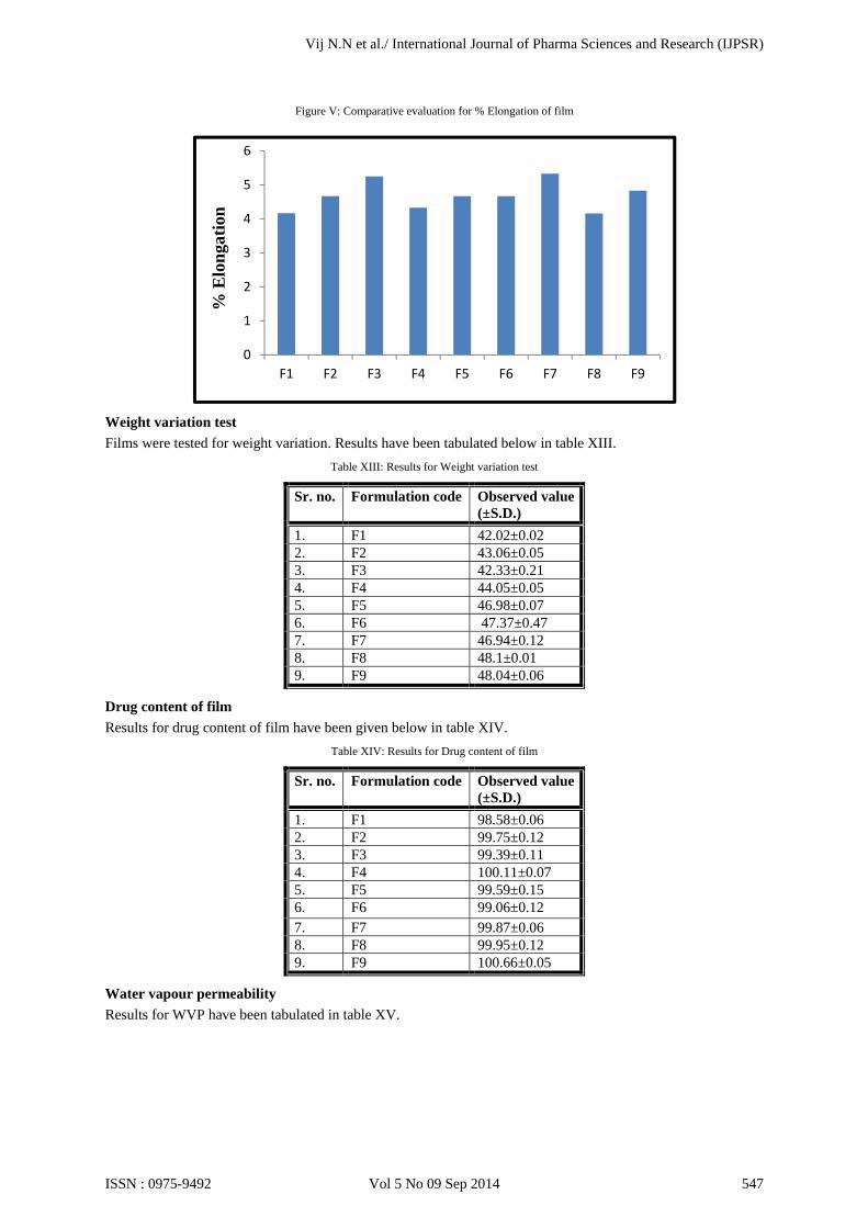
Figure V: Comparative evaluation for % Elongation of film
Weight variation test
Films were tested for weight variation. Results have been tabulated below in table XIII. Table XIII: Results for Weight variation test
Sr. no. Formulation code Observed value (±S.D.)
1. F1 42.02±0.02 2. F2 43.06±0.05 3. F3 42.33±0.21 4. F4 44.05±0.05 5. F5 46.98±0.07 6. F6 47.37±0.47 7. F7 46.94±0.12 8. F8 48.1±0.01 9. F9 48.04±0.06
Drug content of film
Results for drug content of film have been given below in table XIV. Table XIV: Results for Drug content of film
Sr. no. Formulation code Observed value (±S.D.)
1. F1 98.58±0.06 2. F2 99.75±0.12 3. F3 99.39±0.11 4. F4 100.11±0.07 5. F5 99.59±0.15 6. F6 99.06±0.12 7. F7 99.87±0.06 8. F8 99.95±0.12 9. F9 100.66±0.05
Water vapour permeability
Results for WVP have been tabulated in table XV.
0
1
2
3
4
5
6
F1 F2 F3 F4 F5 F6 F7 F8 F9
% E
lon
gati
on
Vij N.N et al./ International Journal of Pharma Sciences and Research (IJPSR)
ISSN : 0975-9492 Vol 5 No 09 Sep 2014 547

Table XV: Results for WVP determination
Sr. no. Formulation code Observed value
(±S.D.) (g cm-2 24h-1)
1. F1 0.0538±0.004 2. F2 0.0523±0.002 3. F3 0.0533±0.008 4. F4 0.0502±0.002 5. F5 0.0512±0.001 6. F6 0.0516±0.005 7. F7 0.0486±0.0005 8. F8 0.0476±0.01 9. F9 0.0478±0.014
According to the British Pharmacopoeia a material can be considered permeable to water vapour when the WVP exceeds 0.05 g cm-2 24h-1. [8] The films displayed such WVP values that show permeability above the limit set in the Pharmacopoeia and can therefore be considered non-occlusive. In-vitro drug release study The In-vitro drug release study of formulations is shown in table XVI.
Table XVI: Cumulative Drug release of formulations
It can be deduced from in vitro diffusion study that formulations F1 to F3 did not sustain the drug release over 24 hours. This may be attributed to low levels of drug release modulating polymers and low viscosity of the formulations. [35] Formulations F4 and F5 sustained drug release over 24 hours. On the other hand, formulations F6 to F9 did not completely release the drug. Drug release was found to be sustained at intermediate levels of hydrophobic polymer, Eudragit and hydrophilic polymer, HPC.
Cumulative Drug Release (%) (±S.D.)
Time (h)
F1 F2 F3 F4 F5 F6 F7 F8 F9
0 0 0 0 0 0 0 0 0 0 0.25 10.01
±0.02 12.64±0.03
24.96±0.03
28.86±0.01
27.88 ±0.04
23.3 ±0.03
25.33 ±0.05
37.49 ±0.04
24.79 ±0.01
0.5 12.7 ±0.02
18.33±0.02
28.53±0.02
32.86±0.42
30.29 ±0.03
29.33 ±0.07
32.61 ±0.02
43.52 ±0.05
30.47 ±0.01
1 19.17±0.07
29.63±0.05
34.43±0.01
38.49±0.05
40.59 ±0.08
37.21 ±0.01
39.85 ±0.03
46.14 ±0.02
39.55 ±0.01
2 27.47±0.03
38.03±0.03
42.59±0.02
47.94±0.05
50.03 ±0.05
43.00 ±0.02
42.51 ±0.04
53.41 ±0.04
46.29 ±0.03
3 34.09±0.02
44.88±0.03
44.35±0.03
54.21±0.05
56.29 ±0.06
47.11 ±0.04
47.21 ±0.04
58.29 ±0.02
50.33 ±0.04
4 40.88±0.06
53.26±0.04
47.35±0.04
59.53±0.03
63.03 ±0.04
56.14 ±0.09
50.86 ±0.06
61.63 ±0.05
56.41 ±0.05
5 49.17±0.02
58.05±0.07
53.78±0.05
63.51±0.06
72.14 ±0.02
61.43 ±0.25
56.5 ±0.02
636.7 ±0.05
61.54 ±0.06
6 60.13±0.02
66.83±0.05
61.89±0.05
71.35±0.05
83.04 ±0.05
67.15 ±0.03
61.97 ±0.04
73.65 ±0.02
64.44 ±0.09
12 96.98±0.03
94.83±0.04
87.94±0.03
91.19±0.03
89.82 ±0.03
79.46 ±0.04
79.87 ±0.01
76.49 ±0.02
74.41 ±0.07
18 96.84±0.03
95.15±0.05
96.24±0.09
95.49±0.02
95.44 ±0.05
88.09 ±0.01
81.71 ±0.01
79.68 ±0.07
77.45 ±0.02
24 97.87±0.03
95.87±0.05
96.93±0.12
97.25±0.36
99.84±0.05
91.69±0.1
84.13±0.01
82.21 ±0.04
79.64±0.03
Vij N.N et al./ International Journal of Pharma Sciences and Research (IJPSR)
ISSN : 0975-9492 Vol 5 No 09 Sep 2014 548

Of the nine formulations, maximum release was found to be for formulation F5 after 24 hours. 99.84% of the drug in the formulation was available for antifungal activity. The composite film had hydrophobic and hydrophilic portions which provide competition for drug release as both the polymers have different release properties. Therefore, as the polymer ratio varies, competition to release drug also varies. Formulation F5 showed steady state release up to 24 hours which also indicates that this formulation would show better contact with biological membrane. In-vitro drug release profile of formulations has been shown in figure VI.
Figure VI: In-vitro drug release profile of formulations F1 to F9
Antifungal activity
The results of antifungal activity of formulations have been shown in table XVII. Table XVII Zone of inhibition and % efficacy of formulations
Sr. no. Formulation Code
Candida albicans
Zone of Inhibition (mm) ±SD
% Efficacy
1. Standard value 24 100 2. F1 22.57±0.21 94.03
3. F2 20.73±0.99 86.39
4. F3 18.23±0.15 75.97
5. F4 23.7±20.2 94.58
6. F5 23.87±0.15 99.44
7. F6 18.9±0.5 78.75
8. F7 17.8±1.31 74.17
9. F8 16.23±0.60 67.64
10. F9 12.4±0.56 51.67
11. Ethanol (control)
1.27±0.23 5.28
12. Drug suspension
23.6±0.61 97.78
13. Commercial cream
17.47±0.40 76.81
The standard value of TH against Candida albicans for zone of inhibition is 24 mm. [36] The study indicates that TH retained its antifungal efficacy when formulated as a film forming dermal gel and drug was active against selected strain of micro-organism. F5 formulation showed a zone of inhibition of 23.87 mm and 99.44%
0
20
40
60
80
100
120
0 500 1000 1500 2000
F-1
F-2
F-3
F-4
F-5
F-6
F-7
F-8
F-9
% C
DR
Time (hrs)
In vitro drug release
Vij N.N et al./ International Journal of Pharma Sciences and Research (IJPSR)
ISSN : 0975-9492 Vol 5 No 09 Sep 2014 549

efficacy. Zone of inhibition for ethanol as a control was also calculated to determine the influence of its inherent antifungal activity. Optimization: [5]
From design expert version 8.0.4.1 thirty nine solutions were found. The batch with Eudragit 12.5 % w/v and HPC 6 % w/v with desirability 1 was found to be optimum. From this data formulation F5 was selected as the optimum formulation. The figures below show the effect of concentration of Eudragit RS PO and Hydroxypropyl cellulose on drug release and antifungal activity. It is shown that both the independent variables have a significant effect on the dependent variables and drug release and antifungal activity decrease as concentration of polymers increases.
Figure VII: Surface response plot showing effect of Eudragit RS PO and Hydroxypropyl cellulose on Drug release
Figure VIII: Surface response plot showing effect of Eudragit RS PO and Hydroxypropyl cellulose on Antifungal activity
Vij N.N et al./ International Journal of Pharma Sciences and Research (IJPSR)
ISSN : 0975-9492 Vol 5 No 09 Sep 2014 550

Figure IX: Contour plot showing effect of Eudragit RS PO and Hydroxypropyl cellulose on Drug release
Figure X: Contour plot showing effect of Eudragit RS PO and Hydroxypropyl cellulose on Antifungal activity
In the figures above, it can be seen that as the concentration of polymers increases the drug release and antifungal activity go on decreasing. Hence it can be concluded that the two factors: X1 and X2 have a combined effect on drug release and antifungal activity. Evaluation of Optimized batch:
Skin irritation study
Skin irritation study on rats showed that after application of the optimized formulation there was no evidence of irritation (erythema and oedema). Hence, the optimized formulation F5 was found to be safe.
Vij N.N et al./ International Journal of Pharma Sciences and Research (IJPSR)
ISSN : 0975-9492 Vol 5 No 09 Sep 2014 551

Figures XXI Photographs of skin irritation test;
A] On application of film B] After removal of film
C] Standard irritant D] Negative Control
Table XVIII: Evaluation table for Skin irritation test according to Draize scoring method
Rat No.
Control group Formulation group Formalin group
Erythema Oedema Erythema Oedema Erythema Oedema
1. 0 0 0 0 2 2 2. 0 0 0 0 2 2 3. 0 0 0 0 2 2
Erythema scale: 0- none; 1-slight; 2- well defined; 3-moderate; and 4- scar formation Oedema scale: 0- none; 1- slight; 2- well defined; 3- moderate; and 4- severe. Best fit kinetic model for optimized formulation The diffusion kinetics of optimized formulation was studied. The best fit model with highest R2 value and least slope value was the first order model.
Table XIX: R2 and slope values for optimized formulation F5
Sr. no. Model R2 Slope
1. Zero order 0.683 0.055 2. First order 0.935 -0.0023. Higuchi 0.899 2.491 4. Korsemeyer-Peppas 0.852 0.562
Stability study
The optimized formulation was evaluated after storage at room temperature and after accelerated stability study at elevated temperature (40oC/75% RH) in stability chamber. [37] Results have been given in table XX and table XXI.
Vij N.N et al./ International Journal of Pharma Sciences and Research (IJPSR)
ISSN : 0975-9492 Vol 5 No 09 Sep 2014 552

Table XX: Stability study data for F5 formulation at room temperature
Sr. no.
Observation Before study During study
3rd month 6th month
1. Clarity Clear Clear Clear 2. pH 5.83±0.01 5.91±0.01 5.94±0.03 3. Viscosity (rpm) 0.3 4877.23 4878.17 4875.23
0.6 4430.43 4416.53 4411.37 1.5 3900.3 3923.67 3903.63 3 3645.07 3643.53 3644.67 6 3299.33 3292.43 3289 12 3033.73 3048.57 3042.7
4. Drug content 99.98±0.13 99.7±0.06 99.59±0.19 5. Antifungal activity 99.44% 98.61% 98.19%
Table XXI: Accelerated Stability study data for F5 formulation
Sr. no.
Observation Before study During study
3rd month 6th month
1. Clarity Clear Clear Clear 2. pH 5.83±0.01 5.9±0.01 5.93±0.02 3. Viscosity (rpm) 0.3 4867.3 4876.33 4875.23
0.6 4419.07 4411.37 4411.37 1.5 3898.23 3913.87 3903.63 3 3624.57 3645.57 3644.67 6 3286.4 3299.13 3289 12 3026.63 3044.03 3042.7
4. Drug content 99.98±0.13 99.62±0.08 99.43±0.15 5. Antifungal activity 99.44% 98.33% 98.06%
CONCLUSION:
Film forming gel of TH was prepared using Eudragit RS PO and hydroxypropyl cellulose. The concentrations of both the polymers were optimized by 32 full factorial design to obtain optimum drug release and antifungal activity. Thus, desirable goals could be achieved by systematic formulation approach. Antifungal study showed that developed film forming gel can reduce the fungal burden and thus, is more effective as compared to commercial product. The film forming dermal gel prepared in this study fulfils all necessary parameters required for topical use. This novel dosage form will improve both the accuracy and the positioning of a delivered dose. The optimized formulation with better bioadhesive property may improve the bioavaibility of topical administration of terbinafine in gel form and can be alternative to the conventionally administered topical formulations.
ACKNOWLEDGEMENTS: The authors duly acknowledge FDC Ltd., Mumbai, India, for providing Terbinafine Hydrochloride as a gift sample, Evonik Degussa India Pvt. Ltd., Mumbai for the gift sample of Eudragit RS PO.
REFERENCES: [1] C.C Long, Common Skin Disorders and their Topical Treatment. In: Walters KA, editor. Dermatological and Transdermal
Formulations. New York: Marcel Dekker Inc., 2002, p. 1-12, 53-54. (Drugs and the Pharmaceutical Sciences, vol 119). [2] S. Buyuktimkin, N. Buyuktimkin, J. Singh, J. Newsam, D. Smith, E. Kisak, inventors, Nuvo Research Inc., assignee. Highly
permeating terbinafine formulation. US patent 2012/0309843 A1. 2012 December 6. [3] C.B. Moore, C.M. Walls, D.W. Denning, In vitro activities of terbinafine against Aspergillus species in comparison with those of
itraconazole and amphotericin B, Antimicrobial Agents and Chemotherapy, 2001, 45(6):1882-85. [4] S. Gungor, M.S. Erdal, B. Aksu, New formulation strategies in topical antifungal therapy. Journal of Cosmetics, Dermatological
Sciences and Applications, 2013, 3:56-65. [5] H. Vaghasiya, A. Kumar, K. Sawant, Development of solid lipid nanoparticles based controlled release system for topical delivery of
terbinafine hydrochloride, European Journal of Pharmaceutics and Biopharmaceutics, 2013, 49:311-22. [6] M. Sen, A. Yakar, Controlled release of antifungal drug terbinafine hydrochloride from poly(N-vinyl 2-pyrrolidone/itaconic
acid)hydrogels, International Journal of Pharmaceutics, 2001, 228:33-41. [7] D.J. Lunter, R. Daniels, New film forming emulsions containing Eudragit NE and/or RS 30D for sustained dermal delivery of
nonivamide, European Journal of Pharmaceutics and Biopharmaceutics, 2012, 82:291-98. [8] I.Z. Schroeder, P. Franke, U.F. Schaefer, C.M. Lehr, Development and characterization of film forming polymeric solutions for skin
drug delivery, European Journal of Pharmaceutics and Biopharmaceutics, 2007, 65:111-21.
Vij N.N et al./ International Journal of Pharma Sciences and Research (IJPSR)
ISSN : 0975-9492 Vol 5 No 09 Sep 2014 553

[9] Li X, R. Zhang, R. Liang, W. Liua, C. Wanga, Z. Su, F. Sun, Y. Li, Preparation and characterization of sustained-release rotigotine film-forming gel, International Journal of Pharmaceutics, 2014, 460(1):273-79.
[10] GELNIQUE [package insert]. Morristown, NJ: Watson Pharmaceuticals, Inc., 2008. [11] BeeGentle [package insert]. West Jordan, UT: CAO Group, Inc., 2010. [12] J.L. Zatz, G.P. Kushla, Gels In: H.A. Lieberman, M.M. Rieger, G.S. Banker, editors. Pharmaceutical Dosage Forms-Disperse System.
2nd ed. New York: Marcel Dekker Inc. p.399-405. (Drugs and the Pharmaceutical Sciences, vol 2). [13] K. Saroha, S. Singh, A. Aggarwal, S. Nanda, Transdermal Gels- An alternative vehicle for drug delivery, International Journal of
Pharmaceutical, Chemical and Biological Sciences 2013, 3(3):495-03. [14] M.A. Attia, H.Y. Badawy, Film forming gel for treatment of oral mucositis: In vitro studies, International Journal of Drug Delivery,
2010, 2:314-321. [15] R. Guo , Du X., Zhang R., Deng L., Dong A., Zhang J., Bioadhesive film formed from a novel organic–inorganic hybrid gel for
transdermal drug delivery system, European Journal of Pharmaceutics and Biopharmaceutics, 2011, 79:574-83. [16] L.K. Souza, C.H. Bruno, L. Lopes, S.H. Pulcinelli, C.V. Santilli, L.A. Chiavacci, Ureasil–polyether hybrid film-forming materials,
Colloids and Surfaces B: Biointerfaces, 2013, 101:156-61. [17] D.M. Jayes, B.S. Nitzan, M.R. Royz, D.M. Barak, O.B. Sholto, R. Zion, S.R. Daudy, inventors, Petah Tikva, assignee, Terbinafine
Formulation, US patent2010/0168233 A1. 2010 July 1. [18] S.M. Mohamed, A.M.E. Masoud, M.D. Elgadir, M.A. Mahdy, Preparation and release charcteristics of itraconazole polymeric films
for topical application, International Journal of Pharmacy and Pharmaceutical Sciences, 2013, 5(3):167-70. [19] B.K. Dey, P.K. Kar, L.K. Nath, Formulation, design, preparation and in vitro-in vivo evaluation of propranolol hydrochloride
transdermal patches using hydrophilic and hydrophobic polymer complex, Research Journal of Pharmacy and Technology, 2009, 2(1):155-60.
[20] Y.B. Ubarchande, T. Regupathy, C. Vijaya, S.V. Deshmane, Formulation and evaluation of mucoadhesive buccal films of losartan potassium, Research Journal of Pharmacy and Technology, 2009, 2(4):833-36.
[21] C. Suja, C. Ramasamy, R. Narayanacharyula, Development and evaluation of lisinopril transdermal patches, Research Journal of Pharmacy and Technology. 2011, 4(8):1260-64.
[22] S.V. Kulkarni, R.P. Kumar, N. Patel, R.B. Someshwara, A.P. Kumar, Development and evaluation of diltiazem HCl transdermal patches by using glycerol and castor oil as plasticizers, Research Journal of Pharmacy and Technology, 2010, 3(3):905-09.
[23] J.R. Kumar, S. Muralidharan, S.A. Dhanaraj, Formulation and in-vitro evaluation of terbinafine hydrochloride transdermal patches, Journal of Pharmaceutical Sciences & Research, 2012, 4(6):1840-43.
[24] D.K. Jain, G.N. Darwhekar, S. Chaurasia, Formulation development and evaluation of transdermal patches of losartan, International Journal of Pharmtech Research, 2012, 4(2):757-64.
[25] D.S. Kumar, R. Sairam, S. Anandbabu, L. Karpagavalli, A. Maheswaran, N. Narayanan, Formulation and evaluation of transdermal patches of salbutamol, Research Journal of Pharmaceutical, Biological and Chemical Sciences, 2012, 3(3):1132-38.
[26] P. Verma, K. Pathak, Nanosized ethanolic vesicles loaded with econazole nitrate for the treatment of deep fungal infections through topical gel formulation, Nanomedicine: Nanotechnology, Biology, and Medicine, 2012, 8:489-96.
[27] S. Amin, S.R. Mir, K. Kohli, A. Ali, Novel polymeric matrix films for transdermal delivery of metoclopramide, International Journal of Pharmaceutical Frontier Research, 2012, 2(1):48-60.
[28] R. Vijaya, S. Sureshkumar, S. Umamaheswari, M. Prakash, T. Senbagapriya, A. Umamaheswari, Preparation of amitriptyline hydrochloride films using eudragit RL 100 and hydroxypropyl methyl cellulose polymers and their in vitro evaluation for effective transdermal delivery, International Journal of Life Science and Pharma Research, 2012, 2(2):7-15.
[29] M. Bharkatiya, R.K. Nema, M. Bhatnagar, Designing and characterization of drug free patches for transdermal application, International Journal of Pharmaceutical Sciences and Drug Research, 2010, 2(1):35-39.
[30] R. Khullar, D. Kumar, N. Seth, S. Saini, Formulation and evaluation of mefenamic acid emulgel for topical delivery, Saudi Pharmaceutical Journal, 2012, 20:63-67.
[31] W. Zhua, C. Guoa, A. Yua, Y. Gaoa, F. Caoa, G. Zhai, Microemulsion-based hydrogel formulation of penciclovir for topical delivery, International Journal of Pharmaceutics, 2009, 378:152-58.
[32] International Conference on Harmonization Steering Committee, ICH Harmonized Tripartite Guideline- Stability Testing of New Drug Substances and Products, ICH Q1A (R2), February 6, 2003.
[33] P.A. McCarron, R.F. Donnelly, A. Zawislak, A.D. Woolfson, Design and evaluation of a water-soluble bioadhesive patch formulation for cutaneous delivery of 5-aminolevulinic acid to superficial neoplastic lesions, European Journal of Pharmaceutical Sciences, 2006, 27:268-79.
[34] D.M. Jayes, B.S. Nitzan, M.R. Royz, D.M. Barak, O.B. Sholto, R. Zion, S.R. Daudy, inventors, Petah Tikva, assignee, Terbinafine Formulation, US patent2010/0168233 A1. 2010 July 1.
[35] P.D. Sawant, D. Luu, R. Ye, R. Buchta, Drug release from hydroethanolic gels, Effect of drug’s lipophilicity (log P), polymer–drug interactionns and solvent lipophilicity, International Journal of Pharmaceutics, 2010, 396:45-52.
[36] H.K. Doddamani, Formulation and validation of organogels as carriers for topical delivery of Terbinafine hydrochloride [dissertation], Bengaluru, India: Ragiv Gandhi University, 2012.
[37] I. Weuts, D. Kempen, A. Decorte, G. Verreck, J. Peeters, M. Brewster, G.V. Mooter, Physical stability of the amorphous state of loperamide and two fragment molecules in solid dispersions with the polymers PVP-K30 and PVP-VA64, European Journal of Pharmaceutical Sciences, 2005, 25:313-20.
Vij N.N et al./ International Journal of Pharma Sciences and Research (IJPSR)
ISSN : 0975-9492 Vol 5 No 09 Sep 2014 554