“Formulation And Texture Characterization Of Environment Friendly ...
FORMULATION, CHARACTERIZATION AND EVALUATION OFNYSTATIN NANOSPONGE GEL FOR THE TREATMENT OF CANDIDIASIS
A Dissertation submittedto
THE TAMIL NADU Dr. M.G.R MEDICAL UNIVERSITYCHENNAI–600032
In partial fulfillment of the requirements for the award of degree of
MASTEROF PHARMACY
Submitted by
Register no:261411263
under the guidance of
Prof.K.Elango,M.Pharm.,(Ph.D.),
ProfessorandHead
Department of Pharmaceutics
COLLEGEOFPHARMACY
MADRAS MEDICAL COLLEGE
CHENNAI–600003
APRIL-2016

DEPARTMENT OF PHARMACEUTICSCOLLEGE OF PHARMACY
MADRASMEDICAL COLLEGECHENNAI-600 003
TAMILNADU
CERTIFICATE
This is to certify that the dissertation entitled “FORMULATION,
CHARACTERIZATION AND EVALUATION OF NYSTATIN NANOSPONGE
GEL FOR THE TREATMENT OF CANDIDIASIS” submitted by the candidate
with RegisterNo.261411263 for the TamilNadu Dr. M.G.R. Medical University
examinations is evaluated.
1.
2.

COLLEGE OF PHARMACY
MADRAS MEDICAL COLLEGE
CHENNAI-600 003
TAMILNADU
CERTIFICATE
This is to certify that the dissertation entitled “FORMULATION,
CHARACTERIZATION AND EVALUATION OF NYSTATIN
NANOSPONGE GEL FOR THE TREATMENT OF CANDIDIASIS”
submitted by the candidate with Register No. 261411263 in partial fulfillment of
the requirements for the award of the degree of MASTER OF PHARMACY in
PHARMACEUTICS by the Tamil Nadu Dr. M.G.R. Medical University is a
bonafide work done by her during the academic year 2015-2016.
Place: Chennai -03.
Date:
( Dr.A.Jerad Suresh, M. Pharm.,M.B.A.,)Ph.D.,

DEPARTMENT OF PHARMACEUTICS
COLLEGE OF PHARMACY
MADRASMEDICAL COLLEGE
CHENNAI-600 003
TAMILNADU
CERTIFICATE
This is to certify that the dissertation entitled “FORMULATION,
CHARACTERIZATION AND EVALUATION OF NYSTATIN
NANOSPONGE GEL FOR THE TREATMENT OF CANDIDIASIS”
submitted by the candidate with RegisterNo.261411263 in partial fulfillment of
the requirements for award of the degree of MASTER OF PHARMACY in
PHARMACEUTICS by the TamilNadu Dr.M.G.R.Medical University is a
bonafide work done by her during the academic year 2015-2016.
Place: Chennai– 03
Date:
(K.Elango)
ACKNOWLEDGEMENT
This thesis is the end of my journey in obtaining my M.Pharm. I have
not travelled in a vacuum in this journey. At the end of my thesis I would like
to thank all those people who made this thesis possible and an unforgettable
experience for me.
I consider this is an opportunity to express my gratitude to all the
dignities who have been involved directly or indirectly with the successful
completion of this dissertation.
First of all I thank the Almighty for giving me strength, endurance and
showering his blessing to undertake this project and pursue with full dedication
and giving us courage always to do hard work.
I consider myself very much lucky with profound privilege and great
pleasure in expressing our deep sense of gratitude to work under the guidance
of Prof. K.Elango, M.Pharm.,(Ph.D.), Head, Department of Pharmaceutics,
College of Pharmacy, Madras Medical College, Chennai for his continuous
guidance, supportive suggestion, innovative ideas, with constant inspiration,
help and encouragement have always propelled us to perform better.
It is my privilege and honour to extend my gratitude and express our
indebtedness to for his enduring support. He has been generous with providing
facilities to carry out this work.
I express my sincere thanks and respectful regards to, in acknowledging
all the facilities provided to us to carry out this work with great ease and
precision.
I express our deepest and very special thanks to, for allowing us to carry
out this project successfully.
I acknowledge my sincere thanks to Prof. Dr. A. Jerad Suresh,
M.Pharm., Ph.D., MBA, Principal, College of Pharmacy, Madras Medical
College, Chennai, for his continuous support in carrying out our project work
in this institution
I am thankful to all of my teaching staff members Asst. Reader Mr.
Ramesh Kumar, M.Pharm., Dr.N.Deattu, M.Pharm., Ph.D.,
Dr.S.DaisyChellakumari, M.Pharm.,Ph.D., Dr. R. Devi Damayanthi,
M.Pharm., Ph.D., of the Department of Pharmaceutics, College of Pharmacy,
Madras Medical College, Chennai -03., for their valuable suggestions,
constant support and encouragement.
I express my heartiest thanks to Mr.N.S.Kumaran, Deputy Manager,
Caplin Point Laboratories, Chennai, for providing the drug Nystatin as gift
sample and Excipients. He gave me full support and encourage throughout my
Project.
I take this an opportunity to thank the Scientist Dr. Hemalatha,
CSIR, Chennai for helping and allowing me to carry out my work in their
premises. I also acknowledge Ms. Arthijeni, for helping me in completion of
my project.
I would like to express my heartfelt thanks to My Parents, My In-
Laws, My Dear Daughters and Husband for their Patience, Sacrifies and
moral supports to successfully carryout my project work.
I take this an opportunity to thank the Scientist, Dr .R. Srinivasan,
Central Leather Research Institute, Chennai for helping me and his
valuable suggestion.
I would like to thank my B.Pharm juniors Dr. Arul Prakash, M.Tech.,
Ph.D., Mr. Ram Prasad, M.Pharm., (Ph.D) and my Senior Mr.StephenM.Pharm.
who stood beside me throughout my project.
I would like to express my thanks to my UG friend Ms. Meenakshi,
M.Pharm., who helped me in my project work.
I would like to give my sincere thanks to my seniors
Chitra,G.Thangalakshmi, S.Kiruthika, D.Renuka, M.Adhilakshmi,
R.Elavarasi, D.Jaison, V.Sundar Raj, B.Prabakaran, K.Gnanasuriyan for
their timely help and co-operation.

I would like to give my sincere thanks to my friends Deepa
Joseph,D.ImmaualNesakumar, M.Meenakshi, D.MohannaPriya,
T.Nandhini, M.Nivedita, D.Saidharshini, C.Saranya, and V.Sundharajan.
I extend my cordial thanks to my juniors for their kind support and co-operation.
I extend my thanks to all non-teaching staff members Mr. R.
Marthandam, Mrs. R. Shankari, Mrs. Razia Sultana, Department of
Pharmaceutics, and Mr. Sivakumar, Department of Pharmaceutical Chemistry,
College of Pharmacy, Madras Medical College, Chennai – 600 003.

ABBREVIATIONS
DDS Drug Delivery System
NDDS Novel Drug Delivery System
mg Milligram
ng Nanogram
ml Millilitre
nm Nanometer
L Litre
PLA Poly Lactic Acid
PLG Poly D,L- Glycolide
PLGA Poly D,L- Lactide co glycolide
PCA Poly Cyano Acrylate
SAS Super Critical Anti-solvent
RESS Rapid Expansion of Critical Solution
DOPE Di Oleolyl Phosphatidyl Ethanolamine
GNP Gliadin Nano Particles
CGNP Clarithromycin Gliadin Nano Particles
OGMP Omeprazole Gliadin Nano Particles
BBB Blood Brain Barrier
CNS Central Nervous System
LDL Low-density Lipoprotein
IBCA Isobutyl 1-cyano acrylate
PVA Polyvinyl Alcohol
UV Ultra-violet
HPLC High Pressure Liquid Chromatography
SEM Scanning Electron Microscope
TEM Transmission Electron Microscope
FT-IR Fourier Transform Infra Red spectroscopy
XRD X-ray diffraction
DSC Differential Scanning Calorimetry
BSA Bovine serum albumin
OPC Oropharyngeal Candidiasis
VVC Vulvovaginal Candidiasis
HIV Human Immuno virus
OTC Over the counter
FDA Food and Drug Administration
PMMA Polymethyl methacrylate
EC Ethyl cellulose
PCS Photon Correlation Spectroscopy
RH Relative Humidity
ICH International Conference on Hormonisation
oC Degree Celsius

TABLE OF CONTENT
S.No. CONTENTS PAGE NUMBER
1.
INTRODUCTION 1DRUG DELIVERY SYSTEM 1NOVEL DRUG DELIVERY SYSTEM 1NANOPARTICLE 5NANOSPONGE 16
2. REVIEW OF LITERATURE 293. AIM AND PLAN OF WORK 384. RATIONALE OF STUDY 395. DISEASE PROFILE 416. DRUG PROFILE 49
7.
EXCIPIENT PROFILE 53Ethyl Cellulose 53Polymethyl Methacrylate 54Polyvinyl Alcohol 56Carbopol 934 58
8. MATERIALS AND METHODS 61Materials used in formulation 61Equipments / Instruments used in formulation 62Compatibility study for Drug and Excipients 63Standard curve for Nystatin 64Formulation Development 64Characterization of Nystatin loaded Nanosponge 65
Determination of Drug Content 65Entrapment efficiency 66In-vitro Drug Release Characteristics 66
Selection and Evaluation of Optimized formulation 66FTIR of Formulation 67Particle Size analysis 67Drug Release Kinetics 67Stability Studies 69
Evaluation of Nanosponge Gel 71Microbiological Study 72
9. RESULTS AND DISCUSSION 7310. SUMMARY AND CONCLUSION 11411. BIBLIOGRAPHY 116
1. INTRODUCTION
Department of Pharmaceutics, Madras Medical College Page 1
1. DRUG DELIVERY SYSTEM
The Drug delivery system refers to a system for transporting pharmaceutical compound
in the body to provide a desired and required therapeutic effect. Drug delivery is a concept
integrated with dosage form and route of administration of pharmaceutical products. The
technologies involved in the formulation of pharmaceutical products modify drug release profile,
absorption, distribution and elimination for the benefit of improving product efficacy and safety,
as well as patient convenience and compliance. Drug release is from: diffusion, degradation,
swelling, and affinity-based mechanisms. Most common routes of administration are non-
invasive peroral (through the mouth), topical (skin), transmucosal (nasal, buccal/sublingual,
vaginal, ocular and rectal) and inhalation routes.1
Newer development of pharmaceutical compounds in the form of targeted delivery
system, in which the drug is only active in the target area of the body (for example, in cancerous
tissues, in brain, in colon), sustained release formulations in which the drug is released over a
period of time in a controlled manner from a formulation.1
2. NOVEL DRUG DELIVERY SYSTEM
The aim of Novel Drug Delivery System is to provide a therapeutic amount of drug to
the appropriate site in the body to accomplish promptly and then maintain the desired drug
concentration. The drug-delivery system should deliver drug at a rate control by the necessarily
of the body over a specified term of treatment.2
This idealized objectives witch to the two aspects most important to drug delivery areas
follows,2
I. Spatial Drug Delivery:
Targeting a drug to a particular organ or tissue.
II. Temporal Drug Delivery:
The drug delivery rate to the target tissue is controlled.

INTRODUCTION
Department of Pharmaceutics, Madras Medical college Page 2
The prime area so research and development for NDDS are:
Liposomes
Niosomes
Nanoparticles
Transdermaldrugdelivery
Implants
Oralsystem
Microencapsulation/Microcapsules
Polymer in drug delivery
Figure-1 :Polymers used in Drug Delivery system.
Efficacy of a drug can be altered by the method of drug delivery into the body. Some
drugs has an optimum concentration in the body and produce maximum therapeutic level,
concentration above or below can produce toxic or no therapeutic action. Slow efficacy of drug
in severe diseases has increases the need for a multidisciplinary approach to the delivery of
therapeutics to targets in tissues. To solve this problem newer development of pharmaceutical
compounds were generated to controlling the pharmacokinetics, pharmacodynamics, non-
specific toxicity, immunogenicity, bio recognition, and efficacy of drugs. The drug delivery
systems (DDS), based on interdisciplinary approaches that combine polymer science,

INTRODUCTION
Department of Pharmaceutics, Madras Medical college Page 3
pharmaceutics, bioconjugate chemistry, and molecular biology are called as Novel drug delivery
system (NDDS).3
To minimize drug degradation and loss, to prevent harmful side-effects and to increase
drug bioavailability and the fraction of the drug accumulated in the required zone, various drug
delivery and drug targeting systems are currently under development. Drug carriers can named
as soluble polymers, microparticles made of insoluble or biodegradable natural and synthetic
polymers, microcapsules, cells, cell ghosts, lipoproteins, liposomes, and micelles. The carriers
can be made slowly degradable, stimuli-reactive (e.g., pH- or temperature-sensitive), and even
targeted (e.g., by conjugating them with specific antibodies against certain characteristic
components of the area of interest).3
ADVANTAGES OF NOVEL DRUG DELIVERY SYSTEM4
Minimization of drug degradation and loss.
Reduction of dosing frequency.
Extension of the duration of action and bioavailability of the drug.
Prevention of drug’s adverse side-effects.
Minimization of drug concentration fluctuations in plasma level.
Improved drug utilization.
Improved patient compliance.
DISADVANTAGES OF NOVEL DRUG DELIVERY SYSTEM4
High cost of final product.
Patients discomfort with DDS device usage.
Possibility of toxicity of the materials.
Harmful degradation products.
Necessity of surgical intervention either on systems application or removal.
INTRODUCTION
Department of Pharmaceutics, Madras Medical college Page 4
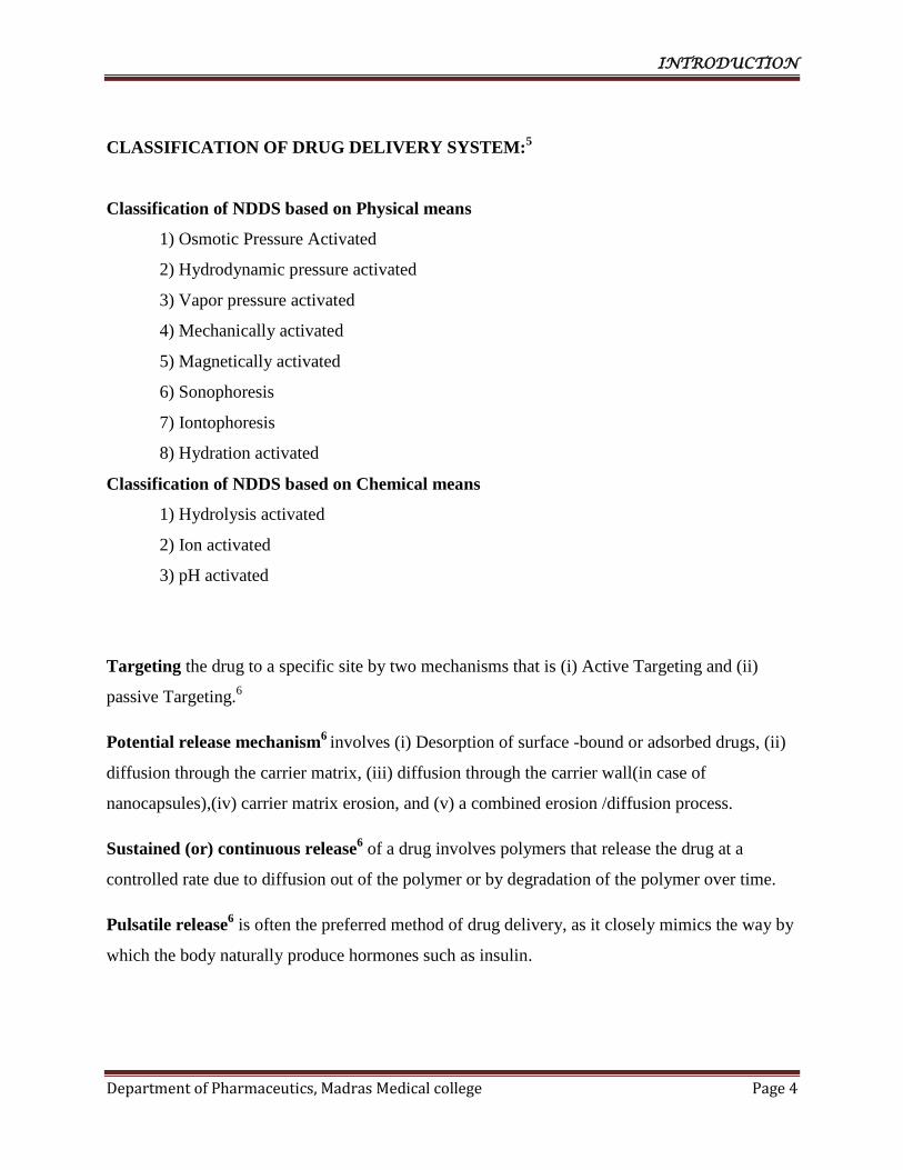
CLASSIFICATION OF DRUG DELIVERY SYSTEM:5
Classification of NDDS based on Physical means
1) Osmotic Pressure Activated
2) Hydrodynamic pressure activated
3) Vapor pressure activated
4) Mechanically activated
5) Magnetically activated
6) Sonophoresis
7) Iontophoresis
8) Hydration activated
Classification of NDDS based on Chemical means
1) Hydrolysis activated
2) Ion activated
3) pH activated
Targeting the drug to a specific site by two mechanisms that is (i) Active Targeting and (ii)
passive Targeting.6
Potential release mechanism6
involves (i) Desorption of surface -bound or adsorbed drugs, (ii)
diffusion through the carrier matrix, (iii) diffusion through the carrier wall(in case of
nanocapsules),(iv) carrier matrix erosion, and (v) a combined erosion /diffusion process.
Sustained (or) continuous release6 of a drug involves polymers that release the drug at a
controlled rate due to diffusion out of the polymer or by degradation of the polymer over time.
Pulsatile release6 is often the preferred method of drug delivery, as it closely mimics the way by
which the body naturally produce hormones such as insulin.

INTRODUCTION
Department of Pharmaceutics, Madras Medical college Page 5
Colloidal drug carrier system6 such as micellar solution, vesicle and liquid crystal dispersion as
well as nanoparticle consisting of small particles of 10-400 nm diameter shows great promise as
drug delivery system.
Micelles6 formed by self - assembly of amphiphilic block co-polymer (5-50 nm) in aqueous
solutions are of great interest for drug delivery application. They can be physically entrapped in
the core of block copolymer micelles and transported at concentration that can exceed their
intrinsic water solubility.
Liposomes3 are a form of vesicles that consist either of many, few or just one phospholipid
bilayers. The polar character of the liposomal core enables polar drug molecules to be
encapsulated. Amphiphilic and lipophilic molecules are solubilized within the phospholipid
bilayer according to their affinity towards the phospholipids. Participation of nonionic
surfactants instead of phospholipids in the bilayer formation results inniosomes. The drugs that
are encapsulated in a nanocage-functionalized with channel proteins are effectively protected
from premature degradation by proteolytic enzymes.
Dendrimers3 are nanometer-sized, highly branched and monodisperse macromolecules with
symmetrical architecture. They consist of a central core, branching units and terminal functional
groups. The core together with the internal units, determine the environment of the nanocavities
and consequently their solubilizing properties, whereas the external groups the solubility and
chemical behaviour of these polymers.
Liquid Crystals3
combine the properties of both liquid and solid states. They can be made to
form different geometries, with alternative polar and non-polar layers (i.e., a lamellar phase)
where aqueous drug solutions can be included.
Nanoparticles7 can be defined as particulate dispersion or solid particle with a size range in 10-
1000 nm.The drug is dissolved , entrapped, encapsulate or attached to a nanoparticle matrix.
Depending upon the method of preparations, nanoparticle, nanospheres, or nanocapsules can be
obtained.

INTRODUCTION
Department of Pharmaceutics, Madras Medical college Page 6
The primary goals for research of nano-bio-technologies in drug delivery include:8
More specific drug targeting and delivery,
Reduction in toxicity while maintaining therapeutic effects,
Greater safety and biocompatibility, and
Faster development of new safe medicines.
Method of preparation:
1. Dispersion of preformed polymers.9
a) Solvent evaporation method.11
b) Spontaneous emulsification or solvent diffusion method.11
2. Polymerization of monomers9 and
3. Ionic gelation or coacervation of hydrophilic polymers.9
4. Supercriticalfluid technology.11
1.Dispersion of preformed polymers:11
It is the most common method used to prepare nanoparticle from poly(lactic acid) (PLA),
poly (D L Glycolide), PLG, Poly(D,L-lactide-co-glycolide) PLGA, and Poly(cyanoacrylates)
PCA.
a) Solvent evaporation method:11
Organic solvent such as Dichloromethane, Chloroform, or ethyl acetate are used to
dissolve the polymer which is also used as solvent to dissolve hydrophobicdrugs. The
drug dissolved or dispersed in a polymer solution is then emulsified in an aqueous
solution containing a surfactant or emulsifying agent to form oil in water emulsion. Once
a stable emulsion is formed the solvent is subjected to evaporation either by reducing the
pressure or by continuous stirring. High speed homogenizer or Ultra sonicator is used for
the preparation of small uniform sized particle.

INTRODUCTION
Department of Pharmaceutics, Madras Medical college Page 7
b) Spontaneous emulsification or solvent diffusion method:11
This is a modification of solvent evaporation method. In this the water miscible solvent
along with water immiscible organic solvent is used as an oil phase. An interfacial
turbulence is formed between the two phases due to spontaneous diffusion of immiscible
solvents leading to formation of small particles. By increasing the concentration of water
miscible solvent decrease in the particle size can be achieved. Both solvent evaporation
and solvent diffusion methods can be used for the hydrophobic and hydrophilic drugs.
2.Polymerization of monomers:11
In this method monomers are polymerized to form nanoparticle in an aqueous solution in which
drug maybe dissolved. Drugs may also be incorporated by adsorption onto the nanoparticles after
polymerization completed. The nanoparticle suspension is then purified by ultracentrifugation
and resuspending in an isotonic surfactant - free medium. This technique is used for preparing
polybutylcyanoacrylateor poly (alkylcyanoacrylate) nanoparticle.
3.Ionic gelation or coacervation of hydrophilic polymers :
The method involves a mixture of two aqueous phases, of which one is the polymer
chitosan, a di-block co-polymer ethylene oxide or propylene oxide (PEO or PPO) and the other is
the polyanion sodium tripolyphospate. Positively charged amino group in chitosan interacts with
negatively charged tripolyphosphate to form coacervates with a size in the range of nanometer.
Coacervates are formed as a result of electrostatic interaction between two phases , whereas,
ionic gelation involves the material undergoing transition from liquid to gel due to ionic
interaction condition at room temperature.
4.Supercritical fluid technology:
Supercritical fluid technology has been investigated as an alternative to prepare
biodegradable micro and nanoparticles. Supercritical fluids are environmentally safe.
A supercritical fluid can be generally defined as a solvent at a temperature above its
critical temperature, at which the fluid remains a single phase regardless of pressure.
Supercritical CO2(SCCO2)is the most widely used supercritical fluid because of its mild critical
conditions (Tc=31.1°C,Pc=73.8bars),non toxicity, non-flammability, and lowprice. The most
INTRODUCTION
Department of Pharmaceutics, Madras Medical college Page 8
common processing techniques involving supercritical fluids are supercritical anti- solvent(SAS)
and rapid expansion of critical solution (RESS).
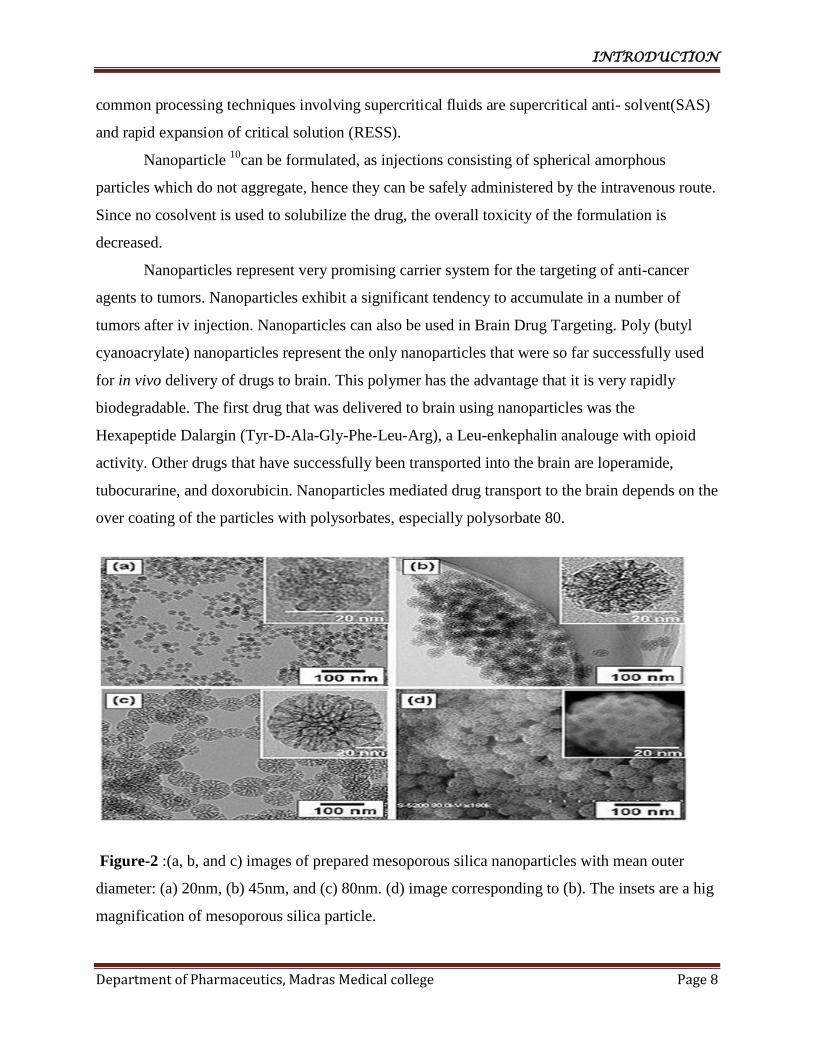
Nanoparticle 10
can be formulated, as injections consisting of spherical amorphous
particles which do not aggregate, hence they can be safely administered by the intravenous route.
Since no cosolvent is used to solubilize the drug, the overall toxicity of the formulation is
decreased.
Nanoparticles represent very promising carrier system for the targeting of anti-cancer
agents to tumors. Nanoparticles exhibit a significant tendency to accumulate in a number of
tumors after iv injection. Nanoparticles can also be used in Brain Drug Targeting. Poly (butyl
cyanoacrylate) nanoparticles represent the only nanoparticles that were so far successfully used
for in vivo delivery of drugs to brain. This polymer has the advantage that it is very rapidly
biodegradable. The first drug that was delivered to brain using nanoparticles was the
Hexapeptide Dalargin (Tyr-D-Ala-Gly-Phe-Leu-Arg), a Leu-enkephalin analouge with opioid
activity. Other drugs that have successfully been transported into the brain are loperamide,
tubocurarine, and doxorubicin. Nanoparticles mediated drug transport to the brain depends on the
over coating of the particles with polysorbates, especially polysorbate 80.
Figure-2 :(a, b, and c) images of prepared mesoporous silica nanoparticles with mean outer
diameter: (a) 20nm, (b) 45nm, and (c) 80nm. (d) image corresponding to (b). The insets are a hig
magnification of mesoporous silica particle.

INTRODUCTION
Department of Pharmaceutics, Madras Medical college Page 9
TYPES OF NANOPARTICLES
1. Quantum Dot
Aquantum dot is a semiconductor nanostructure that confines the motion of conduction
band electrons, valence band holes, or excitons (pairs of conduction band electrons and
valence band holes) in all three spatial directions. The confinement can be due to
electrostatic potentials (generated by external electrodes, doping, strain, impurities), due
to the presence of an interface between different semiconductor materials (e.g. in the case
of self-assembled quantum dots), due to the presence of the semiconductor surface (e.g.
in the case of a semiconductor nanocrystal), or to a combination of these. A quantum dot
has a discrete quantized energy spectrum. A quantum dot contains a small integer number
(of the order of 1-100) of conduction band electrons, valence band holes, or excitons, i.e.,
an integer number of elementary electric charges.
Quantum dots can be contrasted to other semiconductor nanostructures:
1) Quantum wires, which confine the motion of electrons or holes in two spatial
directions and allow free propagation in the third.
2) Quantum wells, which confine the motion of electrons or holes in one direction and
allow free propagation in two directions.
2. Nanocrystalline silicon
Nanocrystallinesilicon(nc-Si) - an allotropic form of silicon - is similar to amorphous
silicon (a-Si), in that it has an amorphous phase. Where they differ, however, is that nc-Si
has small grains of crystalline silicon within the amorphous phase. This is in contrast to
polycrystalline silicon (poly-Si) which consists solely of crystalline silicon grains,
separated by grain boundaries. nc-Si is sometimes also known as microcrystalline
silicon (µc-Si) The difference comes solely from the grain size of the crystalline grains.
Most materials with grains in the micrometre range are actually fine- grained polysilicon,
so nanocrystalline silicon is a better term.
3. Photonic crystal
Photonic crystals are periodic dielectric or metallo-dielectric (nano) structures that are

INTRODUCTION
Department of Pharmaceutics, Madras Medical college Page 10
designed to affect the propagation of electromagnetic waves (EM) in the same way as the
periodic potential in a semiconductor crystal affects the electron motion by defining
allowed and forbidden electronic energy bands.
Since the basic physical phenomenon is based on diffraction, the periodicity of the photonic
crystal structure has to be in the same length-scale as half the wavelength of the EM
waves i.e. ~300 nm for photonic crystals operating in the visible part of the spectrum.
This makes the synthesis cumbersome and complex. To circumvent nanotechnological
methods with their big and complex machinery, different approaches have been followed
to grow photonic crystals as self-assembled structures from colloidal crystals.
Photonic crystals are attractive optical materials for controlling and manipulating the flow of
light. They are of great interest for both fundamental and applied research, and are
expected to find commercial applications soon.
Figure-3: STRUCTURE OF NANOPARTICLES
INTRODUCTION
Department of Pharmaceutics, Madras Medical college Page 11
4. Liposomes
A liposome is a spherical vesicle with a membrane composed of a phospholipid bilayer used to
deliver drugs or genetic material into a cell. Liposomes can be composed of naturally-derived
phospholipids with mixed lipid chains (like egg, phosphatidyl ethanolamine), or of pure
components like DOPE (di oleolyl phosphatidyl ethanolamine).
The lipid bilayer can fuse with other bilayers (e.g., the cell membrane), thus delivering the
liposome contents. By making liposomes in a solution of DNA or drugs,(which would
normally be unable to diffuse through the membrane), they can be (indiscriminately)
delivered past the lipid bilayer.
5. Gliadin nanoparticles
In an effort to improve bioavailability anti-H.pylori effects of antibiotics, mucoadhesive
gliadin nanoparticles (GNP) which have the ability to deliver the antibiotics at the sites
of infection were prepared. GNP bearing clarithromycin(CGNP) and omeprazole(OGNP)
were prepared by desolvation method.
in vivo gastric mucoadhesive studies confirmed the strong mucoadhesive propensity and
specificity and specificity of gliadin nanoparticles towards stomach. Gliadin nanoparticles
show a higher tropism for the gastrointestinal regions and their presence in other intestinal
regions is very low. This high capacity to interact with the mucosa may be explained by
gliadin composition.
In fact, this protein is rich in neutral and lipophilic residues. Neutral amino acid can promote
hydrogen bonding interaction with the mucosa whereas the lipophilic components can
interact within biological tissue by hydrophilic interaction. The related protein gliadin
possessing an amino and disulphide groups on the side chain has a good probability of
developing bonds with mucin gel.
6. Polymeric Nanoparticles
Polymeric nanoparticles have been invented by Speiser et al. They represent interesting
alternative as drug delivery systems to liposomes. They usually exhibit a long shelf life
and a good stability on storage.
INTRODUCTION
Department of Pharmaceutics, Madras Medical college Page 12
These are superior to liposomes in targeting them to specific organs or tissues by adsorbing and
coating their surface with different substances.
Nanoparticles can be prepared either from preformed polymers, such as polyesters (i.e.
polylactic acid), or from a monomer during its polymerization, as in the case of alkyl-
cyanoacrylates.
Most of the methods based on the polymerization of monomers consists in adding a
monomer into the dispersed phase of an emulsion, an inverse microemulsion, or dissolved
in a non-solvent of the polymer.
7. Solid Lipid Nanoparticles (SLN)
Solid lipid nanoparticles have been developed as alternative delivery system to
conventional polymeric nanoparticles. SLNs are sub-micron colloidal carriers (50-
1000nm) which are composed of physiological lipid, dispersed in water or in an aqueous
surfactant solution.
SLNs combine advantages of polymeric nanoparticles, fat emulsions and liposomes, but
avoid some of their disadvantages. They are biodegradable, biocompatible and non-toxic.
8. Nanospheres16
Nanospheres may be defined as solid core spherical particulates, which are nano in size.
They contain drug embedded within the matrix or adsorbed on to the surface.
9. Nanocapsules16
Nanocapsules are the vesicular system in which drug is essentially encapsulated
within central volume surrounded by an embryonic continuous polymeric sheath.
10. Others10
:
Gold nanoparticles stabilized by thiol functionality are extraordinarily stable and therefore are a
great system for studying nanostructure formation. They have many applications. Because gold
nanoparticles are so easy to synthesize they have been studied intensely in recent years.
INTRODUCTION
Department of Pharmaceutics, Madras Medical college Page 13
A common synthesis involves the reduction of a gold salt in the presence of capping agent
molecules such as thiols, citrates or phosphines. The functionalities of these capping
agents can be altered to yield various chemical properties.
Advantages of Nanoparticles
Some of the advantages of using nanoparticles as a drug delivery system are as follows;
1. Ease of manipulation of the particle size and surface characteristics of
nanoparticles so as to achieve both passive and active drug targeting after parenteral
administration.
2. The nanoparticle surface can be modified to alter bio distribution of drugs with
subsequent clearance of the drug so as to achieve maximum therapeutic efficacy with
minimal side effects of the drug.
3. Controlled release and particle degradation characteristics can be readily
modulated by the choice of matrix constituents.
4. Drug loading is relatively high and drugs can be incorporated into the systems
without any chemical reaction; this is an important factor for preserving the drug
activity.
5. Site-specific targeting can be achieved by attaching targeting ligands to surface
of particles or use of magnetic guidance.
6. Liposomes and polymer based nanoparticulates are generally biodegradable, do
not accumulate in the body and so are possibly risk free.
7. Small sized nanoparticles can penetrate through smaller capillaries, which could
allow efficient drug accumulation at the target sites.
8. Various routes of administration are including oral, nasal, parenteral, intra-
ocular etc.
Disadvantages
In spite of these advantages nanoparticles do have limitations like,
1. Altered physical properties which lead to particle–particle aggregation, making physical

INTRODUCTION
Department of Pharmaceutics, Madras Medical college Page 14
handling of nanoparticles difficult in liquid and dry forms due to smaller size and larger
surface area.
2. Smaller the particles size greater the surface area and this property makes nanoparticles very
reactive in the cellular environment.
3. Small particles size results in limited drug loading and burst release. These practical
problems have to be sorted out before nanoparticles can be used clinically or made
commercially available.
ApplicationsofNanoparticles12
In Anti-Microbial Techniques :
One of the earliest nanomedicine applications was the use of nanocrystalline silver
which is as an antimicrobial agent for the treatment of wounds, a nanoparticle cream has been
shown to fight infections.
In Tumor targeting using :
The rational of using nanoparticles for tumor targeting is based on (1) nanoparticles will
be able to deliver a concentrate dose of drug in the vicinity of the tumor targets via the enhanced
permeability and retention effect or active nanoparticles. (2) Nanoparticles will reduce the drug
exposure of health tissues by limiting drug distribution to target organ.
In Gene delivery :
Polynucleotide vaccines work by delivering genes encoding relevant antigens to host
cells where they are expressed, producing the antigenic protein within the vicinity of
professional antigen presenting cells to initiate immune response. Such vaccines produce both
humoral and cell-mediated immunity because intracellular production of protein, as opposed to
extracellular deposition, stimulates both arms of the immune system.
In Cell Repair :
Nanorobots could actually be programmed to repair specific diseased cells, functioning
in a similar way to antibodies in our natural healing processes.

INTRODUCTION
Department of Pharmaceutics, Madras Medical college Page 15
In brain targeting :
The blood-brain barrier(BBB)is the most important factor limiting the development of
new drugs for the central nervous system. Relatively impermeable endothelial cells characterize
the BBB with tight junctions, enzymatic activity and active efflux transport systems. It
effectively prevents the passage of water-soluble molecules from the blood circulation into the
CNS, and canal so reduce the brain concentration of lipid- soluble molecules by the function of
enzymes or efflux pumps. Consequently, the BBB only permits selective transport of molecules
that are essential for brain function. Strategies for nanoparticle targeting to the brain rely on the
presence of and nanoparticle interaction with specific receptor- mediated transport systems in the
BBB.7
Nanoparticles can get access to the brain by two different mechanisms, ie, (1) trans
synaptic transport after inhalation through the olfactory epithelium, and (2) uptake through the
blood-brain barrier.8
In some cases it is reported to mimic molecules that would normally be transported to
brain. For example, polysorbate-coated nanoparticles are thought to mimic low-density
lipoprotein (LDL), allowing them to be transported across the capillary wall and into the
brain by hitching a ride on the LDL receptor.13
In Topical drug delivery:
Nanoemulgel15
:
When nanoemulsion is incorporated into gel it is called as nanoemul gel. Nanoemulsions
are thermodynamically stable transparent (translucent)dispersions of oil and water stabilized by
an interfacial film of surfactant and cosurfactant molecules having a droplet size of less
than100nm. Nanoemulsion formulations possess improved transdermal and dermal delivery
properties in-vitro as well as in-vivo. Nanoemulsions have improved transdermal permeation of
many drugs over the conventional topical formulations such as emulsions and gels e.g.Carvedilol
nanoemul gel was prepared using oleicacid and isopropyl myristate (3:1) as oil phase. Tween20
INTRODUCTION
Department of Pharmaceutics, Madras Medical college Page 16
and Carbitol were used as surfactant and cosurfactant respectively. Carbopol934 was used as
gelling agent15
.
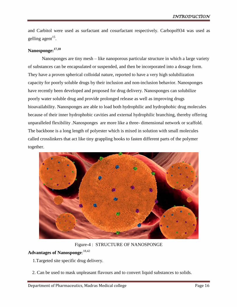
Nanosponge:17,18
Nanosponges are tiny mesh – like nanoporous particular structure in which a large variety
of substances can be encapsulated or suspended, and then be incorporated into a dosage form.
They have a proven spherical colloidal nature, reported to have a very high solubilization
capacity for poorly soluble drugs by their inclusion and non-inclusion behavior. Nanosponges
have recently been developed and proposed for drug delivery. Nanosponges can solubilize
poorly water soluble drug and provide prolonged release as well as improving drugs
bioavailability. Nanosponges are able to load both hydrophilic and hydrophobic drug molecules
because of their inner hydrophobic cavities and external hydrophilic branching, thereby offering
unparalleled flexibility .Nanosponges are more like a three- dimensional network or scaffold.
The backbone is a long length of polyester which is mixed in solution with small molecules
called crosslinkers that act like tiny grappling hooks to fasten different parts of the polymer
together.
Figure-4 : STRUCTURE OF NANOSPONGE
Advantages of Nanosponge:18,42
1.Targeted site specific drug delivery.
2. Can be used to mask unpleasant flavours and to convert liquid substances to solids.

INTRODUCTION
Department of Pharmaceutics, Madras Medical college Page 17
3. Less harmful side effects (since smaller quantities of the drug have contact with healthy
tissue).
4.Nanosponge particles are soluble in water, so the hydrophobic drugs can been capsulated
with in the nanosponge, after mixing with a chemical called an adjuvant reagent.
5. Particles can be made smaller or larger by varying the proportion of cross-linkerto the
polymer.
6. Production through fairly simple chemistry called "click chemistry"(methods for making
the Nanosponge particles and for attaching the linkers).
7. Easy scale-up for commercial production.
8.The drug profiles can be tailored from fast, medium to slow release, preventing over or
under-dosing of the therapy.
9.The material used in this system can provide a protective barrier that shields the drug from
premature destruction with in the body.
10.Improved stability, increased elegance and enhanced formulation flexibility.
11.Nanosponges systems are non-irritating , non-mutagenic, non-allergenic and non-toxic.
12.These are self sterilizing as the average pore sizeis 0.25µm, where bacteria cannot
penetrate.
13.Hydrophobic drugs can be encapsulated within the Nanosponge.
14.Extended release-continuous action up to 12 h.
15.Biodegradable.
Disadvantages
1. The main disadvantage of these nanosponges is their ability to include only small
molecules.

INTRODUCTION
Department of Pharmaceutics, Madras Medical college Page 18
2. The nanosponges could be either Para crystalline or in crystalline form. The loading
capacity of nanosponges depends mainly on degree of crystallization. Para crystalline
nanosponges can show different loading capacities.
3. The nanosponges can be synthesized to be of specific size and to release drugs overtime
by varying the proportion of cross linker to polymer.
Characteristic features of nanosponges56
.
Nanosponges exhibit a range of dimensions (1 µm or less) with tunable polarity of the
cavities. Nanosponges of specific size and adjustable polarity can be synthesized by
varying the crosslinker to polymer proportion.
They could be either para-crystalline or in crystalline form, depending on the process
conditions. Crystal structure of nanosponges plays a very important role in their
complexation with drugs.
The drug loading capacity of nanosponges mainly depends on the degree of
crystallization. Para-crystalline nanosponges have shown various drug loading capacities.
They are nontoxic, porous particles insoluble in most organic solvents and stable at high
temperatures up to 300 °C.
Nanosponges as formulations are stable over the pH range of 1 to 11 and temperature up
to 130 °C.
They form clear and opalescent suspensions in water and can be regenerated by simple
thermal desorption, extraction with solvents, by the use of microwaves and ultrasounds.
Their 3D structure enables capture, transportation and selective release of a vast variety
of substances. They can be targeted to different sites due to their ability to be linked with
different functional groups. Chemical linkers enable nanosponges to bind preferentially
to the target site. They form inclusion and non-inclusion complexes with different drugs.
Magnetic properties can be also imparted to nanosponges (by adding magnetic particles
into the reaction mixture).
Mechanism of Drug Release18
The sponge particles have an open structure and the active is free to move in and out
from the particles and into the vehicle until equilibrium is reached . In case of topical delivery,
once the finished product is applied to the skin, the active that is already in the vehicle will be

INTRODUCTION
Department of Pharmaceutics, Madras Medical college Page 19
absorbed into the skin, depleting the vehicle, which will become unsaturated, therefore
disturbing the equilibrium. This will start a flow of the active from the sponge particle into the
vehicle and from it to the skin until the vehicle is either dried or absorbed .Even after that the
sponge particles retained on the surface of stratum corneum will continue to gradually release
the active to the skin, providing prolonged release overtime.
Figure -5: drug release mechanism in Topical drug delivery.
FACTORS AFFECTING DRUG RELEASE FROM NANOSPONGES18
:
Physical and chemical properties of entrapped actives.
Physical properties of sponge system like pore diameter, pore volume, resiliency etc.
Properties of vehicle in which the sponges are finally dispersed.
Particle size, pore characteristics, composition can be considered as imperative
parameters.
External triggers like temperature, pressure, and solubility of actives.
Pressure: Pressure or rubbing can release active ingredient from Nanosponges onto
skin.
Temperature: Some entrapped actives can be too viscous at room temperature to flow
spontaneously from sponges onto the skin but increased skin or environment can result
in increased flow rate and ultimately drug release.
Solubility: Sponges loaded with water soluble drug like antiperspirants and antiseptic
release the ingredients in the presence of water.
• ACTIVE INGREDIENTS
Sponge
• ACTIVE INGREDIENTS
Vehicle • ACTIVE
INGREDIENTS
Skin

INTRODUCTION
Department of Pharmaceutics, Madras Medical college Page 20
The nanosponges are encapsulate the drug molecule within its core. By the method of
associating with drugs, the nanoparticles can be classified into74
:
1. Encapsulating nanoparticles:- These are represented by nanosponges and
nanocapsules. Nanosponges such as alginate nanosponges, which are sponge
like structure that carry the drug molecules. Nanocapsules such as
poly(isobutyl-cyanoacrylate) (IBCA) are also encapsulating nanoparticles.
They can entrap drug molecule in their aqueous core.
2. Complexing nanoparticles:- These nanoparticle attract the molecule by
electrostatic charges.
3. Conjugating nanoparticles:- These nanoparticles links to drug through strong
covalent bonds.
PREPARATIONOFNANOSPONGES21
.
Nanosponges are prepared depending on type of delivery system, polymers and
nature of drug and solvents. Various approaches used for formation of Nanosponges
are:
1.Polymerization:
The polymerization process leads to the formation of a reservoir type of system, which
opens at the surface through pores. A solution of non-polar drug is made in the
monomer, to which aqueous phase, usually containing surfactant and dispersant to
promote suspension is added. Polymerization is effected, once suspension with the
discrete droplets of the desired size is established; by activating the monomers either by
catalysis or increased temperature.
2.Quasi-emulsionsolventdiffusion
The Nanosponges can also be prepared by quasi-emulsion solvent diffusion method
using the different polymer amounts. To prepare the inner phase, EudragitRS100 was
dissolved in suitable solvent. Then, drug can be added to solution and dissolved under

INTRODUCTION
Department of Pharmaceutics, Madras Medical college Page 21
ultra sonication at 35°C.The inner phase was poured into the PVA solution in water
(outerphase). Following 60min of stirring, the mixture is filtered to separate then
anosponges. The Nanosponges aredried in an air-heated oven at 40 ° C for 12hrs.
3.Emulsion solvent diffusion method
In this method the two phases used are organic and aqueous. Aqueous phase consists of
polyvinyl alcohol and organic phase include drug and polymer. After dissolving drug
and polymer to suitable organic solvent, this phase is added slowly to the aqueous phase
and stirred for two or more hours and then Nanosponges are collected by filtration,
washed and then dried in air at room temp or in vacuum oven at 40°C for 24hours.
Polymers Used in Nanosponge Preparation:
There are various polymers and crosslinkers are used in the preparation of nanosponges,
listedinTable -1 Table -1
Polymers Copolymers
Crosslinkers
Hyper cross linked
Polystyrenes,
Poly(valerolactone Carbonyl diimidazoles,
Carboxylic acid Cyclodextrines and
Alkyloxycarbonyl Cyclodextrins,
allylvalerolactone),Poly
(valerolactone-
allylvalerolactone
dianhydrides,
Diarylcarbonates,
Dichloromethane, Diisocyanates,
Diphenyl
Methylβ-
Cyclodextrin,HydroxyPropyl
β-Cyclodextrins, Ethyl Cellulose.
oxepanedione),Polyvinyl
alcohol.
Carbonate, Epichloridine,
Gluteraldehyde,
Pyromelliticanhydride,2,2-
bis(acrylamido) Acetic acid.

INTRODUCTION
Department of Pharmaceutics, Madras Medical college Page 22
Some drugsformulated as nanosponges18
are given in table -2.
Table -2:Drugs used as Nanosponges.
Drug Nanosponge
vehicle
Therapeutic indication
Econazolenitrate EthylCellulose
Polyvinylalcohol
Antifungal
Paclitaxel β-Cyclodextrin Cancer
Tamoxifen β-Cyclodextrin BreastCancer
Resveratrol β-Cyclodextrin Inflammation
Cardiovascular diseases
Dermatitis
Gonorrhea
Fever
Hyperlipidemia
Dexamethasone β-Cyclodextrin Braintumors
Temozolam ide Poly
(valerolactone-
allylvalerolactone) Poly
(valerolactone-
allylvalerolactone-
oxepanedione)
Braintumors
EVALUATIONOF NANOSPONGES
1. Particle size determination18
:
The particle size can be determined by dynamic light scattering using 90 plus
particle sizer equipped with MAS OPTION particle sizing software (Malvern zeta sizer). From
this the mean diameter and poly dispersity index can be determined.
2. ZetaPotential18
Zeta potential is a measure of surface charge. It can be measured by using additional
electrode in the particle size equipment.
3. LoadingEfficiency18
The loading efficiency of Nanosponges can be determined by the quantitative estimation
of drug Loaded into Nanosponges by UV spectro photometer & HPLC methods.

INTRODUCTION
Department of Pharmaceutics, Madras Medical college Page 23
4. Microscopic study18
Scanning electron microscopy (SEM) and Transmission electron microscopy (TEM) are
used to study the microscopic character of nanosponge. Morphology of the Nanosponge is
studied with SEM.
5. CompatibilityStudies18
The compatibility study of drug with the adjuvants are determined by FTIR,TLC.
Crystalline characteristics can be studied by powder X-ray diffraction (XRD) and Differential
Scanning Calorimetry(DSC).
6.Thin Layer Chromatography18
In TLC, formation of the complex in nanosponge is determined by the Rf
values.
7.Solubility study18
The Inclusion complexation are studied by phase solubility method described
by higuchi and Connors whichexamines the effect of a Nanosponge, on the solubility
of drug.
8.Single Crystal X-Ray Structure18
This method used to determine the detailed inclusion structure and mode of interaction.
The interaction between the host and guest molecules can be identified and the precise
geometrical relationship can be established. This information obtained during the analysis lead
to know about the formation of inclusion complexes.
9.Drug release study18
Release study for the drug is done with dialysis membrane using Franz diffusion cell.
10.Drug kinetic study18
To investigate the mechanism of drug release from Nanosponge the release data could
be analysed using Zero order,
INTRODUCTION
Department of Pharmaceutics, Madras Medical college Page 24
Firstorder, Higuchi, Peppas, Hixon-Crowell, Kopcha and Makoid-Banakar etc. models.
The data can be analysed using graph pad prism software.
11.Porosity19
Porosity study is carried out to check the Nanochannels and nanocavities formed in
Nanosponge. Porosity of Nanosponge is determined by Helium displacement method using
Helium pynometer.
12.Swelling and water uptake19
For swellable polymers like polyamidoamine nanosponges, water uptake can be
determined by soaking the prepared Nanosponges in aqueous solvent. Swelling and water up
take can be calculated using equations.
13.Resiliency(Viscoelasticproperties)19
Resiliency of sponges can be modified to produce beadlets that is softer or firmer
according to the needs of the final formulation. Increased crosslinking tends to slow down the
rate of release. Hence resiliency of sponges will be studied and optimized as per the requirement
by considering the releaseas a function of cross-linking with time.
ComparisonofSomeEffectiveVesicularSystems19
.
Liposome, niosome, ethosome, transferosome and nanosponge are colloidal drug delivery
systems. They all are nanometric in size. Liposome, noisome and transferosomes have some
stability problems which is discuss below in table but Nanosponge enhanced the stability of
drug.

INTRODUCTION
Department of Pharmaceutics, Madras Medical college Page 25
Table-3:Comparison of Nanosponge with Vesicular system.
Liposome Niosome Ethosome Transferosome Nanosponge
Liposome consist of one or more concentric lipid bilayers, which enclose an internal aqueous volume.
Niosomes are non- Ionic surfactant vesicles obtained on hydration of synthetic nonionic surfactants, withor without incorporation of cholesterol or other lipids.
Ethosomes are lipid vesicles containing phospholipids, alcohol(ethanol and isopropylalcohol)in relatively high concentration and water.
Transferosomes are vesicular system consisting of phosphatidyl choline and surfactants.
Nanosponge are novel class of hyper- crosslinked polymer based colloidal structures consisting of solid nanoparticles with colloidal sizes and nanosized cavities.
The composition of liposomes is phospholipids and cholesterol.
They composed of non-ionic surfactants and cholesterol.
They composed mainly of phospholipids, high concentration of ethanol and water.
They consists of phospholipids and surfactants.
They composed of polymers and cross linkers.
Stability problems:
due the formation of ice crystals in liposomes, the subsequent instability of bilayers leads to the leakage of entrapped material. The oxidation of cholesterol and phospholipids also leads to the formulation instability.
Stability problems:
fusion, aggregation, sedimentation and leakage on storage.
The Hydrolysis of encapsulated drug.
Ethosomes has initiated a new area in vesicular research for transdermal drug delivery which can provide better skin permeation and stability than liposomes.
Application of ethosomes provides the advantages such as improved entrapment and physical stability.
Stability problem:
chemically unstable because of their predisposition to oxidative degradation.
Nanosponge are chemically and physically stable. They increase the stability and bioavailability, modify drug release and reduce side- effects.
APPLICATIONS OF NANOSPONGE:
Topical delivery19
:
Local anesthetics, antifungal and antibiotics are among the category of the drugs that
can be easily formulated as topical Nanosponges. A wide variety of substances can be

INTRODUCTION
Department of Pharmaceutics, Madras Medical college Page 26
Incorporated into a formulated product such as gel, lotion, cream, ointment, liquid, or powder.
Enhanced Solublity19
The Nanosponge system has pores, that increase the rate of solubilisation of poorly
soluble drug by entrapping such drugs in pores. Due to nanosize surface area significantly
increased and increase rate of solubilisation.
Nanosponge as chemical sensor19
:
Nanosponges which are the type of “ metaloxides ”act as a chemical sensors which is
used in highly sensitive detection of hydrogen using Nanosponge titania. Nanosponge structure
initially have no point of contact so there is less hinderance to electron transport and it results in
higher 3D interconnect Nanosponges titania which is sensitive to H2 gas.
Used as Taste masking agent.40
Chemotherapy19
The tiny sponges are filled with drug and expose a targeting Peptide that bind to
radiation induced cell surface receptor on tumor. When the sponge encounter tumor cell they
stick to surface and triggered to release cargo. One of the important drug formulated as
Nanosponge is paclitaxel, the active ingredient in the anti-cancer therapy Taxol.
Figure-6: Nanosponge particle attaching to human breast cancer cells16
INTRODUCTION
Department of Pharmaceutics, Madras Medical college Page 27
In anti viral therapy17
:
Nanosponges can be useful in the ocular, nasal and pulmonary administration routes. The
selective delivery of antiviral drugs or small interfering RNA (siRNA) to the nasal epithelia
and lungs can be accomplished by nanocarriers in order to target viruses that infect the RTI such
as respiratory sinctial virus, influenza virus and rhinovirus. They can be used for Human
Immunodeficiency Virus (HIV), HepatitisB Virus (HBV) and Herpes Simplex Virus
(HSV).Drugs used are zidovudine, saquinavir, interferon-α, acyclovir, nelfinavir etc.
In protein drug delivery17
Bovine serum albumin(BSA) protein is unstable in solution form so stored in lyophilized
form. Swellable cyclodextrin based poly(amidoamino)Nanosponge enhanced the stability of
proteins like BSA. Nanosponges have also been used for enzyme immobilization, protein
encapsulation, and subsequent controlled delivery and stabilization.
In oxygen delivery system19,54
Cyclodextrin Nanosponges have also been developed as oxygen delivery system.
Nanosponge has the ability to store and to release oxygen slowly overtime. Oxygen-
filled nanosponges could supply oxygen to the hypoxic tissues which are present in various
diseases.
Characterized by using a, ß and ϒ cyclodextrins and this are suspended in water and get
saturated with water. A silicone form of membrane can also be used for oxygen permeation with
the help of nanosponge/ hydrogel system. They can also applied it to hypoxic tissues caused in
various type of diseases.
Toxin absorbing Nanosponges used to absorb Toxin of staphylococcus aureus which
produce resistance to Methicillin37
.
More effectiveness than direct injection:56
Recent research suggests that nanosponge could be up to five times more effective at
reducing tumor growth than direct injection. The drug delivery system is likened to be filling
virus-sized sponges with an anti-cancer drug, attaching chemical linkers that bond to a receptor
on the surface of tumor cells, then injecting the sponges into the body. When the sponges come
into contact with a tumor cell, they either attach to the surface or are sucked into the cell, where
they off-load their deadly contents in a predictable and controlled manner.

INTRODUCTION
Department of Pharmaceutics, Madras Medical college Page 28
Novel flame retardants containing cyclodextrin nanosponges and phosphorus compounds
to enhance EVA combustion properties69
A novel flame retardant in tumescent system, aimed to improve the fire stability of ethylene
vinyl acetate copolymer (EVA), has been prepared by melt blending of the copolymer and a
complex of cyclodextrin nanosponge-phosphorus compounds. As compared to traditional
systems, this complex which is stable in processing conditions, has the advantage that
nanosponges act as both carbon sources and foam forming agents while the phosphorus
compounds are able to directly generate phosphoric acid in situ. In this context, cyclodextrin
nanosponges undergo dehydration in presence of the acid source, generating water vapour and
char, and thus protecting the copolymer against combustion.
OtherApplications
Biomedical Application,
Analytical Application,
For Hydrogen storage,
In Agriculture,
In Floriculture,
In Food Industry,
For water Purification,
For Oil Cleaning,
As Novel flame Retardants,
Against pore forming Toxins and superbug infections, and
Micro patterning of Mammalian cell.
2. REVIEW OF LITERATURE
Department of Pharmaceutics, Madras Medical College Page 29
1. Dr Prathima srinivas et al 31
formulated and Evaluated Voriconazole Nanosponges for
Oral and topical Delivery as Tablets and Gel. Voriconazole Nanosponges were prepared
by Emulsion Solvent Evaporation Technique with three different Polymers. Final
formulations and Nanosponges were evaluated for Drug content, Entrapment efficiency,
Physical parameters, FTIR Study, In-vitro and in-vivo study, and Anti microbial activity.
From the above evaluation they concluded that the three polymers used were efficient
carriers for Voriconazole Nanosponge showing Diffusion controlled release.
2. Raja CH. NV. et al 36
fabricated and Evaluated Ciprofloxacin loaded Nanosponges for
sustained release. Five batches of Nanosponges using different proportion of Ethyl
cellulose were prepared by solvent evaporation method. The Nanosponges were
evaluated for Characterization by Scanning electron microscopy, Solubility study, In-
vitro dissolution study, and Drug entrapment efficiency. Thereby they concluded that the
Final batch (F5) is considered as a best entrapped and has a greater percentage drug
release.
3. Gautam Seema et al 33
developed and evaluated curcumin loaded Nanosponges for
colon drug delivery. Six batches of Nanosponges were prepared and zeta potential, Drug
content, Drug entrapment efficiency, Surface characteristic by SEM, Particle size, FTIR,
DSC, Stability studies, In-vitro drug release, and Kinetic models of dissolution profiles
were evaluated. Thereby they concluded that curcumin and Eudragit L -100 1:3.5 (F4
batch) has found to be the optimum formulation, that has a significant improvement of
curcumin performance from Nanosponges compared to pure curcumin and other
formulation.
4. P.Suresh Kumar et al 37
formulated and evaluated Miconazole nitrate Loaded
Nanosponges for vaginal drug delivery. Nanosponges were prepared using different
ratios of beta cyclodextrin and Di phenyl carbonate. The Miconazole nitrate was loaded
into the beta cyclodextrin Nanosponges by solvent evaporation technique using various
solvents. Nanosuspension was prepared with the Miconazole Nanosponge. That
nanosuspension is used for the preparation of Gel using Carbopol 934 as gelling agent.
Both the Nanosponge and Gel were evaluated. Thereby they concluded that the
2. REVIEW OF LITERATURE
Department of Pharmaceutics, Madras Medical College Page 30
optimized formulation has good spreadability, extrudability, mucoadhesive nature and
has sustained release.
5. Renuka Sharma et al 34
fabricated and Evaluated Econazole nitrate Nanosponges as
topical hydrogel. Econazole Nanosponge were prepared by using various concentration
of ethyl cellulose and evaluated for entrapment efficiency, in -vitro drug release size
measurement, Rheological properties. For hydrogel the Equilibrium swelling study,
viscosity analysis, texture analysis, in-vitro permeation study has evaluated.
6. Fei Wang, et al38
formulated Hydrogel Retaining Toxin – Absorbing Nanosponges for
Local Treatment of Methicillin Resistant Staphlococcus aureus infection. The
Staphlococcus aureus which are resistant to the methicillin due to their toxin activity
towards the drug. These type of toxins which were forming pore and constitute important
bacterial virulence factors. These toxins disrupt cells by forming pores on cellular
membranes and altering their permeability for bioactivity. Hybrid nanostructures like
Nanosponges may has ability to absorb toxins and by reducing their effect. That can be
proved by in-vitro -toxin Neutralization study, Live whole body imaging of mice to
study Nanosponge retention, in-vivo -toxin Neutralization study and in- vivo
Detoxification efficacy against localized MRSA infection.
7. Gouri Shankar and Y.K. Agarwal40
formulated and Evaluated -cyclodextrin
Nanosponges of a poorly water soluble drug. -cyclodextrin Nanosponge were prepared
and Simavastatin was added to it. The Simvastatin Nanosponges are evaluated for the
Particle size. Polydispersity, Zeta potential, Entrapment efficiency, FT-IR, DSC,XRD,
Mucoadhesive strength ,in-vitro release study, Kinetics study, and Stability study.
Higher Solubilization and prolonged release of Simvastatin Nanosponges served the
purpose of synthesizing Nanosponges. Thereby they concluded that the -cyclodextrin
based Nanosponge was an effective nanocarrier for the delivery of simvastatin.

2. REVIEW OF LITERATURE
Department of Pharmaceutics, Madras Medical College Page 31
8. Monica R. P. Rao et al41
developed and Evaluated the Nanosponge- based Pediatric-
controlled release dry suspension of Gabapentin for reconstitution. Nanosponge of
Gabapentin were formulated using -cyclodextrin by melt method. The Nanosponge drug
complex were characterized by FT-IR, DSC and PXRD as well as evaluated for taste and
saturated solubility. Then it was coated with Ethyl cellulose and Eudragit RS 100. It
shows the release of drug from Nanosponge was in controlled manner, as well as the
taste of Gabapentin was masked. Leaching of drug is insignificant shows the stability of
Nanosponge. Thereby they conclude that the Nanosponge prepared with -cyclodextrin
was an ideal nanocarrier for controlled release.
9. Kurhe, et al42
describes Scaffold based drug delivery system :A Special emphasis on
Nanosponge. Nanosponges were prepared to have ability to encapsulate either
hydrophilic or lipophilic drug and release the drug in a controlled and predictable fashion
at the target site. Thereby they concluded Nanosponges increases the drug solubility
ofpoorly water soluble drugs and protects the drug from physiochemical degradations. It
can be developed asdifferent dosage forms like Paretrals,aerosol, topical, tablet and
capsule. Thus, Nanosponges are a Boon for targeted and site specific drug delivery
system.
10. Shastrulagari Shivani et al43
reviewed Nanosponge – novel emerging drug delivery
system. Types of Nanosponge, advantages, Polymers and cross linker used are reviewed.
Method of preparation of Nanosponge, Factors influencing Nanosponge, characterization
and applications.
11. P.Suresh Kumar. et al 44
prepared and Evaluated Clotrimazole loaded Nanosponges
containing vaginal Gels. Vaginal gel were formulated and the various evaluation are
carried out for Nanosponge and for the Gel. Thereby they concluded that the Nanosponge
gel prepared with HPMC showed good extrudability, homogeneity, spreadability and
required diffusion rate in comparison with other formulations and selected as suitable
carrier to be delivered through vaginal route at controlled rate.
2. REVIEW OF LITERATURE
Department of Pharmaceutics, Madras Medical College Page 32
12. Satyen J. torne, et al 45
prepared and Evaluated for Enhance oral paclitaxel
bioavailability after administration of paclitaxel loaded Nanosponge. The prepared
Nanosponge is evaluated for general test for Nanosponge and the pharmacokinetics of
Paclitaxel through Nanosponge.The plasma concentration of paclitaxel after the oral
administration were significantly higher than the Taxol at the same dose shows the
increased effect than conventional dose/
13. Marzouk M.A., et al 46
prepared and Evaluated the Nystatin Topical formulations with
various Penetration enhancers. All the general parameters are evaluated for the gel and
compared with the plain gel and concluded that the penetration enhancer used Propylene
glycol showed the highest effect in the amount drug permeated followed by
dimethylformamide. The antifungal activity of all Nystatin gels was found to be greater
than that of commercial nystatin cream.
14. Bazigha k. Abdul Rasool, et al47
in-vitro release study of Nystatin from chitosan buccal
gel. Bioadhesive Nystatin gel with chitosan as gelling agent and various enhancers. All
the general tests for gel were evaluated and then the Antifungal effect of Nystatin was
carried out. From the above study thy concluded that the 5% Chitosan with10% w/w of
PG as solubilizing agent has best activity and in-vitro release shows the enhancement of
solubility over commercial product.
15. Reeta T.Metha, et al48
formulated the Liposome encapsulaed Nystatin for systemic
Candidiasis. Liposomes are formulated using various lipids and reagents. That liposomes
are subjected for all the tests and concluded that the formation of liposomes reduces the
toxicity and improve the release.
16. Rodino S, et al49
Nystatin Determined quantitatively by spectrometric method. The
average Nystatin content resulting from the five determination differs from the expected
value not more than 3.15% of the average. Thereby they concluded that the analytical
method based on exploiting photosensitivity and selective photo transformation of the
active substance proved to be reliable for analytical control.
2. REVIEW OF LITERATURE
Department of Pharmaceutics, Madras Medical College Page 33
17. Gabriela Badea, et al50
formulated Nystatin loaded nanostructured carriers for the
influence of basil oil extract on the antioxidant and antifungal activity. Lipid
nanoparticles were prepared with basil oil and evaluated for the anti oxidant activity and
anti microbial activity. Charcterization of size , uniformity, and stability of the
nanoparticle is measured. The antioxidant activity was maintained in the area of a high
ability scavenge free radicals. Thereby they concluded that the NLC based on 2% basil
oil and loaded with 1%nystatin was found to be the most active against Candida albicans.
18. Sachin S. Salunkhe ,et al51
investigated Nystatin loaded Liposomal gel for topical
application by factorial design. Nystatin liposomes were prepared by ethanol injection
method and the effect of variables , size distribution , zeta potential, entrapment
efficiency, and Skin permeation and drug deposition study were done. For the liposomal
gel, the rheological study, drug content, content uniformity and stability studies were
performed. The results were obtained by factorial design and concluded that the
liposomal gel were found to be significantly increase the skin permeation and deposition
in comparison to standard reference product, and has potential application in topical
delivery.
19. Ruba Kello,52
prepared Polyethylene Glycol Novel based Nystatin and Triamcinolone
acetonide ointment. Prepared ointment is subjected to Stability studies, and they
concluded that it polyethylene glycol 1500 and polyethylene glycol 400 can be used as an
alternative ointment base for Nystatin and Triamcinolone acetonide.
20. Tahseen h. Nasti, et al77
prepared and evaluated the pH-sensitive Nystatin liposomes
against Crytococcus neoformans in murine model. The pH sensitive Nystatin liposomes
were prepared by Egg Phosphatidylcholine, cholesterol and mixing the DOPE and
CHEMS in the ratio of 2:3.all the ingredients were mixed in a round bottomed flask and
minimum quantity of chloroform or methanol (1:1) . Solvents were carefully evaporated
to for liposomes. Antifungal susceptibility test were carried out in mice.Results were
compared with Nystatin. As they concluded that the infection was subsided owing to the
use of pHsensitive Nystatin liposomes that release substantial drug at the pH favorable

2. REVIEW OF LITERATURE
Department of Pharmaceutics, Madras Medical College Page 34
for Cryptococcus neoformans. Nystatin , toxic in free form, can be made more effective
and safe entrapping it within pHsensitive liposomes for treatment of fungal disease.
21. Mohammad Barzegar-Jalali,et al53
formulated and evaluated Nystatin suspensions to
determine Effect of different buffered and unbuffered dispersion medium on the physical
and chemical stability. The Prepared nystatin suspensions were evaluated for the
Sedimantation volume, resuspendability, Freeze-thaw cycling test, chemical testing like
Arrhenius accelerated stability test and shelf life. Thereby they concluded that the
buffered cellulosic suspending agent containing nystatin suspension gave stable
suspension when compared to unbuffered vehicle.
22. Kunikazu Mobibe, et al54
formulated and evaluated the Nystatin liposomes for the
Encapsulation characteristics by effects of cholesterol and polyethylene glycol
derivatives. The prepared liposomes are evaluated for the Fluoresence measurement of
nystatin binding. They concluded that the encapsulation of liposomes in relation to the
incorporation of cholesterol and DSPG-PEG, High encapsulation efficiency was obtained
in CH-free PEG-liposomes. Molecular interaction between nystatin and DSPE-PEG
appears to invovlve in the interaction of amino group of nystatin with the phosphate
group of nystatin in the liposomal membrane. These results may help in the development
of effective pharmaceutical formulations for injectable hydrophobic drugs with reduced
side effects.
23. Akhani Jolly R*, et al59
formulated developed and evaluated the in situ gel for vaginal
drug delivery of anti fungal drug. When poloxamers PF 127 and PF 68) were used in
combination for developing thermosensitive and mucoadhesive in situ gel, low to
moderate amounts of HPMC K4M and PF68 were to be used to achieve the desired
gelation temperature, gel strength, mucoadhesion, drug release profile and viscosity
required for a sustained vaginal drug delivery system of nystatin. It was concluded that
the amounts of HPMC K4M had a significant effect on bioadhesion force and gel
strength of the formulated thermosensitive and mucoadhesive in situ gel. The quadratic
mathematical model developed is applicable to predicting in situ gel vaginal
formulations with desired characteristics.
2. REVIEW OF LITERATURE
Department of Pharmaceutics, Madras Medical College Page 35
24. Rawia M. Khalil* et al66
formulated and characterized the nystatin-loaded
nanostructured lipid carriers for topical delivery against cutaneous candidiasis .
Nys NLC is prepared with various surfactants, and Lipids. The prepared NLC
were evaluated for Particle size, Entrapment, TEM, DSC, Stability and Release ,
antimicrobial activity. Thereby they conclude that the prepared Nys NLC is
better than the commercial products.
25. Kunikazu Moribe, et al67
investigated the molecular state of Nystatin
encapsulated in liposomes by spectrometrically. Liposomes with DPPC,
DPPC/CH, DPPC/DSPE-PEG, DPPC/CH/DSPE-PEG, DPPC/CH/DSPE-PEG.
The molecular state of Nystatin in liposomes are subjected to spectrometically,
thereby they concluded that compared with AmB, Nystatin does not readily form
a complex with DSPE-PEG and the enhanced encapsulation of Nystatin would be
attributed to the CH-free liposomes. DPPC liposomes shows the least stability in
these composition.
26. A. Hassan, et al68
confirmed Candida albicans endogenous fungal
endophthalmitis in a patient with chronic Candidiasis. They concluded that
confirmed C. albicans endophthalmitis in a patient with chronic vaginal
Candidiasis, and need a particular importance in patients with additional
immunosuppressive risk factors. So need to carefully monitor patient’s eyes or
administer antifungal prophylaxis in patients with active genital Candidiasis
undergoing procedures. In this case conventional microbiological laboratory
diagnostics were negative and EFE was only confirmed with molecular
techniques. This highlights the need to utilize DNA diagnostics in the early
identifications of at risk patients.
27. Journal of Antimicrobial Chemotherapy69
studied the use of fluconazole and
itraconazole in the treatment of Candida albicans. In this article they concluded
that fluconazole remains a first-line antifungal agent of choice for the treatment
of C. albicans infections, because of its well-known efficacy and safety profile;

2. REVIEW OF LITERATURE
Department of Pharmaceutics, Madras Medical College Page 36
its suitability for use in children, the elderly and patients with impaired immunity; its
range of formulations; and its cost.
28. Mary K. Truhlar,et al 69
quantified the antifungal activity of Nystatin
incorporated in denture liners by new assay technique. Preparation incorporating
higher concentrations of nystatin were more potent inhibitor of Candida albicans
growth, and loss of antifungal activity for all concentrationswas the greatest
from day 0 to 2.Incorporating a single doseof an antimycotic agents into the
reline material of the denturethat allows release of the agent over 2 week period
is a treatment modality that offers considreable promise for the management of
oral candidiasis.
29. B.Indira, et al72
reviewed Nanosponges, a New Era in Drug Delivery. They
Reviewed on the Nanosponge about the Advantages, Disadvantages, Method of
Preparation, Polymers and crosslinkers Used, Factors infiuencing Nanosponge,
Characterization of Nanosponges, and their applications. They concluded that
this new invention, nanosponges will pave a way in overcoming the challenges in
designing of targeted drug delivery systems because of their ability to
accommodate either hydrophilic or lipophilic drugs and release them in a
controlled and predictable manner at specific site in the body. The release rate
can be modulated by controlling the polymer and cross linker ratio. The
nanosponges have been found to possess a profound ability to protect essential
biomarkers in diseases (cancer for example) and biocatalysts from
physicochemical degradation. In near future they stand as milestones in drug
delivery.
30. E. K.Patel et al73
reviewed Nanosponge and micro sponges a Novel drug
delivery system. They reviewed about all the aspect of Nanosponge and
elaborated about the uses of prepared Nanosponges. Camptothecin (CAM), a
plant alkaloid and potent antitumor agent, has a limited therapeutic utility
because of its poor aqueous solubility, lactone ring instability and serious side
effects. Cyclodextrin based nanosponges(NS) are a novel class of cross-linked
2. REVIEW OF LITERATURE
Department of Pharmaceutics, Madras Medical College Page 37
derivatives of cyclodextrins. They have been used to increase the solubility of
poorly soluble actives, to protect the labile groups and control the release. This
study aimed at formulating complexes of Camptothecin with ßcyclodextrin based
nanosponge. They concluded that the Nanosponges are a Nanosized particle
entrap wide variety of products, enhance solubility, provide prolonged use and
provide patient compliance with the formulation.

5. AIM AND PLAN OF WORK
Department of Pharmaceutics, Madras Medical College Page 38
AIM OF WORK
The aim of the present study is
To formulate Nystatin loaded Nanosponge using various polymers.
To provide an efficient dosage form.
PLAN OF WORK
The present work is designed and planned as follows:
Drug-polymer compatibility studies by FT-IR.
Determination of λ max of Nystatin.
Preparation of Calibration curve of Nystatin.
Formulation of Nystatin loaded Nanosponge
Formulation of Nystatin Nanosponge using different polymers (Ethyl cellulose and
Polymethyl methacrylate) at different ratios (Drug:Polymer-1:1,1:2,1:3,1:4and 1:5)
using Solvent Emulsion evaporation Method.
Characterization of Nystatin loaded Nanosponges
Determination of physical properties.
Determination of drug content.
Determination entrapment efficiency.
Determination of particle size distribution.
in-vitro release studies of Nystatin loaded Nanosponge.
Kinetics of drug release.
Morphology of Nanosponge by Scanning electron microscopy(SEM) technique.
Stability studies.
Formulation of Nystatin loaded Nanosponge Gel for optimized product:
Evaluation of Nystatin loaded Nanosponge Gel.
Determination of physical properties.
Determination of drug content.
In-vitro release studies of Nystatin loaded Nanosponge Gel.
Antifungal activity of the Nystatin loaded Nanosponge Gel.

4.Rationale of Study
Department of Pharmaceutics, Madras Medical College Page 39
RATIONALE OF THE STUDY
Fungal Infections have been increasing in incidence in the human population over the
last 15 years or so, largely as a consequence of increased numbers of cancer and immune
compromised patients, who are at risk owing to weakened immune systems and chronic nature of
the disease.
Systemic fungal infections are more serious as they are usually more difficult to
diagnose, are chronic in nature, and in some cases, can become life-threatening. They occur more
frequently in individuals with compromised immune systems (AIDS patients; transplant patients;
cancer patients). Prophylactic treatment is sometimes indicated in AIDS patients and bone
marrow transplant patients, but risk of developing resistance is high. Life-threatening infections
require the use of more potent but much more toxic antifungal.
Superficial fungal infections are almost always caused by dermatophytes or yeasts. In
some instances, they can be rather tenacious, requiring very long treatments, sometimes with
both oral and topical drugs.
RATIONALE OF SELECTION OF DRUG61
Nystatin (Nys) is a polyene antifungal antibiotic, one of the oldest antifungal drug,
produced by Streptomyces noursei strains Nys monomers selectively interact with ergosterol
causing membrane disruption and eventual cell death. Nys possesses a broad spectrum with both
antifungal and fungistatic activity, being effective against azole-resistant strains of Candida and
amphotericin B resistant strains of Candida albicans. Nys is indicated for treatment of Cutaneous
and mucocutaneous fungal infections caused by Candida species, the main yeast capable of
infecting the oral mucosa, C. albicans being the most common species isolated . Oral Candidiasis is
not a lethal disease in healthy patients. It is mainly caused by antibiotic or corticosteroid treatment
and dental prosthesis. However, it must be treated to avoid chronic and systemic invasions of other
tissues, especially among patients with diabetes mellitus, immune compromised or under aggressive
treatments (e.g. chemotherapy) to prevent opportunistic invasive fungal infections.
The presence of a large lactone ring with several double bonds renders it an
amphiphilic and amphoteric molecule with poor solubility in aqueous media (360 mg/L at

Rationale of Study
Department of Pharmaceutics, Madras Medical college Page 40
24˚
C), which reveals great formulation
challenges. This fact support
that
an increasingly
important area of pharmaceutical research is focused
on finding safe and effective methods of
solubilizing poorly soluble
drugs.
Adverse effect of Nys is very rare. From Neonates to Elderly can use Nys, even Pregnant
women can use it safely.62
Cost of Nystatin cream when compared to other antifungals is low.
RATIONALE FOR THE SELECTION OF DOSAGE FORM.20,65
Nanosponge are mainly for Topical drug delivery. It is very much suitable for
antifungal agents. Nanosponge is a nano sized particle, has larger surface area, thereby the
penetration is high when compared to other conventional products. Low solubility drugs
formulated as Nanosponge may increase the solubility thereby increase the release and activity.
Stability of Nanosponge is higher than most of the novel formulation.
Nystatin has only topical action, there is no systemic effect, and it is slightly soluble in
water, hence preparations as Nanosponge increases its effect by increasing solubility.

5.Disease Profile
Department of Pharmaceutics, Madras Medical College Page 41
CANDIDA SPECIES
Candida species is the most common cause of opportunistic fungal infection worldwide.
Candida is the major fungal pathogen of humans, causing diseases ranging from superficial
mucosal infections to disseminated, systemic infections that are often life threatening. A striking
feature of its pathogenicity is ability to grow in yeast, pseudohyphal and hyphal forms. The
hyphal form has an important role in causing disease by invading epithelial cells and causing
tissue damage. Among Candida spp., Candida albicans is the most common infectious agent.
This dimorphic yeast is a commensal that colonizes skin, the gastrointestinal and the
reproductive tracts. Non-C. albicans species are also emerging pathogens and can also
colonize human mucocutaneous surfaces. The pathogenesis and prognosis of candidial infections
are affected by the host immune status and also differ greatly according to disease presentations.
Candidiasis is also technically known as candidosis, moniliasis and oidiomycosis22
.
Table-4 : Candida species implicated in human infections.
Common species Less common species Rare species
Candida albicans
Candida glabrata
Candida dubliniensis
Candida famata
Candida blankii
Candida bracarensis
Candida tropicalis Candida
parapsilosis Candida krusei
Candida inconspicua
Candida lipolytica
Candida metapsilosis
Candida catenulate Candida
chiropterorum Candida ciferri
Candida guilliermondii
Candida lusitaniae
Candida norvegensis
Candida orthopsilosis
Candida eremophila
Candida fabianii
Candida kefyr Candida pelliculosa
Candida rugosa
Candida fermentati
Candida freyschussii
Candida zeylanoides Candida haemulonii
Candida intermedia
Candida lambica
Candida magnolia
Candida membranae faciens
Candida nivariensis
Candida palmioleophila
Candida pararugosa
Candida pseudohaemulonii
Candida pseudorugosa
Candida pintolopesii
Candida utilis
Candida valida
Candida viswanathii

5.Disease Profile
Department of Pharmaceutics, Madras Medical College Page 42
Candida albicans
It is a diploid yeast with two pairs of 8 chromosomes. Its genome size is about 16 Mb. It
possesses 6,159 coding genes. It is the predominant cause of invasive fungal infections. People
at risk include those suffereing from HIV, cancer and intensive care unit patients for example
those undergoing major surgery and organ transplants.
Candida glabrata (Torulopsis glabrata)
It is a small, haploid, monomorphic yeast with 13 chromosomes and a genome size
of 12.3 Mb. It possesses 5283 coding genes. It has become important because of its
increasing incidence worldwide and decreased susceptibility to antifungals. Its emergence is
largely due to an increased immunocompromised patient population and widespread use of
antifungal drugs. In many hospitals, C. glabrata is the second most common cause of
candidemia.
Candida tropicalis
It is a diploid, with 10 to 12 chromosomes and a haploid genome size of 15 Mb. It
possesses 6258 genes. C. tropicalis is the third or fourth most commonly recovered Candida
species from blood cultures. C. tropicalis has progressively been observed to be the
commonest cause of invasive candidiasis in neutropenic patients such as those with acute
leukaemia or those who have under- gone bone marrow transplantation.
Candida parapsilosis
It is a diploid or aneuploid with 14 chromosomes and a genome size of 16 Mb. It
possesses 5733 genes. It is one of the principal causes of invasive Candidiasis. In most parts of
the world, it is the third most common cause of candidemia especially in patients with
intravenous cathe- ters, prosthetic devices, and intravenous drug use. It is one of the most
common causes of candidemia in neonatal intensive care units.
Candida krusei
It is a diploid, with 3-5 chromosomes and genome size of 11 Mb. It is the fifth most
common bloodstream isolate, although, less common (1 to 2%), C. krusei is of clinical
significance because of its intrinsic resistance to fluconazole and reduced susceptibility to most
other antifungal drugs. It is frequently recovered from patients with hematological malignancies
complicated by neutropenia tends to be associated with higher mortality rates (49 vs 28% with C.
albicans), and lower response rates (51 vs 69% with C. albicans..

5.Disease Profile
Department of Pharmaceutics, Madras Medical College Page 43
Candida guilliermondii (teleomorph-Pichia guilliermondii)
It is a haploid. It is reported to have genome size of 12 Mb and possess coding genes
5920. It has been isolated from environmental surfaces and from the skin and nails of
healthcare workers. It has been shown to cause hematogenously disseminated Candidiasis.
Candida lusitaniae (teleomorph-Clavispora lusitaniae)
It is a haploid. It has genome size of 16 Mb. Among, the rare non-albicans Candida
species, it has emerged during the last 20 years as an important nosocomial pathogen
throughout the globe.
Candida kefyr
It consists of 18 chromosomes. Candida kefyr was only isolated from urine cultures
(Weichert et al., 2012). It has been reported to cause systemic Candida infection in patients
with neutropenic leukemia and in a woman with underlying heart disease. Very recently, it has
been described as a pathogen causing invasive fungal enteritis in a patient with underlying
haematological disease following bone marrow transplantation.
Major types of Candidiasis
Candidasis is an acute or chronic infection produced by Candida, generally limited to the
skin and mucous membranes, but it could produce a serious systemic disease.
Mucosal candidiasis
Candidal infections are restricted to non-sterile mucosal surface for example
oropharyngeal and vulvuvaginal Candidiasis.
Oropharyngeal candidiasis (OPC)
Oral candidiasis is one of the most common, oral mucosal infections seen in persons
with HIV. Candida is commensal organism and part of normal oral flora in about 30 to
50% of the population. There are three general factors that may lead to clinically evident oral
candidiasis : immune status of host, oral mucosal environment and particular strain of C.
albicans (hyphal form is usually associated with pathogenic infection).
The ability of the yeasts to overcome host clearance mechanisms and to colonize surfaces
can be considered as a risk factor for oral infection. The balance between Candida colonization
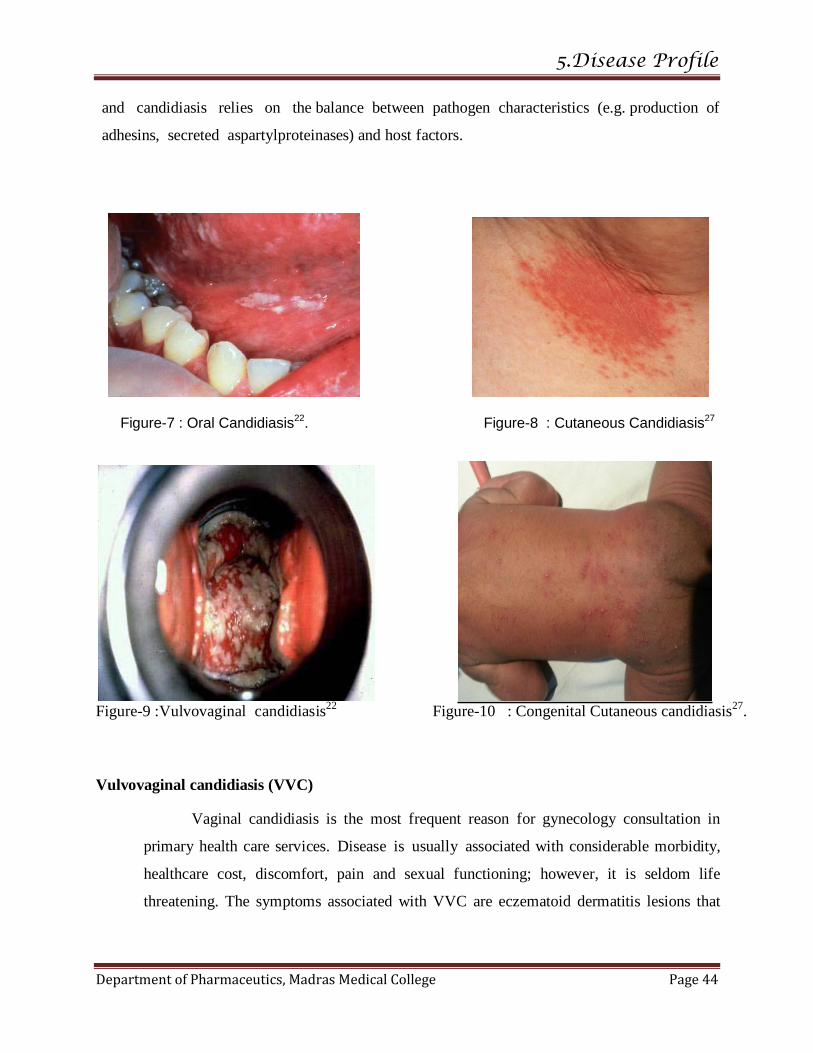
5.Disease Profile
Department of Pharmaceutics, Madras Medical College Page 44
and candidiasis relies on the balance between pathogen characteristics (e.g. production of
adhesins, secreted aspartylproteinases) and host factors.
Figure-7 : Oral Candidiasis22. Figure-8 : Cutaneous Candidiasis27
Figure-9 :Vulvovaginal candidiasis
22 Figure-10 : Congenital Cutaneous candidiasis
27.
Vulvovaginal candidiasis (VVC)
Vaginal candidiasis is the most frequent reason for gynecology consultation in
primary health care services. Disease is usually associated with considerable morbidity,
healthcare cost, discomfort, pain and sexual functioning; however, it is seldom life
threatening. The symptoms associated with VVC are eczematoid dermatitis lesions that
5.Disease Profile
Department of Pharmaceutics, Madras Medical College Page 45
sometimes show vesicular and grey-white psuedomembrane, vulval pruritis, burning,
erythema and curd like discharge.
Cutaneous candidiasis
Cutaneous candidiasis is usually secondary infection of skin and nail (body folds) in
predisposed patients. It occurs as a sub-acute or chronic infection. Disease involvement may be
localised or generalised to the skin or nails. The spectrum of cutaneous candidiasis includes
diaper rash, intertrigo candidiasis, Candida folliculitis, Otomycosis, Onychia and Paronychia. It
usually occurs in warm, moist and creased area, such as axillary folds, inguinal or intergluteal
areas. It is fairly common opportunistic disease and usually lead to maceration and
trauma in skin. It is commonly found in diabetics and obese people. Other predisposing factors
are antibiotic and oral contraceptives become macerated.
Invasive candidiasis
Invasive infections can involve virtually any organ. C. albicans continue to account
for the majority of invasive fungal infections, there has been a recent increase in disease due
to non-albicans Candida species (and antifungal-resistant Candida isolates).
Systemic or disseminated candidiasis
Severe organ invasive or systemic hematogenously disseminated candidiasis is
characterized by spreading of the Candida cells into almost the entire body with a tendency to
create abscesses in vitally important organs, inducing their failure which leads to mortality in
~50% of all cases, irrespective of administration of intensive antifungal therapy.
Risk factors for mucocutaneous candidiasis may include24
:
I. Cutaneous candidiasis
a. Diabetes mellitus
b. Tropical environment
c. Obesity
d. Use of systemic corticosteroids or anti- biotic therapy e. Neutropenia
f. HIV infection
g. Other immunocompromised states

5.Disease Profile
Department of Pharmaceutics, Madras Medical College Page 46
h. Occlusion (e.g., diapers, casts. Dressings)
i. Diseases which disturb the integument(e.g., psoriasis. contact dermatitis)
j. Other
2. Oral candidiasis
a. Use of broad-spectrum antibiotics systemic, topical, and inhalational cortico
steroids; and cytotoxic drugs
b. Radiation therapy
c. HIV Infection
d. Other immunocompromised states
e. Age (i.e , infants and elderly) f. Occlusion (e.g., dentures)
g. Other
Diagnosis23
A. Clinical
I. History may include
a. General medical history, especially but not limited to: 1. History of weight loss or weight gain
2. Endocrine diabetes mellitus
3. Risk factors for HIV disease
4. Presence of other risk factors.
5. Use of systemic medications
6. Other
b. Sexual practices
c. Recurrent infections
d. Duration of condition
e. Current treatment(s) topical and systemic of
1. Mucocutaneous candidiasis
2. Other disease
f. Past treatment(s) topical and systemic of
1. Mucocutaneous candidiasis
2 Other disease
g. Family history of diabetes mellitus and/or mucocutaneous candidiasis
h. Occupation
i. Dermatophytic infection, particularly tinea pedis, tinea cruris j. Drug allergies
k. Other
II. Physical examination may include
a. General physical examination as indicated
b. Examination of the involved area, but special attention may be directed to 1) Skin folds
2) Diaper area

5.Disease Profile
Department of Pharmaceutics, Madras Medical College Page 47
3) Comers of mouth and mucous membranes (e.g., perleche)
4) lnterdigital spaces
5) Scrotum, glans penis and foreskin, crural folds, gluteal area 6) Perianal area
7) Vagina, vulva
8) Axillae
9) Nail unit
10) Other
c. Extent of involvement
d. Clinical appearance
e. Associated findings
1) Secondary bacterial infection
2) Postinflammatory hyperpigmenta- tion and hypopigmentation 3) Onychodystrophy and onycholysis
4) Excoriations
5) Other
f. Other
B. Diagnostic tests
I. Potassium hydroxide preparation (KOH)
Materials is obtained from the site of infection. If the lesion is a pustule, the
purulent material can be used for the specimen. In cases of cutaneous involvement, the
specimen can be obtained from the edge of a lesion. The material is placed on a glass slide
and 1 0% to 20% KOH is added with or without dimethyl sulfoxide. A fungal stain,
such as Chlorazol Black E, or Parker's blue black ink may be used to highlight the
pseudohyphae. The presence of pseudohyphae and yeast forms confirm infection
2.. Fungal culture
Candida species are yeasts rather than molds and therefore grow as yeast
colonies on Sabouraud's glucose agar. The addition of cycloheximide to Sabouraud's agar
may inhibit many species of Candida and other saprophytes. However, C. albicans will
grow on media containing cycloheximide. Examples of such media include Mycosel,
Mycobiotic, and dermatophyte test medium. Although C. albicans will grow on
dermatophyte test media, the agar will not exhibit a red color change as occurs with
dermatophyte growth if read at the appropriate time interval, as indicated by the
manufacturer's instructions.
5.Disease Profile
Department of Pharmaceutics, Madras Medical College Page 48
TREATMENT22,24
Oral Candidiasis
Topical azoles (clotrimazole troches),
Oral azoles (fluconazole, ketoconazole, or itraconazole), or
Oral polyenes (such as nystatin (oral candidiasis in infants, Nystatin suspension [100, 000
units/ml] dropped into the mouth at 4-6 hour intervals or after each feed is usually used) or oral
amphotericin B).
Vulvovaginal candidiasis
Topical agents including azoles (all are used for 1 to 7 days depending on risk
classification: over-the counter (OTC) clotrimazole, OTC butoconazole, OTC miconazole,
OTC tioconazole, terconazole), nystatin [100,000 U per day for 7 to 14 days], oral azoles
(ketoconazole (400 mg for five days), which is not approved in the United States); itraconazole
(200 mg for 1 day, or 200 mg per day for three days), which is not approved in the United
States; and fluconazole (150 mg).

6.DRUG PROFILE
Department of Pharmaceutics, Madras Medical College Page 49
NYSTATIN
Structural formula
CAS Number61
1400-61-9
Synonyms25
Mycostatin, Nistatina,NYS,Nystatin, Nystatine and Nystatinum.
Empirical formula25
C47H75NO17
Chemical name25
(21E,23E,25E,27E,31E,33E)-20-{[(3S,4S,5S,6R)-4-amino-3,5-dihydroxy-6-methyloxan-2-yl]oxy}-
4,6,8,11,12,16,18,36-octahydroxy-35,37,38-trimethyl-2,14-dioxo-1-oxacyclooctatriaconta-
21,23,25,27,31,33-hexaene-17-carboxylic acid
Molecular weight61 : 926.1
Description61,62
Nature : A Yellow to slightly brown powder, Hygroscopic.
Odour : Characteristic.

6.DRUG PROFILE
Department of Pharmaceutics, Madras Medical College Page 50
Solubility : Freely soluble in Dimethylformamide and Dimethyl sulphoxide.
Slightly soluble in Methanol,Very slightly soluble in water, insoluble in
chloroform, in ether, and in Ethanol (95%).
pH : 6.5 to 8, determined in a 3.0 percent w/v.
Melting point : 130˚C
Mechanism of action25
Nystatin exerts its antifungal activity by binding to ergosterol found in fungal cell
membranes. Binding to ergosterol causes the formation of pores in the membrane. Potassium and
other cellular constituents leak from the pores causing cell death. The figure :shows the
Mechanism of action Nystatin with the example of Amphotericin B.
Figure-11: Mechanism of action.57
6.DRUG PROFILE
Department of Pharmaceutics, Madras Medical College Page 51
Pharmacokinetics26
Gastrointestinal absorption of Nystatin is insignificant. Most orally administered Nystatin
is passed unchanged in the stool. In patients with renal insufficiency receiving oral therapy with
conventional dosage forms, significant plasma concentrations of Nystatin may occasionally
occur. Onset of action is rapid, Unknown peak and Half-life, Duration of action for 6-12 hours.59
Therapeutic indications26
Nystatin(topical powder, cream and ointment) is indicated in the treatment of cutaneous
or mucocutaneous mycotic infections caused by Candida albicans and other susceptible Candida
species.
Dosage and administration26
Usual Adult Dose for Cutaneous Candidiasis
Apply nystatin topical cream, ointment or powder in a quantity sufficient to cover the
affected area and immediately surrounding skin 2 to 4 times a day.
The powder formulation is useful for the treatment of moist areas or lesions.
Therapy should be continued for 2 to 8 weeks, depending on the nature and severity of the
infection.
Usual Adult Dose for Vaginal Candidiasis
Insert one Nystatin vaginal tablet (100,000 units) vaginally once a day, preferably at
bedtime, for a total of 14 days.
Usual Pediatric Dose for Cutaneous Candidiasis
Apply Nystatin topical cream, ointment or powder in a quantity sufficient to cover
the affected area and immediately surrounding skin 2 to 4 times a day.
The powder formulation is useful for the treatment of moist areas or lesions.
Therapy should be continued for 2 to 8 weeks, depending on the nature and severity of
the infection.
6.DRUG PROFILE
Department of Pharmaceutics, Madras Medical College Page 52
Usual Pediatric Dose for Vaginal Candidiasis
>13 <18 years: Insert one nystatin vaginal tablet (100,000 units) vaginally once a day,
preferably at bedtime, for a total of 14 days.
Other Comments
For fungal infections of the feet caused by Candida, dust the powder freely on the feet as
well as in shoes and socks.
The vaginal tablets should be used continuously, regardless of initiation of menses.
For external use only. Avoid contact with the eyes.
Contraindication26
The preparation is contraindicated in patients with a history of hypersensitivity to any of
its components.
Drug interactions26
Minor drug interaction with Vitamins only
Side effects26
Nystatin is well tolerated even with prolonged therapy. Oral irritation and sensitization
have been reported.
Nystatin vaginal tablets have included vaginal irritation, burning, and itching. At least
one case of severe vulvovaginitis has also been reported.
Gastrointestinal: Diarrhea (including one case of bloody diarrhea), nausea, vomiting,
gastrointestinal upset/disturbances.
Dermatologic: Rash, including urticaria , Skin irritation, burning, itching, eczema, and pain on
application in less than 0.1% of patients. Stevens-Johnson syndrome has been reported very
rarely.
Other: Tachycardia, bronchospasm, facial swelling, and non-specific myalgia have also been
rarely reported.
Hypersensitivity26
Hypersensitivity side effects associated with Nystatin topical powder,
cream, and ointment have included allergic reactions and allergic contact dermatitis in less than
0.1% of patients.
Storage62
In an airtight container , protected from light.

7.EXCIPIENTS PROFILE
Department of Pharmaceutics, Madras Medical College Page 53
ETHYL CELLULOSE79
Structural formula
Nonproprietary names
BP –Ethylcellulose
PhEur -Ethylcellulose
USP-NF-Ethylcellulose
Synonyms
Aquacoat ECD; Aqualon; Ashacel; E462; Ethocel; ethylcellulosum;Surelease.
Empirical formula
C12H23O6(C12H22O5)nC12H23O5.
Chemical name
Cellulose ethyl ether
CAS Number
[9004-57-3]
Molecular weight
Description
Ethylcellulose is a tasteless, free-flowing, white to light tan-colored
powder.
EXCIPIENTS PROFILE
Department of Pharmaceutics, Madres Medical College Page 54
Solubility
Ethylcellulose is practically insoluble in glycerin, propylene glycol, and water.
Ethylcellulose that contains less than 46.5% of ethoxyl groups is freely soluble in
chloroform,methyl acetate, and tetrahydrofuran, and in mixtures ofaromatic hydrocarbons with ethanol
(95%).
Ethylcellulose that contains not less than 46.5% of ethoxyl groups is freelysoluble in
chloroform, ethanol (95%), ethyl acetate, methanol,and toluene.
Stability and Storage Conditions
Ethylcellulose is a stable, slightly hygroscopic material. It ischemically resistant to
alkalis, both dilute and concentrated, andto salt solutions, although it is more sensitive to acidic
materialsthan are cellulose esters.
Ethylcellulose is subject to oxidative degradation in the presence of sunlight or UV light
at elevated temperatures. This may beprevented by the use of antioxidant and chemical additives
thatabsorb light in the 230–340 nm range.
Ethylcellulose should be stored at a temperature not exceeding32˚C (90˚F) in a dry area
away from all sources of heat. It shouldnot be stored next to peroxides or other oxidizing agents.
Incompatibility
Incompatible with paraffin wax and microcrystalline wax.
Application
Ethylcellulose is widely used in oral and topical pharmaceutical
Hydrophobic coating agent for tablets and granules.
High-viscosity grades of ethylcellulose are used in drug microencapsulation.
Used as Binder
Ethylcellulose is used as a thickening agent in creams, lotions, or gels
Ethylcellulose has been studied as a stabilizer for emulsions.
Microencapsulation 10.0–20.0 %
Sustained-release tablet coating 3.0–20.0%
Tablet coating 1.0–3.0 %
Tablet granulation 1.0–3.0%
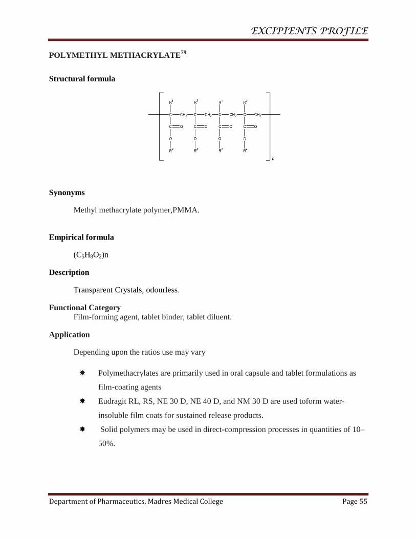
EXCIPIENTS PROFILE
Department of Pharmaceutics, Madres Medical College Page 55
POLYMETHYL METHACRYLATE79
Structural formula
Synonyms
Methyl methacrylate polymer,PMMA.
Empirical formula
(C5H8O2)n
Description
Transparent Crystals, odourless.
Functional Category
Film-forming agent, tablet binder, tablet diluent.
Application
Depending upon the ratios use may vary
Polymethacrylates are primarily used in oral capsule and tablet formulations as
film-coating agents
Eudragit RL, RS, NE 30 D, NE 40 D, and NM 30 D are used toform water-
insoluble film coats for sustained release products.
Solid polymers may be used in direct-compression processes in quantities of 10–
50%.

EXCIPIENTS PROFILE
Department of Pharmaceutics, Madres Medical College Page 56
Polymethacrylate polymers may additionally be used to form the matrix layers of
transdermal delivery systems and have also beenused to prepare novel gel
formulations for rectal administration.
POLYVINYL ALCOHOL79
Structure
Synonyms
Airvol; Alcotex; Elvanol; Gelvatol; Gohsenol; Lemol; Mowiol,Polyvinol; PVA; vinyl
alcohol polymer.
Empirical formula
(C2H4O)n
Molecular weight
Commercially available grades of polyvinyl acohol.
High viscosity -200 000
Medium viscosity -130 000
Low viscosity -20 000
Functional category
Coating agent; lubricant; stabilizing agent; viscosity-increasingagent.
Description
Polyvinyl alcohol occurs as an odorless, white to cream-coloredgranular powder.
Properties
Melting point: 228˚C for fully hydrolyzed grades;
180–190˚C for partially hydrolyzed grades.
Solubility : soluble in water; slightly soluble in ethanol (95%);insoluble in organic
EXCIPIENTS PROFILE
Department of Pharmaceutics, Madres Medical College Page 57
solvents. Dissolution requires dispersion (wetting) of the solid in water at
room temperaturefollowed by heating the mixture to about 90˚C for approximately
5 minutes. Mixing should be continuedwhile the heated solution is cooled to room temperature.
Stability andstorage conditions
Polyvinyl alcohol is stable when stored in a tightly sealedcontainer in a cool, dry place.
Aqueous solutions are stable incorrosion-resistant sealed containers. Preservatives
maybeadded to the solution if extended storage is required.
Polyvinyl alcohol undergoes slow degradation at 100˚C and rapid degradation at
200˚C; it is stable on exposure to light.
Safety
Polyvinyl alcohol is generally considered a nontoxic material. Itis nonirritant to the skin
and eyes at concentrations up to 10%;concentrations up to 7% are used in cosmetics.
Regulatory status
Included in the FDA Inactive Ingredients Guide (ophthalmicpreparations and oral
tablets).Included in nonparenteralmedicines licensed in the UK.Included in the Canadian List of
Acceptable Non-medicinal Ingredients.
Application
Polyvinyl alcohol is used primarily in topical pharmaceutical and ophthalmic
formulation.
It is used as a stabilizing agent for emulsions (0.25–3.0% w/v).
Polyvinyl alcohol is also usedas a viscosity-increasing agent for viscous
formulations such as ophthalmic products.
It is used in artificial tears and contact lens solutions for lubrication purposes.
Used in sustained-release formulations for oral administration,and in transdermal
patches.
Polyvinyl alcohol may be made into microspheres when mixed with a
glutaraldehyde solution.
Used in Emulsions -0.5 % concentration,
Ophthalmic formulations - 0.25–3.00% concentration ,
Topical lotions- 2.5% concentration.

EXCIPIENTS PROFILE
Department of Pharmaceutics, Madres Medical College Page 58
CARBOMER (CARBOPOL 934 )79
Structure
Synonyms
Acrypol; Acritamer; acrylic acid polymer; carbomera; Carbopol;carboxypolymethylene;
polyacrylic acid; carboxyvinyl polymer;Pemulen; TegoCarbomer.
Empirical formula
(C3H4O2) n
Molecular weight
The molecular weight of carbomer is theoretically estimated at 7 – 105to 4 -10
9
Functional category
Bioadhesive material; controlled-release agent; emulsifying agent;emulsion stabilizer;
rheology modifier; stabilizing agent; suspending agent; tablet binder.
Description
Carbomers are white-colored, ‘fluffy’, acidic, hygroscopic powderswith a characteristic
slight odor.
Properties
Melting Point:Decomposition occurs within 30 minutes at 260˚C.

EXCIPIENTS PROFILE
Department of Pharmaceutics, Madres Medical College Page 59
Safety
Carbomers are used extensively in non parenteral products, particularly topical
liquid and semisolid preparations.
Grades polymerized in ethyl acetate may also be used in oral formulations.
There is no evidence of systemic absorption of carbomer polymers following oral
administration.
Acute oral toxicity studies in animals indicate that carbomer 934P has a loworal
toxicity, with doses up to 8 g/kg being administered to dogswithout fatalities
occurring.
Carbomers are generally regarded as essentially nontoxic and nonirritant
materials,there is no evidence in humans of hypersensitivity reactions to
carbomers used topically.
Stability andstorage conditions
Carbomers are stable, hygroscopic materials that may be heated attemperatures
below 104˚C for up to 2 hours without affecting theirthickening efficiency.
Carbomer powder should be stored in an airtight, corrosionresistantcontainer and
protected from moisture. The use of glass, plastic, or resin-linedcontainers is
recommended for the storage of formulations containing carbomer.
Regulatory status
Included in the FDA Inactive Ingredients Database (oral suspensions, tablets;
ophthalmic, rectal, topical, transdermal preparations;vaginal suppositories).
Included in non parenteral medicines licensed in Europe. Included in the
Canadian List of Acceptable Nonmedicinal Ingredients.
Application
In tablet formulations, carbomers are used as controlled release agents and/or as
binders.
Higher viscosity does not result in slower drug release with carbomers.

EXCIPIENTS PROFILE
Department of Pharmaceutics, Madres Medical College Page 60
Uesd as Emulsifying agent -0.1–0.5 %
Gelling agent -0.5–2.0 %
Suspending agent - 0.5–1.0 %
Tablet binder -0.75–3.0 %
Controlled-release agent- 5.0–30.0 %
8.MATERIALS AND METHODS
Department of Pharmaceutics, Madras Medical College Page 61
The List of excipients used and their Manufacturers are in the Table : 5.
Table-5 : List of Materials Used
S.No. DRUG/EXCIPIENTS MANUFACTURER
1. Nystatin Caplin point Laboratories Pvt Ltd.
2. Ethyl cellulose Caplin point Laboratories Pvt Ltd.
3. Polymethyl methacrylate Lab chemicals.
4. Polyvinyl alcohol Lab chemicals.
5. Dichloromethane Chen chems labs.
6. Dimethyl sulfoxide Chen chems labs.
7. Potassium dihydrogen ortho phosphate Supra chemicals, Chennai.
8. Disodium hydrogen ortho phosphate Supra chemicals, Chennai.
9. Methanol Sisco research laboratories Pvt Ltd.
10. Dialysis Bag (LA653) Hi media Lab, Mumbai.
11. Carbopol 934 Sai Mirra Innopharm Pvt. Ltd.
12. Glycerin Microlabs Pvt. Ltd.
13. Triethanolamine Microlabs Pvt. Ltd.

8.MATERIALS AND METHODS
Department of Pharmaceutics, Madras Medical college Page
62
The List of Equipments used in the study and their Manufacturer are in Table- 6
Table-6 : List of Equipments Used.
S.No EQUIPMENTS MANUFACTURER
1. High Speed Homogenizer Remi,Vasai.
2. Ultra sonicator Lark.
3. Electronic balance McDalal,, Chennai.
4. Magnetic Stirrer with Hot plate Remi,Vasai.
5. UV-Visible Spectrophotometer Shimadzu,Japan.
6. High speed cooling centrifuge Remi,Vasai.
7. Binocular microscope Olimpus.
8. Malvern Zeta Sizer Malvern,Germany.
9. Scanning electron microscope Hitachi,Japan.
10. FT-IR Spectrophotometer Nicolet,India.
11. pH meter Symchrony.India.
12. Freeze dryer McDalal,, Chennai.
13. Hot air oven McDalal,, Chennai.
14. Stability Chamber Remi CHM 6+.
8.MATERIALS AND METHODS
Department of Pharmaceutics, Madras Medical college Page
63
METHODOLOGY
COMPATIBILITY STUDIES FOR DRUG AND EXCIPIENTS28
For the development of formulation dosage form, preformulation studies were carried out to
confirm no interaction between the drug and excipients. It gives information needed for selection of
excipients with the drug for the formulation of Nanosponge. Physical compatibility of the drug with
excipients were done. The possibility of drug excipients (Ethyl cellulose and Polymethyl
methacrylate) interaction were investigated by FT-IR Spectrum study.
Physical Compatibility
Physical compatibility of the drug and excipients were carried at Room
temperature and at 40˚C±2˚C/75±5%RH(in days) with the physical admixture of drug and
excipients.
Fourier Transforms Infrared (FT-IR)Spectroscopic studies
The spectroscopic studies were carried out to find out the interaction between pure
drug, excipients and its physical mixture by KBr pellet technique using FT-IR
spectrophotometer. The IR spectrum of the physical mixture is then compared with the
spectrum of pure drug(Nystatin)to assess the compatibility of the excipients and drug. The
scanning range is 450-4000 cm-1
and the resolution is 4cm-1
.
PREPARATION OF PHOSPHATE BUFFER pH 5.569
Solution I – 13.612 g of Potassium dihydrogen phosphate in 1000 ml of Distilled
Water.
Solution II – 35.08 g of Disodium hydrogen phosphate in 1000 ml of Distilled Water.
96.4 ml of Solution I and 3.6 ml of Solution II were mixed together.
8.MATERIALS AND METHODS
Department of Pharmaceutics, Madras Medical college Page
64
STANDARD CURVE OF NYSTATIN28,29
100mg of drug was accurately weighed and dissolved in 30 ml methanol and made
up to 100ml with phosphate buffer pH 5.5. Calibration curve was prepared in a mixture of
phosphate buffer and methanol (7:3) at λ max 305 nm.
FORMULATIONDEVELOPMENT
FORMULATION OF NYSTATIN LOADED NANOSPONGE30
Nystatin Nanosponges were prepared by Emulsion solvent evaporation method. Two
different polymers were used in the formulation. Ethyl cellulose (EC) and Polymethyl
methacrylate (PMMA) were the Polymers used. Polyvinyl alcohol in Distilled water is
used as the aqueous phase. The Drug is dissolved in the required solvent (Dimethyl
sulphoxide) and the Polymers (1:1, 1:2, 1:3, 1:4, and 1:5) were dissolved in
Dichloromethane. The Drug solution was poured into the polymer solution and the mixture
was shaken well. Then the Drug polymer mixture was poured into the aqueous phase and
the mixer is subjected to homogenization using High speed homogenizer in 1500 rpm for 2
hours at 35˚C.The formed Nanosponges were centrifuged by high speed cooling centrifuge
and the residue was freeze dried.
Table-7 : Formulation of Nanosponge
S.No INGREDIENTS F1 F2 F3 F4 F5 F6 F7 F8 F9 F10
1. Drug ( mg) 100 100 100 100 100 100 100 100 100 100
2. EC( mg) 100 200 300 400 500 - - - - -
3. PMMA ( mg) - - - - - 100 200 300 400 500
4. PVA (g) 0.2 0.2 0.2 0.2 0.2 0.3 0.3 0.3 0.3 0.3
5. DCM ( ml) 20 20 20 20 20 20 20 20 20 20
6. Distilled
water(ml)
100 100 100 100 100 100 100 100 100 100
EC-Ethyl cellulose, PMMA – Polymethyl methacrylate, DCM – Dichloromethane, PVA-
Polyvinyl Alcohol.
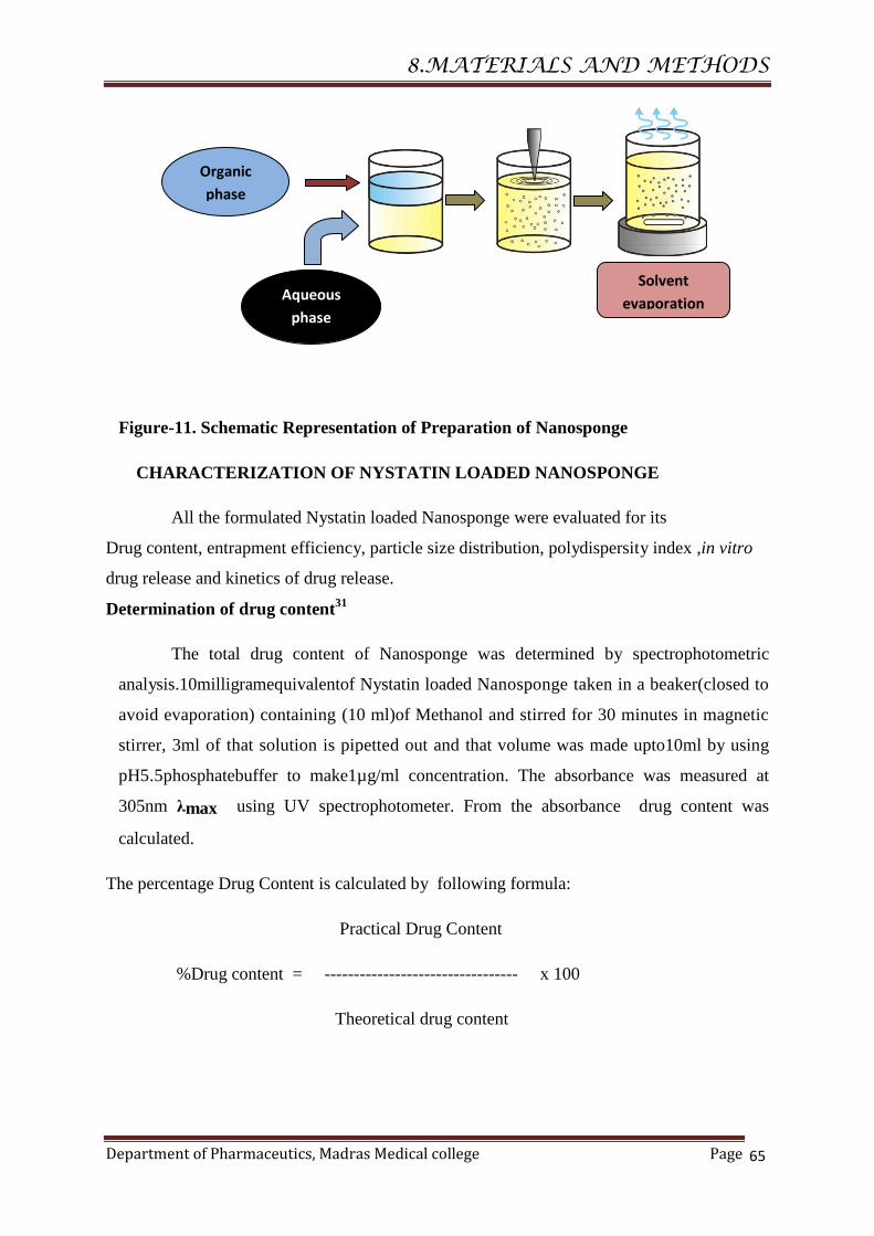
8.MATERIALS AND METHODS
Department of Pharmaceutics, Madras Medical college Page
65
Figure-11. Schematic Representation of Preparation of Nanosponge
CHARACTERIZATION OF NYSTATIN LOADED NANOSPONGE
All the formulated Nystatin loaded Nanosponge were evaluated for its
Drug content, entrapment efficiency, particle size distribution, polydispersity index ,in vitro
drug release and kinetics of drug release.
Determination of drug content31
The total drug content of Nanosponge was determined by spectrophotometric
analysis.10milligramequivalentof Nystatin loaded Nanosponge taken in a beaker(closed to
avoid evaporation) containing (10 ml)of Methanol and stirred for 30 minutes in magnetic
stirrer, 3ml of that solution is pipetted out and that volume was made upto10ml by using
pH5.5phosphatebuffer to make1µg/ml concentration. The absorbance was measured at
305nm λmax using UV spectrophotometer. From the absorbance drug content was
calculated.
The percentage Drug Content is calculated by following formula:
Practical Drug Content
%Drug content = --------------------------------- x 100
Theoretical drug content
Aqueous
phase
Organic
phase
Solvent
evaporation

8.MATERIALS AND METHODS
Department of Pharmaceutics, Madras Medical college Page
66
Determination of drug entrapment efficiency28
The entrapment efficiency was determined by measuring the concentration of the
drug in the supernatant after centrifugation. The unentrapped Nystatin were determined
by adding 10 mg Nystatin loaded Nanosponge in 10 ml of methanol and then the
dispersion were centrifuged at 9,000rpm for 30 minutes at 4ºC using a cooling centrifuge
in order to separate entrapped from the unentrapped drug. The free drug concentration in
supernatant layer after centrifugation is determined at λmax(305nm) using UV
Spectrophotometer
The percentage entrapment efficiency(%EE) is calculated by following formula:
Weight of Initial drug- Weight of free drug
%EE= ------------------------------------------------------------------X 100.
Weight of Initial drug
in- vitro releasestudies28
The in vitro release of Nystatin from Nanosponge was evaluated by the Dialysis Bag
diffusion technique. The release studies of Nystatin from Nanosponge were performed in
Phosphate buffer of pH5.5 and methanol (70:30) 18mg equivalent Nystatin Nanosponge
were suspended in 10 ml of buffer pH 5.5 mixture and placed in the dialysis bag(donor
compartment) and sealed at both ends. The dialysis bag were immersed in receptor
compartment containing 100ml of buffer mixture, which was stirred at 100 rpm and maintain
32 ± 2˚C. The receptor compartment was covered to prevent evaporation of the diffusion
medium. Samples were taken from the receptor compartment and the same amount were
replaced with the diffusion medium. Samples are taken upto 24hrs. Nystatin in the samples
were measured spectrophotometrically at λ 305nm.
Same procedure was carried out for Pure drug and in vitro release were measured
spectrophotometrically at λ 305nm.
SELECTION AND EVALUATION OFOPTIMIZEDFORMULATION
8.MATERIALS AND METHODS
Department of Pharmaceutics, Madras Medical college Page
67
The best formulation selection based on the results obtained from particle size,
entrapment efficiency, in-vitro release studies and kinetics of drug release.
FT-IR spectroscopicstudies
The compatibility study was carried out for optimized formulations(F3 and F7) using
FTIR at wavelength range of 4000to400cm-1
.Spectrum for optimized formulation were taken
and compared.
Morphology ofNanosponge by scanning electronmicroscopy (SEM) technique32
The Surface Morphology of the Nanosponge can be measured by SEM. The
formulations are poured in a circular aluminum stubs using double adhesive tape, and coated
with gold in HUS– 5GB vaccum evaporator and observed in Hitachi S– 3000N SEM at an
acceleration voltage of 10 Kv and a magnification of5000X.
Particle size distribution33
:
Particlesize (z-averagediameter), and polydispersity index (as a measure of the width
of the particle size distribution)of Nystatin loaded Nanosponge dispersion is performed by
dynamic light scattering also known as photon correlation spectroscopy(PCS) using a
Malvern Zeta sizer 3000NanoS(Malvern instruments, UK) at 25°C.
Prior to measurements all samples were diluted using ultra–purified water to yield a
suitable scattering intensity. The diluted nanosponge dispersion was poured into disposable
sizing cuvette which is then placed in the cuvette holder of the instrument and analyzed. Air
bubbles were removed from the capillary before measurement.
Kinetics of drug release78
.
To analyze the drug release mechanism, in-vitro release are fitted into a
Zero-order
First order
Higuchi,
Hixon-Crowell cube root law,
Korsmeyer- peppas model.

8.MATERIALS AND METHODS
Department of Pharmaceutics, Madras Medical college Page
68
1/3 = κt
The zero order rate describes the systems where the drug release rate is
independent of its concentration. It is ideal for the formulation to have release profile
of zero order to achieve pharmacological prolonged action.
Qt= Q0+ K0t
Where, Qtis the amount of drug dissolved in time t,
Q0is the initial amount of drug in the solution (most times, Q0=0)and
K0 is the zero order release constant expressed in units of concentration/time.
The first order rate describes the release from a system where the release rate is
Concentration dependent.
log C = log C0–kt/ 2.303
Where, C is the concentration of the drug at time(t),
C0 is the initial concentration of the drug and
k is the first-order release rate constant.
Higuchi described the release of drugs from porous, insoluble matrix as a square
root of time dependent process based on Fickian diffusion.
Q = kt1/2
Where Q is the amount of drug released in time t.
k constant reflecting design variable system (Differential rate constant) and
t Time in hours
Hixson and Crowell recognized that the particles diameter and regular area is proportional
to the cube root of its volume.
W01/3
–Wt
Where W0 is the initial amount of drug in the pharmaceutical dosage form,

8.MATERIALS AND METHODS
Department of Pharmaceutics, Madras Medical college Page
69
Wtis the remaining amount of drug in the pharmaceutical dosage form at time t and
κ(kappa) is a constant incorporating the surface-volume relation.
Korsmeyer–Peppas describes the fraction of drug release relates exponentially
with respect to time.
Mt/ M∞= Ktn
Where Mt /M∞ is a fraction of drug released at time t,
K is the release rate constant and n is the release exponent.
n is diffusional exponent indicative of the mechanism of the drug release.
The diffusion mechanism is given by the slope (n) value,
Table -8: diffusion mechanism
n value Mechanism
0.45 Fickian diffusion
0.45˂ n˂ 0.89 Anamolous or non-Fickian diffusion
0.89 Case-II transport
n˃ 0.89 Super case -II transport
The models were used to analyze the release of pharmaceutical polymeric dosage
forms when the release mechanism was not known or more than one type of release was
involved, the r2
and K values were calculated for the linear curve obtained by regression
analysis of the above plots.
STABILITY STUDIES
The stability studies of the optimized Nanosponge were performed at different
conditions of temperature and the effect on physical characteristic, entrapment efficiency
and drug content was noted. The Nanosponge were kept in the airtight container and stored
at 40±2˚C and in Relative humidity 75±5% for 45 days. The samples were analyzed for the
above parameter in 15days,30 days and 45days.
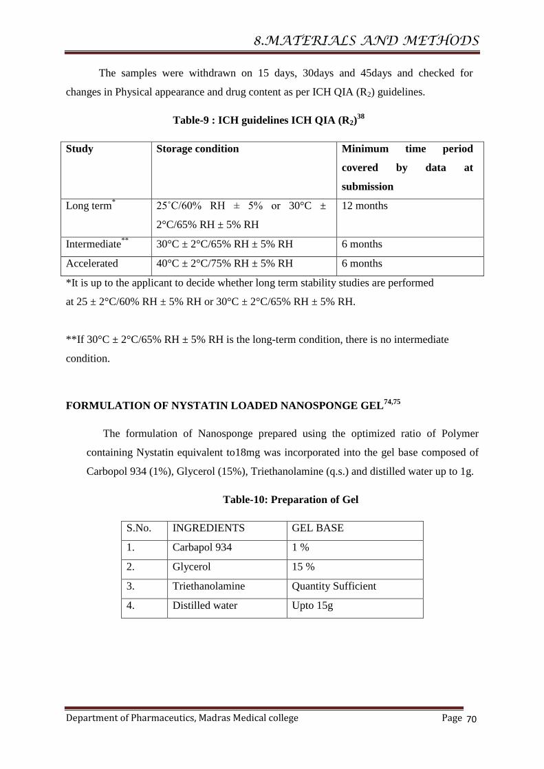
8.MATERIALS AND METHODS
Department of Pharmaceutics, Madras Medical college Page
70
The samples were withdrawn on 15 days, 30days and 45days and checked for
changes in Physical appearance and drug content as per ICH QIA (R2) guidelines.
Table-9 : ICH guidelines ICH QIA (R2)38
Study Storage condition Minimum time period
covered by data at
submission
Long term*
25˚C/60% RH ± 5% or 30°C ±
2°C/65% RH ± 5% RH
12 months
Intermediate**
30°C ± 2°C/65% RH ± 5% RH 6 months
Accelerated 40°C ± 2°C/75% RH ± 5% RH 6 months
*It is up to the applicant to decide whether long term stability studies are performed
at 25 ± 2°C/60% RH ± 5% RH or 30°C ± 2°C/65% RH ± 5% RH.
**If 30°C ± 2°C/65% RH ± 5% RH is the long-term condition, there is no intermediate
condition.
FORMULATION OF NYSTATIN LOADED NANOSPONGE GEL74,75
The formulation of Nanosponge prepared using the optimized ratio of Polymer
containing Nystatin equivalent to18mg was incorporated into the gel base composed of
Carbopol 934 (1%), Glycerol (15%), Triethanolamine (q.s.) and distilled water up to 1g.
Table-10: Preparation of Gel
S.No. INGREDIENTS GEL BASE
1. Carbapol 934 1 %
2. Glycerol 15 %
3. Triethanolamine Quantity Sufficient
4. Distilled water Upto 15g

8.MATERIALS AND METHODS
Department of Pharmaceutics, Madras Medical college Page
71
EVALUATION OF NANOSPONGE GEL
Physical Appearance
The prepared gel was examined for clarity, color, homogeneity and the presence of
foreign particles.
pH
2.5g of gel was accurately weighed and dispersed in 25 ml of distilled water. The pH of
the dispersion was measured by using a digital pH meter.
Rheological study
Viscosity measurement:
Viscosity was determined by Brookfield viscometer. In the present study, spindle
no. S64 with an optimum speed of 0.6 rpm was used to measure the viscosity of the
preparation.
Content Uniformity:
The drug content of the prepared gel was carried out by dissolving accurately weighed
quantity of gel equivalent to 9mg of the drug in a beaker containing 10 ml of methanol,
stir the solution for 30 minutes and centrifuged in High speed cooling centrifuge and 3ml
of the solution is made upto 10 ml with phosphate buffer pH 5.5. The samples were
analyzed spectrophotometrically at λmax 305 nm against blank using UV- Visible
spectrophotometer.
In-vitroDrug Diffusion study
In- vitro drug diffusion study was studied using dialysis bag. The Nanosponge gel
equivalent to 18mg of the drug was placed in a Dialysis Bag having 8 cm length and 3 cm
breath, both the sides are tied with thread. This acted as the donor compartment. Then the
bag was placed in a beaker containing 100 ml phosphate buffered methanol pH 5.5, which
acted as receptor compartment. The temperature of the receptor medium was maintained
at 37°±2°C and the medium was stirred at a speed of 100 rpm using a magnetic stirrer.
5 ml of the samples were collected at a predetermined time and replenished
immediately with the same volume of fresh buffer PB mixture pH5.5. The sink condition

8.MATERIALS AND METHODS
Department of Pharmaceutics, Madras Medical college Page
72
was maintained throughout the experiment. The collected samples were analyzed
Spectrophotometrically at 305 nm using UV- Visible spectrophotometer.
MICROBIOLOGICAL STUDY 49
Antifungal activity for the prepared Nanosponge gel were carried out. Candida
albicans was grown in Malt Extract Agar,(MAE) with medium composition: malt extract 17
g/L and agar 20g/L.
The assay was done using the agar well diffusion method. Sterile agar plates were
prepared by pouring the sterilized media in sterile Petri dishes under aseptic conditions.1
mm of the test organism was spread on agar plates. Using a sterile tube with a diameter of
6mm, the wells were made according to the number of samples. The wells were inoculated
with 80μL of sample. The antimicrobial activity was interpreted based on the size of
inhibition zone (IZ) diameter, which was measured in mm from observation of clear zones
surrounding the wells.

RESULTS AND DISCUSSION
Department of Pharmaceutics, Madras Medical College Page 73
PRE-FORMULATION STUDIES
The optimization of a formulation can be done only after a thorough investigation of
its physic chemical properties of the drug and excipients. The drug and the polymer must be
compatible for a successful formulation.
Physical Compatability
Table-11:Physical compatability
S.No.
Description and condition
Drug (D)
and
excipients
Initial Room
temperature (in
days)
40˚C±2˚C/75±5%
RH(in days)
10 20 30 10 20 30
1. Drug NC NC NC NC NC NC NC
2. D+EC NC NC NC NC NC NC NC
3. D+PMMA NC NC NC NC NC NC NC
4. D+PVA NC NC NC NC NC NC NC
NC – No Change.
Inference
The drug and the excipients of the formulation are physically compatible with each
other. They were evaluated for 10, 20 and 30 days at room temperature and at
40˚C±2˚C/75±5% Relative Humidity.
Chemical compatability
FT-IR spectroscopy gives the possible information about the interaction between the
drug and Polymer.
Drug-Polymer compatibility studies
FT-IR Spectroscopicstudies
The compatibility between drug and Polymer was confirmed using FT-IR
Spectroscopy. Infrared spectroscopic analysis for drug (Nystatin),Polymer, Drug-Polymer
admixture was carried out.
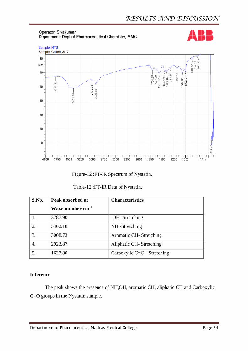
RESULTS AND DISCUSSION
Department of Pharmaceutics, Madras Medical College Page 74
Figure-12 :FT-IR Spectrum of Nystatin.
Table-12 :FT-IR Data of Nystatin.
Inference
The peak shows the presence of NH,OH, aromatic CH, aliphatic CH and Carboxylic
C=O groups in the Nystatin sample.
S.No. Peak absorbed at
Wave number cm-1
Characteristics
1. 3787.90 OH- Stretching
2. 3402.18 NH -Stretching
3. 3008.73 Aromatic CH- Stretching
4. 2923.87 Aliphatic CH- Stretching
5. 1627.80 Carboxylic C=O - Stretching

RESULTS AND DISCUSSION
Department of Pharmaceutics, Madras Medical College Page 75
Figure-13 :FT-IR Spectrum of Ethyl cellulose.
Table-13::FT-IR Data of Ethyl cellulose.
Inference
The peak shoes that the Ethyl cellulose has Characteristics Aliphatic CH and
Methyl CH – streching.
S.No. Peak absorbed at
Wave number cm-1
Characteristics
1. 2977.87 Aliphatic CH - Stretching
2. 1635.52 CH(methyl)-Stretching
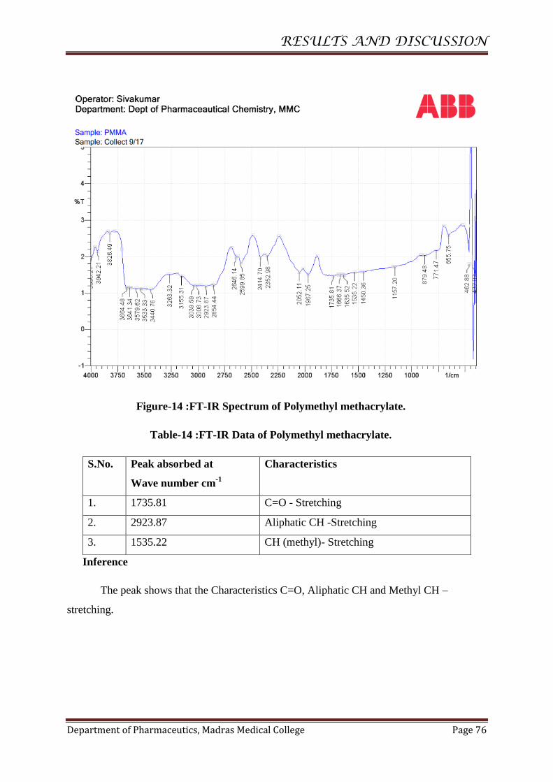
RESULTS AND DISCUSSION
Department of Pharmaceutics, Madras Medical College Page 76
Figure-14 :FT-IR Spectrum of Polymethyl methacrylate.
Table-14 :FT-IR Data of Polymethyl methacrylate.
Inference
The peak shows that the Characteristics C=O, Aliphatic CH and Methyl CH –
stretching.
S.No. Peak absorbed at
Wave number cm-1
Characteristics
1. 1735.81 C=O - Stretching
2. 2923.87 Aliphatic CH -Stretching
3. 1535.22 CH (methyl)- Stretching
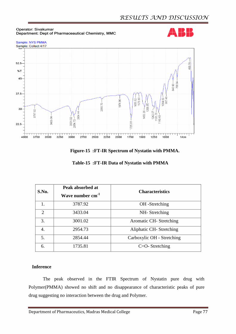
RESULTS AND DISCUSSION
Department of Pharmaceutics, Madras Medical College Page 77
Figure-15 :FT-IR Spectrum of Nystatin with PMMA.
Table-15 :FT-IR Data of Nystatin with PMMA
S.No. Peak absorbed at
Wave number cm-1
Characteristics
1. 3787.92 OH -Stretching
2 3433.04 NH- Stretching
3. 3001.02 Aromatic CH- Stretching
4. 2954.73 Aliphatic CH- Stretching
5. 2854.44 Carboxylic OH - Stretching
6. 1735.81 C=O- Stretching
Inference
The peak observed in the FTIR Spectrum of Nystatin pure drug with
Polymer(PMMA) showed no shift and no disappearance of characteristic peaks of pure
drug suggesting no interaction between the drug and Polymer.
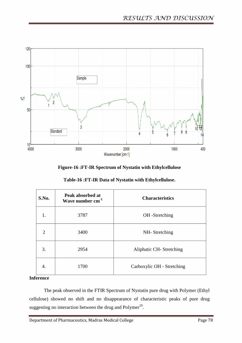
RESULTS AND DISCUSSION
Department of Pharmaceutics, Madras Medical College Page 78
Figure-16 :FT-IR Spectrum of Nystatin with Ethylcellulose
Table-16 :FT-IR Data of Nystatin with Ethylcellulose.
S.No. Peak absorbed at
Wave number cm-1 Characteristics
1. 3787 OH -Stretching
2 3400 NH- Stretching
3. 2954 Aliphatic CH- Stretching
4. 1700 Carboxylic OH - Stretching
Inference
The peak observed in the FTIR Spectrum of Nystatin pure drug with Polymer (Ethyl
cellulose) showed no shift and no disappearance of characteristic peaks of pure drug
suggesting no interaction between the drug and Polymer29
.

RESULTS AND DISCUSSION
Department of Pharmaceutics, Madras Medical College Page 79
STANDARD CURVE OFNYSTATIN
The UV spectrometric method was used to analyse Nystatin. The absorbance of the
drug in phosphate buffered methanol pH 5.5. (70:30) was measured at a wavelength of
305 nm. The results are given in the Table and Figure
Table-17 :Data for calibrationcurve
S.No Concentration (mcg/ml) Absorbanceat305 nm
1
.
0 0
2
.
2 0.148 ± 0.010
3
.
4 0.293 ± 0.0086
4
.
6 0.445 ± 0.026
5
.
8 0.586 ± 0.0061
6
.
10 0.756 ± 0.0214
Figure-17 : Calibration curve for Nystatin.
Inference
It was found that the solutions of N y s t a t i n in Phosphate buffered
methanol pH5.5 show linearity (R2=0.999) in absorbance at concentrations of 2-10 mcg/ml
and obey Beer Lambert’s law 77
.
y = 0.0749x - 0.0034 R² = 0.9994
-0.2
0
0.2
0.4
0.6
0.8
0 2 4 6 8 10 12
Ab
sorb
ance
at
30
5 n
m
CONCENTRATION(mcg/ml)
CALIBRATION CURVE
FOR NYSTATIN
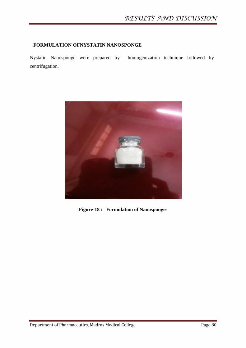
RESULTS AND DISCUSSION
Department of Pharmaceutics, Madras Medical College Page 80
FORMULATION OFNYSTATIN NANOSPONGE
Nystatin Nanosponge were prepared by homogenization technique followed by
centrifugation.
Figure-18 : Formulation of Nanosponges
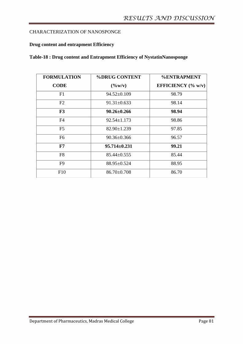
RESULTS AND DISCUSSION
Department of Pharmaceutics, Madras Medical College Page 81
CHARACTERIZATION OF NANOSPONGE
Drug content and entrapment Efficiency
Table-18 : Drug content and Entrapment Efficiency of NystatinNanosponge
FORMULATION
CODE
%DRUG CONTENT
(%w/v)
%ENTRAPMENT
EFFICIENCY (% w/v)
F1 94.52±0.109 98.79
F2 91.31±0.633 98.14
F3 90.26±0.266 98.94
F4 92.54±1.173 98.86
F5 82.90±1.239 97.85
F6 90.36±0.366 96.57
F7 95.714±0.231 99.21
F8 85.44±0.555 85.44
F9 88.95±0.524 88.95
F10 86.70±0.708 86.70
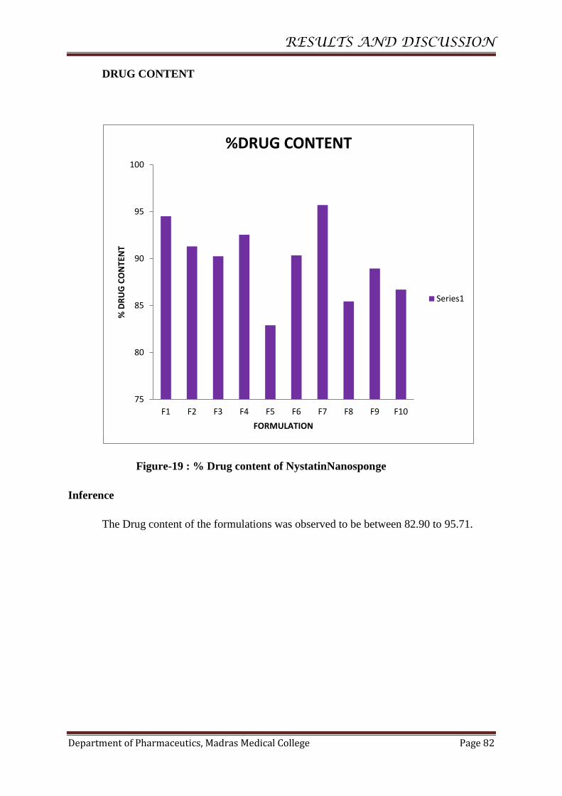
RESULTS AND DISCUSSION
Department of Pharmaceutics, Madras Medical College Page 82
DRUG CONTENT
Figure-19 : % Drug content of NystatinNanosponge
Inference
The Drug content of the formulations was observed to be between 82.90 to 95.71.
75
80
85
90
95
100
F1 F2 F3 F4 F5 F6 F7 F8 F9 F10
% D
RU
G C
ON
TEN
T
FORMULATION
%DRUG CONTENT
Series1

RESULTS AND DISCUSSION
Department of Pharmaceutics, Madras Medical College Page 83
ENTRAPMENTEFFICIENCY
Figure-20 :% Entrapment Efficiency of NystatinNanosponge
Inference
The entrapment efficiency was found to be 97.85 to 99.21. The highest entrapment
efficiency was observed with 99.21 and 98.94 for the formulations F3 and F7.
75
80
85
90
95
100
105
F1 F2 F3 F4 F5 F6 F7 F8 F9 F10
IN %
FORMULATION
% ENTRAPMENT EFFICIENCY
Series1
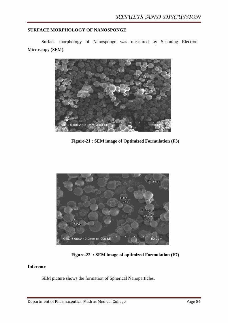
RESULTS AND DISCUSSION
Department of Pharmaceutics, Madras Medical College Page 84
SURFACE MORPHOLOGY OF NANOSPONGE
Surface morphology of Nanosponge was measured by Scanning Electron
Microscopy (SEM).
Figure-21 : SEM image of Optimized Formulation (F3)
Figure-22 : SEM image of optimized Formulation (F7)
Inference
SEM picture shows the formation of Spherical Nanoparticles.

RESULTS AND DISCUSSION
Department of Pharmaceutics, Madras Medical College Page 85
PARTICLE SIZE DISTRIBUTION BY MALVERN ZETA SIZER:
Figure-23 :Size distribution of optimized formulation F3
Inference: The average particle size of Nystatin loaded Nanosponge is 231.1 nm and the
poly dispersity index was found to be 1.000
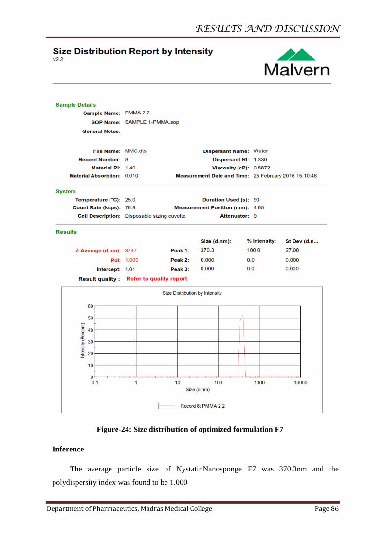
RESULTS AND DISCUSSION
Department of Pharmaceutics, Madras Medical College Page 86
Figure-24: Size distribution of optimized formulation F7
Inference
The average particle size of NystatinNanosponge F7 was 370.3nm and the
polydispersity index was found to be 1.000
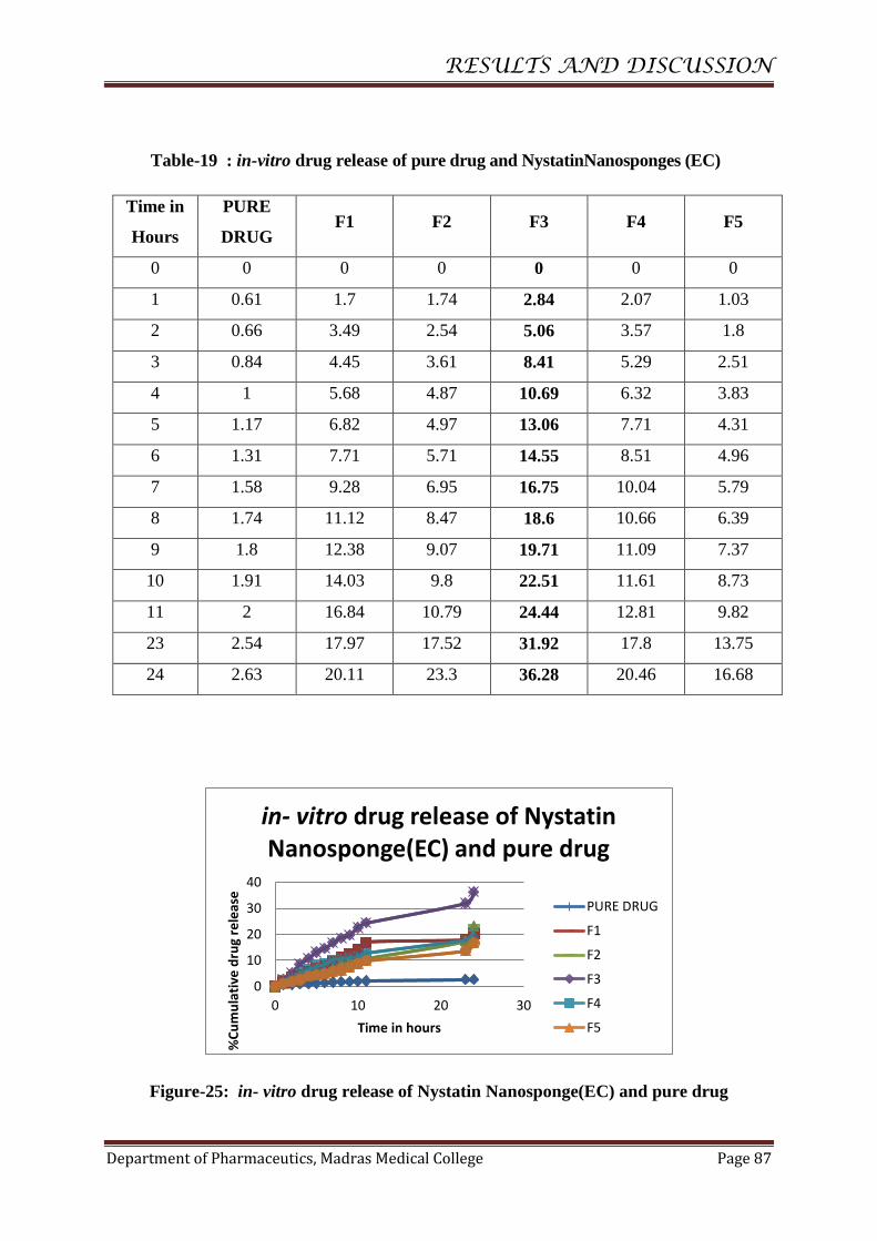
RESULTS AND DISCUSSION
Department of Pharmaceutics, Madras Medical College Page 87
Table-19 : in-vitro drug release of pure drug and NystatinNanosponges (EC)
Time in
Hours
PURE
DRUG F1 F2 F3 F4 F5
0 0 0 0 0 0 0
1 0.61 1.7 1.74 2.84 2.07 1.03
2 0.66 3.49 2.54 5.06 3.57 1.8
3 0.84 4.45 3.61 8.41 5.29 2.51
4 1 5.68 4.87 10.69 6.32 3.83
5 1.17 6.82 4.97 13.06 7.71 4.31
6 1.31 7.71 5.71 14.55 8.51 4.96
7 1.58 9.28 6.95 16.75 10.04 5.79
8 1.74 11.12 8.47 18.6 10.66 6.39
9 1.8 12.38 9.07 19.71 11.09 7.37
10 1.91 14.03 9.8 22.51 11.61 8.73
11 2 16.84 10.79 24.44 12.81 9.82
23 2.54 17.97 17.52 31.92 17.8 13.75
24 2.63 20.11 23.3 36.28 20.46 16.68
Figure-25: in- vitro drug release of Nystatin Nanosponge(EC) and pure drug
0
10
20
30
40
0 10 20 30
%C
um
ula
tive
dru
g re
leas
e
Time in hours
in- vitro drug release of Nystatin Nanosponge(EC) and pure drug
PURE DRUG
F1
F2
F3
F4
F5
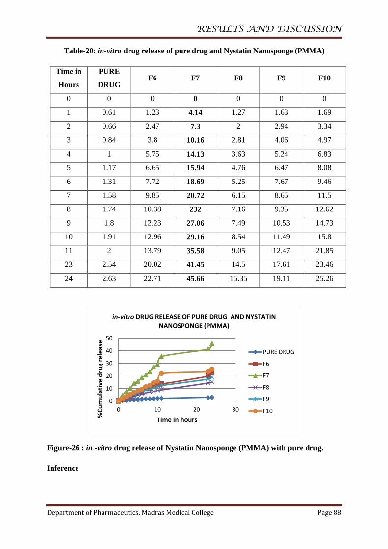
RESULTS AND DISCUSSION
Department of Pharmaceutics, Madras Medical College Page 88
Table-20: in-vitro drug release of pure drug and Nystatin Nanosponge (PMMA)
Time in
Hours
PURE
DRUG F6 F7 F8 F9 F10
0 0 0 0 0 0 0
1 0.61 1.23 4.14 1.27 1.63 1.69
2 0.66 2.47 7.3 2 2.94 3.34
3 0.84 3.8 10.16 2.81 4.06 4.97
4 1 5.75 14.13 3.63 5.24 6.83
5 1.17 6.65 15.94 4.76 6.47 8.08
6 1.31 7.72 18.69 5.25 7.67 9.46
7 1.58 9.85 20.72 6.15 8.65 11.5
8 1.74 10.38 232 7.16 9.35 12.62
9 1.8 12.23 27.06 7.49 10.53 14.73
10 1.91 12.96 29.16 8.54 11.49 15.8
11 2 13.79 35.58 9.05 12.47 21.85
23 2.54 20.02 41.45 14.5 17.61 23.46
24 2.63 22.71 45.66 15.35 19.11 25.26
Figure-26 : in -vitro drug release of Nystatin Nanosponge (PMMA) with pure drug.
Inference
0
10
20
30
40
50
0 10 20 30
%C
um
ula
tive
dru
g re
leas
e
Time in hours
in-vitro DRUG RELEASE OF PURE DRUG AND NYSTATIN NANOSPONGE (PMMA)
PURE DRUG
F6
F7
F8
F9
F10
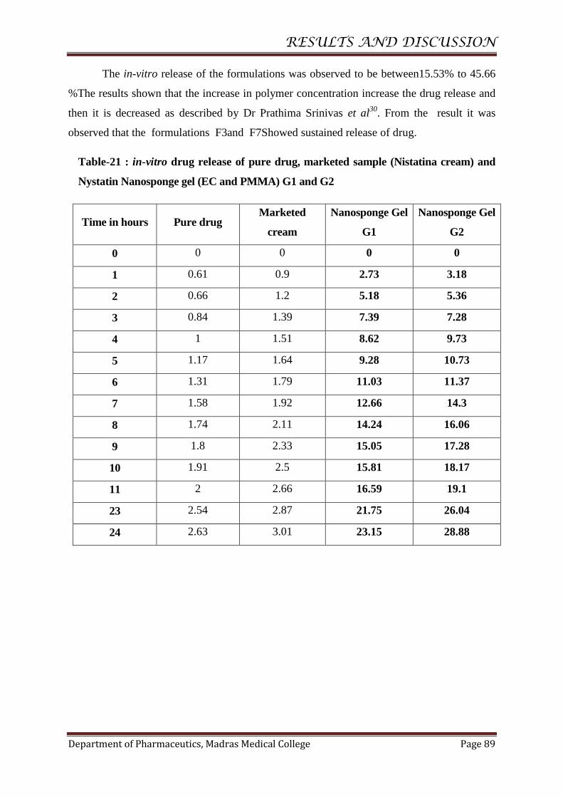
RESULTS AND DISCUSSION
Department of Pharmaceutics, Madras Medical College Page 89
The in-vitro release of the formulations was observed to be between15.53% to 45.66
%The results shown that the increase in polymer concentration increase the drug release and
then it is decreased as described by Dr Prathima Srinivas et al30
. From the result it was
observed that the formulations F3and F7Showed sustained release of drug.
Table-21 : in-vitro drug release of pure drug, marketed sample (Nistatina cream) and
Nystatin Nanosponge gel (EC and PMMA) G1 and G2
Time in hours Pure drug Marketed
cream
Nanosponge Gel
G1
Nanosponge Gel
G2
0 0 0 0 0
1 0.61 0.9 2.73 3.18
2 0.66 1.2 5.18 5.36
3 0.84 1.39 7.39 7.28
4 1 1.51 8.62 9.73
5 1.17 1.64 9.28 10.73
6 1.31 1.79 11.03 11.37
7 1.58 1.92 12.66 14.3
8 1.74 2.11 14.24 16.06
9 1.8 2.33 15.05 17.28
10 1.91 2.5 15.81 18.17
11 2 2.66 16.59 19.1
23 2.54 2.87 21.75 26.04
24 2.63 3.01 23.15 28.88

RESULTS AND DISCUSSION
Department of Pharmaceutics, Madras Medical College Page 90
Figure-27: in-vitro drug release of Pure drug, Marketed sample (Nistatina cream)
and Nystatin Nanosponge gel G1 and G2
Inference
The in–vitro release of the Gel formulation of F3 and F7 was compared with
marketed sample of Nystatin cream, and the release of Nanosponge gel (G1 and G2) was
23.15% and 28.88% respectively.
The release of gel was increased due to the decreased particle size and increased
surface area as described by Rawia M. Khalil et al28
.
Figure-28 : Diffusion Study By Dialysis Bag method
0
5
10
15
20
25
30
35
0 5 10 15 20 25 30
%C
um
ula
tive
dru
g re
leas
e
Time in hours
in-vitro DRUG RELEASE OF PURE DRUG AND MARKETED SAMPLE WITH NYSTATIN NANOSPONGE GEL G1 and G2
PURE DRUG
M1
G1
G2

RESULTS AND DISCUSSION
Department of Pharmaceutics, Madras Medical College Page 91
KINETICS OFDRUG RELEASE
Thein-vitroreleasedatawasappliedtovariouskineticmodelstopredictthemechanism
ofdrugreleaseoftheNanospongeformulations.
Table -22 : Release kinetics of NystatinNanosponge Optimized formulation F3
Time in
Hours
%cum
drug
release
% drug
remaining
Log%cum
drug
remaining
Square
root of
time
Log
time
Cube root
of %
drug
remaining
Log %cum
drug release
0 0 100 2 0 4.641589
1 2.84 97.16 1.98748 1 0 4.597225 0.453318
2 5.06 94.94 1.97744 1.41421 0.30102 4.561941 0.704151
3 8.41 91.59 1.96184 1.73205 0.47712 4.507641 0.924796
4 10.69 89.31 1.95090 2 0.60205 4.469922 1.028978
5 13.06 86.94 1.93921 2.23606 0.69897 4.430028 1.115943
6 14.55 85.45 1.93171 2.44949 0.77815 4.404575 1.162863
7 16.75 83.25 1.92038 2.64575 0.84509 4.366445 1.224015
8 18.6 81.4 1.91064 2.82842 0.90308 4.333859 1.269513
9 19.71 80.29 1.90466 3 0.95424 4.314069 1.294687
10 22.51 77.49 1.88924 3.16227 1 4.263326 1.352375
11 24.44 75.56 1.87829 3.31662 1.04139 4.227633 1.388101
23 31.92 68.08 1.83302 4.79583 1.36172 4.083255 1.504063
24 36.28 63.72 1.80427 4.89897 1.38021 3.994158 1.559667

RESULTS AND DISCUSSION
Department of Pharmaceutics, Madras Medical College Page 92
Figure-29: Zero OrderKineticsof F3
Figure-30 :FirstOrderKinetics of F3
y = 1.3752x + 4.9585 R² = 0.9149
0
10
20
30
40
0 10 20 30
%cu
mu
lati
ve d
rug
rele
ase
Time in hours
Zero order release Kinetics
y = -0.0075x + 1.9811 R² = 0.9448
1.75
1.8
1.85
1.9
1.95
2
2.05
0 10 20 30
log%
cum
ula
tive
dru
g re
mai
nin
g
Time in hours
First order relaese Kinetics
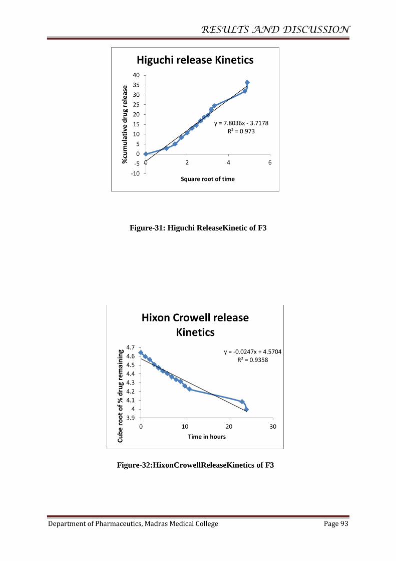
RESULTS AND DISCUSSION
Department of Pharmaceutics, Madras Medical College Page 93
Figure-31: Higuchi ReleaseKinetic of F3
Figure-32:HixonCrowellReleaseKinetics of F3
y = 7.8036x - 3.7178 R² = 0.973
-10
-5
0
5
10
15
20
25
30
35
40
0 2 4 6%cu
mu
lati
ve d
rug
rele
ase
Square root of time
Higuchi release Kinetics
y = -0.0247x + 4.5704 R² = 0.9358
3.9
4
4.1
4.2
4.3
4.4
4.5
4.6
4.7
0 10 20 30
Cu
be
roo
t o
f %
dru
g re
mai
nin
g
Time in hours
Hixon Crowell release Kinetics

RESULTS AND DISCUSSION
Department of Pharmaceutics, Madras Medical College Page 94
Figure-33:KorsmeyerPeppas Release Kinetics of F3
y = 0.7937x + 0.521 R² = 0.975
0
0.5
1
1.5
2
0 0.5 1 1.5log%
cum
ula
tive
dru
g re
leas
e
Log time
Korsmeyer Peppas release kinetics

RESULTS AND DISCUSSION
Department of Pharmaceutics, Madras Medical College Page 95
Table-23 : Release kinetics of F3
S.No.
Release
Kinetics
X-
axis
Y-axis
Slope
Intercept
R2
Linear
Equation
1.
Zero order
equation
Time
in
hours
Cumulative
%drug
release
1.375
4.958
0.914
y=1.375x+4.958
R²=0.914
2.
First order
equation
Time
in
hours
Log%
cumulative
drug
release
-0.007
1.981
0.944
y=-0.007x+1.981
R² = 0.944
3.
Higuchi
release
kinetic
Squar
e root
of
time
Cumulative
%drug
release
7.803
-3.717
0.973
y=7.803x-3.717
R² = 0.973
4.
Hixon
Crowell
equation
Time
in
hours
Cube root
of % drug
release
-0.024
4.570
0.935
y=-0.024x+4.570
R² = 0.935
5
Korsmeyer
Peppas
equation
Log
time
Log %
cumulative
drug release
0.793
0.521
0.975
y=0.793x+0.521
R² = 0.975
Inference
Formulation F3 followed first- kinetics and their R2 value 0.944 indicating the
release to be dose dependent.
The drug release was proportional to the square root of time indicating
that Nystatin release from Nanosponge was diffusion controlled.
The n value for the korsmeyer- peppas for formulation F3 was found to be
0.793. So the formulation F3 non-Fickian type mechanism.

RESULTS AND DISCUSSION
Department of Pharmaceutics, Madras Medical College Page 96
The Drug release pattern from Formulation F3 follows First order release,
Higuchi model and Non-fickian transport.
Table-24 : Release kinetics of Nystatin Nanosponge Optimized formulation F7
Time in
Hours
%cum
drug
release
% drug
remaining
Log%
cum
drug
remain
ing
Square
root of
time
Log
time
Cube root
of %
drug
remaining
Log %cum
drug release
0 0 100 2 0 4.641588
1 4.14 95.86 1.981637 1 0 4.57663 0.617
2 7.3 92.7 1.96708 1.414214 0.30103 4.525777 0.863323
3 10.16 89.84 1.95347 1.732051 0.477121 4.478747 1.006894
4 14.13 85.87 1.933841 2 0.60206 4.411779 1.150142
5 15.94 84.06 1.924589 2.236068 0.69897 4.380561 1.202488
6 18.69 81.31 1.910144 2.44949 0.778151 4.332261 1.271609
7 20.72 79.28 1.899164 2.645751 0.845098 4.295903 1.31639
8 23.2 76.8 1.885361 2.828427 0.90309 4.250634 1.365488
9 27.06 72.94 1.862966 3 0.954243 4.178193 1.432328
10 29.16 70.84 1.850279 3.162278 1 4.137704 1.464788
11 35.58 64.42 1.809021 3.316625 1.041393 4.00873 1.551206
23 41.45 58.55 1.767527 4.795832 1.361728 3.883073 1.617525
24 45.66 54.34 1.73512 4.898979 1.380211 3.787679 1.659536

RESULTS AND DISCUSSION
Department of Pharmaceutics, Madras Medical College Page 97
Figure-34: Zero OrderKineticsof F7
Figure-35: First OrderKineticsof F7
y = 1.7705x + 6.6515 R² = 0.8922
0
10
20
30
40
50
60
0 10 20 30%cu
mu
lati
ve d
rug
rele
ase
Time in hours
Zero order Kinetics
y = -0.0104x + 1.9757 R² = 0.9297
1.7
1.75
1.8
1.85
1.9
1.95
2
2.05
0 10 20 30
log%
cum
ula
tive
dru
g re
mai
nin
g
Time in hours
First order Kinetics

RESULTS AND DISCUSSION
Department of Pharmaceutics, Madras Medical College Page 98
Figure-36:Higuchi Release kinetics of F7
Figure-37:HixonCrowellReleaseKinetics of F7
y = 10.095x - 4.6405 R² = 0.958
-10
0
10
20
30
40
50
0 2 4 6
%C
um
ula
tive
dru
g re
leas
e
Square root of time
Higuchi release kinetics
y = -0.0335x + 4.5482 R² = 0.9186
0
1
2
3
4
5
0 5 10 15 20 25 30
Cu
be
roo
t o
f %
dru
g re
mai
nin
g
Time in hours
Hixon crowell release kinetics
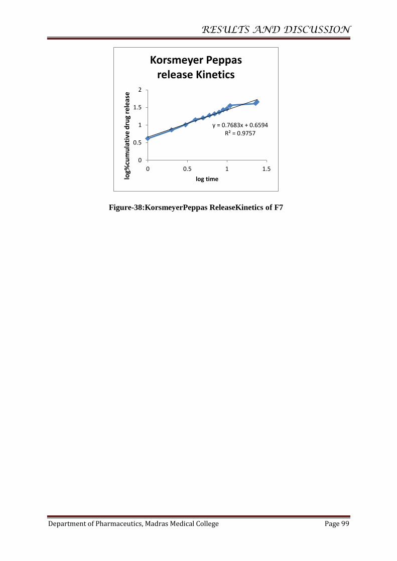
RESULTS AND DISCUSSION
Department of Pharmaceutics, Madras Medical College Page 99
Figure-38:KorsmeyerPeppas ReleaseKinetics of F7
y = 0.7683x + 0.6594 R² = 0.9757
0
0.5
1
1.5
2
0 0.5 1 1.5
log%
cum
ula
tive
dru
g re
leas
e
log time
Korsmeyer Peppas release Kinetics

RESULTS AND DISCUSSION
Department of Pharmaceutics, Madras Medical College Page 100
Table-25: Release kinetics of F7
S.No.
Release
Kinetics
X-axis
Y-axis
Slope
Intercept
R2
Linear
Equation
1.
Zero order
equation
Time
in
hours
Cumulative
% drug
release
1.770
6.651
0.892
y = 1.770x
+ 6.651
R² = 0.892
2.
First order
equation
Time
in
hours
Log%
cumulative
drug
release
-0.010
1.975
0.929
y = -0.010x +
1.975
R² = 0.929
3.
Higuchi
release
kinetic
Square
root of
time
Cumulative
% drug
release
10.09
-4.640
0.958
y = 10.09x
- 4.640
R² = 0.958
4.
Hixon
Crowell
equation
Time
in
hours
Cube root
of % drug
release
-0.033
4.548
0.918
y = -0.033x +
4.548
R² = 0.918
5
Korsmeyer
Peppas
equation
Log
time
Log %
cumulative
drug
release
0.768
0.659
0.975
y = 0.768x
+ 0.659
R² = 0.975
Inference
Formulation F7 followed first- kinetics and their R2 value 0.929 indicating the
release to be dose dependent.
The drug release was proportional to the square root of time indicating
that Nystatin release from Nanosponge was diffusion controlled.
RESULTS AND DISCUSSION
Department of Pharmaceutics, Madras Medical College Page 101
The n value for the korsmeyer- peppas for formulation F7 was found to be
0.768. So the formulation F7 non-Fickian type mechanism.
The Drug release pattern from Formulation F7 follows First order release,
Higuchi model and Non-fickian transport.

RESULTS AND DISCUSSION
Department of Pharmaceutics, Madras Medical college Page 102
Table-26 : Release kinetics of Nystatin Nanosponge Gel- G1
Time in
Hours
%cum
drug
release
% drug
remaining
Log%
cum
drug
remain
ing
Square
root of
time
Log
time
Cube root
of %
drug
remaining
Log %cum
drug release
0 0 100 2 0 4.641589
1 2.73 97.27 1.987979 1 0 4.59896 0.436163
2 5.18 94.82 1.9769 1.414214 0.30103 4.560018 0.71433
3 7.39 92.61 1.966658 1.732051 0.477121 4.524312 0.868644
4 8.62 91.38 1.960851 2 0.60206 4.504193 0.935507
5 9.28 90.72 1.957703 2.236068 0.69897 4.493323 0.967548
6 11.03 88.97 1.949244 2.44949 0.778151 4.464243 1.042576
7 12.66 87.34 1.941213 2.645751 0.845098 4.436812 1.102434
8 14.24 85.76 1.933285 2.828427 0.90309 4.409895 1.15351
9 15.05 84.95 1.929163 3 0.954243 4.395967 1.177536
10 15.81 84.19 1.925261 3.162278 1 4.382818 1.198932
11 16.59 83.41 1.921218 3.316625 1.041393 4.369241 1.219846
23 21.75 78.25 1.893484 4.795832 1.361728 4.277218 1.337459
24 23.15 76.85 1.885644 4.898979 1.380211 4.251556 1.364551
Figure-39: Zero OrderKineticsof G1
y = 0.8545x + 4.7801 R² = 0.8713
0
5
10
15
20
25
30
0 10 20 30
%cu
mu
lati
ve d
rug
rele
ase
Time in hours
Zero order kinetics
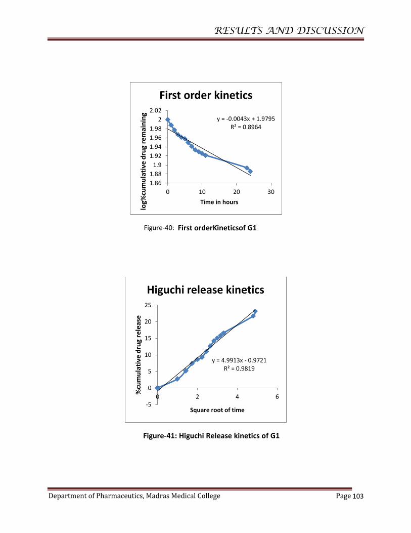
RESULTS AND DISCUSSION
Department of Pharmaceutics, Madras Medical College Page
103
Figure-40: First orderKineticsof G1
Figure-41: Higuchi Release kinetics of G1
y = -0.0043x + 1.9795 R² = 0.8964
1.86
1.88
1.9
1.92
1.94
1.96
1.98
2
2.02
0 10 20 30
log%
cum
ula
tive
dru
g re
mai
nin
g
Time in hours
First order kinetics
y = 4.9913x - 0.9721 R² = 0.9819
-5
0
5
10
15
20
25
0 2 4 6
%cu
mu
lati
ve d
rug
rele
ase
Square root of time
Higuchi release kinetics
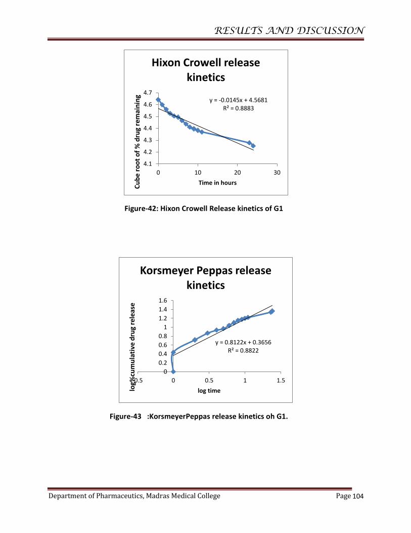
RESULTS AND DISCUSSION
Department of Pharmaceutics, Madras Medical College Page
104
Figure-42: Hixon Crowell Release kinetics of G1
Figure-43 :KorsmeyerPeppas release kinetics oh G1.
y = -0.0145x + 4.5681 R² = 0.8883
4.1
4.2
4.3
4.4
4.5
4.6
4.7
0 10 20 30
Cu
be
roo
t o
f %
dru
g re
mai
nin
g
Time in hours
Hixon Crowell release kinetics
y = 0.8122x + 0.3656 R² = 0.8822
0
0.2
0.4
0.6
0.8
1
1.2
1.4
1.6
-0.5 0 0.5 1 1.5
log%
cum
ula
tive
dru
g re
leas
e
log time
Korsmeyer Peppas release kinetics

RESULTS AND DISCUSSION
Department of Pharmaceutics, Madras Medical College Page
105
Table-27 : Release kinetics of G1
S.No.
Release
Kinetics
X-axis
Y-axis
Slope
Intercept
R2
Linear
Equation
1.
Zero order
equation
Time
in
hours
Cumulative
% drug
release
0.854
4.780
0.871
y =0.854x+4.780
R² = 0.871
2.
First order
equation
Time
in
hours
Log%
cumulative
drug
release
-0.004
1.979
0.896
y = -0.004x + 1.979
R² = 0.896
3.
Higuchi
release
kinetic
Square
root of
time
Cumulative
% drug
release
4.991
-0.972
0.981
y =4.991x -0.972
R² = 0.981
4.
Hixon
Crowell
equation
Time
in
hours
Cube root
of % drug
release
-0.014
4.568
0.888
y = -0.014x + 4.568
R² = 0.888
5
Korsmeyer
Peppas
equation
Log
time
Log %
cumulative
drug release
0.812
0.365
0.882
y =0.812x+0.365
R² = 0.882
Inference
Formulation G1 followed first- kinetics and their R2 value 0.896 indicating the
release to be dose dependent.
The drug release was proportional to the square root of time indicating
that Nystatin release from Nanosponge gel was diffusion controlled.
The n value for the korsmeyer- peppas for formulation G1 was found to be 0.812.
So the formulation G1 non-Fickian type mechanism.
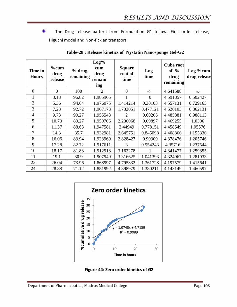
RESULTS AND DISCUSSION
Department of Pharmaceutics, Madras Medical College Page
106
The Drug release pattern from Formulation G1 follows First order release,
Higuchi model and Non-fickian transport.
Table-28 : Release kinetics of Nystatin Nanosponge Gel-G2
Time in
Hours
%cum
drug
release
% drug
remaining
Log%
cum
drug
remain
ing
Square
root of
time
Log
time
Cube root
of %
drug
remaining
Log %cum
drug release
0 0 100 2 0 4.641588
1 3.18 96.82 1.985965 1 0 4.591857 0.502427
2 5.36 94.64 1.976075 1.414214 0.30103 4.557131 0.729165
3 7.28 92.72 1.967173 1.732051 0.477121 4.526103 0.862131
4 9.73 90.27 1.955543 2 0.60206 4.485881 0.988113
5 10.73 89.27 1.950706 2.236068 0.69897 4.469255 1.0306
6 11.37 88.63 1.947581 2.44949 0.778151 4.458549 1.05576
7 14.3 85.7 1.932981 2.645751 0.845098 4.408866 1.155336
8 16.06 83.94 1.923969 2.828427 0.90309 4.378476 1.205746
9 17.28 82.72 1.917611 3 0.954243 4.35716 1.237544
10 18.17 81.83 1.912913 3.162278 1 4.341477 1.259355
11 19.1 80.9 1.907949 3.316625 1.041393 4.324967 1.281033
23 26.04 73.96 1.868997 4.795832 1.361728 4.197579 1.415641
24 28.88 71.12 1.851992 4.898979 1.380211 4.143149 1.460597
Figure-44: Zero order kinetics of G2
y = 1.0748x + 4.7159 R² = 0.9089
0
5
10
15
20
25
30
35
0 10 20 30%cu
mu
lati
ve d
rug
rele
ase
Time in hours
Zero order kinetics
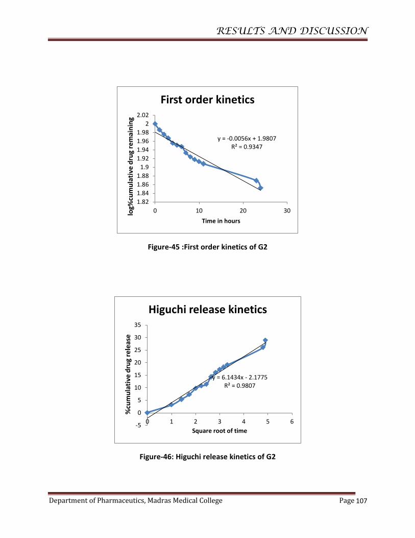
RESULTS AND DISCUSSION
Department of Pharmaceutics, Madras Medical College Page
107
Figure-45 :First order kinetics of G2
Figure-46: Higuchi release kinetics of G2
y = -0.0056x + 1.9807 R² = 0.9347
1.82
1.84
1.86
1.88
1.9
1.92
1.94
1.96
1.98
2
2.02
0 10 20 30log%
cum
ula
tive
dru
g re
mai
nin
g
Time in hours
First order kinetics
y = 6.1434x - 2.1775 R² = 0.9807
-5
0
5
10
15
20
25
30
35
0 1 2 3 4 5 6
%cu
mu
lati
ve d
rug
rele
ase
Square root of time
Higuchi release kinetics

RESULTS AND DISCUSSION
Department of Pharmaceutics, Madras Medical College Page
108
Figure-47 :Hixon Crowell release kinetics of G2
Figure-48:KorsmeyerPeppas release kinetics of G2
y = -0.0187x + 4.5712 R² = 0.9266
4
4.1
4.2
4.3
4.4
4.5
4.6
4.7
0 10 20 30
Cu
be
ro
ot
of
% d
rug
rem
ain
ing
Time in hours
Hixon Crowell release kinetics
y = 0.8572x + 0.3798 R² = 0.89
0
0.5
1
1.5
2
-0.5 0 0.5 1 1.5
log%
cum
ula
tive
dru
g re
leas
e
log time
Korsmeyer Peppas release kinetics

RESULTS AND DISCUSSION
Department of Pharmaceutics, Madras Medical College Page
109
Table-29 : Release kinetics of G2
S.No.
Release
Kinetics
X-axis
Y-axis
Slope
Intercept
R2
Linear
Equation
1.
Zero order
equation
Time
in
hours
Cumulative
% drug
release
1.074
4.715
0.908
y =1.074x+4.715
R² = 0.908
2.
First order
equation
Time
in
hours
Log%
cumulative
drug
release
-0.005
1.980
0.934
y = -0.005x +1.980
R² = 0.934
3.
Higuchi
release
kinetic
Square
root of
time
Cumulative
% drug
release
6.143
-2.177
0.980
y =6.143x -2.177
R² = 0.980
4.
Hixon
Crowell
equation
Time
in
hours
Cube root
of % drug
release
-0.018
4.571
0.926
y = -0.018x + 4.571
R² = 0.926
5
KorsmeyerP
eppas
equation
Log
time
Log %
cumulative
drug release
0.857
0.379
0.89
y =0.857x+0.379
R² = 0.89
Inference
Formulation G2followed first- kinetics and their R2 value 0.934 indicating the
release to be dose dependent.
The drug release was proportional to the square root of time indicating
that Nystatin release from Nanosponge gel was diffusion controlled.
The n value for the korsmeyer- peppas for formulation G2 was found to be 0.857.
So the formulation G2 non-Fickian type mechanism.
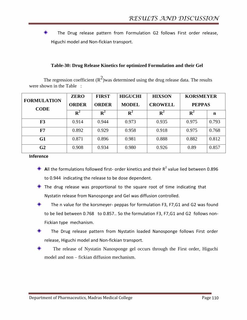
RESULTS AND DISCUSSION
Department of Pharmaceutics, Madras Medical College Page
110
The Drug release pattern from Formulation G2 follows First order release,
Higuchi model and Non-fickian transport.
Table-30: Drug Release Kinetics for optimized Formulation and their Gel
The regression coefficient (R2)was determined using the drug release data. The results
were shown in the Table :
FORMULATION
CODE
ZERO
ORDER
FIRST
ORDER
HIGUCHI
MODEL
HIXSON
CROWELL
KORSMEYER
PEPPAS
R2
R2 R
2 R
2 R
2 n
F3 0.914 0.944 0.973 0.935 0.975 0.793
F7 0.892 0.929 0.958 0.918 0.975 0.768
G1 0.871 0.896 0.981 0.888 0.882 0.812
G2 0.908 0.934 0.980 0.926 0.89 0.857
Inference
All the formulations followed first- order kinetics and their R2 value lied between 0.896
to 0.944 indicating the release to be dose dependent.
The drug release was proportional to the square root of time indicating that
Nystatin release from Nanosponge and Gel was diffusion controlled.
The n value for the korsmeyer- peppas for formulation F3, F7,G1 and G2 was found
to be lied between 0.768 to 0.857.. So the formulation F3, F7,G1 and G2 follows non-
Fickian type mechanism.
The Drug release pattern from Nystatin loaded Nanosponge follows First order
release, Higuchi model and Non-fickian transport.
The release of Nystatin Nanosponge gel occurs through the First order, Higuchi
model and non – fickian diffusion mechanism.

RESULTS AND DISCUSSION
Department of Pharmaceutics, Madras Medical College Page
111
ANTI FUNGAL ACTIVITY
Figure-49 : After inoculation,0 min growth.
Figure-50: After 48 hrs of inoculation of G1

RESULTS AND DISCUSSION
Department of Pharmaceutics, Madras Medical College Page
112
Figure-51:After 48hrs of inoculation for G2
Figure-52: After 48 hrs of inoculation for Marketed sample (Nystatin cream), G1 and G2

RESULTS AND DISCUSSION
Department of Pharmaceutics, Madras Medical College Page
113
Evaluation of Gel
Table-31: Evaluation of Gel
S.No. Code Physical
appearance %Drug content
Viscosity (cps) pH %Drug
release
1. G1 Good 91.10 2.939 x 10⁶ 4.89 25.15
2. G2 Good 94.55 2.853 x 10⁶ 4.92 28.88
Antimicrobial activity of NystatinNanosponge Gel
Table-32: Antimicrobial activity of NystatinNanosponge Gel
S.No. Formulation code Anti fungal activity(Zone of inhibition) in
mm
1. Control 0
2. G1 18±0.8164
3. G2 19.33±1.2472
4. Marketed sample 11.667±0.4714
Figure-53: Antifungal activity.
Inference
The antifungal activity for Nystatin Nanosponge Gel G2 Zone of Inhibition
was 19.33mm, for G1 Zone of Inhibition was 18mm and the zone of inhibition obtained
for Marketed sample was 11.667mm. The antifungal activity of the Nanosponge gel
showed enhancement of Nystatin antifungal effect.
05
1015
20
Anti fungal activity(Zone of inhibition) in mm
Anti fungal activity(Zoneof inhibition) in mm

10.SUMMARY AND CONCLUSION
Department of Pharmaceutics, Madras Medical College Page 114
The purpose of this research was to prepare Nystatin loaded
Nanosponge gel for Sustained release of drug, increase the drug
solubility, increase the drug permeability, to reduce the dosing
frequency and side effects.
The FTIR studies proved that there were no interaction between the
drug and Polymers.
Homogenization technique followed by centrifugation was employed to
prepare Nanosponge using various polymers.
The formulation were prepared using different Polymers
(Ethylcellulose and Polymethyl methacrylate) in different ratios
(Drug:Polymer–1:1,1:2,1:3,1:4 and1:5,) Using dichloromethane as cross
linker as well as solvent.
The formulations were characterized for drug entrapment efficiency.
The entrapment efficiency of the formulations was observed to be
between 97.85 to 99.21. The highest entrapment efficiency was
observed with 99.21 and 98.94 for the formulations F3 and F7.
The formulations were characterized for drug content. The Drug
content of the formulations was observed to be between 82.90 to 95.71.
The particle size analysis done by Malvern Zeta sizer showed that the
average particle size of Nystatin loaded Nanosponge F3 and F7 was
231.1 nm and 370.3 nm respectively.
The SEM analysis of Nanosponge shows the spherical surface of the
particles.

SUMMARY AND CONCLUSION
Department of Pharmaceutics, Madras Medical College Page 115
The in-vitro release of Nystatin Nanopsonge optimized formulation
F3wasfound tobe 36.28% andF7 was 45.66 % at theend of 24 hours.
The drug content of the Gel G1 and G2 was found to be 25.15% and
28.88 % respectively.
The in-vitro release of Nystatin Nanosponge Gel formulation
G1wasfound tobe 23.15% andG2 was 28.88 % at theend of 24 hours.
The pH of the gels G1and G2 was found to be 4.89 and 4.92
respectively.
The Viscosity of the gels G1 and G2 was found to be 2.939x 10⁶ cps
and 2.853x 10⁶ cps respectively.
Anti-fungal activity of Nystatin Nanosponge Gel G2 showed highest
zone of inhibition followed by gel G1 and Marketed Sample as
Marzouk M . A. et al describes the increased zone of inhibition 45
.
It was concluded that the Nystatin loaded Nanosponge Gel may have
increased the solubility, drug release and Antifungal activity(Increase in
Zone of Inhibition), and provide Sus ta ined ef fec t .
FUTURE SCOPE
To perform in-vivo study.
To perform ex-vivo study.
To determine the dosage regimen of the formulation.
To do effect of Anti fungal activity to other species.
To develop the Oral dosage form.
To carry out the IVIVC.
To perform Toxicity studies.

BIBLIOGRAPHY
Department of Pharmaceutics, Madras Medical College Page 116
1. file:///D:/introduction/Drug%20delivery%20%20Wikipedia,%20the%20free%
20encyclopedia.htm
2. Introduction. pdf, inflibnet.ac.in
3. http://www.pharmatutor.org/articles/novel-drug-delivery-system
4. http://www.ncbi.nlm.nih.gov/pmc/articles/PMC3405312/ Jorge F. Coelho,
Drug delivery systems: Advanced technologies potentially applicable in
personalized treatments. EPMA J. 2010 Mar; 1(1): 164–209. Published online
2010 Apr 10. doi: 10.1007/s13167-010-0001-x
5. Suryavanshi Kiran, et al., Fundamentals of drug delivery systems. free
ebooks.net
6. P. Dwaraka nadhar eddy et al., Recent advances in Novel drug delivery
system. Int.J. PharmTech Res.2010; 2(3).
7. Aarti P. Nikam, Mukesh. P. Ratnaparkhiand, Shilpa P. Chaudhari.
Nanoparticle – An Overview. International Journal of Research and
Development in Pharmacy and in Life Sciences, Aug –Sep 2014; Vol - 3, No -
5, pp 1121-1127,ISSN:2278-0238
8. Wim H De Jong et al., Drug delivery and Nanoparticles: Applications and
hazards. Int J Nanomedicine. 2008; Jun; 3(2): 133–149.
9. V.J.Mohanraj et al., Nanoparticle – A Review. Tropic Journal of
Pharmaceutical research, June 2006; 5(1):561-573.
10. file:///d:/introduction/A Review article on nanoparticle PharmaTutor.htm
11. Konwar Ranjit, Ahmed abdul Baquee. Nanoparticle – An overview of
preparation, characterization, and application. Int. Res. J. Pharm.2013; 4(4).
BIBLIOGRAPHY
Department of Pharmaceutics, Madras Medical College Page
117
12. Prabhjotkaur et al., Nanoparticles as a novel drug delivery system:
aAreview.international journal of research in pharmacy and chemistry.2012
;2(3).
13. Gaurav Tiwari. et al., Drug delivery systems: An updated review. Int J Pharm
Investig. 2012; Jan-Mar; 2(1): 2–11.
14. AsishDev, Reha Chodankar, Om Shelke. Emulgels:A Novel
topicaldrugdelivery system. Pharmaceutical andbiologicalevaluations. August
2015; vol.2 (Issue4):64-75.
15. DineshkumarB, et al.,Nanocapsules: A Novel Nano-Drug DeliverySystem.
International journal of research in drug delivery. July2013.
16. MadhuriShringirishi, Sunil Kumar Prajapati, AlokMahor, ShashiAlok,
PoonamYadav, AmitaVerma., Nanosponges : a potential nanocarrier for novel
delivery – a review. Asian Pac J Trop Dis 2015; 5(online first)
17. RiyazAliM.Osmani, ShayleshThirumaleshwar, RohitR.Bhosale and
Parthasarathi K. Kulkarni. Nanosponges: The spanking accession in
drugdelivery- Anupdated comprehensive review.PelagiaResearchLibrary.
DerPharmaciaSinica,2014;5(6):7-21
18. Ujjwal nautiya, Meenakshi Jassal, Jyotsana Kundlas. Nanosponges: As
originated form for targeted drug delivery. International journal of recent
advances in pharmaceutical research. April 2015;5 (2),75-81.
19. GeetaAggarwal, Gurpreet Kaur, S.L.Harikumar. Nanosponge: NewColloidal
Drug Delivery System for Topical Delivery.Igjps,2015; 5(1):53-57.
20. Kamla Pathak and Renuka Sharma. Nanosponges: Emerging Drug Delivery
System .www.iitbhu.ac.in/phe/images/the_pharmstudent.../5.sharma_et_al.pdf
21. VidyaViswanad and Jilsa G. Nanosponges: A Novel approach of drug delivery
system. Int.J.Pharma.sci.Rev.Res. 19(2), Mar-Apr 2013;n-23,119-123.

BIBLIOGRAPHY
Department of Pharmaceutics, Madras Medical College Page
118
22. Parveen Surain Dabas, An approach to etiology, diagnosis and management of
different types of Candidiasis. Journal of Yeast and Fungal Research.
Vol.4(6),pp.63-74,August 2013.
23. Lynn A.Drake, Scott M. Dinehart, Evan R. Farmer, Robert W. Goltz,
Gloria F. Graham, Maria K. Hordinsky, et al., Guidelines of care for
superficial mycotic infections of the skin : Mucocutaneous Candidiasis.
24. David Ellis, Management of cutaneous fungal
infections.www.mycology.adelaide.edu.au
25. file:///D:/introduction/DrugBankNystatin.htm
26. www.drugs.com
27. Dr. Regina Divya, Dr. Sumathi John, Dr. (Col.) C.G.Wilson, Dr. (Col.) Uma
Ramachandran. Atypical presentations of congenital Cutaneous candidiasis-
presenting a cohort of 8cases.IndianJ.Pharm.Biol.Res.2013;1(4):116-118
28. Rawia M. Khalil, Ahmed A. Abd El Rahman, Mahfouz A Kassem, Mohamed
S. El Ridi, Mona M. Abousamra, Ghada E.A. Awad, SoheirS.Mansy.,
Preparation and in vivo Assessment of Nystatin-Loaded solid Lipid
Nanoparticles for Topical Delivery against Cutaneous biomedical and
Pharmaceutical Engineering Vol:8, No:7, 2014.
29. Laith Hamza Samein., Preparation and Evaluation of Nystatin Loaded-Solid-
Lipid Nanoparticles for Topical delivery. International Journal of Pharmacy
and Pharmaceutical Sciences. ISSN- 0975-1491., Vol 6, Suppl 2, 2014.
30. Dr. PrathimaSrinivas , Sreeja. K., Formulational and Evaluation of
Voriconazole Loaded Nanosponges for Oral and Topical Delivery. Int.J. Drug
Dev. & Res., January-March 2013, 5(1):55-69
31. GhadaH.Elosaily., Formulation and in vitro Evaluation of
NystatinNanoemulsion-Based Gel for Topical Delivery.Journal of American
Science 2012;8(12).
BIBLIOGRAPHY
Department of Pharmaceutics, Madras Medical College Page
119
32. GautamSeema., Singh Arun Kumar., BhardwajManoj., Development and
Evaluation of Curcumin Loaded Nanosponges for Colon Drug Delivery.
World Journals of Pharmaceutical Research ., ISSN 2277-7105 Vol 4, Issue ,
1650-1666.
33. Renuka Sharma and KamlaPathak., Polymeric Nanosponges as an alternative
carrier for improved retention of Econazole Nitrate onto the Skin through
topical Hydrogel formulation. Pharmaceutical Development and Technology,
2011; 16(4):
34. Suvakanta dash, Padala narasimha murthy, Lilakantanath, and
Prasantachowdhury. kinetic modeling on drug release from controlled drug
delivery systems. Acta Poloniae Pharmaceutica ñ Drug Research, Vol. 67 No.
3 pp. 217-223, 2010.
35. CH.N.V.Raja, G. Kiran Kumar., Kotapatianusha., Fabrication and Evaluation
of Ciprofloxacin Loaded Nanosponges for Sustained Release. International
Jouanal of Research in Pharmaceutical and Nano Sciences.2(1),2013,1-9.
36. P.Suresh Kumar, N.Hematheerthani.,J.VijayaRatna.,and V.Saikishore.,
Formulation and Evaluation of Miconazole Nitrate Loaded Nanosponges for
Vaginal Drug Delivery. IAJPS 2015,Volume2 (6), 1028-1037.
37. FeiWang,Weiweigao, Soracha Thamphiwatana, Brian T. Luk, Pavimol
Angantikul, Qiangzhe Zhang. et al. Hydrogel Retaining Toxin – Absorbing
Nanosponges for Local Treatment of Methicillin Resistant
Staphlococcusaureus infection. www.Materialviews.com. 2015.Doi:
10.1002/adma.201501071.
38. ICH guidelines. www.ich.gov.Accessed on 28-02-2016.
39. Gouri Shankar and Y.K. Agarwal.2013. Formulation and Evaluation -
cyclodextrin Nanosponges of a poorly water soluble drug. Journal of chemical
and Pharmaceutical Research, 2015,7(4):595-604.
BIBLIOGRAPHY
Department of Pharmaceutics, Madras Medical College Page
120
40. Monica R. P. Rao, RohiniC.Bhingole. Developed and Evaluated the
Nanosponge- based Pediatric-controlled release dry suspension of Gabapentin
for reconstitution. Drug development and Industrial Pharmacy.2015 ISSN:
0363-9045.
41. KurheAjinkya, Kendreprakash and Pandevishal. Scaffold based drug delivery
system :A Special emphasis on Nanosponge.IJPDA. ISSN: 2348-8949, vol:
3,Issue:4
42. ShastrulagariShivani, and Kranthikumarpoladi. Nanosponge – novel emerging
drug delivery system: A review. IJPSR 2015; vol.6(2):529-540.
43. P.Suresh Kumar, N.Hematheerathani, J. VijayaRatna, V.Saikishore.
Preparation and Evaluation of Clotrimazole loaded Nanosponges containing
vaginal Gels. American Journal of Pharmacy and Health Research.2015,
Volume 3, Issue 7.
44. Satyen J. torne, Khalid A. Ansari, Pradeep R. Vavia, Francesco Trotta, and
Roberta Cavalli. Enhanced oral paclitaxel bioavailability after administration
of paclitaxel loadedNanosponges.Drug delivery,2010; 17(6): 419-425.
45. Marzouk M.A.,Ammar A.A, DarwishM.k and El-sayedH.A.Effect of
penetration enhancers on in permeation of of Nystatin from topical
formulations. International Journal of Drug discovery2012;Volume 4,Issue 2,
pp-153-159.
46. Bazigha k. Abdul Rasool, Eman F. AbuGharbieh, RenataA.Awni,
AlaaA.Abdul Rasool. in-vitro Release study of Nystatin Chitosan Buccal
Gel.Jordon Journal of Pharmaceutical Sciences,Volume 3, No 1,2010.
47. Lopez Berestein, Reeta T. Mehta, Roy L. Hopfer, Lori A. Gunner, Rudolph L.
Juliano, and Gabriel , Formulation, toxicity, and Antifungal Activity in vitro
of Liposome – encapsulated Nystatin as Therapeutic Agent for systemic
Candidiasis. Antimicrobial Agents and Chemotherapy, Dec 1987, p 1897-
1900. Vol. 31, No.12.
BIBLIOGRAPHY
Department of Pharmaceutics, Madras Medical College Page
121
48. S.Rodino., M.Butu, C.Negoescu., A.Caunii., R.T.Cristina., M.Butariu.,
Spectrophotometric Method for Quantitative determination of Nystatin
Antifungal Agent in Pharmaceutical Formulations. Digest Journal of
Nanomaterials and Biostructures.,Vol.9,No. 3, July-September 2014 ,p.1215-
1222.
49. Gabriela Badea,Alin Gabriel Bors, IoanaLacatusu, Ovidiu opera, Camelia
Ungureanu, Ralica Stan ,Aurelia Meghea., Influence of Basil oil extract on the
Antioxidant and Antifungal activities of Nanostructured carriers loaded with
Nystatin. http://dx.doi.org/10.1016/j.crci.2014.09.012.
50. Sachin S. Salunkhe , Neela M. Bhatia., Manish S. Bhatia.Rheological
Investigation of Nystatin Loaded Liposomal Gel for Topical Application :
Factorial Design Approach. Int J Pharm Bio Sci 2013 Oct; 4(4):(P) 497-512.
51. RubaKello, Poly ethylene Glycol-Novel Based Nystatin and Triamcinolone
acetonideointment. International Journal of Pharmaceutical Science and
Health Care.Issue 4, Vol 6(Nov-Dec 2014),ISSN 2249-5738.
52. Mohammad Barzegar-Jalali, Jalal Hanaee, Ghobad mohammadi, Zahra
Ahangary, Mohmood Alai-Beirami, Khosro Adibika*
.Effect of different
buffered and unbuffered dispersion medium on the physical and chemical
stability of Nystatin suspension. Pharmacia, vol.1,Issue 1,July 2010.
53. Kunikazu Moribe, Kazuo Maruyuma, Motoharu Iwatsuru. Encapsulation
characteristics of Nystatin in liposomes, effects of cholesterol and
polyethylene glycol derivatives. International journal of Pharmaceutics
188(1999) 193-202.
54. Ajay Vishwakarma, PreetamNikam, RajendraMogal, Swati Talele. Review On
Nanosponges: A Benefication For Novel Drug Delivery.International Journal
of PharmTech Research. Jan-March 2014. Vol.6, No.1, pp 11-20.

BIBLIOGRAPHY
Department of Pharmaceutics, Madras Medical College Page
122
55. Honey Tiwari, AlokMahor, Naveen Dutt Dixit, Manjookushwaha. A review
on nanosponges.World journal of Pharmacy and Pharmaceutical
sciences.2014. Vol 3,Issue 11,219-233.
56. Patel chiraj J, SatyanandTiyagi, Patel Kanu J, Patel Tushar, Patel Harinish K,
Patel Priyanka H. A Recent Approach of Targeted Drug Delivery
System:Nanosponges. Asian Journal of Medical and Pharmaceutical
Sciences.2014;Vol.2(1):102-107.
57. R.S.Myers. Antifungal agents. Immunizing and Antimicrobial Agents.
MEDCH 401. Spring2006.
58. Akhani Jolly R and ModiChetna D. Formulation development and evaluation
of in situ gel for vaginal drug delivery of anti fungal drug. An international
Journal of Pharmaceutical Sciences. Pharma Science Monitor 5(2), Sup-1,
Apr-Jun 2014.
59. Adams.Drugs for Fungal,Protozoan, and Helminthic Infections.Pharmacology
for Nurses 2012;.Pg No. 515.
60. Maria J. Martin, Ana C. Calpena, Francisco Fernandez, MireiaMallandrich,
Patricia Galvez, BeatrizClares. Development of alginate microspheres as
Nystatin carriers for oral mucosa drug delivery.
www.elsevier.com/locate/carbpol. Carbohydrate Polymers 117 (2015) 140–
149.
61. file:///D:/nystatin.html.
62. Indian Pharmacopeia- 2014.Page No:2.4.26
63. British Pharmacopeia- 2004.Page No:1405-1406
64. Ana Coutinho and Manuel Prieto Cooperative Partition Model of Nystatin
Interaction with Phospholipid Vesicles. Biophysical Journal Volume 84 May
2003 3061–3078

BIBLIOGRAPHY
Department of Pharmaceutics, Madras Medical College Page
123
65. Rawia M. Khalil A. Abd- Elbary, Mahfouz A. Kassem, Mohamed S. El
Ridy, Mona M AbouSamra, Ghada E.A. Awad, Soheir S. Mansy.
Formulation and characterization of nystatin-loaded nanostructured lipid
carriers for topical delivery against cutaneous candidiasis . SDI Paper
Template Version 1.6 Date 11.10.2012.Revised-manuscript_version1_7055.
66. Kunikazu Moribe, Kazuo Mauyama, MotoharuIatsaru. Spectroscopic
investigation of the molecular state of Nystatin encapsulated in liposomes.
International Journals of pharmaceutics 201-2000 Page no:37-49.
67. A.hassan, W.Poon, M.Baker,C.Linton,F.A.Muhlschlegel. Confirmed Candida
albicans endogenous fungal endophthalmitis in a patient with chronic
Candidiasis. Journal homepage: www.elsevier.com/locate/mmcr.
68. Mary R Truhlar, Keenath shay, Peter sohnle. Journal of prosthetic Dentistry.
Use of a new assay technique for quantification of antifungal activity of
Nystatin incorporated in denture liners.1994;71:517-24.
69. Flowerlet Mathew, Soumya S Nair, Krishnapriya G Nair, AryaSoman, Mini
Alias , Jeeva Joseph , Ninu Varghese. A review on targeted drug delivery
through Nanosponge .International Journal of Universal Pharmacy and Bio
Sciences 3(4): July-August 2014.
70. Indian Pharmacopiea-2014.Vol.I,4.2, Page No.760
71. B.Indira, Sai Santosh Bolisetti, Ch.Samrat, SamaMallikarjun Reddy,
NeeruduSrikanth Reddy. Nanosponges: A New Era in Drug Delivery :
Review. Journal of Pharmacy Research 2012,5(12),5293-5296
72. E. K.Patel, RJ. Oswal. Nanosponge and micro sponges: A novel drug delivery
system. International Journal of Research in Pharmacy and Chemistry. 2012,
2(2)
73. Baburao A. Bachkar, Laxmikant T. Gadhe, Pratik Battase, NirmalMahajan and
Rahul Wagh. Nanosponges: A potential Nanocarrier for targeted drug
delivery. World Journals of Pharmaceutical Research.2014; Volume 4. Issue

BIBLIOGRAPHY
Department of Pharmaceutics, Madras Medical College Page
124
3. Pg no :751-758..rld Journal of Pharmaceutical Research World Journal of
Pharmaceutical Research.
74. Dheeraj T Bavikar, Yogeshkumar A Biranwar, Kapil R Bare, Venkatesh B
Parik,Mangesh K sapate and Dinesh K Jain. In-vitro and in –vivo Evaluation
of Diclofenac Sodium Gel Prepared with cellulose ether and Carbopol 934P.
Tropical journal of Pharmaceutical Research August 2013; 12(4):489-494.
75. Harshad Vaghasiya, Abhinesh Kumar, Krutika Sawant. Development of Solid
lipid nanoparticles based controlled telease system for topical delivery of
terbinafine hydrochloride. European Journal of pharmaceutical Sciences. April
2013. Page No. 1 to 12.
76. Tahseen H. Nasti, Masood A. Khan and M. Owais. Enhanced efficacy of pH-
sensitive Nystatin Liposomes against Cryptococcus neoformans in murine
Model. Journal of Antimicrobial chemotherapy 2006; 57, 349-352.
77. A.H. Beckett and J. B. Stenlake.Practical Pharmaceutical Chemistry. Fourth
edition., Part 2. Page No. 275-278.
78. D.M. Brahmankar, Sunil B . Jaiswal. Text book Biopharmaceutics and
Pharmacokinetics -A treatise. Second Edition., Page No. 243,397-510.
79. Raymond C. Rowe,Paul S.Sheskey, and Marian E Quinn. Hand book of
Pharmaceutical excipients. Sixth edition.