Formulation and Evaluation of Repaglinide Buccal Tablet: Ex Vivo ...
8
© 2014 Biswajit Biswal et al. This is an open access article distributed under the terms of the Creative Commons Attribution License -NonCommercial-ShareAlike Unported License (http://creativecommons.org/licenses/by-nc-sa/3.0/). Journal of Applied Pharmaceutical Science Vol. 4 (05), pp. 096-103, May, 2014 Available online at http://www.japsonline.com DOI: 10.7324/JAPS.2014.40518 ISSN 2231-3354 Formulation and Evaluation of Repaglinide Buccal Tablet: Ex Vivo Bioadhesion Study and Ex Vivo Permeability Study Biswajit Biswal 1* , Nabin Karna 1 , Bhavesh Bhavsar 2 1 Department of Pharmaceutics, B.Pharmacy College Rampura, Godhra, Gujarat, India. 2 Assistant General Manager, Emcure Pharmaceutical Ltd, Ahmadabad, Gujarat, India. ARTICLE INFO ABSTRACT Article history: Received on: 14/03/2014 Revised on: 05/04/2014 Accepted on: 22/04/2014 Available online: 27/05/2014 The present work was performed to develop and evaluate buccal tablet containing antidiabetic drug (Repaglinide). Ethyl cellulose was used as backing membrane and Carbopol 934p, Polyox wsr N-80 NF, HEC and HPC was used as bucco adhesive polymer. Aspartame was used as sweetener. Thickness, Hardness, weight variation and drug uniformity were investigated. The tablet formulations were also subjected to drug release in 250ml 6.8 phosphate buffer. Ex vivo bioadhesion, retention time and permeation through porcine buccal mucosa membrane. Effects of different bucco adhesive polymer were evaluated on release and bioadhesion, retention time and permeation of drugs. F5 formulations showed maximum amounts of drugs release (87.18%) at the end of 10 h dissolution study. F5 also showed maximum bioadheion (0.0754N) and the resident time of F5 formulation was 9.2 h. It shows 41.52% drug release after 10 h permeation study through porcine buccal mucosa mounted in Franz cell. The tablet also found stable in human saliva after 10hr. The tablet was not showed any type of physical changes after the completion of 10 h. The results of the study suggested that new buccal tablet formulations of combined bucco adhesive polymers can be suitably developed as an alternate to conventional dosage forms. Key words: Repaglinide, Buccal Tablet, ex vivo Bioadhesion, Franz Diffusion Cell. INTRODUCTION In recent years, there has been a growing interest in the use of delivery of therapeutic agent through various transmucosal routes to provide a therapeutic amount of drug to the proper site in body to promptly achieve and then maintain the desired concentration (Langer and Robinson et al., 1985). The oral mucosa may be potential site for controlled or sustained drug delivery. The permeability of the oral mucosa is low; hence, the oral mucosa could be utilized to potent drugs which are required in small doses (Vyas and Khar et al., 2002). Repaglinide is a meglitinide phenylalanine analogue, which is prescribed as an oral antidiabetic drug for the treatment of type II (non-insulin dependent) diabetes mellitus. It acts primarily by decreasing insulin resistance. * Corresponding Author Dr. Biswajit Biswal; Associate Professor, B.Pharmacy College Rampura- Kakanpur, Godhra, Gujarat, India.Email: [email protected] , Mobile no: 09099344198, Fax: 02672286052 Repaglinide is poorly water soluble drug and it belongs to Class II in Biopharmaceutics Classification System. Due to the poor solubility of drug, the dissolution is reduced and hence, it suffers oral bioavailability problems (Sean et al., 2009). Repaglinide, a fast and short-acting meglitinide analog was chosen as the drug candidate since it is indicated for the development of a dosage form with increased GRT (Gastric Residence Time). It has a very short half-life (1 h), low bioavailability (50%) and poor absorption in the upper intestinal tract (Jain et al., 2005). MATERIALS AND METHODS Repaglinide was obtain as a gift sample from Sun pharama Mumbai, Mannitol and Aspartame from Indico remedies Pvt Ltd, Carbopol 934p, Polyox wsr, PVP K-30 from Accurate pharma, Magnesium stearate, HPC, HEC, Ethyl cellulose purchase from Chemdie.
Transcript of Formulation and Evaluation of Repaglinide Buccal Tablet: Ex Vivo ...
© 2014 Biswajit Biswal et al. This is an open access article distributed under the terms of the Creative Commons Attribution License -NonCommercial-ShareAlike Unported License (http://creativecommons.org/licenses/by-nc-sa/3.0/).
Journal of Applied Pharmaceutical Science Vol. 4 (05), pp. 096-103, May, 2014 Available online at http://www.japsonline.com DOI: 10.7324/JAPS.2014.40518 ISSN 2231-3354
Formulation and Evaluation of Repaglinide Buccal Tablet: Ex Vivo Bioadhesion Study and Ex Vivo Permeability Study Biswajit Biswal1*, Nabin Karna1, Bhavesh Bhavsar2
1Department of Pharmaceutics, B.Pharmacy College Rampura, Godhra, Gujarat, India. 2Assistant General Manager, Emcure Pharmaceutical Ltd, Ahmadabad, Gujarat, India.
ARTICLE INFO
ABSTRACT
Article history: Received on: 14/03/2014 Revised on: 05/04/2014 Accepted on: 22/04/2014 Available online: 27/05/2014
The present work was performed to develop and evaluate buccal tablet containing antidiabetic drug (Repaglinide). Ethyl cellulose was used as backing membrane and Carbopol 934p, Polyox wsr N-80 NF, HEC and HPC was used as bucco adhesive polymer. Aspartame was used as sweetener. Thickness, Hardness, weight variation and drug uniformity were investigated. The tablet formulations were also subjected to drug release in 250ml 6.8 phosphate buffer. Ex vivo bioadhesion, retention time and permeation through porcine buccal mucosa membrane. Effects of different bucco adhesive polymer were evaluated on release and bioadhesion, retention time and permeation of drugs. F5 formulations showed maximum amounts of drugs release (87.18%) at the end of 10 h dissolution study. F5 also showed maximum bioadheion (0.0754N) and the resident time of F5 formulation was 9.2 h. It shows 41.52% drug release after 10 h permeation study through porcine buccal mucosa mounted in Franz cell. The tablet also found stable in human saliva after 10hr. The tablet was not showed any type of physical changes after the completion of 10 h. The results of the study suggested that new buccal tablet formulations of combined bucco adhesive polymers can be suitably developed as an alternate to conventional dosage forms.
Key words: Repaglinide, Buccal Tablet, ex vivo Bioadhesion, Franz Diffusion Cell.
INTRODUCTION
In recent years, there has been a growing interest in the use of delivery of therapeutic agent through various transmucosal routes to provide a therapeutic amount of drug to the proper site in body to promptly achieve and then maintain the desired concentration (Langer and Robinson et al., 1985). The oral mucosa may be potential site for controlled or sustained drug delivery. The permeability of the oral mucosa is low; hence, the oral mucosa could be utilized to potent drugs which are required in small doses
(Vyas and Khar et al., 2002). Repaglinide is a meglitinide phenylalanine analogue, which is prescribed as an oral antidiabetic drug for the treatment of type II (non-insulin dependent) diabetes mellitus. It acts primarily by decreasing insulin resistance.
.
* Corresponding Author Dr. Biswajit Biswal; Associate Professor, B.Pharmacy College Rampura-Kakanpur, Godhra, Gujarat, India.Email: [email protected], Mobile no: 09099344198, Fax: 02672286052
Repaglinide is poorly water soluble drug and it belongs to Class II in Biopharmaceutics Classification System. Due to the poor solubility of drug, the dissolution is reduced and hence, it suffers oral bioavailability problems (Sean et al., 2009).
Repaglinide, a fast and short-acting meglitinide analog was chosen as the drug candidate since it is indicated for the development of a dosage form with increased GRT (Gastric Residence Time). It has a very short half-life (1 h), low bioavailability (50%) and poor absorption in the upper intestinal tract (Jain et al., 2005). MATERIALS AND METHODS
Repaglinide was obtain as a gift sample from Sun pharama Mumbai, Mannitol and Aspartame from Indico remedies Pvt Ltd, Carbopol 934p, Polyox wsr, PVP K-30 from Accurate pharma, Magnesium stearate, HPC, HEC, Ethyl cellulose purchase from Chemdie.
Biswal et al. / Journal of Applied Pharmaceutical Science 4 (05); 2014: 096-103 97
Preparation of buccal tablets Direct compression method has been employed to
prepare buccal tablets of Repaglinide using Carbopol 934p, Polyox WSR, HPC and HEC as muccoadhesive polymers and ethyl cellulose act as a backing membrane. Method
All the ingredients including drug, polymers and excipients were weighed accurately according to the batch formula shown in Table 1. The drug is thoroughly mixed with mannitol on a butter paper with the help of a stainless steel spatula. Then all the ingredients except lubricants were mixed in the order of ascending weights and blended for 10 min in an inflated polyethylene pouch. After uniform mixing of ingredients, lubricant was added and again mixed for 2 min. The prepared blend (100 mg) of each formulation was precompressed, on 10-station rotary tablet punching machine at a low compression force to form single layered flat-faced tablet of 9 mm diameter. Then, 50 mg of ethyl cellulose powder was used as backing layer to prevent the bidirectional flow and final compression was done at a high compression force. Compatibility of Excipients
Fourier transform infrared (FT-IR) spectroscopy was employed to characterize the possible interactions between the drug and the carriers in the solid state on Shimadzu Spectrum by the conventional KBr pellet method. The spectra were scanned over a frequency range 4000–400 cm-1 (Patel et al., 2011) Evaluation of buccal tablets
Twenty tablets were selected at random and weighed individually. The individual weights were compared with the average weight for determination of weight variation, Thickness, Hardness and friability of the tablets were determined by using Monsanto hardness tester and Roche friabilator respectively (Swamy et al., 2011). Uniformity of drug content
Accurately weighed quantity of the powder tablet equivalent to 2 mg of the drug was transferred to 100 ml volumetric flask. 50 ml of buffer solution of pH 6.8 was added. Mix with the aid of ultrasonicator for 10 min, and then the volume was made up to 100 ml with the same buffer solution, mixed solution was filtered through the membrane filter disc with an average pore diameter not greater than 0.45μm. 5 ml of the filtrate was diluted to 100 ml with same buffer solution and examined under U.V Spectrophotometer at 247 nm (Thanda et al., 2012). Determination of surface pH
The Surface pH of the prepared Repaglinide buccal tablets was determined to evaluate the possible irritation effects on the mucosa. The Repaglinide buccal tablets were placed in glass tubes and allowed to swell in contact with distilled water (12ml) and the pH was measured by bringing the pH paper, in contact
with the surface of the tablet and allowing it to equilibrate for 1 min (Sellappan et al., 2013). Moisture absorption study
Agar (5% w/v) was dissolved in hot water, transferred into Petri plates and allowed to solidify. Six Repaglinide buccal tablets from each batch were placed in vacuum over night prior to the study to remove moisture if any and weighed initially, laminated on one side with cellophane tape (impermeable backing membrane). Then buccal tablets were placed on the surface of the agar and incubated at 37 0C for 1 hour. At the end of test, buccal tablets were reweighed and the percentage moisture absorption was calculated using the following formula (Velmurugan et al., 2010). % Moisture Absorption = Final weight – Initial weight X 100
Initial weight Water absorption ratio and wetting time
A piece of tissue paper folded twice was placed in a Petridish containing 5ml of water. A pre weighed tablet was placed on the paper and the time for complete wetting was measured which is characterized by colouring of tablet. The wetted tablet was then weighed. Water absorption ration R was determined according to the following formula (Desai and Kumar et al., 2004).
R = (WA - WB/ WB) 100 Where, WA = weight of tablet after absorption of water WB = weight of tablet before absorption of water Bioadhesive Strength
A modified physical balance was used for determining the bioadhesive strength. The left pan was removed. The buccal tablet was then stuck to glass stopper through its backing membrane using an adhesive (Feviquick). To left arm of balance the glass stopper along with the tablet was hanged. A clean glass mortar was placed below hanging glass stopper. The balance was so adjusted that right hand-side was exactly 5 g heavier than the left. Fresh porcine buccal mucosa was obtained from a local slaughterhouse and used within 2 hours of slaughter. The mucosal membrane was separated by removing the underlying fat and loose tissues, washed with distilled water and then with phosphate buffer pH 6.8 at 37°C. The fresh porcine buccal mucosa was cut into pieces and washed with phosphate buffer pH 6.8. A piece of buccal mucosa was tied with the mucosal side upwards using thread over the hollow cylinder, the cylinder was used because it was gave strength to the buccal mucosa and it was not float during the adhesion. The cylinder was then lowered into the glass beaker (250ml) which was then filled with 200ml phosphate buffer pH 6.8 kept at 37 ± 0.5°C to keep mucosal membrane moist. This was then kept below the left-hand setup of the balance. The tablet to be tested for bioadhesion was then stuck with a little moisture. The 5 g weight on the right pan was removed. This lowered the tablet over the mucosa, with a force of 5g. The balance was kept in this
98 Biswal et al. / Journal of Applied Pharmaceutical Science 4 (05); 2014: 096-103
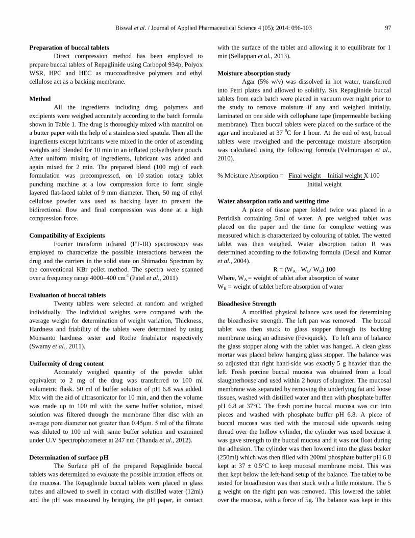
position for 3 minutes and then the weight was added slowly to the right hand pan until the tablet detached from the mucosal surface (Fig. 1). The detachments force gave the bioadhesive strength of the buccal tablet in gm (Vaidya et al., 2009). From the bioadhesive strength, force of adhesion and then the averages of three determinations were calculated. Force of adhesion (F) = [W x g] / 1000 Where g is acceleration due to gravity (9.80665 m/sec2)
Fig. 1: Determination of Bioadhesive Strength of Repaglinide Buccal Tablet. Residence Time
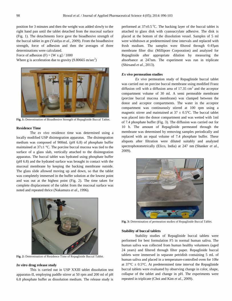
The ex vivo residence time was determined using a locally modified USP disintegration apparatus. The disintegration medium was composed of 900mL (pH 6.8) of phosphate buffer maintained at 37±1 °C. The porcine buccal mucosa was tied to the surface of a glass slab, vertically attached to the disintegration apparatus. The buccal tablet was hydrated using phosphate buffer (pH 6.8) and the hydrated surface was brought in contact with the mucosal membrane by keeping the backing membrane outside. The glass slide allowed moving up and down, so that the tablet was completely immersed in the buffer solution at the lowest point and was out at the highest point (Fig. 2). The time taken for complete displacement of the tablet from the mucosal surface was noted and repeated thrice (Nakamura et al., 1996).
Fig. 2: Determination of Residence Time of Repaglinide Buccal Tablet. In vitro drug release study
This is carried out in USP XXIII tablet dissolution test apparatus-II, employing paddle stirrer at 50 rpm and 200 ml of pH 6.8 phosphate buffer as dissolution medium. The release study is
performed at 37±0.5 oC. The backing layer of the buccal tablet is attached to glass disk with cyanoacrylate adhesive. The disk is placed at the bottom of the dissolution vessel. Samples of 5 ml were withdrawn at predetermined time intervals and replaced with fresh medium. The samples were filtered through 0.45μm membrane filter disc (Millipore Corporation) and analyzed for Repaglinide after appropriate dilution by measuring the absorbance at 247nm. The experiment was run in triplicate (Shirsand et al., 2013). Ex vivo permeation studies
Ex vivo permeation study of Repaglinide buccal tablet was carried out on porcine buccal membrane using modified Franz diffusion cell with a diffusion area of 17.35 cm2 and the acceptor compartment volume of 30 ml. A semi permeable membrane (porcine buccal mucosa membrane) was clamped between the donor and acceptor compartments. The water in the acceptor compartment was continuously stirred at 100 rpm using a magnetic stirrer and maintained at 37 ± 0.5°C. The buccal tablet was placed into the donor compartment and was wetted with 1ml of 7.4 phosphate buffer (Fig. 3). The diffusion was carried out for 10 h. The amount of Repaglinide permeated through the membrane was determined by removing samples periodically and replaced with an equal volume of 7.4 phosphate buffer. These aliquots after filtration were diluted suitably and analyzed spectrophotometrically (Elico, India) at 247 nm (Shanker et al., 2009).
Fig. 3: Determination of permeation studies of Repaglinide Buccal Tablet. Stability of buccal tablets
Stability studies of Repaglinide buccal tablets were performed for best formulation F5 in normal human saliva. The human saliva was collected from human healthy volunteers (aged 24 years) and filtered through filter paper. Repaglinide buccal tablets were immersed in separate petridish containing 5 mL of human saliva and placed in a temperature-controlled oven for 10hr at 37°C ± 0.5°C. At predetermined time interval the Repaglinide buccal tablets were evaluated by observing change in color, shape, collapse of the tablet and change in pH. The experiments were repeated in triplicate (Choi and Kim et al., 2009).
Biswal et al. / Journal of Applied Pharmaceutical Science 4 (05); 2014: 096-103 99
RESULTS AND DISCUSSION
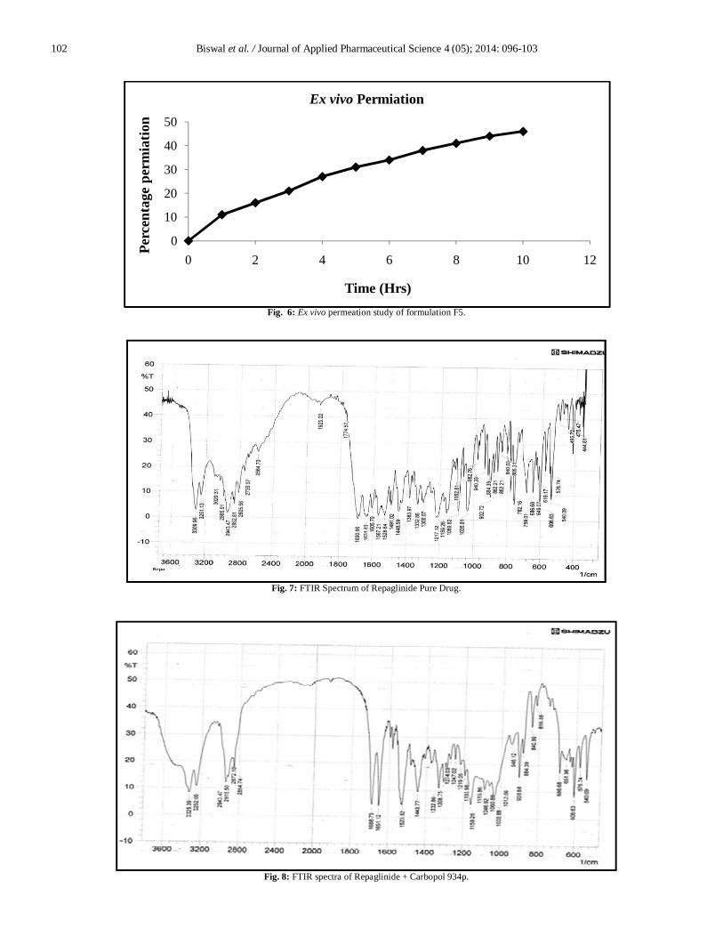
The main goal of this work was to develop new mucoadhesive buccal tablets of Repaglinide, an antidiabetic drug for the treatment of type II (non-insulin dependent) diabetes mellitus, consisting of drug free non-adhesive protective layer. The purity of drug sample was identified by scanning the drug sample on IR spectrophotometer. The peaks of the IR spectra of drug sample were found to be similar with the standard IR spectra of pure Repaglinide as reported. Figure 6 shows the I.R. spectra of Repaglinide. The I.R. spectra of mixture of drug and polymer indicated no incompatibility between drug and polymers, hence Carbopol 934p, Polyox WSR N-80NF were chosen as polymers for further investigations. The spectrum of drug shows absorption bands at 3309.96 cm-1 N-H stretching, 1690.66cm-1 N-H bending , 2943.47 cm-1 C-H2 Stretching. The Drug and Carbopol 934p absorption peaks found at 3325.39 cm-1, 1688.73 cm-1, 2915.50 cm-1. The Drug and Polyox wsr N-80NF absorption peaks found at 3308.03 cm-1, 1605.79 cm-1, 2934.79 cm-1. The double layered structure design was expected to provide drug delivery in an unidirectional fashion to the mucosa and to avoid loss of drug due to wash out by saliva, release drug immediately to produce a prompt pharmacological action and remain in oral cavity and provide a sustained release of enough drug over an extended period of time. A total of twelve formulations of mucoadhesive buccal tablets of Repaglinide were prepared and evaluated for biological, physical and mechanical parameters. The hardness of prepared buccal tablets of Repaglinide was found 4.43 to 4.59 kg/cm2. Mean thickness and weights were found to be uniform as indicated by the low values of standard deviation. The thickness and weight of the prepared buccal tablets were found to be in the range of 2.02 to 2.28 mm and 149 to 151mg respectively.
Friability values less than 1% indicate good mechanical strength to withstand the rigors of handling and transportations. Results are given in Table 2. The drug content of buccal tablets was quite uniform as seen in the above mentioned table. The average drug content of the buccal tablets was found to be within the range of 98.17 to 99.93 % and the low values of standard deviation and coefficient of variation (<2) indicate uniform distribution of the drug within the prepared buccal tablets. The surface pH was determined in order to investigate the possibility of any side effects, in the oral cavity as acidic or alkaline pH is bound to cause irritation to the buccal mucosa. Surface pH of all formulations was found to be almost in neutral pH and ranged between 6.03 and 6.71 and no mucosal irritation was expected. The surface pH of all the formulations is shown in Table 3. The swelling index of mucoadhesive tablets are shown in Table 3. It is manifest that an increase in the amount of polyox wsr N-80NF causes increases in swelling index along with the decreasing amount of HPC and HEC refer Table 1. Among all the formulations, F1 showed maximum swelling index of 57% after 8th and followed by F5, F7, F4, F8, F9, F2, F3 and F6. From the swelling index results as given in Table 3, it was concluded that swelling increases as the time proceeds because the polymer gradually absorb water due to hydrophilicity of polymer. The outermost hydrophilic polymer get hydrated which results in and swelling and a gel barrier is formed at the outer surface. As the gelatinous layer progressively dissolves or disperses. The swelling behaviour provides an idea regarding the moisture intake capacities of polymers and the differences in swelling of the hydrophilic polymers may be due to the difference in resistance of the matrix network structure. The bioadhesion strength and residence time of Repaglinide buccal tablets are shown in Table 3. Adhesion occurs shortly after the beginning of swelling but the bond formed between mucosal layer and polymer
Table. 1: Formulation Variables of Buccal Tablet.
Ingredient formulation code (Qty. mg/tab) F1 F2 F3 F4 F5 F6 F7 F8 F9
Repaglinide 2 2 2 2 2 2 2 2 2 Mannitol 34 34 34 34 34 34 34 34 34
Carbopol 934p 25 25 25 25 25 25 25 25 25 Polyox WSR N-80NF 25 -- -- 12.5 12.5 -- 15 15 --
HPC -- 25 -- 12.5 -- 12.5 10 -- 10 HEC -- -- 25 -- 12.5 12.5 -- 10 15
PVP K30 10 10 10 10 10 10 10 10 10 Aspartame 1 1 1 1 1 1 1 1 1
Mag. stearate 3 3 3 3 3 3 3 3 3 Ethyl cellulose 50 50 50 50 50 50 50 50 50 Total weight 150 150 150 150 150 150 150 150 150
Table. 2: Physicochemical Property of Buccal Tablet.
Formulation code Weight Variation (mg) N=20
Thickness (mm) N=10
Hardness (kg/cm2) N=10
Friability (%w/w) N=3
F1 149 ± 1.97 2.19 ± 0.0059 4.59 ± 0.0262 0.62 ± 0.03 F2 150 ± 1.92 2.15 ± 0.0173 4.57 ± 0.0202 0.72 ± 0.01 F3 150 ± 1.27 2.09 ± 0.0042 4.59 ± 0.0225 0.68 ± 0.02 F4 151 ± 1.87 2.11 ± 0.0379 4.54 ± 0.0322 0.65 ± 0.01 F5 149 ± 1.11 2.17 ± 0.0031 4.41 ± 0.0290 0.60 ± 0.03 F6 150 ± 1.95 2.08 ± 0.0352 4.45 ± 0.0312 0.68 ± 0.06 F7 150 ± 1.82 2.28 ± 0.0442 4.43 ± 0.0448 0.62 ± 0.01 F8 151 ± 1.99 2.17 ± 0.0031 4.44 ± 0.0451 0.54 ± 0.04 F9 149 ± 1.94 2.02 ± 0.0052 4.43± 0.0284 0.42 ± 0.03
The values are expressed as mean±SD.
100 Biswal et al. / Journal of Applied Pharmaceutical Science 4 (05); 2014: 096-103
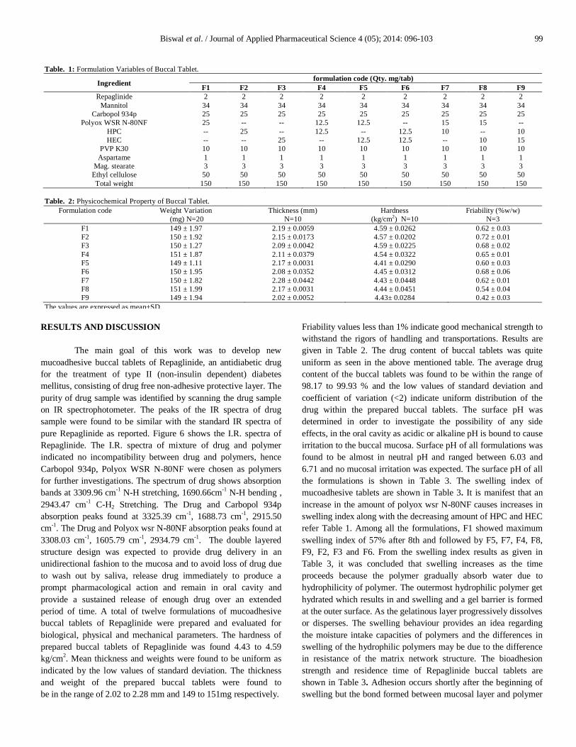
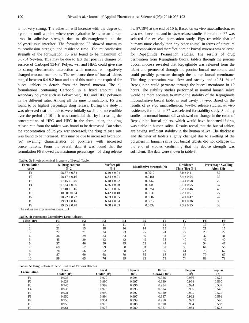
is not very strong. The adhesion will increase with the degree of hydration until a point where over-hydration leads to an abrupt drop in adhesive strength due to disentanglement at the polymer/tissue interface. The formulation F5 showed maximum mucoadhesion strength and residence time. The mucoadhesive strength of the formulation F5 was found to be maximum of 0.0754 Newton. This may be due to fact that positive charges on surface of Carbopol 934-P, Polyox wsr and HEC, could give rise to strong electrostatic interaction with mucous or negatively charged mucous membrane. The residence time of buccal tablets ranged between 6.4-9.2 hour and noted this much time required for buccal tablets to detach from the buccal mucosa. The all formulations containing Carbopol in a fixed amount. The secondary polymer such as Polyox wsr, HPC and HEC polymers in the different ratio. Among all the nine formulations, F5 was found to be highest percentage drug release. During the study it was observed that the tablets were initially swell and no erodible over the period of 10 h. It was concluded that by increasing the concentration of HPC and HEC in the formulation, the drug release rate from the tablets was found to be decreased. But when the concentration of Polyox wsr increased, the drug release rate was found to be increased. This may be due to increased hydration (or) swelling characteristics of polymers with increased concentrations. From the overall data it was found that the formulation F5 showed the maximum percentage of drug release
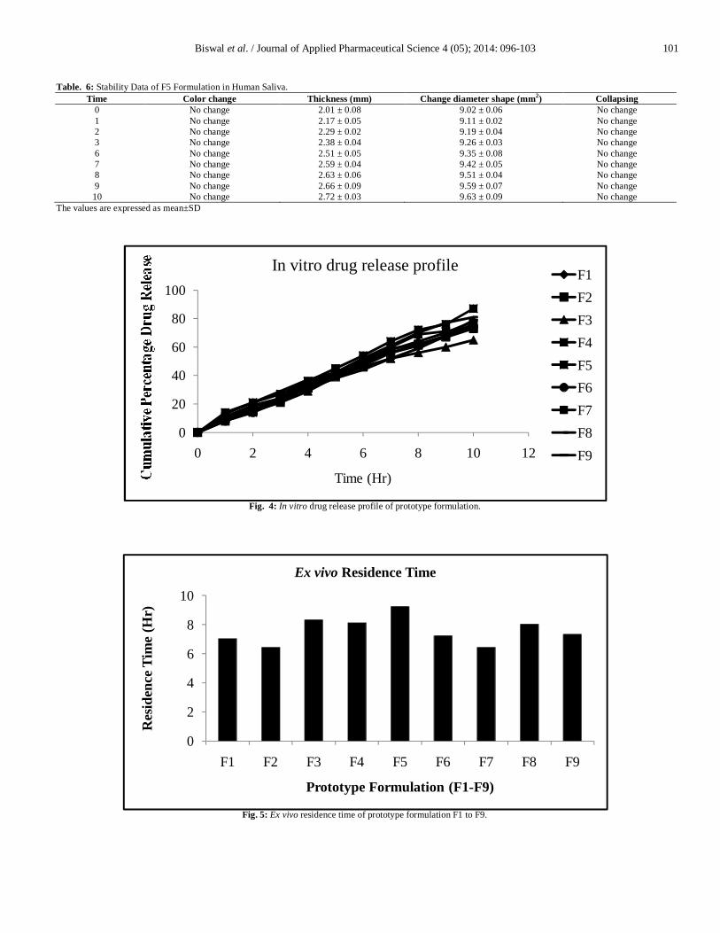
i.e. 87.18% at the end of 10 h. Based on ex vivo mucoadhesion, ex vivo residence time and in-vitro release studies formulation F5 was selected for ex vivo permeation study. Pigs resemble that of humans more closely than any other animal in terms of structure and composition and therefore porcine buccal mucosa was selected for Repaglinide Permeation studies. The results of drug permeation from Repaglinide buccal tablets through the porcine buccal mucosa revealed that Repaglinide was released from the tablet and permeated through the porcine buccal membrane and could possibly permeate through the human buccal membrane. The drug permeation was slow and steady and 42.51 % of Repaglinide could permeate through the buccal membrane in 10 hours. The stability studies performed in normal human saliva would be more accurate to mimic the stability of the Repaglinide mucoadhesive buccal tablet in oral cavity in vivo. Based on the results of ex vivo mucoadhesion, in-vitro release studies, ex vivo residence, formulation F5 was selected for stability study. Stability studies in normal human saliva showed no change in the color of Repaglinide buccal tablets, which would have happened if drug was stable in human saliva. Results reveal that the buccal tablets are having sufficient stability in the human saliva. The thickness and diameter of tablets slightly changed due to swelling of the polymers in human saliva but buccal tablets did not collapse till the end of studies confirming that the device strength was sufficient. The data were shown in table 6.
Table. 3: Physicochemical Property of Buccal Tablet. Formulation
code % Drug content
N=2 Surface pH
N=3 Bioadhesive strength (N) Residence Time (Hr) N=3
Percentage Swelling After 8Hr
F1 98.57 ± 0.84 6.19 ± 0.04 0.0512 7.0 ± 0.41 57 F2 98.17 ± 0.16 6.34 ± 0.01 0.0481 6.4 ± 0.54 32 F3 97.15 ± 1.46 6.18 ± 0.02 0.0667 8.3 ± 0.58 29 F4 97.34 ± 0.86 6.36 ± 0.30 0.0608 8.1 ± 0.55 37 F5 97.40 ± 1.16 6.71 ± 0.06 0.0754 9.2 ± 0.46 46 F6 100.01±0.84 6.42 ± 0.10 0.0530 7.2 ± 0.51 27 F7 98.72 ± 0.72 6.03 ± 0.05 0.0507 6.4 ± 0.47 42 F8 99.93 ± 0.16 6.14 ± 0.04 0.0612 8.0 ± 0.36 36 F9 99.35 ± 0.78 6.08 ± 0.03 0.0532 7.3 ± 0.55 33
The values are expressed as mean±SD Table. 4: Percentage Cumulative Drug Release .
Time (Hr) F1 F2 F3 F4 F5 F6 F7 F8 F9 1 14 8 11 11 9 11 8 13 9 2 21 15 18 16 14 19 14 21 15 3 27 21 24 23 25 24 22 29 22 4 36 29 34 33 36 31 33 37 31 5 45 39 42 42 45 38 40 42 40 6 57 46 50 49 53 44 49 54 47 7 69 52 59 58 68 52 56 64 56 8 78 56 62 68 72 59 61 73 62 9 87 60 68 79 85 68 68 79 67 10 99 65 76 89 93 78 74 83 73
Table. 5: Drug Release Kinetic Studies of Various Batches
Formulation Zero Order (R2)
First Order (R2)
Higuchi (R2)
Hixon Crowell (R2)
Peppas (R2)
Peppas (n)
F1 0.936 0.970 0.994 0.986 0.986 0.525 F2 0.928 0.990 0.997 0.980 0.994 0.530 F3 0.945 0.992 0.996 0.984 0.994 0.537 F4 0.938 0.973 0.995 0.984 0.996 0.545 F5 0.931 0.990 0.997 0.980 0.995 0.525 F6 0.952 0.994 0.997 0.987 0.992 0.591 F7 0.958 0.951 0.999 0.960 0.993 0.590 F8 0.955 0.978 0.988 0.990 0.984 0.583 F9 0.961 0.978 0.980 0.987 0.964 0.623
Biswal et al. / Journal of Applied Pharmaceutical Science 4 (05); 2014: 096-103 101
Table. 6: Stability Data of F5 Formulation in Human Saliva. Time Color change Thickness (mm) Change diameter shape (mm2) Collapsing
0 No change 2.01 ± 0.08 9.02 ± 0.06 No change 1 No change 2.17 ± 0.05 9.11 ± 0.02 No change 2 No change 2.29 ± 0.02 9.19 ± 0.04 No change 3 No change 2.38 ± 0.04 9.26 ± 0.03 No change 6 No change 2.51 ± 0.05 9.35 ± 0.08 No change 7 No change 2.59 ± 0.04 9.42 ± 0.05 No change 8 No change 2.63 ± 0.06 9.51 ± 0.04 No change 9 No change 2.66 ± 0.09 9.59 ± 0.07 No change 10 No change 2.72 ± 0.03 9.63 ± 0.09 No change
The values are expressed as mean±SD
Fig. 4: In vitro drug release profile of prototype formulation.
Fig. 5: Ex vivo residence time of prototype formulation F1 to F9.
0
20
40
60
80
100
0 2 4 6 8 10 12
Time (Hr)
In vitro drug release profile F1F2F3F4F5F6F7F8F9
0
2
4
6
8
10
F1 F2 F3 F4 F5 F6 F7 F8 F9
Res
iden
ce T
ime
(Hr)
Prototype Formulation (F1-F9)
Ex vivo Residence Time
102 Biswal et al. / Journal of Applied Pharmaceutical Science 4 (05); 2014: 096-103
Fig. 6: Ex vivo permeation study of formulation F5.
Fig. 7: FTIR Spectrum of Repaglinide Pure Drug.
Fig. 8: FTIR spectra of Repaglinide + Carbopol 934p.
0
10
20
30
40
50
0 2 4 6 8 10 12
Perc
enta
ge p
erm
iatio
n
Time (Hrs)
Ex vivo Permiation
Biswal et al. / Journal of Applied Pharmaceutical Science 4 (05); 2014: 096-103 103
CONCLUSION
The mucoadhesive strength were influenced by the nature and proportions of the mucoadhesive polymers used in the formulations .In all the formulations, as the mucoadhesive polymer mixture concentration increased, the mucoadhesive strength also increased. Buccal tablets formulated (F5) with a mixture of carbopol 934p, Polyox WSR N-80NF and HEC showed comparatively higher bioadhesion than that of all other formulataion. Very strong mucoadhesion could damage the epithelial lining of the buccal mucosa. Mucoadhesive strength exhibited by the formulation F5 tablets can be considered satisfactory for maintaining them in the oral cavity for 12hrs. The surface pH was determined in order to investigate the possibility of any side effects, in the oral cavity. The surface pH of the buccal tablets depends on the nature and composition of mucoadhesive polymers. Surface pH of the all the formulation were found to be in the range of 5.9 to 6.5. This pH is near to the neutral, so the buccal tablet does not cause any irritation on the mucosa. The Ex vivo residence time was determined by using modified physical balance method. Formulations F5 to F3 showed Higher Bioadhesive time when compared to the other formulations. As the concentration of the Carbopol 934p same in all formulation polyox wsr and HEC shows greater bioadhesive time. the residence time also increased. This test reflectsthe mucoadhesive capacity of polymers used in formulations. The results revealed that the mixture of carbopol 934p and HEC containing formulations showed better bioadhesion than the mixture of all other muccoadhesive polymer in the formulations. Based on ex vivo mucoadhesion, ex vivo residence time and in-vitro release studies formulation F5 was selected for ex vivo permeation study. REFERENCES
Choi HG, Kim CK. Development of omeprazole buccal adhesive tablets with stability enhancement in human saliva. J Control Release, 2000; 68:397–404.
Desai KGH, Kumar TMP. Preparation and evaluation of a novel buccal adhesive systems. AAPS Pharm Sci Tech, 2004; 5:1–9.
Jain SK, Awasthi AM, Jain NK, Agrawal GP. Calcium silicate
based microspheres of repaglinide for gastroretentive floating drug delivery: Preparation and in vitro characterization. J. Control. Release, 2005; 107:300–309.
Langer MA, Robinson RJ. 1985 Sustained release drug delivery systems In: Remington's Pharmaceutical Sciences. New York: Mack Publishing Company 1644-61.
Nakamura F, Ohta R, Machida Y, Nagai T. In vitro and in vivo nasal mucoadhesion of water soluble polymers. Int. J. Pharm, 1996; 134: 173-181.
Patel M, Pandya N, Bhaskar V.H. Preparation, Characterization and In Vitro Evaluation of Repaglinide Binary Solid Dispersions with Hydrophilic Polymers. International Journal of Drug Development & Research 2011; 3:111-121.
Sean CS. 2009. Martindale the Complete Drug Reference. Pharmaceutical Press. Sellappan V, Srinivas P. Formulation and in vitro evaluation of losartan potassium mucoadhesive buccal tablets. Asian J Pharm Clin Res 2013; 6:125-130.
Shanker G, Kumar CK, Gonugunta CSR, Kumar BV, Veerareddy PR. Formulation and evaluation of bioadhesive buccal drug delivery of tizanidine hydrochloride tablets. AAPS PharmSciTech, 2009; 10:530–9.
Shirsand SB, Wadageri GV, Raju SA, Kolli G, Reddy V. Design and evaluation of mucoadhesive bilayer buccal tablets of nebivolol. RGUHS J Pharm Sci, 2013; 3:40-47.
Swamy PV, Kinagi MB, Biradar SS, Gada S, Shilpa H. Formulation Design and Evaluation of Bilayer Buccal Tablets of Granisetron Hydrochloride. Ind J Pharm Edu Res, 2011; 45:242-247.
Thanda V, Firoz S, Yerram C, Vikram A, Divya SK, Murali KT. Design and characterisation sustained release matrix tablets of Repaglinide using natural polymers. International Journal of Pharmacy, 2012; 2:73-83.
Vaidya M, Manwar V, Mahajan M, Sakarkar M. Design and in vitro evaluation of mucoadhesive buccal tablets of terbutaline sulphate. Int. J. Pharm. Tech. Res, 2009; 1: 588-597.
Velmurugan S, Deepika B, Nagaraju K, Sundar V. Formulation and in‐vitro Evaluation of Buccal Tablets of Piroxicam. International Journal of PharmTech Research, 2010; 2:1958‐1968.
Vyas SP, Khar RK. 2002. Controlled drug delivery concepts and advances. New Delhi: Vallabh Prakashan.
How to cite this article:
Biswajit Biswal, Nabin Karna, Bhavesh Bhavsar., Formulation and Evaluation of Repaglinide Buccal Tablet: Ex Vivo Bioadhesion Study And Ex Vivo Permeability Study. J App Pharm Sci, 2014; 4 (05): 096-103.
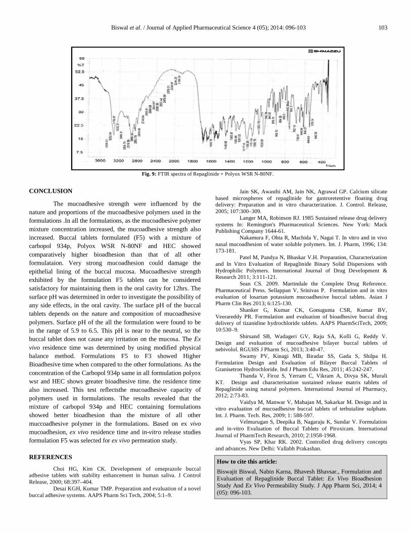
Fig. 9: FTIR spectra of Repaglinide + Polyox WSR N-80NF.