![Retirement List - WordPress.com€¦ · NECKLACE $45 [6187] MONOGRAM INITIAL NECKLACE Antique Pewter, Antique Gold $48 [6185] HORIZON NECKLACE $48 [6149] ENCHANTED CROSS NECKLACE](https://static.fdocuments.in/doc/165x107/5f453a3e5ac36e55ec0eb842/retirement-list-necklace-45-6187-monogram-initial-necklace-antique-pewter.jpg)
Formation of Pearl-Necklace Monomorphic G-Quadruplexes … · Formation of Pearl-Necklace...
26
Formation of Pearl-Necklace Monomorphic G-Quadruplexes in the Human CEB25 Minisatellite Samir Amrane, †,‡,∥ Michael Adrian, †,∥ Brahim Heddi, † Alexandre Serero, § Alain Nicolas, § Jean-Louis Mergny,* ,‡ and Anh Tua ̂ n Phan* ,† † School of Physical and Mathematical Sciences, Nanyang Technological University, 637371 Singapore ‡ University of Bordeaux, European Institute of Chemistry and Biology, INSERM U869, 33600 Pessac, France § Institut Curie, Centre de Recherche, UMR3244 CNRS, Universite ́ Pierre et Marie Curie, 75248 Paris, France * S Supporting Information ABSTRACT: CEB25 is a human minisatellite locus, com- posed of slightly polymorphic 52-nucleotide (nt) tandem repeats. Genetically, most if not all individuals of the human population are heterozygous, carrying alleles ranging from 0.5 to 20 kb, maintained by mendelian inheritance but also subject to germline instability. To provide insights on the biological role of CEB25, we interrogated its structural features. We report the NMR structure of the G-quadruplex formed by the conserved 26-nt G-rich fragment of the CEB25 motif. In K + solution, this sequence forms a propeller-type parallel-stranded G-quadruplex involving a 9-nt central double-chain-reversal loop. This long loop is anchored to the 5′-end of the sequence by an A·T Watson−Crick base pair and a potential G·A noncanonical base pair. These base pairs contribute to the stability of the overall G-quadruplex structure, as measured by an increase of about 17 kcal/mol in enthalpy or 6 °C in melting temperature. Further, we demonstrate that such a monomorphic structure is formed within longer sequence contexts folding into a pearl-necklace structure. ■ INTRODUCTION G-quadruplexes are a family of nucleic acid structures formed by G-rich DNA or RNA sequences, built from the stacking of G-tetrads, each being a planar association of four guanines held together by eight hydrogen bonds and stabilized by cations, such as Na + or K + . 1−4 G-quadruplex structures from many human genomic regions including oncogenic promoters and telomeres have been elucidated based on short truncated G-rich fragments derived from natural sequences. 2 These structures show a variety of G-quadruplex folding topologies with respect to strand orien- tations, glycosidic conformations of guanine bases, and connecting loops. Several G-quadruplex structures of oncogenic promoter sequences exhibit unique folding topologies. For instance, a five- G-tract sequence from the c-myc promoter folds into a snap-back configuration with a stable triad-containing diagonal loop, 5 and a G-rich sequence from the c-kit promoter forms an unprecedented snap-back parallel G-quadruplex involving an isolated guanine and a 5-nt stem loop. 6 Human telomeric repeats have been found to adopt multiple G-quadruplex conformations depending on the length of the fragments, flanking sequences, and crowding con- dition. 4,7 Furthermore, the formation of a G-quadruplex structure containing an extended loop has been observed for sequences spanning five or more human telomeric repeats. 8 An array of G-quadruplexes interconnected by short linkers (TTA) could be formed on a long telomeric sequence. 9 Intramolecular G-quadruplexes adopted by a single DNA strand have attracted much interest since they could be formed within biologically relevant regions of the genome. 10−13 Although the biological role of the numerous potential G-quadruplex- forming sequences in the genome remains to be characterized, such structures which can be formed on single-stranded DNA and RNA, for example, during replication, recombination, and tran- scription, likely play a role and interfere with these key cellular processes. 13−15 The remarkable G-quadruplex structure adopted by the human CEB25 sequence (locus D10S180), a polymorphic minisatellite in the human population maintained by mendelian inheritance but also subject to germline instability, 16 is under the scope of this paper. Minisatellites consist of head-to-tail arrays of identical or slightly polymorphic 10−100-bp long motifs. They are present in prokaryote and eukaryote genomes and might constitute as much as 10% of the human genome. 17 Often, the overall size of the minisatellite loci varies between individuals and can range from 0.5 to >15 kbp. These variations correspond to simple or complex gain or loss of repeat units and have initially been used as genetic DNA typing markers. 16,18 The large number of minisatellite loci, the variety of their nucleotide sequences, and their persistence in the genome suggest that they likely possess Received: September 24, 2011 Published: February 29, 2012 Article pubs.acs.org/JACS © 2012 American Chemical Society 5807 dx.doi.org/10.1021/ja208993r | J. Am. Chem. Soc. 2012, 134, 5807−5816
Transcript of Formation of Pearl-Necklace Monomorphic G-Quadruplexes … · Formation of Pearl-Necklace...

Formation of Pearl-Necklace Monomorphic G-Quadruplexes in theHuman CEB25 MinisatelliteSamir Amrane,†,‡,∥ Michael Adrian,†,∥ Brahim Heddi,† Alexandre Serero,§ Alain Nicolas,§
Jean-Louis Mergny,*,‡ and Anh Tuan Phan*,†
†School of Physical and Mathematical Sciences, Nanyang Technological University, 637371 Singapore‡University of Bordeaux, European Institute of Chemistry and Biology, INSERM U869, 33600 Pessac, France§Institut Curie, Centre de Recherche, UMR3244 CNRS, Universite Pierre et Marie Curie, 75248 Paris, France
*S Supporting Information
ABSTRACT: CEB25 is a human minisatellite locus, com-posed of slightly polymorphic 52-nucleotide (nt) tandemrepeats. Genetically, most if not all individuals of the humanpopulation are heterozygous, carrying alleles ranging from 0.5to 20 kb, maintained by mendelian inheritance but also subjectto germline instability. To provide insights on the biologicalrole of CEB25, we interrogated its structural features. Wereport the NMR structure of the G-quadruplex formed by theconserved 26-nt G-rich fragment of the CEB25 motif. In K+
solution, this sequence forms a propeller-type parallel-strandedG-quadruplex involving a 9-nt central double-chain-reversalloop. This long loop is anchored to the 5′-end of the sequence by an A·T Watson−Crick base pair and a potential G·Anoncanonical base pair. These base pairs contribute to the stability of the overall G-quadruplex structure, as measured by anincrease of about 17 kcal/mol in enthalpy or 6 °C in melting temperature. Further, we demonstrate that such amonomorphic structure is formed within longer sequence contexts folding into a pearl-necklace structure.
■ INTRODUCTIONG-quadruplexes are a family of nucleic acid structures formedby G-rich DNA or RNA sequences, built from the stacking ofG-tetrads, each being a planar association of four guanines heldtogether by eight hydrogen bonds and stabilized by cations,such as Na+ or K+.1−4 G-quadruplex structures from many humangenomic regions including oncogenic promoters and telomereshave been elucidated based on short truncated G-rich fragmentsderived from natural sequences.2 These structures show a varietyof G-quadruplex folding topologies with respect to strand orien-tations, glycosidic conformations of guanine bases, and connectingloops. Several G-quadruplex structures of oncogenic promotersequences exhibit unique folding topologies. For instance, a five-G-tract sequence from the c-myc promoter folds into a snap-backconfiguration with a stable triad-containing diagonal loop,5 and aG-rich sequence from the c-kit promoter forms an unprecedentedsnap-back parallel G-quadruplex involving an isolated guanine anda 5-nt stem loop.6 Human telomeric repeats have been found toadopt multiple G-quadruplex conformations depending on thelength of the fragments, flanking sequences, and crowding con-dition.4,7 Furthermore, the formation of a G-quadruplex structurecontaining an extended loop has been observed for sequencesspanning five or more human telomeric repeats.8 An array ofG-quadruplexes interconnected by short linkers (TTA) couldbe formed on a long telomeric sequence.9
Intramolecular G-quadruplexes adopted by a single DNAstrand have attracted much interest since they could be formedwithin biologically relevant regions of the genome.10−13 Althoughthe biological role of the numerous potential G-quadruplex-forming sequences in the genome remains to be characterized,such structures which can be formed on single-stranded DNA andRNA, for example, during replication, recombination, and tran-scription, likely play a role and interfere with these key cellularprocesses.13−15 The remarkable G-quadruplex structure adoptedby the human CEB25 sequence (locus D10S180), a polymorphicminisatellite in the human population maintained by mendelianinheritance but also subject to germline instability,16 is under thescope of this paper.Minisatellites consist of head-to-tail arrays of identical or
slightly polymorphic 10−100-bp long motifs. They are presentin prokaryote and eukaryote genomes and might constitute asmuch as 10% of the human genome.17 Often, the overall size ofthe minisatellite loci varies between individuals and can rangefrom 0.5 to >15 kbp. These variations correspond to simple orcomplex gain or loss of repeat units and have initially beenused as genetic DNA typing markers.16,18 The large number ofminisatellite loci, the variety of their nucleotide sequences, andtheir persistence in the genome suggest that they likely possess
Received: September 24, 2011Published: February 29, 2012
Article
pubs.acs.org/JACS
© 2012 American Chemical Society 5807 dx.doi.org/10.1021/ja208993r | J. Am. Chem. Soc. 2012, 134, 5807−5816

biological roles. Interestingly, the yeast natural minisatellites arelocated in the coding region of cell-wall proteins and variationof the intragenic repeats may have a role in adaptation withinenvironmental changes.19 In the human genome, numerousG-rich minisatellite loci are located in subtelomeric locations,but internal minisatellite repeats also exist and are associatedwith numerous disorders, such as epilepsy related to the EPM1gene,20 diabetes associated with the insulin gene,21 and bipolardisorders linked to the serotonin transporter gene 5-HTT,22
and might also predispose to cancer related to the function ofthe H-ras gene.23 On a mechanistic perspective, linked to theunderlying mechanisms promoting size variation in somaticand/or germline cells by replication, repair, or recombinationmechanisms,24 it is also suspected that the nature of the DNAsequence itself carries structural properties at the DNA andchromatin levels,25 as observed for DNA sequences implicatedin trinucleotide-repeat expansion diseases,26−28 able to formnoncanonical DNA structures such as hairpin, triplex,quadruplex, or sticky DNA.29,30 Therefore, structural study ofminisatellite sequences might provide fundamental insights toelucidate their functions.Along this line, here we characterize the sequence poly-
morphism of the G-rich human CEB25 minisatellite loci anduncover the remarkable structural features of the G-quadruplexforming motif which contains successive guanines prone toG-quadruplex formation, in particular, a segment, AAGGGTGGG-TGTAAGTGTGGGTGGGT, composed of four GGG tracts(underlined) separated by linkers of 1, 9, and 1 nucleotides,respectively. We show that this sequence forms in K+ solution apropeller-type parallel-stranded G-quadruplex involving a9-nucleotide (nt) central double-chain-reversal loop. This longloop is anchored to the 5′ end of the sequence by an A·TWatson−Crick base pair and a potential G·A noncanonical basepair, contributing to the stability of the overall G-quadruplexstructure. Further, we demonstrate that such a structure isformed in longer sequence contexts and, along the line of its invivo organization in tandem arrays, fold into a monomorphicpearl-necklace structure.
■ METHODSDNA Sample Preparation. Unlabeled and site-specific labeled
DNA oligonucleotides (Tables 1 and S1, Supporting Information) werechemically synthesized on an ABI 394 DNA/RNA synthesizeror purchased from Eurogentec (Seraing, Belgium). Samples werepurified and dialyzed successively against potassium chloride solutionand water as previously described.8 Unless otherwise stated, DNAoligonucleotides were dissolved in solution containing 70 mMpotassium chloride and 20 mM potassium phosphate, pH 7. DNAconcentration was expressed in strand molarity using a nearest-neighbor approximation for the absorption coefficients of the unfoldedspecies.31
Sequencing of Human CEB25 Alleles. CEB25 regions wereamplified from human genomic DNA with primers specific to theflanking chromosomal regions (CEB25Up 5′-AAAGACAATGACT-CAGGGTGG-3′ and CEB25LowII 5′-CCGGCACAAACCCTG-CTGCTGGGAGTAAGAGGG-3′). Due to the high GC content of theCEB25 sequence, PCR reactions were performed as previouslydescribed32 and resuspended in a final volume of 15 μL. The PCRproducts were analyzed by electrophoresis on 1.0% agarose gels toverify the size determined by prior Southern blot analysis, purified,cloned into pGEMT-Easy (Promega), and sequenced by using theBig Dye Terminator version 3.1 kit (Perkin-Elmer) and 20% betaine(Sigma).Analysis of the CEB25 Size Polymorphism in CEPH Families.
Lyophilized DNA from five CEPH families (#1334, 1354, 1355, 1357,
and 1417) was suspended in 200 μL of TE buffer. The genomic DNAwas purified by phenol/chlorophorm/isoamyl alcohol extraction andethanol precipitation and resuspended in 20 μL of TE. For each indi-vidual, 15 μg of genomic DNA was digested with ApaI (3 h at 37 °C)and treated with RNase A for 30 min. The resulting fragments wereseparated by electrophoresis on 0.8% agarose gels and transferredunder vacuum (Qbiogene) onto Hybond N+ membranes (Pharmacia).The membranes were hybridized with the radiolabeled naturalCEB25−1.7 fragment. According to the data provided by the NCBIresources, human CEB25 sequence is flanked by ApaI restriction sites,36 bp upstream and 843 bp downstream, respectively. Therefore, thesize of the CEB25 locus is 879 bp smaller than the ApaI band detectedby Southern blot.
UV-Melting Experiments. The thermal stability of DNAstructures was characterized in heating/cooling experiments byrecording the UV absorbance at 295 nm as a function of temperature33
using a UVIKON XL UV/vis spectrophotometer. UV-melting experi-ments were conducted as previously described34 at variable DNAstrand concentrations ranging from 1 to 125 μM in a 20 mM potassiumphosphate buffer (pH 7) containing 70 mM KCl. The heating andcooling rates were 0.2 °C/min. Experiments were performed with 1 and0.2 cm path length quartz cuvettes.
Thermal Difference Spectra. Thermal difference spectra (TDS)were obtained by taking the difference between the absorbance spectra
Table 1. List of DNA Sequences Analyzed in This Worka
sequence name sequence (5′−3′)
26CEB AAGGGTGGGTGTAAGTGTGGGTGGGT
m1T TAGGGTGGGTGTAAGTGTGGGTGGGTm2T ATGGGTGGGTGTAAGTGTGGGTGGGTm10C AAGGGTGGGCGTAAGTGTGGGTGGGTm11T AAGGGTGGGTTTAAGTGTGGGTGGGTm12A AAGGGTGGGTGAAAGTGTGGGTGGGTm13T AAGGGTGGGTGTTAGTGTGGGTGGGTm14T AAGGGTGGGTGTATGTGTGGGTGGGTm15T AAGGGTGGGTGTAATTGTGGGTGGGTm16A AAGGGTGGGTGTAAGAGTGGGTGGGTm17T AAGGGTGGGTGTAAGTTTGGGTGGGTm18C AAGGGTGGGTGTAAGTGCGGGTGGGTm(1,2)T TTGGGTGGGTGTAAGTGTGGGTGGGTm(11,13−15)T AAGGGTGGGTTTTTTTGTGGGTGGGTm(11,13−15,17)T AAGGGTGGGTTTTTTTTTGGGTGGGTm(1,2,11,13−15,17)T TTGGGTGGGTTTTTTTTTGGGTGGGTm1Δ -AGGGTGGGTGTAAGTGTGGGTGGGT
m(1,2,26)Δ --GGGTGGGTGTAAGTGTGGGTGGG-
m1Δ2T -TGGGTGGGTGTAAGTGTGGGTGGGTI7 AAGGGTIGGTGTAAGTGTGGGTGGGT26CEBmut AAGTGTGTGTGTAAGTGTGTGTGTGT52CEB(5′) 26CEB−GTGAGTGTGGGTGTGGAGGTAGATGT52CEB(3′) GTGAGTGTGGGTGTGGAGGTAGATGT-26CEB
57CEB(mid) TGGGTGTGGAGGTAGATGT-26CEB-GTGAGTGTGGGT
78CEB(5′,3′) 52CEB(5′)−26CEB78CEB(5′,3′I7) 52CEB(5′)−I778CEB(5′,3′mut) 52CEB(5′)−26CEBmut78CEB(5′mut,3′) 26CEBmut−52CEB(3′)78CEB(mid) GTGAGTGTGGGTGTGGAGGTAGATGT−
52CEB(5′)
130CEB(5′,mid,3′) 52CEB(5′)−26CEB−52CEB(3′)130CEB(5′I7,mid,3′) I7−52CEB(3′)−52CEB(3′)130CEB(5′,midI7,3′) 52CEB(5′)−I7−52CEB(3′)130CEB(5′,mid,3′I7) 52CEB(5′)−52CEB(5′)−I7
aLong sequences can be read by connecting DNA fragments ofrespective names and sequences. Mutations (variations from 26CEB)are shown in boldface.
Journal of the American Chemical Society Article
dx.doi.org/10.1021/ja208993r | J. Am. Chem. Soc. 2012, 134, 5807−58165808

from unfolded and folded oligonucleotides that were respectivelyrecorded much above and below its melting temperature (Tm). TDSprovide specific signatures of different DNA structural conforma-tions.35 Spectra were recorded between 220 and 320 nm on a JASCOV-650 UV/vis spectrophotometer using 1 cm path length quartzcuvettes. The DNA oligonucleotides (∼5 μM) were prepared in a20 mM potassium phosphate buffer (pH 7) containing 70 mM KCl.For each experiment, an average of three scans was taken, and the datawere zero-corrected at 320 nm.Circular Dichroism. Circular dichroism (CD) spectra were recorded
on a JASCO-810 spectropolarimeter using 1 cm path length quartzcuvettes with reaction volume of 600 μL. The DNA oligonucleotides(∼5 μM) were prepared in a 20 mM potassium phosphate (pH 7)buffer containing 70 mM KCl. For each experiment, an average ofthree scans was taken, the spectrum of the buffer was subtracted, andthe data were zero-corrected at 320 nm.Differential Scanning Calorimetry (DSC). Microcalorimetry
experiments were performed on a Nano DSC-II microcalorimeter aspreviously described.26−28 The oligonucleotides were prepared atconcentrations ranging from 100 to 150 μM in a 20 mM potassiumphosphate buffer (pH 7) containing 70 mM KCl. An average of sixdifferential scanning calorimetric (DSC) heating and cooling profileswas taken.NMR Spectroscopy. NMR experiments were performed on 400,
600, 700, and 800 MHz Bruker spectrometers at 25 °C, unlessotherwise specified. The strand concentration of the NMR sampleswas typically 0.2−2.0 mM. Resonances for guanine residues wereassigned unambiguously by using site-specific low-enrichment 15Nlabeling,36 site-specific 2H labeling,37 and through-bond correlations atnatural abundance.38 Spectral assignments were completed by NOESY,TOCSY, {13C−1H}-HMBC, {13C−1H}-HSQC and {31P−1H}-HSQC, aspreviously described.39 Interproton distances were deduced from NOESYexperiments performed in H2O (mixing time, 200 ms) and 2H2O (mixingtimes, 100, 150, 200, and 350 ms).One-bond C1′−H1′, C8−H8, C6−H6, and C2−H2 residual dipolar
couplings (RDC) were obtained by comparing the 13C−1H splitting innondecoupled {13C−1H}-HSQC spectra in aligned and isotropicconditions (see example in Figure S1, Supporting Information) mea-sured on unlabeled DNA. The aligned medium was obtained byadding 20 mg/mL of Pf1 bacteriophage (ALSA Biotech) in an un-labeled DNA sample.All spectral analyses were performed using the FELIX (Felix NMR,
Inc.) and SPARKY (T. D. Goddard and D. G. Kneller, SPARKY 3,University of California, San Francisco) programs.Structure Calculation. Structure computations were performed
using the XPLOR-NIH program with RDC restraints. The procedurelies in two general steps essentially as previously described:40 (i) dis-tance geometry simulated annealing and (ii) distance-restrained molec-ular dynamics refinement. RDC restraints, hydrogen-bond restraints,interproton distance restraints, dihedral restraints, planarity restraints,and repulsive restraints were imposed during structure calculations.The components of the alignment tensor were calculated using
optimized structures derived from NMR distance restraints. Only theresidues forming the core of the G-quadruplex were used to find thetensor parameters. Structure refinement has improved the correlationbetween the calculated and measured RDCs (Figure S2, SupportingInformation). The dihedral restraints were based on intraresiduesNOE of H1′-H6/8 cross-peak intensities (glycosidic dihedral angles)and observed phosphorus chemical shifts (backbone dihedral ε andβ angles). The planarity restraints were used in agreement withpreviously observed G-quadruplex X-ray structures. The repulsiverestraints were applied to several pairs of well-resolved protons, whichdid not give observable NOE cross-peaks. Structures were displayedusing the PyMOL program.Data Deposition. The coordinates of ten lowest-energy
d[AAGGGTGGGTGTAAGTGTGGGTGGGT] (26CEB) G-quadruplexstructures have been deposited in the Protein Data Bank (accession code2LPW).
■ RESULTS
Features of the Human CEB25 Polymorphism. ThehCEB25 locus is localized on chromosome 10q26.3, approx-imately 1.3 Mb away from the chromosome end, within theintron 9 of the LRRC27 (leucine-rich-repeat containing protein27) gene of unknown function. The sequence of the originallyreported 0.8-kb-long CEB25 allele (accession numberAL096806) is shown in Figure S3, Supporting Information. Itis composed of 17 motifs of approximately 52 nucleotides longwith a 53% GC content and a strong GC asymmetry; 92% ofthe G nucleotides are on the same strand.16 To characterize thelength polymorphism of the CEB25 locus in individuals of thehuman population, we examined genomic DNA from 81 indi-viduals from five CEPH families (Figure S4, SupportingInformation). Remarkably, the size of the CEB25 locus isextremely variable, ranging from approximately 0.5 to 20 kb,corresponding to ∼10−400 motifs, respectively. Genetically, allindividuals are heterozygotes carrying two different paternaland maternal size alleles segregating with mendelian inher-itance. Along the three generations examined, we found no denovo size variants (0/81). This indicates that CEB25 has a lowgermline instability, previously estimated to 2%, from the geno-typing of approximately 500 children of other CEPH families(G. Vergnaud, personal communication). Recent studies correlatedmammalian meiotic hot spot activity to the local presence ofPRDM9 sequence-specific binding site,41−43 only few motifs ofthe CEB25 alleles have sequences that correspond to a highaffinity binding site of the human PRDM9 protein (Figure S3,Supporting Information). Thus, not surprisingly, CEB25germline instability is not as high as other minisatellites suchas CEB144 but remains sufficient to generate the diversity ofCEB25 alleles in the human population.To further characterize CEB25, we cloned and sequenced a
second 1.7-kb-long hCEB25 allele. From the comparison of theCEB25−0.8 and CEB25−1.7 alleles and motifs (Figure S3,Supporting Information), several conclusions can be drawn: (i)both CEB25 alleles are canonical minisatellites composed oftandem repeats, (ii) the size of the motifs is highly conservedwith a large majority of 52-bp-long repeats but few 36-bp-longmotifs, and (iii) the sequence of the motifs is slightly polymorphic.Altogether, we identified 21 and 16 polymorphic sites within theCEB25−0.8 and CEB25−1.7 alleles, respectively. They are mostlysingle-base substitutions. Specifically, the 31 repeats of theCEB25−1.7 allele include 16 different motifs, and the motifsthat are not unique are present in two to four copies. Relevantto the potential of CEB25 motifs to form G-quadruplexes, mostrepeats (81%) contain at least four of their five guanine triplets(Figure S5, Supporting Information). These G-tracts representapproximately 30% of the sequence of the CEB25−1.7 G-strand,and only 10% (15/155) are affected by single-base substitutionssuggesting that the G-tracts are conserved under evolutionaryconstraint.
G-Rich Segment from CEB25 Minisatellite FormsStable G-Quadruplex in K+ Solution. The 26-nt G-richsequence d[AAGGGTGGGTGTAAGTGTGGGTGGGT](named 26CEB) harboring four G-tracts was taken from the52-nt full repeating unit of the CEB25 minisatellite (locusD10S180 or accession number AL096806) (Table 1). In K+
solution, the proton NMR spectrum of 26CEB shows 12 well-resolved imino proton peaks at 10−12 ppm (Figure 1A),indicating the formation of a G-quadruplex involving threeG-tetrad layers (four imino proton peaks for each G-tetrad
Journal of the American Chemical Society Article
dx.doi.org/10.1021/ja208993r | J. Am. Chem. Soc. 2012, 134, 5807−58165809

layer).45 The thermal absorption difference spectrum (TDS) of26CEB displays typical patterns of a G-quadruplex structurewith a negative minimum at 295 nm and two positive maximaat 240 and 275 nm (Figure 1B).35 The CD profile of 26CEB issuggestive of a parallel-stranded G-quadruplex with a negativeminimum at 240 nm and a positive maximum at 260 nm(Figure 1C).46 Interestingly, despite the presence of a 9-ntlinker between the two central G-tracts (i.e., longer than the
maximum loop length of 7 nt generally accepted for bio-informatics studies47), the 26CEB structure is very stable with amelting temperature (Tm) of 76.5 °C in ∼100 mM K+ solution.The independence of the melting temperature on DNA con-centration, ranging from 1 to 125 μM (data not shown),implies the monomeric nature of the structure.
Folding Topology of the 26CEB G-QuadruplexDetermined by NMR. Guanine imino (H1) and aromatic(H8) protons of 26CEB were unambiguously assigned by site-specific low-enrichment 15N labeling,36 site-specific 2H labeling,37
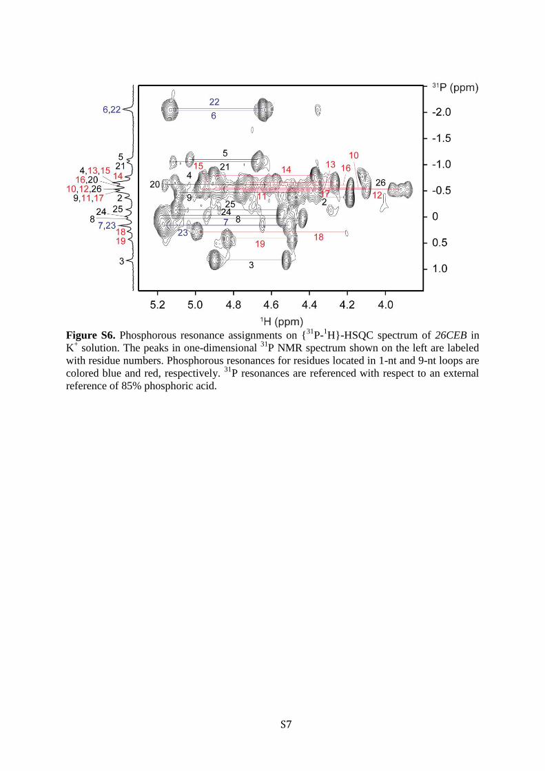
and through-bond correlations at natural abundance ({13C−1H}-HMBC) (Figure 2).38 These unambiguous assignments wereused cooperatively with other through-bond correlation experi-ments including TOCSY, {13C−1H}-HSQC, and {31P−1H}-HSQC to trace the H8/H6−H1′NOE sequential connectivityand to assign cross-peaks in NOESY spectra (Figures 3 and S6,Supporting Information).The characteristic imino-H8 proton cyclic NOE connectivity
patterns around G-tetrads (Figure 3A,B) established a three-layered parallel-stranded G-quadruplex structure comprisingG3·G7·G19·G23, G4·G8·G20·G24, and G5·G9·G21·G25tetrads. The assignment of the G4·G8·G20·G24 tetrad tothe middle layer is consistent with imino protons of the corre-sponding guanines being the most protected from the ex-change with solvent (Figure 2A). Four G-stretches of the
Figure 1. Spectroscopic characteristics of G-quadruplex formation bythe 26CEB G-rich fragment from the CEB25 minisatellite in K+
solution: (A) imino proton NMR, (B) TDS, and (C) CD spectra of26CEB.
Figure 2. Imino and H8 proton assignments of 26CEB in K+ solution. (A) Imino proton assignments from 15N-filtered spectra of samples, 2% 15N-enriched at indicated positions. Peaks marked by asterisks stayed more than 15 h in 2H2O solvent at 25 °C. (B) H8 proton assignments by site-specific 2H labeling at indicated positions. The reference spectra (ref.) of imino and aromatic protons are shown at the top. (C) Long-rangeJ-couplings between imino/H8 protons and 13C5. (D) Heteronuclear multi-bond correlations (HMBC) spectra at natural abundance. Assignmentsare labeled with residue numbers.
Journal of the American Chemical Society Article
dx.doi.org/10.1021/ja208993r | J. Am. Chem. Soc. 2012, 134, 5807−58165810

G-tetrad core, i.e., G3-G4-G5, G7-G8-G9, G19-G20-G21, andG23-G24-G25, are oriented in the same direction defining fourmedium-size grooves (Figure 3D). The structure has threedouble-chain-reversal loops: the first and third loops, eachconsists of a single nucleotide, while the central loop consists ofnine nucleotides. The nonparticipation of guanines from thecentral 9-nt loop in the G-tetrad core formation was supportedby NMR data of modified sequences with these residuesbeing substituted by an inosine, in which downfield inosineimino protons could not be observed (Figure S7, SupportingInformation). The intensity of intraresidue H8/6-H1′ NOEcross-peaks indicated a syn conformation only for the A1 residue(Figure S8, Supporting Information).A2·T18 Watson−Crick and Potential A1·G17 Base
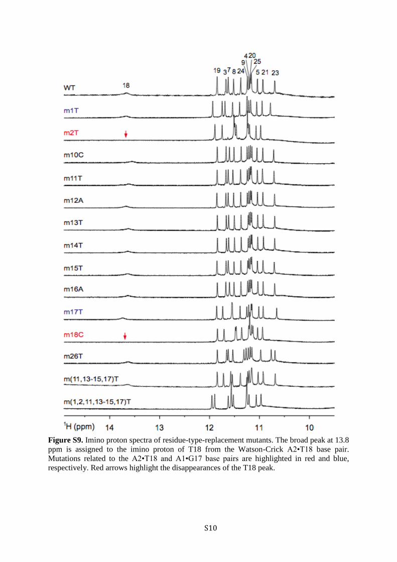
Pairs Anchor 9-nt Double-Chain-Reversal Loop on Topof 5′ G-Tetrad. Previous studies showed the destabilizingeffect of long double-chain-reversal loops on a G-quadruplexstructure.48−53 The unexpected high thermal stability (Tm =76.5 °C) of the 26CEB G-quadruplex, which contains a 9-ntdouble-chain reversal loop, suggested the occurrence of extrahydrogen-bond interactions involving this loop. Indeed, thepresence of a low-field peak at 13.8 ppm (Figure 4) indicatedthe formation of a Watson−Crick base pair.54 Among a numberof single-residue modified sequences (Table 1), only mutationsaffecting A2 and T18, such as m2T and m18C, resulted in thedisappearance of this peak (Figure 4), suggesting the formationof the A2·T18 Watson−Crick base pair. In addition, structurecalculation suggested the possibility of pairing alignmentbetween A1 and G17, which is consistent with a number ofobserved NOEs and the structure destabilization when thesebases are mutated (see discussion below). Mutations ofresidues 10−16 resulted in nearly identical imino protonspectra to that of 26CEB (Figure S9, Supporting Information),
suggesting the absence of significant interaction between theseresidues with the G-tetrad core. Nonetheless, the appearance ofextra imino proton peaks in the spectrum of 26CEB at low pH(Figure S10, Supporting Information), suggested that iminoprotons of the 9-nt loop are somewhat protected from theexchange with solvent due to some secondary structure ofthis loop.
Solution Structure of the 26CEB G-Quadruplex. TheG-quadruplex structure adopted by the 26CEB sequence in K+
solution (presented in stereoview in Figure 5) was calculatedon the basis of NMR restraints derived from the buildup ofseveral NOESY spectra (mixing times 100, 150, 200, and350 ms) and one-bond 13C−1H RDCs (see Table 2). Thestructure of the parallel-stranded three-layer G-tetrad core iswell-defined, while that of the long loop is less well con-verged (Figure 5).
Figure 3. (A) The imino-H8 proton cyclic connectivities on NOESY spectrum (mixing time, 300 ms) of 26CEB. The arrangements of G-tetradswere identified from framed cross-peaks with the residue number of imino protons labeled in the first position and that of H8 protons in the secondposition. (B) Gα·Gβ·Gγ·Gδ tetrad showing the proximity of imino and H8 protons (red arrows). (C) The H8/6-H1′ sequential connectivities onNOESY spectrum (mixing time, 350 ms) of 26CEB. Intraresidue H8/6-H1′ cross-peaks are labeled with residue numbers. Missing sequentialconnectivities are marked with asterisks. Connectivities through 1-nt and 9-nt loops are colored blue and red, respectively. (D) Schematic structureof the 26CEB G-quadruplex satisfying the NOE connectivities shown in parts A and C. Guanines in the G-tetrad core are colored cyan. Thebackbones of the core, 1-nt loops, and 9-nt loop are colored black, blue, and red, respectively.
Figure 4. Identification of the A2·T18 Watson−Crick base pair. Iminoproton spectra of the natural 26CEB sequence (WT) and modifiedsequences, m2T and m18C. Red arrows highlight the disappearances ofthe imino proton peak at 13.8 ppm, characteristic of a Watson−Crickbase pair.
Journal of the American Chemical Society Article
dx.doi.org/10.1021/ja208993r | J. Am. Chem. Soc. 2012, 134, 5807−58165811

At the 3′ end, the terminal base T26 stacks and caps underthe bottom G-tetrad. At the 5′ end, A2 and T18 form a
Watson−Crick base pair, which stacks on top of the G-tetradcore (Figures 5 and 6), consistent with the observation of theT18 imino proton and NOEs between A2(H2) and T18(H6/CH3) (Figure S11, Supporting Information), A2(H2/H8) andG7/G23(H1), as well as T18(H6/CH3) and G3/G7/G19(H1)(data not shown). The base of A1, which is in the synconformation, is positioned over the center of the A2·T18 pair,while its sugar is located directly above the A2 five-memberedring, in agreement with numerous NOEs from A2(H2/H8) toA1(H1′/H2′/H2″/H3′) (Figure S11, Supporting Information).Constrained by NOEs observed between G17(H8) andT18(H6/CH3) (Figure S11, Supporting Information), G17 isstaggered above T18. The proximity of A1 and G17 was furthersupported by the observation of a weak NOE from A1(H2) toG17(H1′) and A1(H8) to T18(CH3) (Figure S11, SupportingInformation). In some structures, A1 and G17 are in such spatialpositions to form a noncanonical base pair, in which aminoprotons of A1 is hydrogen-bonded to G17(N3) (Figure 6A,B).Aside from G17 and T18, little interaction was detected for
other residues in the central 9-nt loop (Figure 6A; see thermo-dynamic analysis below). T10 points toward the G-quadruplexcore, while the remaining residues are less well-converged, withG11, T12, A13, A14, G15, and T16 bases sampling multipleconformations characterized by roughly similar backboneprogression geometries (Figure 5A).Bases T6 and T22 of the first and third single-nucleotide double-
chain-reversal loops protrude outward from the G-tetrad core(Figure 6D). T6 and T22 show similar NMR patterns with brokenH8/H6−H1′ NOE sequential connectivities and nearly degeneratedchemical shifts (Figures 3C and S6, Supporting Information).
Figure 5. Stereoviews of the parallel-stranded G-quadruplex structure of 26CEB in K+ solution. (A) Ten superimposed structures. (B) Ribbon viewof a representative structure. Guanines are colored cyan; adenines, green; thymines, orange; backbone and sugar, gray; O4′ atoms, yellow;phosphorus atoms, red. Bases of non-interacting loop residues are not shown for clarity.
Table 2. Statistics of the Computed Structures of the CEB25Minisatellite Sequence d[AAGGGTGGGTGTAAGTGTGG-GTGGGT]
(A) NMR Restraints
distance restraints 2H2O H2Ointraresidue 521 2sequential (i, i + 1) 289 16long-range (i, ≥i + 2) 43 57
other restraintshydrogen bond 52dihedral angle 32repulsive 11RDC 56
(B) Structure Statistics for 10 Molecules Following Molecular DynamicsRefinement
NOE violationsnumber (>0.2 Å) 0.2maximum violation (Å) 0.151 ± 0.077rmsd of violations (Å) 0.012 ± 0.002rmsd from RDC restraints (Hz) 3.345 ± 0.195
deviations from the ideal covalent geometrybond lengths (Å) 0.003 ± 0.000bond angles (deg) 0.691 ± 0.009impropers (deg) 0.385 ± 0.015
pairwise all heavy atom rmsd values (Å)G-tetrad core 0.68 ± 0.11G-tetrad core and A1, A2, G17, and T18 1.22 ± 0.30all residues 3.10 ± 0.83
Journal of the American Chemical Society Article
dx.doi.org/10.1021/ja208993r | J. Am. Chem. Soc. 2012, 134, 5807−58165812
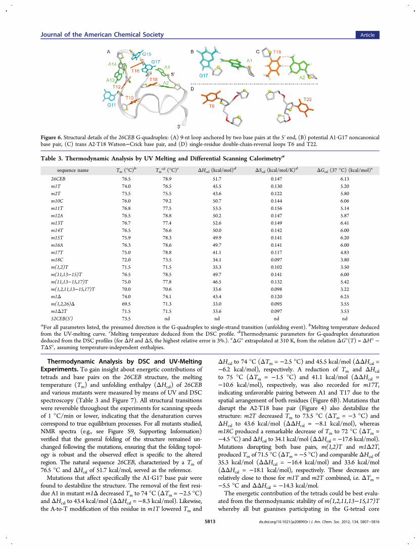
Thermodynamic Analysis by DSC and UV-MeltingExperiments. To gain insight about energetic contributions oftetrads and base pairs on the 26CEB structure, the meltingtemperature (Tm) and unfolding enthalpy (ΔHcal) of 26CEBand various mutants were measured by means of UV and DSCspectroscopy (Table 3 and Figure 7). All structural transitionswere reversible throughout the experiments for scanning speedsof 1 °C/min or lower, indicating that the denaturation curvescorrespond to true equilibrium processes. For all mutants studied,NMR spectra (e.g., see Figure S9, Supporting Information)verified that the general folding of the structure remained un-changed following the mutations, ensuring that the folding topol-ogy is robust and the observed effect is specific to the alteredregion. The natural sequence 26CEB, characterized by a Tm of76.5 °C and ΔHcal of 51.7 kcal/mol, served as the reference.Mutations that affect specifically the A1·G17 base pair were
found to destabilize the structure. The removal of the first resi-due A1 in mutant m1Δ decreased Tm to 74 °C (ΔTm = −2.5 °C)and ΔHcal to 43.4 kcal/mol (ΔΔHcal = −8.3 kcal/mol). Likewise,the A-to-T modification of this residue in m1T lowered Tm and
ΔHcal to 74 °C (ΔTm = −2.5 °C) and 45.5 kcal/mol (ΔΔHcal =−6.2 kcal/mol), respectively. A reduction of Tm and ΔHcal
to 75 °C (ΔTm = −1.5 °C) and 41.1 kcal/mol (ΔΔHcal =−10.6 kcal/mol), respectively, was also recorded for m17T,indicating unfavorable pairing between A1 and T17 due to thespatial arrangement of both residues (Figure 6B). Mutations thatdisrupt the A2·T18 base pair (Figure 4) also destabilize thestructure: m2T decreased Tm to 73.5 °C (ΔTm = −3 °C) andΔHcal to 43.6 kcal/mol (ΔΔHcal = −8.1 kcal/mol), whereasm18C produced a remarkable decrease of Tm to 72 °C (ΔTm =−4.5 °C) and ΔHcal to 34.1 kcal/mol (ΔΔHcal = −17.6 kcal/mol).Mutations disrupting both base pairs, m(1,2)T and m1Δ2T,produced Tm of 71.5 °C (ΔTm = −5 °C) and comparableΔHcal of35.3 kcal/mol (ΔΔHcal = −16.4 kcal/mol) and 33.6 kcal/mol(ΔΔHcal = −18.1 kcal/mol), respectively. These decreases arerelatively close to those for m1T and m2T combined, i.e. ΔTm =−5.5 °C and ΔΔHcal = −14.3 kcal/mol.The energetic contribution of the tetrads could be best evalu-
ated from the thermodynamic stability of m(1,2,11,13−15,17)Twhereby all but guanines participating in the G-tetrad core
Figure 6. Structural details of the 26CEB G-quadruplex: (A) 9-nt loop anchored by two base pairs at the 5′ end, (B) potential A1·G17 noncanonicalbase pair, (C) trans A2·T18 Watson−Crick base pair, and (D) single-residue double-chain-reversal loops T6 and T22.
Table 3. Thermodynamic Analysis by UV Melting and Differential Scanning Calorimetrya
sequence name Tm (°C)b Tmcal (°C)c ΔHcal (kcal/mol)d ΔScal (kcal/mol/K)d ΔGcal (37 °C) (kcal/mol)e
26CEB 76.5 78.9 51.7 0.147 6.13m1T 74.0 76.5 45.5 0.130 5.20m2T 73.5 75.5 43.6 0.122 5.80m10C 76.0 79.2 50.7 0.144 6.06m11T 76.8 77.5 53.5 0.156 5.14m12A 76.5 78.8 50.2 0.147 5.87m13T 76.7 77.4 52.6 0.149 6.41m14T 76.5 76.6 50.0 0.142 6.00m15T 75.9 78.3 49.9 0.141 6.20m16A 76.3 78.6 49.7 0.141 6.00m17T 75.0 78.8 41.1 0.117 4.83m18C 72.0 73.5 34.1 0.097 3.80m(1,2)T 71.5 71.5 35.3 0.102 3.50m(11,13−15)T 76.5 78.5 49.7 0.141 6.00m(11,13−15,17)T 75.0 77.8 46.5 0.132 5.42m(1,2,11,13−15,17)T 70.0 70.6 33.6 0.098 3.22m1Δ 74.0 74.1 43.4 0.120 6.25m(1,2,26)Δ 69.5 71.3 33.0 0.095 3.55m1Δ2T 71.5 71.5 33.6 0.097 3.5352CEB(5′) 73.5 nd nd nd nd
aFor all parameters listed, the presumed direction is the G-quadruplex to single-strand transition (unfolding event). bMelting temperature deducedfrom the UV-melting curve. cMelting temperature deduced from the DSC profile. dThermodynamic parameters for G-quadruplex denaturationdeduced from the DSC profiles (for ΔH and ΔS, the highest relative error is 3%.). eΔG° extrapolated at 310 K, from the relation ΔG°(T) = ΔH° −TΔS°, assuming temperature-independent enthalpies.
Journal of the American Chemical Society Article
dx.doi.org/10.1021/ja208993r | J. Am. Chem. Soc. 2012, 134, 5807−58165813

formation were replaced by thymines. The mutant was char-acterized with Tm of 70 °C (ΔTm = −6.5 °C) and ΔHcal of33.6 kcal/mol (ΔΔHcal = −18.1 kcal/mol), rather similar valuesto those of m(1,2)T or m1Δ2T. Taken together, A2·T18 andA1·G17 stabilize the G-quadruplex by 6 ± 1 °C in Tm and by17 ± 1 kcal/mol in ΔHcal.
Mutations of other loop residues, including single-residuemodifications (m10C, m11T, m12A, m13T, m14T, m15T, andm16A) and the multiple-residue modification m(11,13−15)Tonly marginally affect the thermodynamic stability of thestructure (|ΔTm| < 0.5 °C and |ΔΔHcal| < 2.0 kcal/mol), whilethe effect observed for m(11,13−15,17)T (ΔTm = −1.5 °C andΔΔHcal = −5.2 kcal/mol) could be explained by the disruptionof the A1·G17 base pair.
Formation of the 26CEB G-Quadruplex in LongerSequence Contexts. To test whether the structure observedfor the 26-nt G-rich 26CEB fragment is formed within longersequence contexts, we recorded NMR spectra of extendedsequences comprising several 52-nt full-repeating units. Iminoproton spectra of the 78- and 130-nt sequences 78CEB(5′,3′)and 130CEB(5′,mid,3′) (Table 1) displayed a similar pattern tothat of 26CEB (Figure 8A), indicating that the 26CEB segmentsin these sequences form the same G-quadruplex structure asthat of an isolated 26CEB, while the remaining parts are essentiallyunstructured. The spectral broadening and minor shifts of somepeaks in 78CEB(5′,3′) and 130CEB(5′,mid,3′) as compared tothose of 26CEB are due to larger molecular sizes and different5′ or 3′ flanking sequences. The imino proton spectrum of the78CEB(5′,3′) sequence containing two 26CEB segments(Figure 8A) closely resembles the calculated weighted sum ofthe two spectra of the 78CEB(5′mut,3′) and 78CEB(5′,3′mut)sequences (Figure 8B) containing mutations that disruptG-quadruplex formation at the 5′ and 3′ side, respectively, sup-porting the formation of two G-quadruplex blocks connectedby a 26-nt linker in the 78CEB(5′,3′) sequence. Formation ofG-quadruplex blocks at different locations of long CEB25sequences was also supported by NMR data for sequences
Figure 8. (A) Imino proton spectra of CEB25 minisatellite sequences of various lengths. Proposed folding topologies are shown by the schematicswith G-tetrads being highlighted in cyan. (B) Imino proton spectra of 78-nt CEB25 minisatellite sequences containing mutations that disruptG-quadruplex formation at the 3′ side (top) and 5′ side (bottom), respectively, as well as their weighted sum (middle) that simulates the spectrum ofthe 78CEB(5′,3′) sequence containing two G-quadruplex blocks. Note the similarity between the latter and the observed spectrum of 78CEB(5′,3′) inpart A. (C) CEB25 minisatellite pearl-necklace model.
Figure 7. Histogram representation of changes in the meltingtemperature (filled bars) and unfolding enthalpy (open bars) valuesobserved for various mutants with respect to 26CEB. Most
Journal of the American Chemical Society Article
dx.doi.org/10.1021/ja208993r | J. Am. Chem. Soc. 2012, 134, 5807−58165814

containing a single inosine substitution in one G-quadruplexblock (Figure S12, Supporting Information) and other longsequences (Figure S13, Supporting Information). On the basisof these results, a “pearl-necklace” model of stable G-quadruplexblocks interconnected by non-quadruplex-forming sequenceson a long single-stranded CEB25 repeats is proposed (Figure 8C).
■ DISCUSSIONOver the past two decades, many different G-quadruplexes havebeen described in telomeres and oncogenic promoters.2 Inthis work, we have shown that the G-rich sequence of theCEB25 minisatellite forms a propeller-type parallel-strandedG-quadruplex involving two single-nucleotide and one 9-ntdouble-chain-reversal loops. There are additional base pairsformed between bases of the long loop and those at the 5′-end,anchoring this loop to the top of the G-tetrad core.Previously, an inverse correlation between the length of a
propeller loop Tn in a G-quadruplex and the stability of thestructure has been described: each added base leads to a 2 °Cdrop in Tm or 0.3 kcal/mol lost in ΔG.52,53 The G-quadruplexin CEB25 minisatellite, consisting of a 9-nt loop, is more stablethan expected from previous studies. Our thermodynamicanalysis have shown that the two base pairs between the longloop and the 5′-end sequence contribute to the stability of theoverall structure by about 17 kcal/mol in enthalpy or 6 °C inmelting temperature, highlighting the importance of the loopand flanking sequences in stabilizing G-quadruplex folds.The structure of the CEB25 G-quadruplex can be readily
compared with the form I of the c-kit2 promoter sequence.55
A noncanonical C1·A13 base pair formed in the c-kit2 structurebetween the last base of a 5-nt central double-chain-reversalloop and a 5′-end flanking base is comparable to the A2·T18base pair in the CEB25 structure, suggesting a general rule forthe formation of a base pair between the last residues of adouble-chain-reversal loop and the 5′-end flanking bases.However, there is no equivalence of the second base pairA1·G17 of the CEB25 structure in the c-kit2 G-quadruplex. Itwill be interesting to address the question regarding the context(loop length and sequence) necessary for the formation of twoor more base pairs.In contrast to telomeric G-quadruplexes,4 the CEB25 G-
quadruplex has been observed to be monomorphic and robust.This was evident from the observation of only one majorconformation identified following a number of modificationsand extensions made on the sequence. It is likely that the firstand third single-residue loops drive the whole strand to adoptthe propeller-type parallel G-quadruplex independent of thesize and sequence of the flanking and the central loop segments,consistent with the previous observation that a single-residuelinker between G-tracts favors a double-chain-reversal loop.47−52
The “beads-on-a-string” formation of G-quadruplexes on a longtelomeric sequence has been suggested earlier.9,56,57 ConsecutiveG-quadruplexes have been observed to be separated rather irre-gularly from each other by AFM,58 and each four-repeat frag-ment can adopt multiple conformations.4 On the contrary, theproposed pearl-necklace model for human CEB25 minisatelliterepeats is based on the observation of a single G-quadruplexconformation by NMR. Arrays of G-quadruplexes might beformed regularly in CEB25 repeats interlinked by essentiallyunstructured strings and thus provide higher-ordered structureand biological functions which can be specific to tandem arrayscompared to an isolated G-quadruplex. For example, a high localconcentration of intramolecular G-quadruplexes might provide
additive or synergistic probability of G-quadruplex formationand/or interference in resolution influencing the array stabilitydependent on replication.13,59
■ CONCLUSIONWe have solved the NMR structure of a three-layered G-quadruplex structure formed by the G-rich fragment of eachCEB25 minisatellite repeat in K+ solution. This is a propeller-type parallel-stranded G-quadruplex involving a 9-nt centraldouble-chain-reversal loop. This long loop is anchored to the 5′end of the sequence by an A·T Watson−Crick base pair and apotential G·A noncanonical base pair. Thermodynamic andmutation analyses have established the role of these base pairsin the stability of the G-quadruplex structure. We show thatsuch a structure can be formed in long sequence contexts,providing evidence for a pearl-necklace of G-quadruplexesformed by single-stranded CEB25 minisatellite.
■ ASSOCIATED CONTENT*S Supporting InformationAdditional experimental data (Table S1 and Figures S1−S13)and complete ref 25. This material is available free of charge viathe Internet at http://pubs.acs.org.
■ AUTHOR INFORMATIONCorresponding [email protected]; [email protected] Contributions∥These authors contributed equally to this workNotesThe authors declare no competing financial interest.
■ ACKNOWLEDGMENTSWe thank Mr. Kah Wai Lim and Dr. Serge Bouaziz for theirinitial help with the structure calculation. The research in ATPgroup was supported by Singapore Ministry of Education grants(RG62/07 and RG72/10). M.A. is supported by the YousefJameel scholarship. J.L.M acknowledges support from theAssociation pour la recherche sur le cancer (programme libreA.R.C.), the Region Aquitaine, the Fondation pour laRecherche Medicale (F.R.M.), INCa and ANR grants (F-DNAand G4-TOOLBOX). The research in A.N.’s team is supportedby grants from the Ligue Nationale contre le Cancer (LNCC;Equipe Labellisee EL2007.LNCC/AN and EL2010.LNCC/AN)and a postdoctoral fellowship to A.S. from the F.R.M.
■ REFERENCES(1) Davis, J. T. Angew. Chem., Int. Ed. 2004, 43, 668.(2) Patel, D. J.; Phan, A. T.; Kuryavyi, V. Nucleic Acids Res. 2007, 35,7429.(3) Neidle, S. Curr. Opin. Struct. Biol. 2009, 19, 239.(4) Phan, A. T. FEBS J. 2010, 277, 1107.(5) Phan, A. T.; Kuryavyi, V.; Gaw, H. Y.; Patel, D. J. Natl. Chem.Biol. 2005, 1, 167.(6) Phan, A. T.; Kuryavyi, V.; Burge, S.; Neidle, S.; Patel, D. J. J. Am.Chem. Soc. 2007, 129, 4386.(7) Heddi, B.; Phan, A. T. J. Am. Chem. Soc. 2011, 133, 9824.(8) Yue, D. J.; Lim, K. W.; Phan, A. T. J. Am. Chem. Soc. 2011, 133,11462.(9) Yu, H. Q.; Miyoshi, D.; Sugimoto, N. J. Am. Chem. Soc. 2006,128, 15461.(10) Maizels, N. Nat. Struct. Mol. Biol. 2006, 13, 1055.(11) Lipps, H. J.; Rhodes, D. Trends Cell Biol. 2009, 19, 414.
Journal of the American Chemical Society Article
dx.doi.org/10.1021/ja208993r | J. Am. Chem. Soc. 2012, 134, 5807−58165815

(12) Paeschke, K.; Capra, J. A.; Zakian, V. A. Cell 2011, 145, 678.(13) Lopes, J.; Piazza, A.; Bermejo, R.; Kriegsman, B.; Colosio, A.;Teulade-Fichou, M. P.; Foiani, M.; Nicolas, A. EMBO J. 2011, 30,4033.(14) Duquette, M. L.; Handa, P.; Vincent, J. A.; Taylor, A. F.;Maizels, N. Genes Dev. 2004, 18, 1618.(15) Cahoon, L. A.; Seifert, H. S. Science 2009, 325, 764.(16) Vergnaud, G.; Denoeud, F. Genome Res. 2000, 10, 899.(17) Bois, P.; Jeffreys, A. J. Cell. Mol. Life Sci. 1999, 55, 1636.(18) Kashi, Y.; King, D. G. Trends Genet. 2006, 22, 253.(19) Verstrepen, K. J.; Jansen, A.; Lewitter, F.; Fink, G. R. Nat. Genet.2005, 37, 986.(20) Lalioti, M. D.; Scott, H. S.; Antonarakis, S. E. Nat. Genet. 1997,17, 17.(21) Benett, I. Diabet. Med. 1995, 12, 452.(22) Klenova, E.; Scott, A. C.; Roberts, J.; Shamsuddin, S.; Lovejoy,E. A.; Bergmann, S.; Bubb, V. J.; Royer, H. D.; Quinn, J. P. J. Neurosci.2004, 24, 5966.(23) Phelan, C. M.; Rebbeck, T. R.; Weber, B. L.; Devilee, P.;Ruttledge, M. H.; Lynch, H. T.; Lenoir, G. M.; Stratton, M. R.; Easton,D. F.; Ponder, B. A. J.; CannonAlbright, L.; Larsson, C.; Goldgar,D. E.; Narod, S. A. Nat. Genet. 1996, 12, 309.(24) Jeffreys, A. J.; Barber, R.; Bois, P.; Buard, J.; Dubrova, Y. E.;Grant, G.; Hollies, C. R.; May, C. A.; Neumann, R.; Panayi, M.;Ritchie, A. E.; Shone, A. C.; Signer, E.; Stead, J. D.; Tamaki, K.Electrophoresis 1999, 20, 1665.(25) Law, M. J.; et al. Cell 2010, 143, 367.(26) Panigrahi, G. B.; Lau, R.; Montgomery, S. E.; Leonard, M. R.;Pearson, C. E. Nat. Struct. Mol. Biol. 2005, 12, 654.(27) Amrane, S.; Sacca, B.; Mills, M.; Chauhan, M.; Klump, H. H.;Mergny, J. L. Nucleic Acids Res. 2005, 33, 4065.(28) Amrane, S.; Mergny, J. L. Biochimie 2006, 88, 1125.(29) Yauk, C. L.; Dubrova, Y. E.; Grant, G. R.; Jeffreys, A. J. Mutat.Res. 2002, 500, 147.(30) Napierala, M.; Bacolla, A.; Wells, R. D. J. Biol. Chem. 2005, 280,37366.(31) Cantor, C. R.; Warshaw, M. M.; Shapiro, H. Biopolymers 1970,9, 1059.(32) Jeffreys, A. J.; Wilson, V.; Neumann, R.; Keyte, J. Nucleic AcidsRes. 1988, 16, 10953.(33) Mergny, J. L.; Phan, A. T.; Lacroix, L. FEBS Lett. 1998, 435, 74.(34) Mergny, J. L.; Lacroix, L. Oligonucleotides 2003, 13, 515.(35) Mergny, J. L.; Li, J.; Lacroix, L.; Amrane, S.; Chaires, J. B. NucleicAcids Res. 2005, 33, e138.(36) Phan, A. T.; Patel, D. J. J. Am. Chem. Soc. 2002, 124, 1160.(37) Huang, X.; Yu, P.; LeProust, E.; Gao, X. Nucleic Acids Res. 1997,25, 4758.(38) Phan, A. T. J. Biomol. NMR 2000, 16, 175.(39) Phan, A. T.; Gueron, M.; Leroy, J. L. Methods Enzymol. 2001,338, 341.(40) Schwieters, C. D.; Kuszewski, J. J.; Tjandra, N.; Clore, G. M.J. Magn. Reson. 2003, 160, 65.(41) Myers, S.; Bowden, R.; Tumian, A.; Bontrop, R. E.; Freeman,C.; MacFie, T. S.; McVean, G.; Donnelly, P. Science 2010, 327, 876.(42) Baudat, F.; Buard, J.; Grey, C.; Fledel-Alon, A.; Ober, C.;Przeworski, M.; Coop, G.; de Massy, B. Science 2010, 327, 836.(43) Berg, I. L.; Neumann, R.; Lam, K. W.; Sarbajna, S.; Odenthal-Hesse, L.; May, C. A.; Jeffreys, A. J. Nat. Genet. 2010, 42, 859.(44) Buard, J.; Bourdet, A.; Yardley, J.; Dubrova, Y.; Jeffreys, A. J.EMBO J. 1998, 17, 3495.(45) Feigon, J.; Koshlap, K. M.; Smith, F. W. Methods Enzymol. 1995,261, 225.(46) Gray, D. M.; Wen, J. D.; Gray, C. W.; Repges, R.; Repges, C.;Raabe, G.; Fleischhauer, J. Chirality 2008, 20, 431.(47) Huppert, J. L.; Balasubramanian, S. Nucleic Acids Res. 2005, 33,2908.(48) Phan, A. T.; Modi, Y. S.; Patel, D. J. J. Am. Chem. Soc. 2004, 126,8710.
(49) Hazel, P.; Huppert, J.; Balasubramanian, S.; Neidle, S. J. Am.Chem. Soc. 2004, 126, 16405.(50) Rachwal, P. A.; Findlow, I. S.; Werner, J. M.; Brown, T.; Fox,K. R. Nucleic Acids Res. 2007, 35, 4214.(51) Bugaut, A.; Balasubramanian, S. Biochemistry 2008, 47, 689.(52) Guedin, A.; De Cian, A.; Gros, J.; Lacroix, L.; Mergny, J. L.Biochimie 2008, 90, 686.(53) Guedin, A.; Gros, J.; Alberti, P.; Mergny, J. L. Nucleic Acids Res.2010, 38, 7858.(54) Patel, D. J.; Tonelli, A. E. Biopolymers 1974, 13, 1943.(55) Kuryavyi, V.; Phan, A. T.; Patel, D. J. Nucleic Acids Res. 2010, 38,6757.(56) Xu, Y.; Ishizuka, T.; Kurabayashi, K.; Komiyama, M. Angew.Chem., Int. Ed. 2009, 48, 7833.(57) Martadinata, H.; Heddi, B.; Lim, K. W.; Phan, A. T. Biochemistry2011, 50, 6455.(58) Wang, H.; Nora, G. J.; Ghodke, H.; Opresko, P. L. J. Biol. Chem.2011, 286, 7479.(59) Ribeyre, C.; Lopes, J.; Boule, J. B.; Piazza, A.; Guedin, A.;Zakian, V. A.; Mergny, J. L.; Nicolas, A. PLoS Genet. 2009, 5,e1000475.
Journal of the American Chemical Society Article
dx.doi.org/10.1021/ja208993r | J. Am. Chem. Soc. 2012, 134, 5807−58165816

Formation of pearl-necklace monomorphic G-quadruplexes
in the human CEB25 minisatellite
Samir Amrane
1,2,#, Michael Adrian
1,#, Brahim Heddi
1, Alexandre Serero
3,
Alain Nicolas3, Jean-Louis Mergny
2,* and Anh Tuân Phan
1,*
1School of Physical and Mathematical Sciences, Nanyang Technological University,
Singapore, 2University of Bordeaux, INSERM U869, European Institute of Chemistry and
Biology, 33600 Pessac, France, and 3Institut Curie, Centre de Recherche, UMR3244 CNRS,
Université Pierre et Marie Curie, 75248 Paris, France
#These authors contributed equally to this work
*Corresponding authors: [email protected]; [email protected]
SUPPORTING INFORMATION

S1
Table S1. List of site-specific labeled DNA sequences analyzed in this worka-d
Name Sequence (5'-3')
26CEB AAGGGTGGGTGTAAGTGTGGGTGGGT
I3 AAIGGTGGGTGTAAGTGTGGGTGGGT
I4 AAGIGTGGGTGTAAGTGTGGGTGGGT
I5 AAGGITGGGTGTAAGTGTGGGTGGGT
I7 AAGGGTIGGTGTAAGTGTGGGTGGGT
I8 AAGGGTGIGTGTAAGTGTGGGTGGGT
I9 AAGGGTGGITGTAAGTGTGGGTGGGT
I11 AAGGGTGGGTITAAGTGTGGGTGGGT
I15 AAGGGTGGGTGTAAITGTGGGTGGGT
I17 AAGGGTGGGTGTAAGTITGGGTGGGT
I19 AAGGGTGGGTGTAAGTGTIGGTGGGT
I20 AAGGGTGGGTGTAAGTGTGIGTGGGT
I21 AAGGGTGGGTGTAAGTGTGGITGGGT
I23 AAGGGTGGGTGTAAGTGTGGGTIGGT
I24 AAGGGTGGGTGTAAGTGTGGGTGIGT
I25 AAGGGTGGGTGTAAGTGTGGGTGGIT 15N-G4 AAGG*GTGGGTGTAAGTGTGGGTGGGT
15N-G8 AAGGGTGG*GTGTAAGTGTGGGTGGGT
15N-G9 AAGGGTGGG*TGTAAGTGTGGGTGGGT
15N-G15 AAGGGTGGGTGTAAG*TGTGGGTGGGT
15N-G19 AAGGGTGGGTGTAAGTGTG*GGTGGGT
15N-G20 AAGGGTGGGTGTAAGTGTGG*GTGGGT
15N-G24 AAGGGTGGGTGTAAGTGTGGGTGG*GT
15N-G25 AAGGGTGGGTGTAAGTGTGGGTGGG*T
2D-G15 AAGGGTGGGTGTAAG#TGTGGGTGGGT 2D-G17 AAGGGTGGGTGTAAGTG#TGGGTGGGT 2D-G19 AAGGGTGGGTGTAAGTGTG#GGTGGGT 2D-G24 AAGGGTGGGTGTAAGTGTGGGTGG#GT
a Individual guanines 2%-
15N-labeled are marked by asterisks.
b Individual guanines
2H-labeled at the H8 position are marked by hash signs.
c I is inosine.
d Labeled and modified residues are shown in boldface.

S2
Figure S1. C1’-H1’ region of the non-decoupled {
13C-
1H}-HSQC spectra in isotropic (left
spectrum, blue contours) and aligned media (right spectrum, red contours).

S3
Figure S2. Correlation between the measured (Dmeas) and calculated (Dcalc) RDCs for
structures derived from NMR restraints except RDCs (A, best structure, and C ensemble of
10 best structures) and with RDCs (B, best structure, and D ensemble of 10 best structures).
Blue dots represent RDCs of the guanines residues from the G-tetrad core; red dots represent
RDCs of other residues.

S4
Figure S3. Structure and sequences of human CEB25 alleles. (A) Sequence of the CEB25
repeated motif according to.16
The sequence of the 26CEB oligonucleotide used in this study
and the potent PDRM9 binding sites are underscored with full and dashed lines, respectively.
(B) and (C) Complete sequence of the G-strand of two human CEB25 alleles : on the left is
represented the CEB25-0.8 kb allele sequenced by G. Vergnaud and available in web data
released by the Genome Reference Consortium (Accession number AL096806). On the right
is shown the full sequence of the CEB25-1.7 kb allele cloned by G. Vergnaud and sequenced
in this study. Polymorphic DNA bases are highlighted and the 26CEB G-quadruplex-prone
sequence is underscore. Among the 48 sequenced motifs belonging to both CEB25 alleles 9
are unique (A, D, L, N, P, Q, R, T and U).

S5
Figure S4. Southern blot analysis of CEB25 inheritance in
the 1334 CEPH family through 3 generations. Lane 1 to 6: 6
children ; lane 7 and 8 the father and the mother,
respectively ; lane 9 and 10, the mother’s parents ; lane 11
and 12, the fathers’s parents. 15 µg of genomic DNA was
digested of by ApaI and hybridized with a CEB25-1.7 probe.

S6
Figure S5. Analysis of the number of G-triplets contained per motif among the sequenced
CEB25 motifs. 48 repeats were identified in the complete sequences of both CEB25-0.8 and -
1.7.
S7
Figure S6. Phosphorous resonance assignments on {
31P-
1H}-HSQC spectrum of 26CEB in
K+ solution. The peaks in one-dimensional
31P NMR spectrum shown on the left are labeled
with residue numbers. Phosphorous resonances for residues located in 1-nt and 9-nt loops are
colored blue and red, respectively. 31
P resonances are referenced with respect to an external
reference of 85% phosphoric acid.

S8
Figure S7. Imino proton spectra of inosine mutants. Inosine substitution of each guanine in
the G-tetrad core is marked by low-field shift of the corresponding imino peak (seen in I3-I5,
I7-I9, I19-I21 and I23-I25). No inosine-modified peak observed on imino spectra of I11, I15
and I17 (in red), implying that G11, G15 and G17 are not part of G-tetrads.

S9
Figure S8. Stack view of NOESY spectrum (mixing time, 100 ms) showing strong intensity
of intra-residue H8-H1’ cross-peak from residue A1 (marked by asterisk).
S10
Figure S9. Imino proton spectra of residue-type-replacement mutants. The broad peak at 13.8
ppm is assigned to the imino proton of T18 from the Watson-Crick A2•T18 base pair.
Mutations related to the A2•T18 and A1•G17 base pairs are highlighted in red and blue,
respectively. Red arrows highlight the disappearances of the T18 peak.

S11
Figure S10. Imino proton spectra of 26CEB in K
+ solution at different pH. New peaks
appeared at low pH are labeled with asterisks.

S12
Figure S11. Regions of NOESY spectrum (mixing time, 350 ms) showing cross-peaks that
define the formation of base pairs. Staggering of G17 above T18 is determined from cross-
peak (i); A2T18 base pairing, (ii); proximity of A1 and G17, (iii) and (xvii); A1 and A2
relative positions, (iv), (v), (vii), (viii), (x), (xi), (xiii) and (xiv); intra-residue cross peaks of
A1 and G17, (vi), (ix), (xii), (xv) and (xvi).

S13
Figure S12. Folding of multiple G-quadruplexes linked by 26-nt single-strands. Spectrum
degeneracy was broken by inosine substitution (at I7 position) of indicated G-quadruplex.

S14
Figure S13. Imino spectra of CEB25 minisatellite sequences of various lengths: (A) 26CEB,
(B) 52CEB(5’), (C) 78CEB(5’,3’mut), (D) 52CEB(3’), (E) 78CEB(5’mut,3’), (F)
78CEB(5’,3’). (G) 57CEB(mid), (H) 78CEB(mid) and (I) 130CEB(5’,mid,3’). Proposed
folding topologies are shown by the schematics. Guanine tetrads are highlighted in cyan.

S15
REFERENCES
(25) Law, M. J.; Lower, K. M.; Voon, H. P.; Hughes, J. R.; Garrick, D.; Viprakasit, V.;
Mitson, M.; De Gobbi, M.; Marra, M.; Morris, A.; Abbott, A.; Wilder, S. P.; Taylor, S.;
Santos, G. M.; Cross, J.; Ayyub, H.; Jones, S.; Ragoussis, J.; Rhodes, D.; Dunham, I.; Higgs,
D. R.; Gibbons, R. J. Cell 2010, 143, 367.


![G-Quadruplexes: Prediction, Characterization, and ... · 1A) to form complex structural motifs known as G-quadruplexes [2] (Figure 1B). G-quadruplexes are of growing interest in chemistry](https://static.fdocuments.in/doc/165x107/5ce92d8c88c99308268cad7b/g-quadruplexes-prediction-characterization-and-1a-to-form-complex-structural.jpg)