Forensic Sercalow, Immunology, Biochemistry - NCJRS · become a part of forensic serology in the...
44
U. S. Department uf Justice National Institute of Justice Forensic Sercalow, Immunology, and Biochemistry a publication of the National Institute of Justice
Transcript of Forensic Sercalow, Immunology, Biochemistry - NCJRS · become a part of forensic serology in the...

U.S. Department uf Justice National Institute of Justice
Forensic Sercalow, Immunology, and Biochemistry
a publication of the National Institute of Justice

About the National Institute of Justice
The National lnstitute of Justice is a research branch of the U.S.Department of Justice. The Institute's mission is to develop knowledge about crime. its causes and control. Priority is given to policy-relevant research that can yield approaches and information State and local agencies can use in preventing and reducing crime. Established in 1979 by the Justice System Improvement Act. NIJ builds upon the foundation laid by the former National lnstitute of Law Enforcement and Criminal Justice. the first major Federal research program on crime and justice.
Carrying out the mandate assigned by Congress, the National lnstitute of Justice:
Sponsors research and development to improve and strengthen the criminal justice system and related civil justice aspects. with a balanced program of basic and applied research.
Evaluates the effectiveness of federally funded justice improvement programs and identifies programs that promise to be successful if continued or repeated.
Tests and demonstrates new and improved approaches to strengthen the justice system. and recommends actions that can be taken by Federal. State, and local governments and private organizations and individuals to achieve this goal.
Disseminates information from research. demonstrations. evaluations. and special programs to Federal. State. and local governments; and serves as an international clearinghouse of justice information.
Trains criminaljustice practitioners in research and evaluation findings. and assists the research commun-ity through fellowships and special seminars.
Authority for administering the lnstitute and awarding grants. contracts. and cooperative agreements is vested in the NIJ Director. An Advisory Board. appointed by the President. assists the Director by recom-mending policies and priorities and advising on peer review procedures.
Reports of NIJ-sponsored studies are reviewed hy lnstitute officials and staff. The views of outside experts knowledgeable in the report's subject area are also obtained. Publication indicates that the report meets the Institute's standards of technical quality, but it signifies no endorsement of conclusions or recommendations.
James K.Stewart Direcror
U.S. Department ofJustice National Institute of Justice
Sourcebook in Forensic Serology, Immunology, and Biochemistry
R.E.Gaensslen, Ph.D. Professor of Forensic Science University of New Haven West Haven. Connecticut
August 1983
wiLA 1989 Update

This project was supported by Grant Numbers 76-NI-99-0107 and 78-N1-AX-0001. awarded to R. E. Gaensslen, Ph.D.. by the National Institute of Justice, U.S. Department of Justice. under the Omnibus Crime Convol and Safe Streets Act of 1968. as amended. Additional financial support was provided by the Research Foundation of the City University of New York. Points of view or opinions stated in this document are those of the authors and do not necessarily represent the official position or policies of the U.S. Department of Justice, or of the City University of New York.
The author merves the right to reproduce. publish. translate. or otherwise use and to authorize others to publish and use all or any pan of the copyrighted material contained in this publication.
Copyright @ 1983 by the Research Foundation of the City Univasity of New York on behalf of R.E. Gaensslen.

Sourcebook Errata
On page 17, the equation following line 11, after the words "The above equation may be rewritten:" should read as follows:
k l [ES] = --------- [ E l [S l
k2 + k3
The equation following lines 16 and 17, after the words "Solving for ES and substituting in the previous equation yields" should read as follows:
On page 426, lines 29 and 30 giving the correspondences of different PGM subtype nomenclatures should read as follows:

PREFACE TO THE REPRINT EDITION
In the six years or so since this Sourcebook appeared, the stock
of copies was exhausted. There were indications from a number of
sources, however, that there was still a demand for copies and that
the book still has a valuable place as a reference work in forensic
biology. Accordingly, the National Institute of Justice is to be
commended for its decision to reprint the book on a demand basis
through the National Criminal Justice Reference Service. In this
respect, I am grateful to Mr. James K. Stewart, Director of the
Institute, and to Dr. Richard M. Rau, manager of the Forensic Science
and Criminal Justice Technology Program. As a result of their
continued support, this book will continue to be available to those
interested in it.
It was not realistic to consider undertaking a complete and
systematic update and review of all the literature that has appeared
since the book's publication. I was asked, however, to provide a brief
summary of some of the more recent information in the field in this
new Preface, and that is its major purpose. Because the literature
grows with such rapidity, a number of more recent reviews as well as
some specific papers are cited here and an effort has been made to
related these to specific subject areas covered in the book. In
addition, an introduction to the rapidly developing field of molecular
biology and DNA analysis that has recently become a part of forensic
biology is given, and some references provided. DNA analysis has
become a part of forensic serology in the relatively few years since
the book was written. .
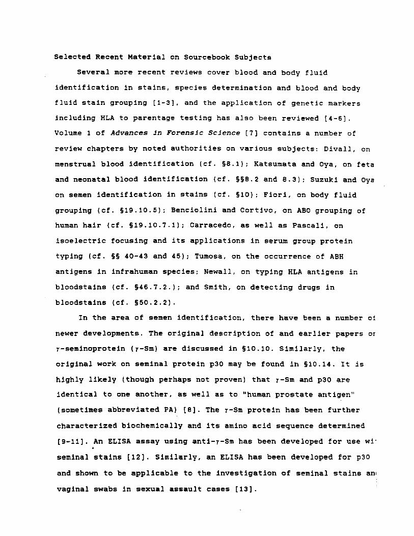
Selected Recent Material on Sourcebook Subjects
Several more recent reviews cover blood and body fluid
identification in stains, species determination and blood and body
fluid stain grouping [I-31, and the application of genetic markers
including HLA to parentage testing has also been reviewed [ 4 - 6 1 .
Volume 1 of Advances in Forensic Science [?I contains a number of
review chapters by noted authorities on various subjects: Divall, on
menstrual blood identification (cf. S8.1); Katsumata and Oya, on feta
and neonatal blood identification (cf. SS8.2 and 8.3); Suzuki and Oya
on semen identification in stains (cf. 810): Fiori, on body fluid
grouping (cf. 319.10.5): Benciolini and Cortivo, on ABO grouping of
human hair (cf. S19.10.7.1); Carracedo, as well as Pascali, on
isoelectric focusing and its applications in serum group protein
typing (cf. §S 40-43 and 45); Tumosa, on the occurrence of ABH
antigens in infrahuman species; Newall, on typing HLA antigens in
bloodstains (cf. S46.7.2.); and Smith, on detecting drugs in
bloodstains (cf. S50.2.2).
In the area of semen identification, there have been a number of
newer developments. The original description of and earlier papers or
7-seminoprotein (7-Sm) are discussed in 910.10. Similarly, the
original work on seminal protein p30 may be found in 310.14. It is
highly likely (though perhaps not proven) that y-Sm and p30 are
identical to one another, as well as to "human prostate antigen"
(sometimes abbreviated PA) [a]. The y-Sm protein has been further
characterized biochemically and its amino acid sequence determined
[9-111. An ELISA assay using anti-7-Sm has been developed for use wi' 1
seminal stains [12]. Similarly, an ELISA has been developed for p30
and shown to be appllcable to the investigation of seminal stains ant
vaginal swabs in sexual assault cases 1131.

Another smaller seminal protein of prostatic origin, called
8-microseminoprotein (8-MSP) has been isolated and extensively
characterized by Hara and his collaborators in Japan [14-171. I am
indebted to Prof. Dr. Mitsuwo Hara at the Kurume University School of
Medicine for making copies of his more recent work available. Another
human seminal protein of seminal vesicle origin, known as MHS-5, has
been purified and a monoclonal antibody prepared against It [ l B ] . The
monoclonal anti-MHS-5 has been used as the basis for an ELISA test for
human semen identification.
The theory underlying absorption-inhibition testing as well as a
novel two-dimensional A-I method are discussed in a paper by Lee and
collaborators [19]. More recent material on the biochemical genetics
of and relationships between ABO, Lewis, Secretor and related antigens
(cf. 5 19.9) may be found in reviews by Watkins and by Oriol and
coworkers 120-221. Extensive reviews of the application of the
polymorphic isoenzyme (and other) systems in forensic serology (cf.
Unit VI) have been published by Sensabaugh [23-261.
The U.S. population data for various genetic marker systems that
are included in the book have been updated and analyzed in a series of
three papers 127-291. In addition, several papers have discussed the
application of population genetic marker data to stain typing
information as might be obtained in particular case situations [24,
30-321.
The forthcoming third volume of Advances i n Forens ic Science 1331
offers reviews of several important subjects, in addition to its
extensive coverage of DNA typing (about which more below). Schanfield a
extensively reviews immunoglobulin allotyping (cf. §44), Butler
reviews and updates the Ag system (cf. §45.1.2), and Mayr reviews the
application of HLA typing in disputed parentage cases (cf. 546.7.1).

In addition, Fletcher reviews enzyme-linked immunsorbent assay (ELISA
as applied to forensic blood and body fluid identification and
grouping problems. ELISA applications have come along sufficiently
recently that there is nothing about them in the book.
Molecular Biology and DNA Typing
Evidence that DNA is in fact the genetic material, the structure
of the nucleic acids, and the manner in which DNA controls protein
synthesis are briefly reviewed in $1.2.2 of this book. In the past
decade or so, extraordinary advances have been made in the field of
molecular biology. These advances have enabled the development of wha.
is often called genetic engineering. Perhaps the most significant
advances in molecular biology from the point of view of forensic
biology have been: (1) the discovery and characterlo ion of a large
variety of restriction endonucleases and their widespread
availability: (2) the discovery and refinement of techniques for
cloning manageable-sized fragments of DNA into vectors; (3) the
discovery of restriction fragment length polymorphisms and the
availability of human DNA probes for their detection; and (4) the
description and refinement of polymerase chain reaction techniques,
and their use in connection with allele specific oligonucleotide
probes.
The large array of restriction endonucleases (restriction
enzymes; RE) allows very large DNA to be cleaved into smaller,
manageable fragments for subsequent characterization and/or
manipulation. Knowledge of the RE cleavage recognition sequences in
DNA has meant that sequence information is available about the ends c 1
the fragments produced.
Some of the REs produce blunt ends, but many others produce "jagged"
cuts in double stranded DNA producing fragments in which a few bases
from one strand protrude as a single strand beyond the terminus of the
other strand. These few base single stranded ends are sometimes called
"sticky," because if another piece of double stranded DNA with a
complementary single stranded sticky end is produced, the two
fragments of DNA can be recombined into a single doble stranded
molecule using appropriate ligases. Variations of this procedure form
the basis of genetic engineering. Using these techniques, fragments of
human DNA can be introduced into vectors (usually plasmids or
cosmids). Then, by subsequent cloning, these human DNA fragments can
be reproduced in any desirable quantity in perpetuity, and in addition
they can be isolated and recovered from the vectors.
In recent years it has been recognized that the human genome
contains substantial segments of repetitive sequence DNA [ 3 4 , 3 5 ] . Some
repetitive DNA occurs in the form of relatively short, highly repeated
sequences that have been called lminisatellites.' Certain
minisatellite loci have been found in the human genome at which there
is substantial variation between individuals in the number of times
the core sequence is repeated. If a RE is used to cleave DNA outside
the repeat region, fragments of differing size are produced according
to the number of repeats occurring in the region. Human DNA loci of
this kind are termed "variable number of tandem repeat" or "VNTR"
loci. Separation of the RE-digested VNTR fragments according to size
by electrophoresis, transfer of the fragments to a nitrocellulose or
nylon membrane, and hybridization with a labeled human DNA probe that . recognizes the core sequence produces banding patterns that are
characteristic of the individual from whom the DNA came.
6
This phenomenon is called "restriction fragment length polymorphism"
or "RFLP" and is the basis of most current "DNA typingw as it is
applied in forensic serology.
In 1980, Botstein, White, Skolnik and Davis [36] recognized that
RFLP could be used as a basis for genetic mapping, and this approach
has indeed yielded considerable information [37]. Wyman and White [38]
soon described a highly polymorphic VNTR locus, and a large number of
other such loci are now known. Not much notlce of these developments
was taken by the forensic science community until Jeffreys described
several multilocus probes [39] that could be employed to produce what
were described as DNA 'fingerprints' [40]. The importance of these
findings in terms of their applications both to disputed parentage
problems and to individual identification problems was quickly
recognized 141,423.
In just the past few years, Jeffreys has further characterized
the multilocus probes [43] and cloned a series of single locus probes
recognizing several of the loci detected by the original probes
144-461. These probes are used exclusively by Cellmark Diagnostics in
their DNA typing work in the U.S. Other DNA probes are used by the
Lifecodes Corp. in their DNA typing work, and some information about
their probes and procedures has been published [47-491. A number of
DNA probes from GenMark are available through Promega, and still other
probes are available from Collaborative Research, Inc. Recently, the
FBI Laboratory initiated DNA typing in casework after a lengthy
research and development effort aimed at selecting a typing system,
appropriate probes, and validating the procedures that are to be used .

7
Another DNA analysis procedure that has already found limited
application in forensic serology, and is certain to be significant in
the future, is the polymerase chain reaction (PCR) technique. PCR was
developed by Erlich and collaborators at the Cetus Corp. in California
[ 5 2 - 5 3 1 . With PCR, specific sequences of DNA can be replicated to
produce hundreds of thousands to millions of copies provided specific
primers are available. The primers are constructed from knowledge of
the sequences flanking the region of interest. PCR has been applied to
the diagnosis of genetic disorders 1541 and to the analysis of
polymorphism at specific subregions of the HLA locus [ 5 5 ] . DNA
analysis of the HLA-DQa polymorphism has been described in single
human hair roots 1561. Many samples of forensic interest are limited
in quantity and may also have been subjected to environmental
conditions that degrade the DNA. As a result, RFLP analysis may not be
possible. PCR techniques are attractive for forensic analysis because
so little DNA is required for analysis, and experience has shown that
some samples which were unsuitable for RFLP analysis could be analyzed
using PCR procedures. At the present time, PCR procedures are used in
conjunction with allele-specific oligonucleotide (ASO) probes. The
loci currently detected do not show the degree of polymorphism
exhibited by RFLP loci. The information obtained at present from PCR
analysis is thus very valuable as an exclusionary tool, but less
valuable in inclusionary cases. Efforts are underway in many
laboratories, however, to develop primers that will enable PCR
amplification of VNTR loci [ 5 7 ] . Further research and development will
be necessary to evaluate the forensic applications of PCR techniques, a
as very few forensic laboratories have had much experience with PCR at
the present time.
Further information about DNA typing and its forensic
applications may be obtained from a number of currently available -sources. Volume 3 of Advances in Forensic Science [33] will have &-gM
chapters on DNA polymorphisms and their forensic applications. The
Banbury Conference on forensic applications of DNA has papers by a
number of authorities in the field [ 5 8 ] . The FBI Forensic Science
Research and Training Center at Quantico has sponsored two major
symposia on the forensic applications of DNA [ 5 9 , 6 0 ] . The proceedings
of the first of these symposia are available on videotape, and the
proceedings of the second will be published. An extensive report on
forensic applications of DNA typing is currently in preparation by the
Office of Technology Assessment of the U.S. Congress, and should be
delivered sometime in 1989.
DNA typing is undoubtedly the most exciting development in
forensic serology in many years, and arguably the most exciting
development ever. It will take some time for techniques and procedures
to be worked out and tested on a relatively wide scale. The need for
standardization of methodology and for some general agreement on
procedures for the interpretation of RFLP typing results has recently
been discussed [ 5 8 , 6 1 ] . In the next few years, molecular and forensic
biologists working together will undoubtedly establish guidelines and
standards for reliable and reproducible DNA typing procedures that can
be widely employed in the analysis of both disputed parentage and
identification cases.
West Haven, CT July, 1989 ..

References
1. Gaensslen, R.E. and F.R. Camp. 1982. "Forensic Serology: Analysis of Bloodstains and Body Fluid Stains", in: C.H. Wecht (ed.) Forensic Sciences, vol. 2, Chpt. 29, Matthew Bender, New York
2. Gaensslen, R.E., H.C. Lee and P.J. Desio. 1986. "Genetic-Marker Systems: Individualization of Blood and Body Fluids," in: G. Davies (ed.) Forensic Science, 2nd ed. (rev. 6r expd.), Chpt. 15, pp. 209-240, American Chemical Society, Washington, D.C.
3. Lee, H.C. 1982. Identification and Grouping of Bloodstains, in: Saferstein, R. (ed.) Forensic Science Handbook, pp. 267-337, Prentice Hall, Englewood Cliffs, NJ
4. Gaensslen, R.E. and the late F.R. Camp, Jr. 1986. "Forensic Serology: Parentage Testing," an extensive revision and update of the original (1982) version entitled "Forensic Serology: Paternity Testing and Transfusion Reactions", by F.R. Camp, Jr. and R.E. Gaensslen in: C.H. Wecht (ed.) Forensic Sciences, vol. 2, Chpt. 30, Matthew Bender, New York
5. Silver, H. (ed.) Probabili ty of Inclusion in Paternity Testing, American Association of Blood Banks, Arlington, VA, 1982
6. Walker, R.H. (ed. ) Inclusi on Probabf lities in Parentage Testing, American Association of Blood Banks, Arlington, VA, 1983
7. Lee, H.C. and R.E. Gaensslen (eds.) Advances in Forensfc Sciences, Volume 1. Forensic Serology, PSG Publishing Co., Littleton, MA [formerly Biomedical Publications, Inc. Foster City, CAI, 1985
8. Wang, M.C., L.A. Valenzuela, G.P. Murphy and T.M. Chu. 1979. Purification of a human psotate specific antigen. Invest. Urol. 17(2): 159-163
9. van Halbeek, H., G.J. Gerwig, J.F.G. Vliegenthart, R. Tsuda, M. Hara, K. Akiyama and K. Schmid. 1985. Occurrence of the Y determinant on the N-glycosidic carbohydrate units of human y-seminoprotein, Biochem. Biophys. Res. Comm. 131(1): 507-514
10. Akiyama, K., T. Nakamura, S. Iwanaga and M. Hara. 1987. The chrymotrypsin-like activity of human prostate-specific antigen, y-seminoprotein, FEBS Letters 225(1,2): 168-172
11. Schaller, J., K. Akiyama, R. Tsuda, M. Hara, T. Marti and E.E. Rickli. 1987. Isolation, characterization and amino acid sequence of y-seminoprotein, a glycoprotein from human seminal plasma, Eur. J. Bfochem. 170: 111-120
12. Tsuda, R:, H. Iki and M. Hara. 1985. Demonstration of seminal stains by enzyme immunoassay using peroxidase-conjugated anti-y- Seminoprotein (7-Sm) Fabl. Forensic immunological studies of body fluids and secretion. Report XXIV, Jpn. J. Leg. Med. 39(1): 30-34

13. Graves, H.C.B., G.F. Sensabaugh and E.T. Blake. 1985. Postcoital detection of a male-specific semen protein. Application to the investigation of rape, New Engl. J . Med. 312(6): 38-43
14. Tsuda, R., T. Inoue and M. Hara. 1982. A seminal plasma specific antigen of prostate gland. Forensic immunological studies of body fluids and secretion. Report XVIII, Jpn. J. Leg. Med. 36(5): 703-709
15. Tsuda, R., M. Hara, T. Inoue and T. Okabe. 1983. Immunochemical localization of 7-seminoprotein and 0-microseminoprotein in prostatic glands. Forensic immunological studies of body fluids and secretion. Report XIX, Jpn. J . Leg. Med. 37(1): 16-19
16. Ito, Y., N. Yoshitake, R. Tsuda and M. Hara. 1987. Immunohistochemical localizations of 7-seminoprotein and 8-microseminoprotein in human prostatic tissue. Forensic immunological studies of body fluids and secretion. Report XXIX, Jpn. J . Leg. Med. 41(5): 418-421
17. Akiyama, K., Y. Yoshioka, K. Schmid, G.D. Offner, R.F. Troxler, R. Tsuda and M. Hara. 1985. The amino acid sequence of human 0-microseminoprotein, Biochim. Biophys. Acta 8 2 9 : 288-294
18. Herr, J.C., T.A. Summers, R.S. McGee, W.M. Sutherland, M. Sigman and R.J. Evans. 1986. Characterization of a monoclonal antibody to a conserved epitope on human seminal vesicle-specific peptides: a novel probe/marker system for semen identification, Biol. Reprod. 35: 773- 784
19. H.C. Lee, R.E. Gaensslen, E.M. Pagllaro and B. Novitch. 1988. Two Dimensional Absorption-Inhibition. J. Forensic S c i . 33(5): 1127-1138
20. Watkins, W.M. 1980. Biochemical genetics of the ABO, Lewis and P systems, Adv. Hum. Genet. 10: 1-136
21. Oriol, R., J. Le Pendu and R. Mollicone. 1986. Genetics of ABO, H. Lewis, X an related antigens, VOX Sang. 51: 161-171
22. Oriol, R. 1989. The biochemical genetics of the ABH and Lewis cellular antigens, i n : Proceedings of the Internat ional Symposium on Forensic Immunology, sponsored/hosted by Federal Bureau of Investigation, Forensic Science Research & Training Center, Quantico, VA, June 1986, U.S. Government Printing Office, Washington, DC
23. Sensabaugh, G.F. 1981. Uses of polymorphic red cell enzymes in forensic science, C l i n . Haematol. lO(1): 185-207
24. Sensabaugh, G.F. 1982. Biochemical Markers of Individuality, i n : Saferstein, R. (ed.) Forensic Science Handbook, pp. 267-337, Prentice Hall, Englewood Cliffs, NJ
25. Sensabaugh; G.F. 1982. Isozymes in Forensic Science, in: M.C. Rattazi, J.G. Scandalios and G.S. Whitt (eds.), Isozymes: Current Topics i n B io log ica l and Medical Research, vol. 6 , pp. 247-282, Alan R. Liss, New York

26. Sensabaugh, G.F. 1983. The Utilization of Polymorphic Enzymes in Forensic Science, i n : M.C. Rattazi, J.G. Scandalios and G.S. Whltt (eds.), I s o z y m e s : C u r r e n t T o p i c s I n B i o l o g i c a l a n d M e d i c a l R e s e a r c h , vol. 11, pp. 137-154, Alan R. Liss, New York
27. Gaensslen, R.E., S.C. Bell and H.C. Lee. 1987. Distributions of Genetic Markers in United States Populations. I. Blood Group and Secretor Systems. J. F o r e n s i c S c i . 32(4): 1016-1058
28. Gaensslen, R.E.,S.C. Bell and H.C. Lee. 1987. Distributions of Genetic Markers in United States Populations. 11. Isoenzyme Systems. J. F o r e n s i c S c i . 32(5): 1348-1381
29. Gaensslen, R.E., S.C. Bell and H.C. Lee. 1987. Distributions of Genetic Markers in United States Populations. 111. Serum Group Systems and Hemoglobin Variants. J. F o r e n s i c S c i . 32(6): 1754-1774
30. Gaensslen, R.E. 1985. When blood is their argument: Use and interpretation of population genetic marker frequency data in forensic serology, FBI C r i m e Lab. D i g e s t 12(4): 75-81
31. Buckleton, J.S., K.A.J. Walsh, G.A.F. Seber and D.G. Woodfield. 1987. A stratified approach to blood group frequency surveys, J. F o r e n s i c S c i . S o c . 27: 103-112
32. Walsh, K.A.J. and J.S. Buckelton. 1988. A discussion of the law of independence and its application to blood group frequency data, J. F o r e n s d c S c f . S o c . 28 : 95-98
33. Lee H.C. and R.E. Gaensslen (eds.) A d v a n c e s i n F o r e n s i c S c i e n c e s , Volume 3 . DNA and Other Polymorphisms, Year Book Medical Publishers, Inc., Chicago, IL, in press for early 1990
34. Hardman, N. 1986. Structure and function of repetitive DNA in eukaryotes, B i o c h e m . J. 234 : 1-1 1
35. Fowler, J.C.S., L.A. Burgoyne, A.C. Scott, and H.J.W. Harding. 1988. Repetivive deoxyribonucleic Acid (DNA) and human genome variation--A concise review relevant to forensic biology, J. F o r e n s i c S c i . 33(5): 1111-1126
36. Botstein, D., R.L. White, M. Skolnick and R.W. Davis. 1980. Construction of a genetic linkage map in man using restriction fragment length polymorphisms, Am. J. Hum. G e n e t . 32: 314-331
37. White, R., M. Leppert, T. Bishop, D. Barker, J. Berkowitz, C. Brown, P. Callahan, T. Holm and L. Jerowski. 1985. Construction of linkage maps with DNA markers from human chromosomes, N a t u r e 313: 101
38. Wyman, 4 . R . and R. White. 1980. A highly polymorphic locus in human DNA, P r o c . N a t . Acad. S c f . USA 77: 6754-6758
39. Jeffreys, A.J., V. Wilson, and S.L. Thein. 1985. Hypervariable 'minisatellite' regions in human DNA, N a t u r e 314: 61-13

40. Jeffreys, A.J., V. Wilson and S.L. Thein. 1985. Individual specific 'fingerprints' of human DNA, Nature 316: 76-79
41. Dodd, B.E. 1985. DNA Fingerprinting in matters of family and crime, Nature 318: 506-507
42. Gill, P., A.J. Jeffreys D.J. Werrett. 1985. Forensic application of DNA 'fingerprints,' Nature 318: 577-579
43. Jeffreys, A.J., V. Wilson, S.L. Thein, D.J. Weatherall and B.A.J. Ponder. 1986. DNA 'fingerprints' and segregation analysis of multiple markers in human pedigrees, Am. J. Hum. Genet. 39: 11-24
44. Wong, z., V. Wilson, A.J. Jeffreys and S.L. Thein. 1986. Cloning a selected fragment from a human DNA 'fingerprint': Isolation of an extremely polymorphic minisatellite, Nucl. Acids Res . 14(11): 4605-4616
45. Wong, Z., V. Wilson, I. Patel, S. Povey and A.J. Jeffreys. 1987. Characterization of a panel of highly variable minisatellites cloned from human DNA, Ann. Hum. Genet . 51: 269-288
46. Jeffreys, A.J., N.J. Royle, V. Wilson, V. and 2 . Wong. 1988. Spontaneous mutation rates to new length alleles at tandem repetitive hypervariable loci in human DNA, Nature 332: 278-281
47. Baird, M., I. Balasz, A. Giusti, L. Miyazaki, L. Nicholas, K. Wexler, E., Kanter, J. Glassberg, F. Allen, P. Rubinstein and L. Sussman. 1986. Allele frequency distribution of two highly polymorphic DNA sequences in three ethnic groups and its application to the determination of paternity, Am. J. Hum. Genet. 39: 489-501
48. Kanter, E., M.. Baird, R. Shaler, and I. Balasz. 1986. Analysis of restrlction fragment length polymorphisms in deoxyribonucleic acid (DNA) recovered from dried bloodstains, J. Forens i c S c i . 31(2): 403-408
49. Giusti, A,, M. Baird, S. Pasquale, I. Balasz and J. Glassberg. 1986. Application of deoxyribonucleic acid (DNA) polymorphisms to the analysis of DNA recovered from sperm, J. Forens ic S c i . 31(2): 409-417
50. Baechtel, F.S. 1988. A primer on the methods used in the typing of DNA, FBI C r i m e Labora tory D i g e s t 15 (Suppl. 1): 3-11
51. Budowle, B., H. Deadman, R. Murch and F.S. Baechtel. 1988. An introduction to the methods of DNA analysis under investigation in the FBI Laboratory, FBI C r i m e Laboratory Diges t lS(1): 8-21
52. Erlich, H.A., D.H. Gelfand and R.K. Saiki. 1988. Specific DNA amplification, Nature 331: 461-462
53. Marx., J.L. 1988. Multiplying genes by leaps and bounds, Sc ience 240: 1408-1410
54. Saiki, R.K., S. Scharf, F. Faloona, K.B. Mullis, G.T. Horn, H.A. Erlich, H.A. and N. Arnheim. 1985. Enzymatic amplification of 8-globin genomic sequences and restriction site analysis for diagnosis of sickle cell anemia, Science 230: 1350-1354
55. Saiki, R.K., T.L. Bugawan, G.T. Horn, K.B. Mullis and H.A. Erlich. 1986. Analysis of enzymatically amplified 8-globin and HLA-DQa DNA with allele-specific oligonucleotide probes, N a t u r e 324: 163-166
56. Higuchi, R., C. von Beroldingen, G.F. Sensabaugh and H.A. Erlich. 1988. DNA typing from single hairs, N a t u r e 332: 543-546
57. Jeffreys, A.J., V. Wilson, R. Neumann and J. Keyte. 1988. Amplification of human minisatellites by the polymerase chain reaction: Towards DNA fingerprinting of single cells, N u c l . Acids Res. 16(23): 10953-10971
58. 3 . Ballantyne, G. Sensabaugh and 3 . Wltkowski (eds.) DNA Technology and Forensic Science, Banbury Report 32, Cold Spring Harbor Laboratory Press, Cold Spring Harbor, NY, 1989
59. DNA Technology in Forensic Science, Seminar sponsored and hosted by Che FBI Forensic Science Research and Training Center, Quantico, VA, May, 1988. Selected presentations available on videotape from the National Audiovisual Center, 8700 Edgeworth Dr., Capital Heights, MD 20743 at nominal cost
60. Proceedings of the International Symposium on the Forensic Aspects of DNA Analysis, sponsored and hosted by the FBI Forensic Science Research and Training Center, Quantico, VA, June, 1989, to be published by the U.S. Government Printing Office
61. Lander, E.S. 1.989. DNA fingerprinting on trial, Nature 339: 501-505
FOREWORD
The National Institute of Justice is pleased to publish this important reference work for forensic serologists. The late John 0.Sullivan, manager of the Institute's forensic science program from 1975 to 1981, played a key role in encouraging and supporting development of this publication. It is a particularly fitting legacy of Mr. Sullivan's con- tributions to advancing the state of the art in the forensic sciences.
James K.Stewart Direrror

PREFACE For a number of years, I have thought it would be desirable to have available a
comprehensive review of the literature of the many subjects that now comprise forensic serology, immunology and biochemistry. My appointment as a Visiting Fellow in the National Institute of Law Enforcement and Criminal Justice (now the National Institute of Justice) in 1976afforded me the opportunity to prepare this review. I trust that the product may be a useful reference work for forensic serologists working in various laboratories, particularly in this country.
1have taken a more or less historical approach to each of the major subjects, in part because I thought it would provide continuity, and in part because I thought it would be more interesting. Accordingly, the different subject areas are discussed from the time of their origins in the published literature up to the present time. Much of the material is now of purely historical interest, and does not represent the current under- standing of the subjects. I hope that the distinctions between older notions of purely historical interest, and current ones, have been clearly made.
There are many excellent reviews of the subjects covered here by specialists in those fields. They treat the various topics more comprehensively and better than I have been able to do, and I have cited them in the reference lists. In this work, I have attempted to treat all the subjects of interest in present-day forensic serology, and to combine the historical developments, the essential background information, and the forensic applications under the same cover.
This work has been entitled a "sourcebook", because it is quite simply a narrative review of the scientific literature. Because I regard this book primarily as a guide to the published literature, careful attention has been paid to the accuracy of the refer- ence lists which appear at the end of each unit.
The book is divided into a total of nine units. The first unit consists of background material in serology, immunology, biochemistry, genetics and methods that are em- ployed in the field. I was persuaded that this material should be included, and that it might serve a useful pupse . Units I1 and I11 have to do with the identification of blood and body fluids, respectively, and Unit IV has to do with species determination. These make up most of the identification sections. Units V, VI and VII have to do with the different classes of genetic markers in blood and body fluids, and make up most of the individualization sections. Unit VIII is concerned with the sexing of bloodstains, and with efforts to individualize blood using non-genetic markers. Unit IX consists of a set of translations of original papers of historical interest in the field. The rationale for the translations set is discussed in the Preface to that unit, which is self-contained. The eight units of the sourcebook are further divided into sections and subsections.
References are compiled at the end of each unit. Because of the large number of references, some consistent bibliographic style had to be selected, and in arriving at these conventions, I have made an effort to provide as much information as possible for readers who wish to find particular references. An effort was made to consult every reference which is cited here. References which could not be examined have a notation of the source that was used. These are indicated as "cited by" or "through". If another reference contains similar information to the one cited, or an abstract of it, I have indicated this fact with the words "and see".
The A.I.B.S. convention has been followed in citing all the references [Council of Biological Editors, Committee on Form and Style: CBE Style Manual, 3rd ed., American Institute of Biological Sciences, Washington, D.C.,19721. References are cited in the text by the name(s) of the author(s) and the year the paper was published. The use of the name(s) and year as part of a sentence constitutes a citation. Papers written by more than two authors are cited in the text by the last name of the first author, and "et al. ", followed by the year. In cases where the same author(s) wrote

several papers in the same year, they are distinguished in the text and in the reference lists by lower case arabic letters, e.g. 1971a, 1971b, etc. In some cases, a senior author with two or more coauthors, not always the same people, wrote more than one paper in a given year. The year and lower case letter convention is used to distinguish these, even though the full list of names on the papers is not the same. Thus, for example, if A. Smith, B. Jones and C. Williams wrote a paper in 1960, and A. Smith, B. Jones, C. Johnson and D. Williams had another paper in the same year, the former would be cited in the text as "Smith et al., 1960aV, the latter as "Smith et al.. 1960b". The arabic letters are used in the reference list as well as in the text in these cases. The reference lists are in strict alphabetical order by first letter of last name of 6rst author, including institutional authors. Editorials are cited as "Editorial", unless they were signed, and it was clear who wrote them. In the older literature, first name(s) or initial(s) of authors were not always given. There was a tendency to use titles. Authors' initials which are given in parentheses in the reference Kits were supplied, and did not appear in the original article. Titles of articles are given in full in the original language, except in cases where the original language does not use the Latinic alphabet. I have tried to retain accent and diacritical marks in citing authors' names and article titles. Russian and Japanese journals generally provide an English translation of the names of authors and the title of the article. I have usually given these in English. Trans- literation of author names and article titles from sources in languages using Cyrillic alphabets follow the U.S. Government Printing Ofice Style Manual (1973). Abbrevi- ations of journal titles have been taken from Bibliographic Guide for Editors and Authors, American Chemical Society, Washington, D.C., 1974, or from BZOSZS List of Serials. BioSciences Information Service of Biological Abstracts, Philadelphia, PA, 1976. In cases where these sources did not provide a standard abbreviation, I have followed the guidelines given in ANSI Standard 239.5-1969 (R1974) of the Arneri- can National Standards Institute in arriving at the usage which appears.
In some libraries, foreign journals are catalogued according to their foreign titles. Where I encountered this practice, footnotes were added to the reference lists giving the appropriate information. Similarly, many journals have undergone title changes over the years, many have been superceded by other journals, and some have been divided up into a number of separate parts, and so forth. In cases where I thought these changes might cause difficulty in locating an article in a library, I have added explan- atory bibliographic footnotes. The principal Russian medicolegal journal Cyne6~0-MeAHUWHCKaR E~cllep~Hsais uniformly cited in the reference lists as "Sud. Med. Ekspert.". Journal title abbreviations are set in italic type, and volume numbers are in boldface type. In many cases, journals have been issued in several series over the years. Sometimes, the original volume numbering was dropped when a new series was issued, but in other cases it was retained. The series in which the cited volume number appeared is given in parentheses following the volume number. "N.S." means "new series" and this series is always the second one. In German language journals, the word "Folge" indicates a series; thus, "N.F." means "neue Folge", "3F" means third "Folge", and so on. If the original volume number was retained in the journal, even though a subsequent series designation was being used, both designations are given. For example, "21 (2 ser. 6)" means that the piece is the 21si volume of the journal. and is also the 6th volume of the second series. An arabic numeral in parentheses following the volume number is the number of the journal within the particular volume (or over-all). Thus, "14(12)" indicates volume 14, nurnber 12. I included this in some cases because it was common in the older literature to cite references by nurnber only, rather than by volume and page number. Thus an author might cite "Berl. Klin. Wochenschr., 1906, No. 6". I would cite this reference as "Berl. Klin. Wochenschr. 43(6): pages". In this way, a reader could verify that the two papers were the same, though cited differently. Full pagination for each article has been given as called for by the A.I.B.S. convention. A single page number indicates that the reference occupies only one page. Deviations from these conventions are in the direction of giving more information about the reference. I hope that the use of well defined conventions, and explanatory footnotes where they seem to be necessary, will help readers to find references in which they are interested more easily than I was able to do in many cases.

Papers in the reference lists marked with the symbol f have been tramkited into English as part of the translations set, which appears as Unit IX.
The term "substrate" is sometimes used in forensic serology to mean the object or material upon which a stain was deposited. The term also has the technical biochem- ical meaning of the reactant(s) in enzyme-catalyzed reactions. I have restricted the use of "substrate" to the biochemical meaning. Objects or materials upon which stains have been deposited are "substrata" (singular: "substratum").
In many of the respective sections dealing with genetic marker systems, I have compiled as much U.S. population data as I could find with a reasonable amount of effort. Some criteria had to be used in selecting and presenting this data. Since this book was prepared with forensic serologists in this country in mind, I have included only U.S. population data. I also decided, arbitrarily, not to include any data published before 1950, The data are presented in tables in essentially the same form as given by the original author(s). The only additions I have made are percentages of individuals representing various phenotypes, in cases where the author(s) gave only numbers. I have not tried to calculate numbers if the author(s) presented percentages. The popu- lation sampled is described in the terms used by the original author(s). At the present time, the single, most comprehensive reference work on population data ever compiled is the 1976 edition of The Distribution of the Human Blood Groups and Other Polymorphisms, by Mourant, Kopei: and Domaniewska-Sobczak (cited in the text asMourant et al., 1976). No one seriously interested in human blood group population data can do without this reference. In the older literature, a comprehensive tabulation wasprepared by W.C. Boyd in 1939. ABO and MN frequencies for many of the world's populations which had been studied were given.
Because this book took considerably longer to complete than was originally antic- ipated, some more recent references may be cited in later units, and not in earlier ones, even though they contain information on the subjects covered in both places. I have made some effort to remedy this problem in revision, but may not have succeeded entirely.
A large number of people have been helpful to me in many different ways in the course of this project. I take pleasure in acknowledging their help and assistance in the remainder of the preface. I am grateful to the following for granting their kind permission to use material from figures and tables in published sources: American Association for the Advancement of Science [publishers of Science]; Dr. V. A. Mc- Kusick; W. B. Saunders & Co.; Elsevier Sequoia, S.A.; Prof. Dr. Hiroshi Hirose in Japan; Interscience Publishers, Division of John Wiley & Sons; and Rutgers Univer- sity Press.
The following were very helpful in locating, reproducing and/or obtaining permis- sions to reprint the photographs of well-known investigators: A special word of thanks to Lucinda Keister, Curator of the Photographic Collection, National Library of Medicine. She was extremely helpful and cooperative throughout the entire project. Col. Frank R. Camp, Jr. provided me with the photographs of L. Hirszfeld, F. Schiff, R. R. Race and Ruth Sanger, and I am very grateful to Dr. R. R. Race who took time to grant me permission to publish the last-mentioned of these pictures. Vergesellschaft Otto Spatz, MUnchen, kindly granted permission to reprint the photograph of Prof. Dr. 0. Prokop, which originally appeared in Muenchener Medizinische Wochenschrift. Edizioni Minerva Medica, Torino, kindly granted permission for reprinting the photo- graph of Prof. L. Lattes which originally appeared in Minerva Medicolegale.
As the project was largely bibliographic, I could not have managed it without the assistance of many people associated with various libraries I used. Morton Goren and Lavonne Wienke of the LEAA Library were very helpful in obtaining interlibrary loan materials. I owe a special debt of thanks to Mr. Albert Berkowitz and hi staff at the National Library of Medicine in Bethesda. They provided me with space to work, and a most congenial environment in which to do so, for more than two years time. The NLM staff treated me as a colleague during may stay in the library. I would particu- larly thank Doralee Agayoff, Jeanne Crosier, Edith Blair, Paula Strain, Maxine Henke, Peggy Beavers, Richard Mumford, John Broadwyn, Dr. Stephen Kim, Char-lotte Kenton, and Dorothy Hanks. All of them went out of their way to assist me in
various ways. Mr. Stanley Lewicki and Mr. Alexander Draznowski helped me in the translations of languages of which I have no knowledge. Frank Liebersky was helpful in translating foreign languages, and in locating documents in the collection as well.
I want to thank the Chief of the Stack and Reader Division, U.S. Library of Congress, for providing me access to the collection.
A number of people provided me with references and translations to which I would otherwise not have had access. Col. Kiel in Texas sent me translations of several articles from Chinese. Mr. Walter Hoetzer of the International Association for Identification provided me with several articles from Identification News. Mr. Richard Tanton of West Palm Beach, FL, provided me with a translation of an article from Czechoslovakian. The libraries of Toulouse University in France, and of the Julius- Maximilian University in Germany, sent me copies of theses. Dr. Patricia Tippett kindly allowed me to consult her doctoral thesis when I was in London, and spent some time talking with me as well. Many colleagues and friends made me aware of refer- ences as the project progressed, and I want to thank all of them collectively.
My fellow visiting fellows at the National Institute of Justice were wonderful colleagues for two years, and they have since become good friends. It was indeed a pleasure to have been associated with them: Abraham Miller, Paul Wice, Joan Jacoby and Kent Miller.
I want to thank Gary Siglar and W. C. Stuver for supplying me with population data for U.S. cities prior to its publication elsewhere, and for granting me their permission to use the data in the tables of this book.
I am very grateful to Dr. Peter Shenkin of the Department of Mathematics, John Jay College of Criminal Justice of the City University of New York, for working with me as a co-author for section 1.2.8.
A number of people agreed to read various sections of the manuscript during its preparation, and they gave me many useful and constructive suggestions for im- provements and modifications. In this respect, I want to thank Joanne Slaunwhite, Frances Gdowski, W. C. Stuver, Dr. John Thornton, Dr. Henry Lee, Dr. Charles Tumosa, Dr. Robert Shaler, Dr. Cornelius McWright, Dr. George Sensabaugh and Dr. Patrick Lincoln. Dr. Lincoln was exceptionally helpful with his comments on a number of major sections, and both he and Dr. Barbara Dodd have been extremely kind and helpful to me over the years in many ways. In expressing my thanks to these expert readers, I hasten to point out that the responsibility for any remaining deficiencies and shortcomings is mine alone.
I am very grateful to the National Institute of Justice for the support provided to me for this project. Without it, the project would certainly never have been under- taken. I am grateful to my Government project monitors, Winifred L. Reed and the late John 0.Sullivan, for their patience and support over the several years. They not only tolerated my presence for two years, but have always been supportive and under- standing about the many delays the project has experienced.
I reserve very special praise and gratitude for Maureen Swift and for Danice Gomien. Ms. Swift typed the entire manuscript, in some cases more than once. Ms. Gomien prepared all the figures and tables. Both of them navigated hundreds of pages of dficult material skillfully and well, somehow managing to make sense out of the many curious symbols and usages that are found in this field. There is no doubt that the work could not have been completed but for their continuing cooperation and perseverance.
West Haven, Connecticut December, 1980
CONTENTS
PREFACE ............................................................. v
UNlT I.BACKGROUNDMATERIAL SECTION 1 . BACKGROUND MATERIAL IN BIOCHEMISTRY.
GENETICS AND IMMUNOLOGY 1.1 Biochemistry...................................................... 1
1.1.1 Classes of Biologically Important Organic Compounds ............... 1 1.1.1.1 Carbohydrates ............................................. 1 1.1.1.2 Lipids .................................................... 1 1.1.1.3 Amino Acids and Proteins ...................................11 1.1.1.4 Nucleotides and Nucleic Acids ............................... 12
1.1.2 Proteins ...................................................... 12 1.1.2.1 Protein Structure ...........................................12 1.1.2.2 Protein Purification .........................................14 1.1.2.3 Estimation of Protein ....................................... 15 1.1.2.4 Criteria of Purity and Molecular Weight Determination ..........16
1.1.3 Enzymes...................................................... 16 1.1.3.1 Introduction ...............................................16 1.1.3.2 Enzyme Nomenclature ...................................... 16 1.1.3.3 Kinetics of Enzyme Catalyzed Reactions .......................16 1.1.3.4 Enzyme Catalyzed Reactions and Cofactors .................... 19
1.1.4 Metabolism ................................................... 20 1.2 Genetics.......................................................... 20
1.2.1 Introduction ...................................................20 1.2.2 Gene Action at the Biochemical Level .............................20
1.2.2.1 Development of the One Gene-One Enzyme Hypothesis- The Beginning of Present Day Understanding ..............20
1.2.2.2 Evidence that DNA is the Genetic Material .................... 21 1.2.2.3 Structure of DNA and RNA ................................. 21 1.2.2.4 Replication of DNA ........................................ 23 1.2.2.5 RNA. Protein Synthesis and the Genetic Code ..................23
1.2.3 Chromosomes ................................................. 25 1.2.3.1 Mitosis.................................................... 26 1.2.3.2 Meiosis ................................................... 26 1.2.3.3 Human Chromosomes and Sex Determination ..................26
1.2.4 Patterns of Inheritance .......................................... 27 1.2.4.1 Simple Patterns ............................................27 1.2.4.2 Variable Expressivity. Codominance and Multiple
Allelic Systems ......................................... 30 1.2.4.3 Linkage. Crossing Over and Genetic Mapping ..................32 1.2.4.4 Sex-Related Inheritance ..................................... 33
1.2.5 Mutation ..................................................... 38 1.2.6 Polymorphism ................................................. 38 1.2.7 Methods in Human Genetics ..................................... 39 1.2.8 Population Genetics [written with Dr .Peter Shenkin. Dept .of
Mathematics. John Jay College of C.U.N.Y.] .................. 39 1.2.8.1 Hardy-Weinberg Equilibrium ................................ 39 1.2.8.2 Significance of Marker Systems .............................. 41
1.3 Immunology and Serology .......................................... 43 1.3.1 Introduction ................................................... 43 1.3.2 Antigens...................................................... 43

1.3.2.1 Nature of Antigens .........................................43 1.3.2.2 Conditions of Antigenicity ...................................43 1.3.2.3 Types of Antigens ..........................................44 1.3.2.4 Antigen Specificity and the Nature of the
Antigenic Determinant ..................................44 1.3.3 Antibodies ....................................................45
1.3.3.1 Formation of Antibodies-The Immune Response ...............45 1.3.3.2 Types and Structure of Antibody Molecules ....................45
1.3.4 Antigen-Antibody Reactions ......................................48 1.3.4.1 Agglutination..............................................49 1.3.4.2 Precipitation ...............................................53 1.3.4.3 Radioimmunoassay .........................................54
1.3.5 Complement and Complement-Mediated Reactions .................54 1.3.5.1 Introduction ............................................... 54 1.3.5.2 Nature and Properties of Complement .........................54 1.3.5.3 Complement Fixation .......................................55
1.3.6 Hypersensitivity................................................55 1.3.6.1 Immediate Hypersensitivity ..................................56 1.3.6.2 Delayed Hypersensitivity ....................................56
SECTION 2. SURVEY OF SELECTED METHODS 2.1 Introduction ....................................................... 57 2.2 Immunodiffusion ..................................................57
2.2.1 Single ImmunodilTusion .........................................57 2.2.2 Double ImmunodiEusion ........................................57
2.3 Electrophoresis ....................................................58 2.3.1 Introduction...................................................58 2.3.2 Factors Influencing Migration and Separation ...................... 58 2.3.3 Paper Electrophoresis ...........................................59 2.3.4 Starch Gel Electrophoresis ......................................59 2.3.5 Agar Gel Electrophoresis ........................................60 2.3.6 Cellulose Acetate Electrophoresis .................................60 2.3.7 Polyacrylamide Gel Electrophoresis ...............................61
2.3.7.1 Polyacrylamide Disc Gel Electrophoresis ....................... 61 2.3.7.2 Polyacrylamide Flat Gel Electrophoresis .......................62
2.4 Immunoelectrophoresis ..............................................62 2.4.1 Simple Immunoelectrophoresis ........................ :..........62 2.4.2 Some Variations of Immunoelectrophoresis ........................62 2.4.3 Quantitative Immunoelectrophoresis .............................. 63
2.4.3.1 Rocket Electrophoresis ......................................63 2.4.3.2 Crossed Immunoelectrophoresis ...............................63 2.4.3.3 Other Methods of Quantitative Immunoelectrophoresis ...........65
2.5 Isoelectric Focusing and Isotachophoresis .............................65 2.5.1 Isoelectric Focusing ............................................ 65 2.5.2 Isotachophoresis ...............................................65
UNIT 11.IDENTIFICATIONOF BLOOD SECTION 3. HISTORY AND DEVELOPMENT OF MEDICOLEGAL
EXAMINATION OF BLOOD .......................................73 SECTION 4. CRYSTAL TESTS
4.1 Structure and Nomenclature of Porphyrins and Hematin Compounds .....77 4.2 Crystat Tests ......................................................79
4.2.1 Introduction...................................................79 4.2.2 Hematin Crystal Tests ..........................................80 4.2.3 Acetone Chlorhemin Crystal Test ................................85 4.2.4 Hemochromogen Crystal Tests ................................... 85

SECTION 5. SPECTRAL AND MICROSCOPICAL METHODS 5.1 Spectroscopic and Spectrophotometric Methods ........................ 89 5.2 Spectrofluorimetric Methods ........................................ 94 5.3 Microscopical Methods ............................................. 94
5.3.1 Blood Identification by Microscopical Techniques ................... 94 5.3.2 Biological Stains and Dyes ...................................... 97
SECTION 6. CATALYTIC TESTS 6.1 Guaiacum Test .................................................... 101 6.2 Aloin Test ........................................................103 6.3 Phenolphthalin Test ................................................ 103 6.4 Benzidine Test ....................................................105 6.5 Leucomalachite Green and Leucocrystal Violet Tests ...................108 6.6 Other Catalytic Tests .............................................. 109
6.6.1 Peroxide ......................................................109 6.6.2 Eusii.........................................................109 6.6.3 2,7-Diaminofluorene. ...........................................109 6.6.4 Rhodamine B ..................................................109 6.6.5 p-Phenylenediamine ............................................110 6.6.6 o-Tolidme and o-Toluidine ......................................110 6.6.7 o-Dianisidine ..................................................110 6.6.8 Amidopyrine .................................................. 110 6.6.9 Benzylidine Dimethylaniline .....................................111 6.6.10 3,3',5,5 '-Tetramethylbenzidine ..................................111 6.6.11 Chlorpromazine............................................... 111 6.6.12 Diphenylamine ...............................................112 6.6.1 3 Fluorescin ...................................................112
6.7 Luminol Test .....................................................112 6.8 Catalytic Tests-General Consideration...............................114
SECTION 7. OTHER TESTS 7.1 Immunological Tests with Anti-human Hemoglobin Sera ................117 7.2 Chromatographic Methods ......................................... -119 7.3 Electrophoretic Methods ............................................ 120 7.4 Heating Test ......................................................120
SECTION 8. IDENTIFICATION OF BLOOD FROM PARTICULAR SOURCES
8.1 Identihation of Menstrual Blood .................................... 121 8.1.1 Microscopical and Histological Methods ...........................121 8.1.2 Methods Based on Fibrinolytic Properties ..........................122 8.1.3 Immunological Methods ........................................124 8.1.4 Methods Based on Menstrual Blood Toxicity .......................1% 8.1.5 Determination of LDH Isoenzymes ............................... 125
8.2 Identification of Retroplacental Blood. Blood Shed at Parturition and in Abortion. and the Forensic Diagnosis of Pregnancy ...........126
8.2.1 Methods Based on Pregnancy Hormones ............ ;. ............ 126 8.2.2 Methods Based on Pregnancy-Associated Proteins ..................127 8.2.3 Methods Based on Leucine Aminopeptidase and Cystine
Arninopeptidase............................................128 8.2.4 Methods Based on Alkaline Phosphatase ..........................128
8.3 Identification of Fetal Blood and Blood from Children ..................129 8.3.1 Fetal Hemoglobin ..............................................129 8.3.2 Methods Based on a-Fetoprotein ................................. 129 8.3.3 Miscellaneous Methods ..........................................130
SECTION 9. DETERMINATION OF THE AGE OF BLOODSTAINS ...................................................131

UNIT III. IDENTIFICATION OF BODY FLUIDS SECTION 10. IDENTIFICATION OF SEMEN
10.1 Introduction .....................................................149 10.2 Detection and Identification of Spermatozoa ........................... 150
10.2.1 Isolation and Identification of Spermatozoa from Seminal Stains ...........................................150
10.2.2 Survival of Spermatozoa in the Vagina ...........................152 10.2.3 Spermatozoan Morphology-Medicolegal Implications .............154
10.3 Seminal (Prostatic) Acid Phosphatase and Vaginal Acid Phosphatase ..............................................155
10.3.1 Introduction..................................................155 10.3.2 Seminal Acid Phosphatase Detection for Medicolegal
Semen Identification .......................................155 10.3.3 Persistence of Acid Phosphatase .................................160 10.3.4 Acid Phosphatase Assay Techniques and Activity Units ............ 162 10.3.5 Specificity of the Acid Phosphatase Test-The Problem
of Vaginal Acid Phosphatase ...............................163 10.3.6 Purification, Properties and Molecular Heterogeneity
of Prostatic Acid Phosphatase ...............................166 10.3.7 Identification of Vaginal Secretions ..............................168
10.4 Immunological Methods ...........................................169 ..
10.4.1 Precipitin Tests ...............................................169 10.4.2 Other Immunological Methods ..................................171
10.5 Crystal Tests .....................................................172 10.5.1 Florence Test .................................................172 10.5.2 Barberio Test .................................................173 10.5.3 Puranen's Test ................................................174 10.5.4 Other Crystal Tests ...........................................174
10.6 Chromatographic and Electrophoretic Methods .......................174 10.7 Creatine Phosphokinase ...........................................175 10.8 Lactic Dehydrogenase-X Isoenzyme ................................. 176 10.9 Sperm and Seminal Fluid Esterases .................................176 10.10 y-Seminoprotein.................................................177 10.1 1 Other Methods ..................................................177 10.12 Seminal Stain Fluorescence .......................................178 10.13 The composition of Semen .......................................178
10.13.1 Sperm Cell Antigens .........................................179 10.13.2 Seminal Plasma Proteins and Antigens ..........................179 10.13.3 Enzymes and Low Molecular Weight Components ................ 180
10.14 Seminal Protein p30..............................................180 SECTION 11. IDENTIFICATION OF SALIVA
11.1 Identification of Inorganic Ions .....................................183 11.1.1 Thiocyanate..................................................183 11.1.2 Nitrite ......................................................183
11.2 Alkaline Phosphatase .............................................183 11.3 Amylase ........................................................184
11.3.1 Application of Amylase Detection to Saliva Stain Identification .............................................184
11.3.2 Some Properties of Amylase and Starch .......................... 187 11.4 Immunological Methods ........................................... 187 11.5 Microscopical Methods ............................................189 11.6 Fluorescence of Saliva Stains under Ultraviolet Light ..................189
SECTION 12. IDENTIFICATION OF URINE 12.1 Microscopical Methods. Ultraviolet Light and Odor ...................191 12.2 Inorganic Ions ...................................................191 12.3 Urea............................................................191

12.4 Creatinine .......................................................193 12.5 Indican .........................................................193 12.6 Chromatographic Methods .........................................1% 12.7 Immunological Methods ...........................................1%
SECTION 13. IDENTIFICATION OF FECAL MATTER .................. 197
SECTION 14. IDENTIFICATION OF OTHER BODY FLUIDS AND SECRETIONS .......................................199
UNIT IV. DETERMINATION OF SPECIES OF ORIGIN SECTION 15. OLDER METHODS
15.1 Introduction .....................................................215 15.2 Chemical Methods ................................................215 15.3 Micrometric Methods .............................................215
SECTION 16. IMMUNOLOGICAL METHODS FOR BLOOD AND BLOODSTAINS
16.1 The Pretii~itin Test ...............................................221 16.1.1 Development of the Precipitin Test and Its
Medicolegal Application ................................... 221 16.1.2 More Recent Developments-Gel Methods .......................224 16.1.3 Effects of Some External Influences ..............................225 16.1.4 Tests with Anti-human Hemoglobin Sera .........................227 16.1.5 The Antigen-Antibody Reaction-Optimization of
Reactant Concentrations . . . . . . . . . . . . . . . . . . . . . . . . . . . . . . . . . . .227 16.2 Anti-human Globulin Serum Inhibition Technique .....................227 16.3 Passive Hemagglutination Techniques ...............................229 16.4 Mixed Antiglobulin Technique .....................................231 16.5 Sensitized Particle Techniques ......................................231
16.5.1 Sensitized Colloidon Particles ...................................231 16.5.2 Sensitized Latex Particles ......................................233
16.6 Other Immunological Methods .....................................233 16.6.1 Complement Fixation Tests .................................... .233 16.6.2 Anaphylaxis (Hypersensitivity) Tests .............................234 16.6.3 Hemolysins ..................................................235 16.6.4 Agglutinins .................................................. 236 16.6.5 Serum-Hemoglobin Precipitation ................................236 16.6.6 Phytoprecipitation and Phytagglutination Methods .................237 16.6.7 Gamma-globulin Deviation .....................................237 16.6.8 Fluorescent Antibody Techniques ................................237
16.7 Immunoelectrophoresis ............................................238 16.8 Cross Reactions of Antisera and the Problem of Closely
Related Species Differentiation .................................238 16.9 Serum Protein Structure and Phylogeny-Taxonomic
Serology and Immunology .....................................241 SECTION 17. OTHER METHODS FOR SPECIES
DETERMINATION OF BLOOD AND BLOODSTAINS, BODY FLUIDS AND TISSUES
17.1 Differential Denaturation of Hemoglobin with Alkali ..................243 17.2 The Fibrin Plate Method ..........................................243 17.3 Hemoglobin Separation by Chromatography or Electrophoresis ..........244 17.4 Isoenzyme Patterns ...............................................244 17.5 Species Diagnosis in Other Body Fluids and in Tissues .................244
UNIT V.BLOOD GROUPS SECTION 18. INTRODUCTION TO THE FORENSIC
APPLICATION OF GENETIC MARKER SYSTEMS TO IDENTIFICATION AND DISPUTED PARENTAGE PROBLEMS ....................................................... 257

SECTION 19. THE ABO AND SECRETOR SYSTEMS 19.1 Origins and Earlier Studies ........................................ 261 19.2 Inheritance of the ABO Blood Groups ...............................264 19.3 Subgroups in the ABO System ..................................... 266
19.3.1 Subgroups of A ...............................................266 19.3.1.1 A, and A2Subgroups ......................................266 19.3.1.2 Subgroup A3 ..............................................268 19.3.1.3 Further Subgroups of A .................................... 268. ........................... 26919.3.1.4 So-called Intermediate A (A,,, A,) 19.3.1.5 Additional Subgroups of A .................................269 19.3.1.6 Quantitative Approaches ................................... 269
19.3.2 Variants of B .................................................270 19.4 Antibodies of the ABO System .....................................270
19.4.1 Anti-A and Anti-B ............................................270 19.4.2 Anti-H and "Anti.0 ...........................................272 19.4.3 Isoagglutinins in Body Fluids Other Than Serum ..................273
19.5 Quantitative and Physicochemical Approaches ........................ 274 19.6 The Bombay Phenotype ...........................................275 19.7 Some Other Complexities in the ABO System ........................276
19.7.1 So-called "Cis-AB ........................................... 276 19.7.2 Cross Reacting Anti-A-Like and Anti-&Like Antibodies
in Group 0 Serum-Blood Group C ................. :....... 276 19.7.3 Acquired B ..................................................279
19.8 The Secretor System ..............................................280 19.8.1 Group Specific Substances in Body Fluids ........................280 19.8.2 Inheritance of the Secretor Characteristic ........................281 19.8.3 Further Studies on Group Substances in Body Fluids ............... 282
19.8.3.1 Saliva....................................................282 19.8.3.2 Seminal Plasma and Spermatozoa ...........................284
19.8.4 Inhibition Tests for Group Substances in Body Fluids ..............285 19.9 Biochemical Studies on the ABO System ............................. 286
19.9.1 Early Studies .................................................286 19.9.2 Chemical Nature of the Blood Group Substances .................. 287 19.9.3 Biochemical Genetics of the ABO. Secretor and Lewis Systems......290
19.10 Medicolegal Applications ......................................... 293 19.10.1 Introduction and Disputed Parentage Testing ....................293
19.1 0.1.1 General Introduction ......................................293 19.10.1.2 Early Developments in the Medicolegal Application
of Blood Groups ......................................2% 19.10.1.3 Disputed Parentage ........................................2%
19.10.2 Early Studies on Grouping Bloodstains ..........................297 19.10.3 Further Developments-Bloodstain Grouping Methods ............298
19.10.3.1 Detection of Isoagglutinins-Lattes Test .....................298 19.10.3.2 Detection of Agglutinogens in Bloodstains by Absorption
Technique-Absorption-Inhibition or Agglutinin Binding .............................................300 a Development of the Technique ........................300 aSensitivity .........................................301 aThe Use of 0 (Anti.A. B) Sera .......................301 o Interpretation ......................................302 o Inhibition With a Doubling Dilution Titration
Series of Antiserum .............................302 mThe Problem of Nonspecific Absorption and
Interference Due to Contamination ................302.Lectins............................................304 a Testing of the Immunoglobulins in Antisera ............305 oGamma Globulin Deviation Procedure ..................305

19.10.3.3 Detection of Agglutinogens in Bloodstains by ...............................Inhibition of Hemolysis 305
19.10.3.4 Detection of Agglutinogens in Bloodstains by ..........................Absorption-Elution Technique 305
. ........................Development of the Technique 305 ...............................Further Modifications 307
.........................................Sensitivity 307 ...............Interference by Adventitious Substances 308
........Selection and Evaluation of Antisera for Elution 308 Reliability and Specificity............................310
a Other Methods of Eluting Antibodies..................310 19.10.3.5 Detection of Agglutinogens in Bloodstains by Mixed
Agglutination Technique ..............................310 19.10.3.6 Detection of Agglutinogens in Bloodstains by Fluorescent
...............and Ferritin ~abelledAntibody Technique 312 .........................19.10.3.7 Use of Formalin Treated Red Cells 312 ........................19.10.3.8 Reversible Agglomeration Technique 312
....................19.10.4 Determination of A Subgroups in Bloodstains 312 ......................19.10.5 ABO Grouping in Body Fluids and Tissues 313
19.10.5.1 Introduction ............................................. 313 ........19.10.5.2 Methods of ABO Grouping in Body Fluids and Tissues 313
..........................................- Inhibition 313 Mixed Agglutination ................................ 314 Elution ............................................ 314 Other Techniques................................... 315
..........19.10.5.3 ABO Grouping of Different Body Fluids and Tissues 315 19.10.5.4 Problems in the Grouping of Body Fluids and Tissues..........316 19.10.5.5 Fractionation of Soluble ABH Substances from
........................Secretor Fluids (and Red Cells) 319 19.10.6 Factors Affecting the Success of Medicolegal Blood
....................................Group Determinations 320 .............................19.10.7 ABO Grouping of Hair Nails and Teeth 320
.......................................19.10.7.1 ABO Grouping of Hair 320 19.10.7.2 ABO Groupingin Nails and Teeth..............................321
19.1 1 The Distribution of ABO Groups and Secretors in ............................................U.S.Populations 322
SECTION 20. THE LEWIS SYSTEM ......................................................20.1 Introduction 329
20.2 Discovery and Development ........................................ 329 20.3 Lewis Antigens on the Red Cell .................................... 330
....................20.4 Lewis Substances in Saliva and Other Body Fluids 330 .............................20.5 Some Complexities of the Lewis System 330
20.5.1 Antigens of Le(a -b -) Red Cells............................... 330 20.5.2 Lex .........................................................331 20.5.3 Anti.AILeb ................................................... 331
20.6 Lewis Antisera ................................................... 331 20.6.1 Anti-Lea..................................................... 331 20.6.2 Anti-Leb..................................................... 331 20.6.3 Other Lewis Antisera .;........................................333
20.7 Theories About the Lewis System....................................333 20.8 Biochemical Studies............................................... 333 20.9 Medicolegal Applications ..........................................333
.................20.10 Distribution of Lewis Phenotypes in U.S. Populations 333
SECTION 21. THE MNSs SYSTEM ...........21.1 Discovery and Mode of Inheritance of the MN Blood Groups 335
..............................................21.2 The S and s Factors 335

21.3 Recombination and Mutation in the MNSs System .................... 335 21.4 The Variant S"-The Problem of U ..................................337 21.5 Complexities of the MN System ....................................337
21.5.1 Variants of M and N ..........................................337 21.5.2 Other Associated Antigens ......................................338
21.6 Antisera to MNSs Antigens ........................................338 21.7 Heterozygous Advantage and MN ..................................338 21.8 Biochemical Studies on the MN System ............................. 338
21.8.1 Introduction..................................................338 21.8.2 The Thomsen Phenomenon .....................................339 21.8.3 The Nature of the MN Receptors ............................... 339
21.9 Medimlegal Applications .......................................... 342 21.9.1 Introduction-Disputed Parentage Applications ................... 342 21.9.2 Detection of MN in Bloodstains ................................. 343 21.9.3 More Recent Developments in MN Grouping in Bloodstains ........ 344 21.9.4 The Problem of Anti-N Cross Reactivity .........................344 21.9.5 The Detection of Ss and Other Antigens in Bloodstains............. 345
21.10 Distribution of MNSs Phenotypes in U.S. Populations ................ 346
SECTION 22. THE Rh SYSTEM 22.1 Introduction .....................................................349 22.2 Discovery and Development of the Rh System ........................ 349 22.3 Rh Nomenclature ................................................ 350 22.4 The Incomplete Rh Antibody ....................................... 352 22.5 Complexities of the Rh System-Furthe1 Rh Factors .................. 353
22.5.1 Subdivisions of D or Rh,....................................... 353 22.5.2 Variations in C or rh'. ......................................... 353 22.5.3 Variations in E or rb" and e or hr" .............................. 354 22.5.4 Compound or Complex Antigens and/or Antisera ..................354 22.5.5 The LW Antigen ............................................. 355 22.5.6 Suppressions, Deletions and Modifiers ............................ 356
22.6 Inheritance of the Rh Factors and Further Nomenclature Considerations ............................................... 357
22.6.1 Rh Inheritance ............................................... 357 22.6.2 The Numerical System of Nomenclature .........................357
22.7 Biochemical Studies on the Receptors-Biochemical Genetics ........... 358 22.7.1 The Number of Rh Antigenic Sites on Red Cells .................. 358 22.7.2 Relationship of the Rh Antigens to the Erythrocyte Membrane ...... 359 22.7.3 Isolation and Purification of the Rh Antigens ..................... 359 22.7.4 Biochemical Genetics of Rh .................................... 361
22.8 Medicolegal Applications of Rh ..................................... 361 22.9 Distribution of Rh Phenotypes in U.S.Populations ....................364
SECTION 23. THE KELL, DUFFY AND KIDD BLOOD GROUP SYSTEMS
23.1 Kell System ..................................................... 369 23.1.1 The K and k Antigens ......................................... 369 23.1.2 Complexities of the Kell System ................................. 369 23.1.3 Numerical Notation and Nomenclature for the Kell System ......... 370 23.1.4 Genetics of the Kell System .................................... 370 23.1.5 Kell Antibodies ...............................................371 23.1.6 Medicolegal Applications of Kell ................................ 371
23.2 Duffy System ................................................... -372 23.2.1 Discovery and Development .................................... 372 23.2.2 Extension of the Duffy System ...................................373 23.2.3 Other Aspects of the Duffy System .............................. 373 23.2.4 Medicolegal Applications ....................................... 373
23.3 Kidd System .....................................................373

23.3.1 Discovery and Development .................................... 373 23.3.2 The Jk(a-b-) Phenotype .....................................374 23.3.3 Kidd Antibodies ..............................................374 23.3.4 Medicolegal Applications .......................................375
23.4 Distribution of Kell, Duffy and Kidd Phenotypes in U.S. Populations .............................................375
SECTION 24. THE P AND LUTHERAN SYSTEMS 24.1 P System ........................................................ 381
24.1.1 Discovery and Development ....................................381 24.1.2 Extension of the P System......................................381 24.1.3 Blood Factor Q ...............................................383 24.1.4 Additional Notes about the P System ............................383 24.1.5 Biochemical Studies on the P System ............................383 24.1.6 Medicolegal Applications .......................................385
24.2 Lutheran System .................................................386 24.2.1 Discovery and Development .................................... 386 24.2.2 The Lu(a- b-) Phenotype.....................................387 24.2.3 Extension of the Lutheran System ...............................387 24.2.4 Development of Lutheran Antigens ..............................387 24.2.5 Medicolegal Applications .......................................387
SECTION 25. SOME OTHER BLOOD GROUP SYSTEMS 25.1 Introduction ..................................................... 389 25.2 The Ii System ....................................................389 25.3 Diego ...........................................................389 25.4 Yt..............................................................390 25.5 Auberger ........................................................ 390 25.6 Dombrock .......................................................390 25.7 Colton ..........................................................390 25.8 Sid and Cad ..................................................... 390 25.9 Some General Considerations on Blood Groups .......................391
UNIT VI.ISOENZYMES SECTION 26. INTRODUCTION TO ISOENZYMES ...................... 423
SECTION 27. PHOSPHOGLUCOMUTASE 27.1 Recognition of PGM ..............................................425 27.2 PGM Polymorphism .............................................. 425
27.2.1 PGM, ....................................................... 425 27.2.2 PGM2....................................................... 426 27.2.3 PGM3.......................................................427 27.2.4 Linkage Relationships of the PGM Loci ..........................427
27.3 Biochemical Studies on PGM Isoenzymes ............................ 427 27.3.1 Properties of the PGM Enzyme .................................427 27.3.2 Studies on the Enzymes Produced by the Different
PGM Loci ............................................... 429 27.4 Medicolegal Applications ..........................................430
27.4.1 Disputed Parentage ........................................... 430 27.4.2 PGM Grouping in Bloodstains ..................................431
27.4.2.1 Development of Methods ...................................431 27.4.2.2 Survival of PGM Isoenzymes in Blood........................ 432 27.4.2.3 Problems in PGM Grouping of Blood ........................433
27.4.3 PGM Grouping of Semen, Vaginal Secretions and Other Tissues ............................................433
27.5 Distribution of PGM Phenotypes in U.S. Populations .................. 435
SECTION 28. ADENYLATE KINASE 28.1 Recognition of Adenylate Kinase ................................... 439 28.2 AK Polymorphism ................................................439

28.2.1 AKI.........................................................439 28.2.2 Additional AK Loci-Linkage Relations .........................440
28.3 Biochemical Studies on AK ........................................440 28.4 Medicolegal Applications ..........................................441
28.4.1 Disputed Parentage ...........................................441 28.4.2 AK Phenotyping in Bloodstains .................................441 28.4.3 Survival of AK in Blood and Bloodstains .........................442 28.4.4 AK Phenotyping in Other Tissues ...............................442
28.5 Distribution of AK Phenotypes in U.S. Populations ....................442 SECTION 29 . ERYTHROCYTE ACID PHOSPHATASE
29.1 Recognition of Acid Phosphatase in Blood ............................445 29.2 ACP Polymorphism ...............................................445 29.3 Additional Genetic Loci Determining Acid Phosphatase
Enzymes-Tissue Acid Phosphatases ............................446 29.3.1 Human Tissue Acid Phosphatase Isoenzymes ......................446 29.3.2 Some Comparisons of the Various Acid Phosphatases ..............448 29.3.3 Linkage Relations of the ACP Loci ..............................449
29.4 Studies on the ACPl Isoenzymes (EAP Isoenzymes) ...................449 29.5 Medicolegal Applications ..........................................450
29.5.1 Disputed Parentage ...........................................450 29.5.2 ACP Phenotyping in Dried Bloodstains ...........................450 29.5.3 Methods of Phenotyping ACP Isoenzymes ........................452 29.5.4 Red Cell ACP Phenotyping in Aged Whole Blood Samples ..........453
29.6 Distribution of ACPl Phenotypes in U.S. Populations ..................453 SECTION 30 . ADENOSINE DEAMINASE
30.1 Recognition of Adenosine Deaminase ................................457 30.2 ADA Polymorphism in Human Red Cells ............................457 30.3 ADA Isoenzymes in Other Tissues . . . . . . . . . . . . . . . . . . . . . . . . . . . . . . . . . .458 30.4 Purification and Properties of Red Cell ADA .........................460
30.4.1 Detection and Assay of ADA . . . . . . . . . . . . . . . . . . . . . . . . . . . . . . . . . . .460 30.4.2 Studies on Red Cell ADA ......................................460
30.5 Medicolegal Applications ..........................................461 30.5.1 Disputed Parentage ...........................................461 30.5.2 ADA Phenotyping in Dried Bloodstains ..........................461 30.5.3 ADA Phenotyping in Other Tissues ..............................462
30.6 Distribution of ADA Phenotypes in U.S. Populations ..................462 SECTION 3 1 . ESTERASES
31.1 Introduction to Esterases ..........................................463 31.2 Cholinesterases ...................................................463
31.2.1 Recognition and Classification of Cholinesterases ..................463 31.2.2 Red Cell Acetylcholinesterase (E.C. 3.1.1.7) ......................463 31.2.3 Plasma (Serum) Cholinesterase (ChE; Pseudocholinesterase;
PCE; E.C. 3.1.1.8. acylcholine acyl-hydrolase) ................464 31.2.3.1 Early Studies on Plasma ChE ............................... 464 31.2.3.2 Genetically Controlled Variation in Plasma ChE ...............464 31.2.3.3 Further Genetic Variation in Plasma ChE .....................465 3 1.2.3.4 Electrophoretically Detectable Genetic Variation in Plasma
ChE-A Second Plasma ChE Locus .....................465 31.2.3.5 Silent Alleles at E, ........................................466 31.2.3.6 Molecular Heterogeneity of Plasma ChE-
Biochemical Studies ...................................467 31.2.3.7 Assay Methods for ChE-Detection of El
Phenotype and Screening Techniques .....................468 31.3 Carboxylesterases .................................................469
31.3.1 Classification of Carboxylesterases ...............................469 31.3.2 Red Cell Carboxylesterases .....................................469
31.3.3 Genetic Variation of the Red Cell A-Esterases ....................471 .31.3.4 Esterase D and Its Polymorphism ...............................471 31.3.5 Biochemical Studies on Esterases .................................471 3 1.3.6 Tissue Esterases-Carboxyl Esterase Classification
and a Genetic Interpretation ................................472 31.4 Medicolegal Applications of the Esterases ............................473
31.4.1 Disputed Parentage ...........................................473 31.4.2 Plasma Cholinesterase Phenotyping in Bloodstains .................473 31.4.3 ESD Phenotyping in Bloodstains ................................475 31.4.4 ESD Phenotyping in Other Tissues ..............................475
3 1.5 Distribution of Cholinesterase and ESD Phenotypes in U.S. Populations .............................................475
SECTION 32 . CARBONIC ANHYDRASE 32.1 Recognition of Carbonic Anhydrase .................................479 32.2 Multiple Forms of Carbonic Anhydrase ..............................479
32.2.1 Recognition of CA Isoenzymes ..................................479 32.2.2 Genetics and Nomenclature of CA Isoenzyrnes .................... 480
32.3 Genetic Variation of CA ...........................................480 32.3.1 Genetic Variation at the CAI Locus..............................480 32.3.2 Genetic Variation at the CAI, Locus .............................481
32.4 Biochemical Studies on the CA Isoenzymes ..........................481 32.5 Red Cell CA Variation in Infrahuman Species-Phylogenetic
Relationships ................................................482 32.6 Medicolggal Applications ..........................................482 32.7 Distribution of CAI, Phenotypes in U.S. Populations ...................482
SECTION 33 . GLUCOSE-6-PHOSPHATE DEHYDROGENASE AND 6-PHOSPHOGLUCONATE DEHYDROGENASE
33.1 Glucose-dPhosphate Dehydrogenase (GlcaPD; Gd; Zwischenferment; D-Glc-6P:NADP' 1-oxidoreductase; E.C. 1 .1.1.49). ...............483
33.1.1 Recognition of Glc-6PD ........................................483 33.1.2 The Relationship of Glc-6PD and Hemolytic Anemia-
Recognition of Genetic Variants in Glc-6PD ..................483 33.1.3 The Genetic Variants of Glc-6PD ..............................-484 33.1.4 X-linkage of the Gd Locus-X-Chromosome Inactivation ...........486 33.1.5 Biochemical Studies on Glc-6PD ................................486 33.1.6 Medicolegal Applications .......................................487 33.1.7 Distribution of Common Gd Phenotypes in U.S. Black Populations ...487
33.2 6-Phosphogluconate Dehydrogenase .................................488 33.2.1 Recognition of PGD ...........................................488 33.2.2 Genetic Variants of PGD ......................................488
33.2.2.1 Variants Exhibiting Normal Activity .........................488 33.2.2.2 Variants Exhibiting Deficiencies in Activity-PGDO and PGDw ..490 33.2.2.3 The Nomenclature of PGD Variants .........................490 33.2.2.4 Linkage Relations and Chromosomal Localization of PGD ..... -492
33.2.3 Biochemical Studies on PGD ...................................492 33.2.4 Medicolegal Applications .......................................492 33.2.5 Distribution of PGD Phenotypes in U.S.Populations ...............493
SECTION 34 . GLYOXALASE I 34.1 Recognition of Glyoxalase .........................................495 34.2 Genetic Variation of Glyoxalase I ...................................495 34.3 Assay and Detection Methods for GLO ..............................495 34.4 Biochemical Studies on Glyoxalase I ................................4% 34.5 Medicolegal Applications ..........................................4%
34.5.1 Disputed Parentage ...........................................497 34.5.2 Electrophoretic Methods and Bloodstain Typing ...................497
34.6 Distribution of GLO Phenotypes in U.S. Populations ..................497

SECTION 35. GLUTAMIC-PYRUVIC TRANSAMINASE 35.1 Recognition of GPT ...............................................499 35.2 Genetic Variation of GPT .........................................499 35.3 Procedures for GPT Phenotyping ...................................500 35.4 Biochemical Studies on GPT .......................................501 35.5 Medicolegal Applications ..........................................SO2
35.5.1 Disputed Parentage ...........................................502 35.5.2 GPT Phenotyping in Bloodstains ................................502 35.5.3 GPT in Other Tissues .........................................502
35.6 Distribution of GPT Phenotypes in U.S. Populations ...................502
SECTION 36. PEPTIDASES 36.1 Introduction to Peptidases .........................................503 36.2 Peptidases of Human Red Cells and Tissues-Multiple Genetic Loci
Determining Peptidases ........................................503 36.3 Detection of Peptidase Activity .....................................504 36.4 Genetic Variation of the Peptidases .................................506
36.4.1 PEPA .......................................................506 36.4.2 PEPB .......................................................507 36.4.3 PEPC .......................................................507 36.4.4 PEPD ....................................................... 507 36.4.5 Linkage Relation of the PEP Loci ............................... 507
36.5 Biochemical Studies on the Peptidases ...............................507 36.6 Medicolegal Applications ..........................................507
SECTION 37. OTHER ISOENZYME SYSTEMS 37.1 Introduction ..................................................... 509 37.2 Alkaline Phosphatase .............................................509
37.2.1 Alkaline Phosphatases of Serum and Tissues ......................509 37.2.2 Placental Alkaline Phosphatase (PL) ............................509 37.2.3 Other Notes on Alkaline Phosphatases ...........................510
37.3 a-Amylase.......................................................510 37.3.1 Activity and Occurrence of a-Amylase ...........................510 37.3.2 Genetics of Salivary and Pancreatic Amylases (AMY, and AMY,) ... 510 37.3.3 Biochemical Studies on the Amylase Isoenzymes ...................511
37.4 Superoxide Dismutase ..............................................512 37.5 Galactose-1-Phosphate Uridylyl Transferase ..........................512
37.5.1 Metabolic Role of the Enzyme ..................................512 37.5.2 Genetic Variation of GALT ....................................513
37.5.2.1 Galactosemia .............................................513 37.5.2.2 Further Genetic Variants of GALT .......................... 513
37.5.3 Population Studies on the GALT Variants-A Further Type of "Galactosemia" and a GALK Variant ........................ 514
37.5.4 Other Notes on GALT ........................................515 37.6 Glucose Phosphate Isomerase ....................................... 515 37.7 Glutathione Reductase ............................................516 37.8 Glutathione Peroxidase ............................................516 37.9 Hexokinase ......................................................516 37.10 Lactate Dehydrogenase ...........................................517 37.1 1 Pepsinogen (Pg) .................................................517 37.12 Uridine Monophosphate Kinase ....................................518 37.13 Diaphorases ....................................................518
37.13.1 DIAl and DIAz ..............................................518 37.13.2 DIAp (Sperm Diaphorase) .....................................519
37.14 Phosphoglycolate Phosphatase .....................................520
.
UNlT M.HEMOGLOBIN.SERUM GROUP SYSTEMS. HLA AND OTHER GENETIC MARKERS
SECTION 38. HEMOGLOBIN 38.1 Introduction ..................................................... 545 38.2 Hemoglobin Structure ............................................. 545
38.2.1 Normal Adult Hemoglobins .................................... 545 38.2.2 Normal Embryonic and Fetal Hemoglobins ....................... 547 38.2.3 Genetic Variants of Hemoglobin ................................ 549
38.2.3.1 Introduction .............................................. 549 38.2.3.2 a Chain Variants .......................................... 550 38.2.3.3 0 Chain Variants ........................................... 550 38.2.3.4 y Chain Variants .......................................... 550 38.2.3.5 Other Variants and Other Hemoglobins ......................552 38.2.3.6 Hemoglobin Nomenclature .................................553
38.3 Biochemical Genetics of Hemoglobin ................................554 38.4 Methods of Separating and Characterizing Hemoglobins ...............554 38.5 Medicolegal Applications ..........................................555
38.5.1 Disputed Parentage ........................................... 555 38.5.2 Hb Typing in Bloodstains ...................................... 555
38.6 Distribution of Common Hemoglobin Variants in U.S. Populations ......558 SECTION 39. INTRODUCTION TO SERUM (PLASMA) PROTEINS ......563 SECTION 40. HAPTOGLOBIN
40.1 Recognition of Haptoglobin (Hp) ...................................569 40.2 Haptoglobin Physiology ........................................... 569 40.3 Genetics and Biochemistry of Haptoglobins ...........................569
40.3.1 Genetic Variation in Haptoglobin ...............................569 40.3.2 Additional Genetic Variation at the Hp' Locus-Haptoglobin
LLSubtypes"............................................... 570 40.3.3 Other Hp Variants and Hp 0 ................................... 570
40.3.3.1 Quantitative Variants ...................................... 570 40.3.3.2 Qualitative Variants .......................................572
40.3.4 Structure of the Haptoglobins ...................................573 40.3.5 Subunit and Polypeptide Chain Structure .........................575 40.3.6 Biochemical Genetics .......................................... 575
40.4 Medicolegal Applications ..........................................5 n 40.4.1 Disputed Parentage ...........................................577 40.4.2 Haptoglobin Typing in Dried Bloodstains .........................577
40.5 Distribution of Hp Phenotypes in U.S. Populations ....................579 SECTION 4 1. GROUP SPECIFIC COMPONENT
41.1 Recognition-Genetic Variation ....................................581 41.2 Further Gc Phenotypes ............................................581 41.3 Additional Genetic Variation at the Gc' Locus-Gc "Subtypes" .........583 41.4 Methods of Phenotyping Gc ........................................583 41.5 Physiological and Biochemical Studies on Gc .........................584
41.5.1 The Function and Properites of Gc Protein .......................584 41.5.2 Biochemical Studies ...........................................584
41.6 Medicolegal Applications ..........................................584 41.6.1 Disputed Parentage ...........................................584 41.6.2 Gc Phenotyping in Bloodstains ..................................585
41.7 Distribution of Gc Phenotypes in U.S. Populations ...................-586 SECTION 42. TRANSFERRIN (SIDEROPHILIN)
42.1 Introduction .....................................................589 42.2 Genetic Variation ................................................. 589 42.3 Further Genetic Heterogeneity in Tf-Tf C Subtypes ..................590
42.4 Biochemical Studies on Transferrin ................................. 592 42.4.1 Structure of Transferrin ........................................592 42.4.2 Structural Differences Among the Tf Variants .................... 592 42.4.3 Metal Biding Properties .......................................592
42.5 Medicolegal Applications .......................................... 593 . 42.6 Distribution of Tf Phenotypes in U.S. Populations .....................593 SECTION 43 . al-ANTITRYPSIN (Pi).
43.1 Introduction .....................................................595 43.2 Genetic Variation .................................................595
43.2.1 Multiple Alleles Controlling a,-Antitrypsin .......................595 43.2.2 Quantitative Variation and Complete Deficiency ................... 5% 43.2.3 Refinements of Pi Typing Methods, Isoelectric Focusing, Pi M
. . Subtypes and Other Variants ............................... 5% 43.2.4 Standardization of Nomenclature and Techniques ................. 597
. 43.3 Relationship of a,-Antitrypsin Deficiency and Disease ..................597 43.4 Biochemical Studies ............................................... 597 43.5 Medicolegal Applications .......................................... 598 43.6 Distribution of Pi Phenotypes ....................................... 598
SECTION 44. GENETIC MARKERS OF THE IMMUNOGLOBULINS 44.1 Introduction ..................................................... 601 44.2 Genetic Variation in the y Chains of IgG-The Gm System ............601
44.2.1 The Gm Factors .............................................. 601 44.2.2 Assignment of Gm Factors to IgG Subclasses .....................604 44.2.3 Isoallotypic Markers of Immunoglobulins-The Nonmarkers ........604 44.2.4 Gm Genetics .................................................605
44.3 Light Chain Markers-The Km System ............................. 607 44.4 Other Immunoglobulin Markers .................................... 607 44.5 Determination of Immunoglobulin Types ............................. 607 44.6 Medicolegal Applications .......................................... 608
44.6.1 Disputed Parentage ...........................................608 44.6.2 Gm and Km Typing in Bloodstains .............................. 608 44.6.3 Gm.and Km Typing in Body Fluids and Tissues ................... 609
44.7 Distribution of Gm and Km Phenotypes in Populations ..................610 SECTION 45 . OTHER SERUM PROTEIN GENETIC MARKERS
45.1 Serum Lipoproteins-The Ag, Lp and Ld Systems ....................611 . 45.1.1 Introduction..................................................611 . 45.1.2 The Ag System ............................................... 611
45.1.3 The Lp System ............................................... 613 . 45.1.4 The Ld System ............................................... 614 . . 45.2 Complement Components ...........................................614
.. 45.2.1 Introduction .................................................. 614 : 45.2.2 C2 Component ............................................... 614
45.2.3 C3 Component ............................................... 615 45.2.4 C4 Component ............................................... 615 45.2.5 C6 Component ............................................... 616 45.2.6 C7 Component ............................................... 616 45.2.7 C8 Component ............................................... 616 45.2.8 -Properdin Factor B (Bf) .......................................617
45.3 a2-Macroglobulin (a2M)--Xm, ALM and a2M....................... 617 45.4 a,-Acid Glycoprotein (Orosomucoid) ................................617
. 45.5 Ceruloplasmin (Cp) ............................................... 618 45.6 Transcobalamin II(TcI1) ..............................................619 45.7 Thyroxine Binding a-Globulin (TBG) ...............................619 45.8 Albumin ........................................................ 620 45.9 &-Glycoprotein I (Bg) ............................................621
SECTION 46. THE HLA SYSTEM AND THE HUMAN MAJOR HISTOCOMPATIBILITY COMPLEX
46.1 Introduction .....................................................623 46.2 Definition and Function of HLA Specilicities .........................624 46.3 Genetics and Nomenclature of the Serologically Defined HLA Loci......624
46.3.1 HLA-A and HLA-B Antigens ..................................6% 46.3.2 Cross Reactivity of Antisera and "Splits" .........................625 46.3.3 Formal Genetics and Chromosomal Localization ...................625 46.3.4 The HLA-C Locus............................................626
46.4 Lymphocyte Defined Antigens-A Further HLA Locus................627 46.5 HLA Testing and Typing Procedures ................................628 46.6 Biochemical Studies on HLA Antigens ..............................628 46.7 Medicolegal Applications ..........................................629
46.7.1 Disputed Parentage ........................................... 629 46.7.2 HLA Antigen Typing in Dried Blood ............................ 630
SECTION 47. POLYMORPHIC PROTEINS OF HUMAN SALIVA 47.1 Introduction ..................................................... 631 47.2 Polymorphic Salivary Protein Systems ...............................631
47.2.1 Acidic Protein (Pa System) .....................................631 47.2.2 The Pr and Db Systems and their Relation to Pa ..................632 47.23 Basic Protein (Pb) ............................................632 47.2.4 Other Polymorphic Salivary Proteins .............................633
47.2.4.1 The Major Parotid Salivary Glycoprotein (Gl) .................633 47.2.4.2 Pm ......................................................633 47.2.4.3 Ph ..................................................... -633 47.2.4.4 Sal I and Sal I1 ...........................................633
47.3 Polymorphic Salivary Enzyme Systems ..............................634 47.4 Typing Salivary Polymorphic Proteins ...............................634 47.5 Medicolegal Applications ..........................................635
SECTION 48. POLYMORPHIC PROTEINS IN HAIR ....................637
UNIT VIII.DETERMINATION OF SEX OF ORIGIN. NONCENETIC MARKERS AND BLOOD COMPONENT PROFILING
SECTION 49. DETERMINATION OF SEX OF ORIGIN 49.1 Introduction .....................................................66S 49.2 Determination of Sex Chromatin....................................665
49.2.1 Sex Chromatin Determination in Blood and Bloodstains...............665 49.2.2 Sex Chromatin Determination in Epithelial Cells and Hair..............666
49.3 Y-Body (F-Body ) Determinations ................................... 666 49.3.1 F-Body Determinations in Dried Blood and Bloodstains .............646 49.3.2 F-Body Determinations in Other Tissues .........................6 7
49.4 Sex Hormone Level D e t d o n s .....................................668 SECTION 50. NON-GENETIC MARKERS AND BLOOD COMPONENT
PROFILING 50.1 Introduction .....................................................669 50.2 Detection of Specific Components of Blood ...........................669
50.2.1 Antibodies ...................................................669 50.2.2 Drugs and Plasma Metabolites ..................................610 50.2.3 Hepatitis Antigens ............................................610
50.3 Serum Protein Profiling ........................................... 671

FIGURES 1.1 D-Series of Aldoses ................................................ 8 1.2 Haworth Formulas for the Anomers of Glucose ........................ 9 1.3 Maltose .......................................................... 9 1.4 Cellobiose ........................................................ 9 1.5 A Triglyceride .................................................... 9 1.6 A Phosphatidic Acid ............................................... 9 1.7 A Phosphatidyl Derivative ..........................................10 1.8 Sphingosine....................................................... 10 1.9 A Sphingomyelin .................................................. 10 1.10 N-Acetyl Neuraminic Acid ......................................... 10 1.1 1 Perhydrocyclopentanophenanthrene .................................. 10 1.12 Cholesterol ....................................................... 11 1.13 Testosterone ......................................................11 1.14 Estradiol .........................................................12 1.15 Isoprene..........................................................12 1.16 @-Carotene....................................................... 12 1.17 Ionic Forms of an Amino Acid ...................................... 14 1.18 Initial Velocity of Enzyme Catalyzed Reaction as a Function of Substrate
Concentration..................................................... 17 1.19 Lineweaver-Burk Plot Showing Hypothetical Curves for Enzyme Catalyzed
Reaction and Behavior with Competitive and Noncompetitive Inhibitors ...18 1.20 Purine and Pyrimidine Bases ........................................22 1.21 Ribose and Deoxyribose Sugars ......................................23 1.22 Nucleosides.......................................................23 1.23 Nucleotides.......................................................24 1.24 A Deoxyribose Polynucleotide Chain ................................. 25 1.25 Double Stranded DNA .............................................26 1.26 Double Helical DNA .............................................. 27 1.27 DNA Replication .................................................. 28 1.28 Structure of Alanine-t-RNA .....;.................................. 28 1.29 Overall Scheme of Protein Synthesis ................................. 29 1.30 Mitosis........................................................... 31 1.31 Meiosis ..........................................................31 1.32 Illustration of Segregation .......................................... 32 1.33 Illustration of Independent Assortment ............................... 33 1.34 Illustration of Linkage ............................................. 34 1.35 Illustration of Crossing Over ........................................ 35 1.36 Diagrammatic Summary of the Gene Map of Human Chromosomes ......37 1.37 Hypothetical Pedigree for a Simple Mendelian Recessive Trait ...........40 1.38 Hypothetical Pedigree for an X-linked Recessive .......................41 1.39 Prediction of Outcome of Crosses in a Population with 50% AA and 50% Aa
Individuals Under Random Mating ................................... 42 1.40 Prediction of Outcome of Crosses in a Population with 25% AA :50% Aa :25%
aa Individuals Under Random Mating ................................ 42 1.41 Diagrammatic Structure of Immunoglobulin Molecule .................. 47 1.42 Proteolytic Cleavage of IgG ......................................... 49 1.43 The Complement Cascade ..........................................56 2.1 Precipitin Patterns in Double Diffusion ............................... 59 2.2 Rocket Electrophoresis ............................................. 64 2.3 Schematic Representation of Pattern of Normal Serum Examined by Crossed
Electrophoresis .................................................... 64

4.1 Porphin .......................................................... 77 4.2 Porphin-Shorthand Notation ....................................... 78 4.3 Structural Isomers of Etioporphyrin .................................. 78 4.4 Equivalent Representations of Protoporphyrin IX ...................... 80 4.5 Iron Protoporphyrin . . . . . . . . . . . . . . . . . . . . . . . . . . . . . . . . . . . . . . . . . . . . . . . .80 4.6 Interrelationships Among Hemoglobin Derivatives ...................... 82 5.1 Absorption Spectra of Some Hemoglobin Derivatives (after Lemberg and
Legge, 1949)...................................................... 91 5.2 Absorption Bands for Hemoglobin and Some Derivatives .................92 5.3 Biebrich Scarlet ...................................................100 5.4 Hematoxylin (A) and Hematein (B) ..................................100 5.5 Eosin ............................................................100 5.6 Methylene Blue ...................................................100 6.1 Barbaloin ........................................................104 6.2 Phenolphthalin Oxidation and Phenolphthalein .........................104 6.3 Benzidine ........................................................1M 6.4 Course of Benzidine Oxidation by Peroxide ............................106 6.5 Crystal Violet .....................................................108 6.6 Malachite Green ..................................................108 6.7 2.7.Diaminofluorene ...............................................109 6.8 Rhodamine B ..................................................... 110 6.9 o-Tolidine ........................................................110 6.10 o-Toluidine .......................................................111 6.11 o-Dianisidine......................................................111 6.12 Amidopyrine ...................................................... 112 6.13 3,3',5,5'.Tetramethylbenzidine .......................................112 6.14 Luminol.......................................................... 113 6.15 Luminol Structures in Solution ......................................114 6.16 Albrecht Mechanism for Luminol Light Reaction ......................115 6.17 Shevlin and Neufeld Mechanism for Luminol Light Reaction ............116 8.1 Fibrinolytic System ................................................123 8.2 Fibrinogen Degradation by Plasmin ..................................125 9.1 Scheme for Determination of a-Ratio ................................132 9.2 Scheme for Determination of a,-Ratio ................................132 10.1 Reaction of P-Naphthol with Stabilized Diazonium Salt .................158 10.2 Reaction of a-Naphthol with Stabilized Diazonium Salt .................158 10.3 ZnC1, Double' Salt of Tetrazolium Chloride of Diazo Blue B (Fast Blue B) 159 10.4 ZnC1, Double Salt of Diazo Red RC (Fast Red RC) ................... 159 10.5 Spermine.........................................................174 10.6 Choline .......................................................... 174 10.7 Naphthol Yellow S (A) and Flavianic Acid (B) ........................174 11.1 Amylose ......................................................... 188 11.2 Amylopectin ......................................................188 12.1 Reaction of Xanthydrol with Urea ...................................192 12.2 Jaffe Reaction Product (after Blass et al.. 1974) .......................193 15.1 Relationships of Red Cell Sizes of Many Species (after Gulliver, 1875)....218 16.1 Scheme of Agglutination of Sensitized Rh+ Cells by AHG Serum ........228 16.2 Scheme of Testing Bloodstains for Human Origin by AHG Inhibition . . . . .230 16.3 Scheme of Testing Bloodstains for Human Origin by Mixed Antiglobulin
Technique . . . . . . . . . . . . . . . . . . . . . . . . . . . . . . . . . . . . . . . . . . . . . . . . . . . . . . . .232 16.4 Quantitative Precipitin Analysis by Reflectometry (after Boyden, 1957) ...239 19.1 Determination of Inhibitive Titer for an Antiserum with Secretor Saliva Con-
taining the Corresponding Group Substance ...........................285 19.2 a-L-Fucose .......................................................287 19.3 N-Acetyl-Galactosamine............................................ 287 19.4 Lacto-N-Fucopentaose I1 ...........................................288 19.5 Lacto-N-Fucohexaose I .............................................288

19.6 Melibiose.........................................................289 19.7 Composition of the Carbohydrate Fragments Responsible for Blood Group
Activities A (a). B (b). and H (c) ....................................289 19.8 Composition of the Carbohydrate Fragments Responsible for Le-pecificity
(a) and LebSpecificity (b) .......................................... 290 19.9 Overall Scheme for the Genetically Controlled Synthesis of A. B. H and
Lea ..............................................................292 19.10 Conversion of Type 1 Chain to Group Substances ......................293 19.11 Conversion of Type 2 Chain to Group Substances ......................294 19.12 Comparison of Results on Inhibition Test by Holzer vs .Hirszfeld and Amzel-
Kind Techniques ..................................................303 21.1 Terminal Oligosaccharide Structure of .M. N. T and Tn Immunodominant
Groups (according to Springer and Desai, 1974) .......................341 21.2 Amino Terminal Sequences of the First 23 Amino Acid Residues in the Peptide
Moieties of Glycophorin A from MM. MN and NN Red Cells and of Gly- cophorin B from Red Cells, Regardless of MN Type (after Furthmayr. 1978 and Tomita et al.. 1978)............................................343
24.1 Biosynthetic Scheme for the P System Antigens ........................386 27.1 Electrophoretic Patterns of PGM Types ............................... 428 27.2 Scheme for Phosphoglucomutase Reaction ............................430 27.3 Detection Reaction Sequence for PGM ...............................431 28.1 Detection Reaction Sequences for AK ................................439 28.2 Electrophoretic Patterns of AK Phenotypes at pH 7 ....................440 29.1 Electrophoretic Patterns of ACP, Phenotypes in pH 6 Buffers Containing
Citrate ...........................................................447 30.1 Electrophoretic Patterns of ADA Phenotypes ..........................458 30.2 Segregation of a Silent ADA Allele (after Chen et al.. 1974) ............459 30.3 Detection Reaction Sequence for ADA ...............................461 31.1 Succinyl Dicholine .................................................464 31.2 Dibucaine ........................................................464 31.3 Solanine..........................................................465 31.4 R02-0683 ........................................................469 31.5 Relative Electrophoretic Mobilities of the Red Cell Carboxylesterases .....472 31.6 Electrophoretic Patterns of ESD Phenotypes .......................... .47 3. 32.1 Designations of Minor Isozymes in Relation to the Major Isozymes of CA
(after Tashian. 1969) ..............................................480 32.3 Electrophoretic Patterns of CA I1 Isoenzymes . Electrophoresis in Gels at
pH 8.6 and Staining with Fluorescein Diacetate ........................ 482 33.1 Simplified Representation of Major Red Cell Pathways of Glucose
Metabolism.......................................................485 33.2 Basis of Detection Reaction Sequences for Glc-6-PD and PGD ...........487 33.3 Electrophoretic Patterns of Glc-6-PD Common Variant Isoenzymes . . . . . . .488 34.1 Electrophoretic Patterns of GLO Phenotypes ..........................4% 34.2 Detection Reaction Sequences for GLO ...............................497 35.1 Electrophoretic Patterns of GPT Phenotypes ...........................500 35.2 Detection Reaction Sequences for GPT ...............................501 36.1 Relative Electrophoretic Mobilities of Peptidases A through F and S (after
Hams and Hopkinson. 1976) .......................................-504 36.2 Schemes for the Detection of Peptidase Activity ........................506 36.3 Electrophoretic Patterns of PEPA Phenotypes in Black Populations ...... -508 37.1 Electrophoretic Patterns of AMY, and AMYz Phenotypes Following Vertical . Polyacrylamide Gel Electrophoresis (after Merritt et al.. 1973a)..........511 37.2 Principal Reactions of Galactose Metabolism ...........................513 37.3 Electrophoretic Patterns of GALT Variants (after Ng et al.. 1973) .......514 38.1 a and /3 Chain Sequences in Hemoglobin .............................546 38.2 Sequences of the a. 8. y and 8 Chains of Human Hemoglobins ...........548 38.3 Tetrameric Fonnulas of Adult. Fetal and Embryonic Hemoglobins ........ 549
Relative Electrophoretic Mobility of Some Hemoglobins at pH 8.6 ....... 556
Diagrammatic Representation of Possible Unequal Crossovers in Hp a
Electrophoretic Patterns of Some Gc Phenotypes (after Immunofmation Ag-
Arrangements of Transferrin Variants According to Relative Electrophoretic Mobility on (A) Polyacrylamide Gels (Sutton and Jamieson, 1972). and (B)
Relative Electrophoretic Mobility of Some Hemoglobins at pH 6 ......... 557 Plasma (Serum) Protein Profiles by Various Techniques ................. 567 Electrophoretic Patterns of Hp Types and Subtypes ....................571 Electrophoretic Patterns of Some Hp Variants .........................573 Diagrammatic Structure of Monomeric Haptoglobin ....................574
Chains ...........................................................576
arose Gel Electrophoresis at pH 8.6) .................................582 Relative Electrophoretic Mobility of Some Tf Variants at Alkaline pH . . . .590
Agarose Gels after High Voltage Runs (Rittner and Rittner, 1975) .......591 Distribution of the Genetic Markers on the IgG Subclasses ..............606
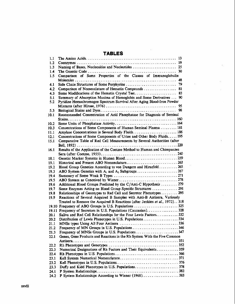
TABLES 1.1 The Amino Acids .................................................. 13 1.2 Coenzymes .......................................................19 1.3 Naming of Bases. Nucleosides and Nucleotides ........................ 25 1.4 The Genetic Code ................................................. 30 1.5 Comparison of Some Properties of the Classes of Immunoglobulin
Molecules ........................................................ 48 4.1 Side Chain Structures of Some Porphyrins ............................79 4.2 Comparison of Nomenclature of Hematin Compounds .................. 81 4.3 Some Modifications of the Hematin Crystal Test ....................... 83 5.1 Summary of Absorption Maxima of Hemoglobin and Some Derivatives ... 90 5.2 Pyridine Hemochromogen Spectrum Survival After Aging Blood-Iron Powder
Mixtures (after Hirose. 1976) ....................................... 95 5.3 Biological Stains and Dyes ..........................................98
10.1 Recommended Concentration of Acid Phosphatase for Diagnosis of Seminal Stains............................................................160
10.2 Some Units of Phosphatase Activity ..................................164 10.3 Concentrations of Some Components of Human Seminal Plasma ......... 181 11.1 Amylase Concentrations in Several Body Fluids ........................186 12.1 Concentrations of Some Components of Urine and Other Body Fluids .....195 15.1 Comparative Table of Red Cell Measurements by Several Authorities (after
Bell, 1892) .......................................................220 16.1 Results of the Application of the Coetzee Method to Human and Chimpanzee
Sera (after Coetzee. 1955) ..........................................240 18.1 Genetic Marker Systems in Human Blood .............................259 19.1 Historical and Present ABO Nomenclature ............................263 19.2 Blood Group Genetics According to von Dungern and Hirszfeld . . . . . . . . . .265 19.3 ABO System Genetics with Al and A2 Subgroups ...................... 267 19.4 Summary of Some Weak B Types ................................... 271 19.5 ABO System as Conceived by Wiener ................................278 19.6 Additional Blood Groups Predicted by the C/Anti-C Hypothesis .........279 19.7 Some Enzymes Acting on Blood Group Specific Structures ..............291 19.8 Relationships of Genotypes to Red Cell and Secretor Phenotypes .........295 19.9 Reactions of Several Acquired B Samples with Anti-B Antisera. Variously
Treated to Remove the Acquired B Reactions (after Jenkins et al., 1972) .. 318 19.10 Frequency of ABO Groups in U.S. Populations ........................325 19.11 Frequency of Secretors in U.S. Populations (Caucasian) .................328 20.1 Saliva and Red Cell Relationships for the Four Lewis Factors ............332 20.2 Distribution of Lewis Phenotypes in U.S. Populations ...................334 21.1 MNSs types Using All Four Antisera ................................336 21.2 Frequency of MN Groups in U.S. Populations ........................- 3 4 6 21.3 Frequency of MNSs Groups in U.S. Populations .......................347 22.1 Genes. Gene Products and Reactions in the Rh System With the Five Common
Antisera..........................................................351 22.2 Rh Phenotypes and Genotypes .......................................352 22.3 Numerical Designations of Rh Factors and Their Equivalents ............359 22.4 Rh Phenotypes in U.S. Populations ...................................366 23.1 Kell System Numerical Nomenclature ................................371 23.2 Kell Phenotypes in U.S. Populations ..................................376 23.3 Duffy and Kidd Phenotypes in U.S. Populations ........................378 24.1 P System Relationships .............................................382 24.2 P System Relationships According to Wiener (1968)....................383

Names and Structures of Glycosphingolipids of Erythrocytes (after Schwarting er al 385
Nomenclature and Properties of Various T i u e Esterases (after Coates et al.,
Nomenclature and Properties of PGD Variants ....................... -491
Activities of Various Peptidases with a Number of Substrates (after Rapley
.. 1977) ...................................................... Distribution of PGMl Phenotypes in U.S. Populations ...................436 Distribution of AK Phenotypes in U.S. Populations .....................443 Distribution of ACPl Phenotypes in U.S. Populations ................... 454 Distribution of ADA Phenotypes in U.S. Populations ................... 462 Nomenclature and Properties of the Plasma Cholinesterase Variants ......466 Classification of Carboxylesterases ...................................470
1975)............................................................ 474 Distribution of Cholinesterase and ESD Phenotypes in U.S. Populations ...476 CAI Locus Variants ................................................ 481 Distribution of Common Gd Phenotypes in U.S. Black Populations .......489
Distribution of PGD Phenotypes in U.S. Populations ....................494 Distribution of GLO Phenotypes in U.S. Populations ................... 498
et al 505.. 1971) ...................................................... Some Hemoglobin Variants ......................................... 551 Distribution of Common Hb Variants in U.S. Populations ...............560 Properties of Some Serum (Plasma) Proteins ..........................565 Distribution of Hp Phenotypes in U.S. Populations .....................580 Gc Variant Nomenclature Equivalents ................................ 583 Distribution of Gc Phenotypes in U.S. Populations ..................... 587 Distribution of Tf Phenotypes in U.S. Populations ......................594 Genetic Markers of the Immunoglobulins .............................602