
Forensic Fang et al., - OMICS International | Open … et al., J Forensic Res 2011, S2 STR Profiling...
10
Special Issue 2 • 2011 J Forensic Res ISSN: 2157-7145 JFR, an open access journal Open Access Research Article Forensic Research Fang et al., J Forensic Res 2011, S2 http://dx.doi.org/10.4172/2157-7145.S2-005 STR Profiling of Human Cell Lines: Challenges and Possible Solutions to the Growing Problem Rixun Fang 1 , Jaiprakash G. Shewale 1 , Vivian T. Nguyen 1 , Holly Cardoso 2 , Mavis Swerdel 2 , Ronald P.Hart 2 and Manohar R. Furtado 1 * 1 Life Technologies Corporation, 850 Lincoln Centre Drive, Foster City, CA 94404M, USA 2 W.M. Keck Center for Collaborative Neuroscience, Rutgers University, Piscataway, NJ 08854, USA *Corresponding author: Manohar R Furtado, Ph.D.Life Technologies 850 Lincoln Centre Drive,Foster City, CA 94404, USA Phone: 650 554 2670; Fax:650 554 6795; E-mail: [email protected] Received September 09, 2011; Accepted October 10, 2011; Published October 19, 2011 Citation: Fang R, Shewale JG, Nguyen VT, Cardoso H, Swerdel M, et al. (2011) STR Profiling of Human Cell Lines: Challenges and Possible Solutions to the Growing Problem. J Forensic Res S2:005. doi:10.4172/2157-7145.S2-005 Copyright: © 2011 Fang R, et al. This is an open-access article distributed under the terms of the Creative Commons Attribution License, which permits unrestricted use, distribution, and reproduction in any medium, provided the original author and source are credited. Abstract Genomic DNA preparations from 60 human cell lines from the National Cancer Institute (NCI), 48 human cell lines from American Type Culture Collection (ATCC) and 19 embryonic cell lines were profiled for autosomal short tandem repeat (STR) loci using the AmpFℓSTR ® Identifiler ® kit. Each DNA sample was profiled at least twice to ensure consistency and reproducibility of results. The resulting STR profiles were compared with the STR profiles in the database of ATCC. The allele calls for the common loci between the Identifiler ® kit and the database were identical except for one DNA sample, which we attribute to amplification artifacts. We have observed a high percentage of the STR loci exhibiting allelic imbalance. Certain STR loci for some cell lines exhibited 3 or more alleles. This type of observation can result from a unique profile for a given cell line or as the result of clonotypic heterozygosity and is not necessarily due to contamination. Our study demonstrates that STR based technologies are useful for cell line authentication applications. These data, combined with data from other researchers, will enable the development of a standard genotyping protocol for cell line authentication. Keywords: Cell line identification; Cell line authentication; Cell line contamination; STR; SNP; Cell line ID Abbreviations: STR: Short Tandem Repeat; RFLP: Restriction Fragment Length Polymorphism; ISSR: Inter-simple Sequence Repeat; VNTR: Variable Number of Tandem Repeat; SNP: Single Nucleotide Polymorphism; CODIS: Combined DNA Index System Introduction Cell culture technology is a vital research tool used in diverse areas such as basic research, medicine, genetics, cell biology, vaccine development, reconstructive medicine, toxicity testing, and drug discovery, screening and development. e utility of cell lines became popular in different areas of science because of widespread availability and improvements in cell culture technologies. ough the history of cell culture dates back to 1885, HeLa was the first human cancer cell line to be established in culture in 1952 [1,2]. e cell lines used for biomedical research can be grouped into primary cell cultures, continuous cell cultures (established cell cultures), and stem cells. Cell line misidentification or contamination, similar to microbial contamination, are routine and obvious laboratory issues. However, from the literature it is evident that many cell biologists, unlike microbiologists, have not resolved these issues. e problem of cell line contamination and misidentification is highlighted in several articles, spanning several decades [3-7]. It is estimated that the results from studies published in as many as 20% of scientific publications using cultured cells could be of questionable significance [8] because 15 – 36% of the cell lines used were either misidentified or cross-contaminated with another cell line [9,10]. Dr. Roland M. Nardone submitted a letter in July 2007, signed by 18 other leading scientists, to the Secretary, US Department of Health and Human Services, highlighting the incidences and severity of this issue [7]. Lack of cell authentication results in wasted resources and impedes scientific progress, including drug discovery. ere are multiple sources of cell line contamination or misidentification [3-5,7]. Some common reasons are: inadvertent mixing of cell lines, mislabeling, over-subculturing, the pleiomorphic nature of the cells in culture and differential proliferation rates (resulting in a given strain taking over the culture). Lack of proper training of personnel handling the cell lines and transfer of unauthenticated cell lines from one investigator to another can compound the problem. Moreover, scientists may assume that changes in cell cultures resulted from the in vitro environment and a change in gene expression, rather than from mislabeling or the unintended introduction of an alien cell. e absence of distinctive morphological, biochemical and genetic markers for distinguishing one cell line from another can make identifying contamination difficult. e need for cell line authentication was proposed as early as the 1970s by W. Nelson-Reese [11]. Confirmation of the identity and purity of cell lines is now becoming a prerequisite for distribution. All reputable cell banks now employ methods to confirm the identity and origin of the cell lines that they distribute [12]. Isoenzyme analysis, karyotyping, fluoroscent antibody staining, HLA typing, verification of species origin, and tests for microbial contamination are some of the methods for cell authentication and characterization [13-18]. e use of polymorphic genetic markers in the authentication of cell lines is becoming more popular because of their discrimination power and commercial availability of easy to use kits for this purpose. ese genetic markers [19] are useful in genotyping assays such as restriction fragment length polymorphism (RFLP), retro-element insertion polymorphism [20], inter-simple sequence repeat (ISSR) [21],
Transcript of Forensic Fang et al., - OMICS International | Open … et al., J Forensic Res 2011, S2 STR Profiling...

Special Issue 2 • 2011J Forensic ResISSN: 2157-7145 JFR, an open access journal
Open AccessResearch Article
ForensicResearch
Fang et al., J Forensic Res 2011, S2http://dx.doi.org/10.4172/2157-7145.S2-005
STR Profiling of Human Cell Lines: Challenges and Possible Solutions to the Growing ProblemRixun Fang1, Jaiprakash G. Shewale1, Vivian T. Nguyen1, Holly Cardoso2, Mavis Swerdel2, Ronald P.Hart2 and Manohar R. Furtado1* 1Life Technologies Corporation, 850 Lincoln Centre Drive, Foster City, CA 94404M, USA2W.M. Keck Center for Collaborative Neuroscience, Rutgers University, Piscataway, NJ 08854, USA
*Corresponding author: Manohar R Furtado, Ph.D.Life Technologies 850 Lincoln Centre Drive,Foster City, CA 94404, USA Phone: 650 554 2670; Fax:650 554 6795; E-mail: [email protected]
Received September 09, 2011; Accepted October 10, 2011; Published October 19, 2011
Citation: Fang R, Shewale JG, Nguyen VT, Cardoso H, Swerdel M, et al. (2011) STR Profiling of Human Cell Lines: Challenges and Possible Solutions to the Growing Problem. J Forensic Res S2:005. doi:10.4172/2157-7145.S2-005
Copyright: © 2011 Fang R, et al. This is an open-access article distributed under the terms of the Creative Commons Attribution License, which permits unrestricted use, distribution, and reproduction in any medium, provided the original author and source are credited.
AbstractGenomic DNA preparations from 60 human cell lines from the National Cancer Institute (NCI), 48 human cell
lines from American Type Culture Collection (ATCC) and 19 embryonic cell lines were profiled for autosomal short tandem repeat (STR) loci using the AmpFℓSTR® Identifiler® kit. Each DNA sample was profiled at least twice to ensure consistency and reproducibility of results. The resulting STR profiles were compared with the STR profiles in the database of ATCC. The allele calls for the common loci between the Identifiler® kit and the database were identical except for one DNA sample, which we attribute to amplification artifacts. We have observed a high percentage of the STR loci exhibiting allelic imbalance. Certain STR loci for some cell lines exhibited 3 or more alleles. This type of observation can result from a unique profile for a given cell line or as the result of clonotypic heterozygosity and is not necessarily due to contamination. Our study demonstrates that STR based technologies are useful for cell line authentication applications. These data, combined with data from other researchers, will enable the development of a standard genotyping protocol for cell line authentication.
Keywords: Cell line identification; Cell line authentication; Cell line contamination; STR; SNP; Cell line ID
Abbreviations: STR: Short Tandem Repeat; RFLP: Restriction Fragment Length Polymorphism; ISSR: Inter-simple Sequence Repeat; VNTR: Variable Number of Tandem Repeat; SNP: Single Nucleotide Polymorphism; CODIS: Combined DNA Index System
Introduction
Cell culture technology is a vital research tool used in diverse areas such as basic research, medicine, genetics, cell biology, vaccine development, reconstructive medicine, toxicity testing, and drug discovery, screening and development. The utility of cell lines became popular in different areas of science because of widespread availability and improvements in cell culture technologies. Though the history of cell culture dates back to 1885, HeLa was the first human cancer cell line to be established in culture in 1952 [1,2]. The cell lines used for biomedical research can be grouped into primary cell cultures, continuous cell cultures (established cell cultures), and stem cells. Cell line misidentification or contamination, similar to microbial contamination, are routine and obvious laboratory issues. However, from the literature it is evident that many cell biologists, unlike microbiologists, have not resolved these issues. The problem of cell line contamination and misidentification is highlighted in several articles, spanning several decades [3-7]. It is estimated that the results from studies published in as many as 20% of scientific publications using cultured cells could be of questionable significance [8] because 15 – 36% of the cell lines used were either misidentified or cross-contaminated with another cell line [9,10]. Dr. Roland M. Nardone submitted a letter in July 2007, signed by 18 other leading scientists, to the Secretary, US Department of Health and Human Services, highlighting the incidences and severity of this issue [7]. Lack of cell authentication results in wasted resources and impedes scientific progress, including drug discovery.
There are multiple sources of cell line contamination or misidentification [3-5,7]. Some common reasons are: inadvertent mixing of cell lines, mislabeling, over-subculturing, the pleiomorphic
nature of the cells in culture and differential proliferation rates (resulting in a given strain taking over the culture). Lack of proper training of personnel handling the cell lines and transfer of unauthenticated cell lines from one investigator to another can compound the problem. Moreover, scientists may assume that changes in cell cultures resulted from the in vitro environment and a change in gene expression, rather than from mislabeling or the unintended introduction of an alien cell. The absence of distinctive morphological, biochemical and genetic markers for distinguishing one cell line from another can make identifying contamination difficult.
The need for cell line authentication was proposed as early as the 1970s by W. Nelson-Reese [11]. Confirmation of the identity and purity of cell lines is now becoming a prerequisite for distribution. All reputable cell banks now employ methods to confirm the identity and origin of the cell lines that they distribute [12]. Isoenzyme analysis, karyotyping, fluoroscent antibody staining, HLA typing, verification of species origin, and tests for microbial contamination are some of the methods for cell authentication and characterization [13-18]. The use of polymorphic genetic markers in the authentication of cell lines is becoming more popular because of their discrimination power and commercial availability of easy to use kits for this purpose. These genetic markers [19] are useful in genotyping assays such as restriction fragment length polymorphism (RFLP), retro-element insertion polymorphism [20], inter-simple sequence repeat (ISSR) [21],

Special Issue 2 • 2011J Forensic ResISSN: 2157-7145 JFR, an open access journal
Citation: Fang R, Shewale JG, Nguyen VT, Cardoso H, Swerdel M, et al. (2011) STR Profiling of Human Cell Lines: Challenges and Possible Solutions to the Growing Problem. J Forensic Res S2:005. doi:10.4172/2157-7145.S2-005
Page 2 of 10
variable number of tandem repeat (VNTR) [22,23], single nucleotide polymorphism (SNP) [24] and short tandem repeat (STR) [25-30] for identification of cell lines. STRs with 2-6 bases have been routinely used in human identification laboratories for applications such as paternity testing, forensic casework, and the identification of victims of mass disaster for more than two decades [31-34].
In the past few years, application of STRs in human cell line identification has received a good deal of attention due to its usefulness in human identification [25-29]. Use of a SNP panel for human identification is an alternate genotyping system [35]. SNP genotyping offers several advantages over STRs such as the abundance (~9 million in the human genome), distribution throughout the genome (about 1 every 300 bp), 90% of human genetic variation come from SNPs, low mutation rate (1 x 10⁻⁹ per locus per generation), biallelic nature, high multiplex capability, and ease of data analysis. Though STRs are predominantly used for human identification, SNPs are commonly used for the identification of missing persons, victims of mass disaster, and resolving complex paternity cases [36]. The TaqMan Genotyping assays can detect single SNPs and the GenPlex™ HID system enables simultaneous amplification of 48 SNPs for such applications. The utility of this system in human identification has been demonstrated in multiple laboratories [35,37,38].
We describe the usefulness of the Identifiler® kit (for the analysis of 15 autosomal STR loci and Amelogenin) in human cell line authentication. The Identifiler® kit is currently used for human
identification in forensic laboratories and provides a standard metric for human cell line authentication. Cell line authentication procedures will become relevant in source attribution for cultures in research laboratories and biological sample tracking in cell based therapeutics. These tools provide a standard methodology to assess the origin of a given cell, determine if contamination or clonotypic heterozygosity are an issue, as well as determine if there are unique genotypic features in a given cell line, particularly cancer cells. We also address several important considerations when analyzing genotyping data.
Materials and MethodsGenomic DNA from the panel of 60 human cancer cell lines (NCI-
60) utilized in the National Cancer Institute’s (NCI) anti-cancer drug screen was kindly provided by the NCI (National Cancer Institute, Bethesda, MD). Genomic DNA from 48 human cell lines was obtained from American Type Culture Collection (ATCC, Manassas, VA). Genomic DNA from 19 embryonic stem cell lines (provided by Dr. Uma Lakshmipathy, Invitrogen, Inc., Carlsbad, CA) was isolated with the DNeasy® Blood and Tissue Kit (QIAGEN, Valencia, CA). The cell lines are listed in Tables 1, 2, and 3 of the supplementary material.
The Quantifiler® Duo DNA quantification kit, Identifiler® kit, 7500 Real-time PCR System, 3130xl Genetic Analyzer and associated software, and GeneMapper® ID v3.2.1 software were from Applied Biosystems (Foster City, CA). PowerPlex® 16 was obtained from Promega (Madison, WI). All other chemicals used in this study were of analytical grade.
Figure 1: Typical STR profile with allele designation for genomic DNA extracted from the NCI cell line HOP-62. The horizontal axis represents the values in base pairs and the vertical axis represents the values in relative fluorescent units (RFU). The 6-FAM channel (blue) shows the loci D8S1179, D7S820, D21S11, and CSF1P0; the VIC channel (green) contains the loci D3S1358, TH01, D13S317, D16S539, and D2S1338; the NED channel (yellow) shows the loci D19S433, vWA, THO1, and D18S51; the PET channel (red) contains the FGA, D5S818, and Amelogenin.

Special Issue 2 • 2011J Forensic ResISSN: 2157-7145 JFR, an open access journal
Citation: Fang R, Shewale JG, Nguyen VT, Cardoso H, Swerdel M, et al. (2011) STR Profiling of Human Cell Lines: Challenges and Possible Solutions to the Growing Problem. J Forensic Res S2:005. doi:10.4172/2157-7145.S2-005
Page 3 of 10
Quantitation of human DNA
The quantity of human DNA in the preparations was determined with the Quantifiler® Duo DNA Quantification Kit (Applied Biosystems, Foster City, CA) using 2 µL of the DNA extract [39]. PCR was performed on the 7500 Real Time PCR System and the data were analyzed using 7500 System SDS Software v1.2.3 (Applied Biosystems, Foster City, CA).
STR analysis
The DNA samples were amplified using the Identifiler® kit as described previously [40,41]. The quantity of template DNA was 1 ng unless otherwise mentioned in the text. The amplified products were analyzed on a 3130xl Genetic Analyzer (Applied Biosystems, Foster City, CA) and data analysis was performed using GeneMapper® ID Software v3.2.1 (Applied Biosystems, Foster City, CA) using 50 rfu (relative fluorescence units) as the threshold.
Results We profiled genomic DNA from 60 human cell lines (NCI-60 cell
lines), 48 human cell lines obtained from ATCC, and 19 embryonic stem cell lines. The genotypes are provided as supplementary data in Tables 1, 2, and 3, respectively. A typical Identifiler® profile from a cell line is presented in Figure 1. The genotype results obtained from our analysis were compared with the genotypes for 44 cell lines available from the ATCC website for the overlapping loci. Out of a total of 387 alleles from the 44 cell line DNA preparations, only one discrepant genotype was observed. The genotype for the D5S818 locus for 22Rv1 cell line DNA obtained in our laboratory was 11, 13, and is reported as 11, 12, 13 at the ATCC’s website. The genotype 11, 13 at this locus was confirmed by re-amplification of this DNA preparation using 2 ng of template. Such comparison for the profiles generated from NCI-60 cell line DNA preparations could not be made because the genotypes generated at NCI for 22Rv1 were not available.
MarkerCell lines Cell lines
NCI-ADR-RES OVCAR-8 M14 MDA-MB-435AMEL X X X X
CSF1PO 11 11 11 11
D13S317 12 12 12 12D16S539 13 13 9,13 13
D18S51 14 14 13,17 13,17D19S433 14,16 14,16 14,15 14D21S11 28 28 30 30D2S1338 19,23 19,23 19,24 19,24
D3S1358 18 16,18 14,16 14
D5S818 12 12 11,12 11,12
D7S820 12 12 8,10 8,10D8S1179 10 10 13 13FGA 20 20 21 21TH01 7 7 6,7 6,7TPOX 8 8 8,11 8,11vWA 16,17 16,17 16,18 16,18
Table 4: STR profiles for NCI 60 cell lines exhibiting similar STR profiles.
MarkerCell line
NCI-BL1395 NCI-H1395
AMEL X X
CSF1PO 12 12D13S317 10,14 10,14D16S539 11,13 11,13D18S51 12,14 12,14D19S433 13,16 13,16D21S11 28,29 29D2S1338 17,24 17,24D3S1358 15,18 15D5S818 12 12D7S820 8 8D8S1179 12,14 12,14FGA 18,23 18,23TH01 6,9.3 6,9.3TPOX 8 8vWA 14,17 14,17
Table 5: STR profiles for the two ATCC cell lines.

Special Issue 2 • 2011J Forensic ResISSN: 2157-7145 JFR, an open access journal
Citation: Fang R, Shewale JG, Nguyen VT, Cardoso H, Swerdel M, et al. (2011) STR Profiling of Human Cell Lines: Challenges and Possible Solutions to the Growing Problem. J Forensic Res S2:005. doi:10.4172/2157-7145.S2-005
Page 4 of 10
Of the NCI cell lines studied, the genotype for the DNA from NCI-ADR-RES was identical to the profile from OVCAR-8 except for the locus D3S1358 (Table 4). The genotype for the DNA from M14 was identical to the profile from MDA-MB-435 except for loci D16S539, D19S433, and D3S1358 (Table 4).
ATCC cell lines NCI-BL1395 and NCI-H1395 were from the same individual, NCI-BL-H1395 was from the EBV transformed B lymphoblast and NCI-H1395 was from an adenocarcinoma. However, the STR profiles for the DNA preparations from these cell lines was different for D21S11 and D3S1358 loci when amplified by the Identifiler® kit (Table 5). The genotypes of DNA preparations obtained from ATCC and NCI were compared; the STR profile for five cell lines obtained from ATCC matched that of the DNA preparations obtained from NCI (Table 6).
We observed that the STR profiles of DNA from the NCI and ATCC cell lines had some unique features not usually present in human reference samples. Three examples are provided here. The profile generated from HCC38 cell line DNA (ATCC) exhibited high peak imbalance at multiple heterozygous loci (Figure 2a). The loci D8S1179, D21S11, D13S317, D16S539, and D2S1338 exhibited high peak imbalance (high heterozygote peak height ratio) of 30, 43, 34, 43, and 43%, respectively. In contrast, the alleles at D19S433, vWA, and TPOX loci exhibited good peak balance of 90, 95, and 87, respectively. The observation was confirmed by re-amplification (Figure 2b). The high peak imbalance pattern was also confirmed by amplification using the PowerPlex® 16 genotyping system (data not shown), which eliminates the primer binding and other PCR amplification related effects.
Figure 3a presents the profile for the HCC1143 cell line DNA wherein the allele 17 at the D18S51 locus was amplified at low amplitude compared to allele 14 resulting in higher peak imbalance. As a result, homozygous allele 14 was the genotype called. Allele 17 was recovered (above the analysis parameter threshold) using 2ng of template DNA (Figure 3b). Correct genotype for the HCC1143 cell line DNA at the D18S51 locus is 14, 17.
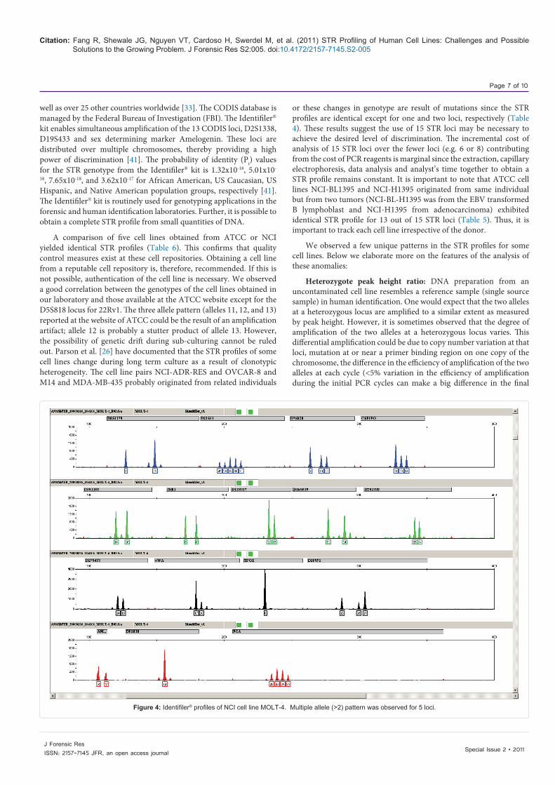
Multi-allele profiles were observed for a few cell lines. For example, the MOLT-4 profile has multiple alleles at the loci D21S11, D7S820, CSF1PO, FGA, and D18S51 (Figure 4). This multiple allele pattern was confirmed by repeat amplification using 2 ng of template DNA (data not shown). The multi allele pattern was also confirmed by amplification using the PowerPlex® 16 genotyping system (data not shown).
In a separate study, we profiled 19 embryonic stem cell lines analyzed in a previous study [42] using the Identifiler® kit (supplementary data (Table 3). All profiles were conclusive. Results confirmed that several stably-transfected derivatives of BG01V (coded with YW or YA) appropriately matched the parental line and that differentiated cultures (EB) matched undifferentiated cultures. STR typing proved to be a valuable method for tracking cellular genotype without being affected by minor genetic modification or differentiation.
The ability of STR profiling for detection of a mixture was investigated by generating mixture of DNA preparations from HCT15 and ACHN cell lines at different ratios up to 1:15. The results are summarized in Figure 5. Mixtures can be tracked by amplitude of unshared alleles. In general, it is possible to detect mixture of two individuals in a sample up to a ratio of 1:10.
DiscussionThere are several advantages to using autosomal STRs in human
identity testing. From a technical point of view, the amplicons are relatively short, the size difference between two alleles at a locus from a heterozygous individual is small, development of megaplex PCR for simultaneous amplification of 16 to 20 loci simultaneously and the techniques to separate fragments with single base length variation are relatively straight forward and kits and software are available commercially. As a tool for the identification of a unique person, STRs are ideal because they have a highly polymorphic nature, are scattered through out the genome and STRs that reside on different chromosomes and are not linked provide a unique genotype with high discrimination power [31-33]. A core set of 13 autosomal STR loci, termed CODIS (combined DNA index system) loci, have been selected for the generation of a database of convicted offenders in the USA, as
Marker
Cell Line and Source
Hs578T MDA-MB-231 PC-3 T-47D MCF7
ATCC NCI ATCC NCI ATCC NCI ATCC NCI ATCC NCI
AMEL X X X X X X X X X XCSF1PO 13 13 12,13 12,13 11 11 11,13 11,13 10 10D13S317 11 11 13 13 11 11 12 12 11 11D16S539 9,12 9,12 12 12 11 11 10 10 11,12 11,12D18S51 16 16 11,16 11,16 14,15 14,15 17 17 14 14D19S433 14,15 14,15 11,14 11,14 14 14 14 14 13,14 13,14
D21S11 29,32.2 29,32.2 33.2 33.2 29,31.2 29,31.2 28,31 28,31 30 30
D2S1338 17,26 17,26 20,21 20,21 18,20 18,20 24 24 21,23 21,23
D3S1358 16,17 16,17 16 16 16 16 15,17 15,17 16 16
D5S818 11 11 12 12 13 13 12 12 11,12 11,12D7S820 10 10 8,9 8,9 8,11 8,11 11 11 8,9 8,9D8S1179 13 13 13 13 13 13 13 13 10,14 10,14FGA 23,24 23,24 22,23 22,23 24 24 23 23 23,25 23,25
TH01 9,9.3 9,9.3 7,9.3 7,9.3 6,7 6,7 6 6 6 6TPOX 8 8 8,9 8,9 8,9 8,9 11 11 9,12 9,12vWA 17 17 15,18 15,18 17 17 14 14 14,15 14,15
Table 6: STR profile concordance between the same cell line DNA obtained from ATCC and NCI.

Special Issue 2 • 2011J Forensic ResISSN: 2157-7145 JFR, an open access journal
Citation: Fang R, Shewale JG, Nguyen VT, Cardoso H, Swerdel M, et al. (2011) STR Profiling of Human Cell Lines: Challenges and Possible Solutions to the Growing Problem. J Forensic Res S2:005. doi:10.4172/2157-7145.S2-005
Page 5 of 10
Figure 2: Identifiler® profiles of ATCC cell line HCC38. Genomic DNA from the cell line was amplified twice, (a) 1 ng DNA and (b) 2 ng DNA, to confirm the accuracy of STR genotyping.
A
B

Special Issue 2 • 2011J Forensic ResISSN: 2157-7145 JFR, an open access journal
Citation: Fang R, Shewale JG, Nguyen VT, Cardoso H, Swerdel M, et al. (2011) STR Profiling of Human Cell Lines: Challenges and Possible Solutions to the Growing Problem. J Forensic Res S2:005. doi:10.4172/2157-7145.S2-005
Page 6 of 10
Figure 3: Identifiler® profiles of ATCC cell line HCC1143. Different amount of genomic DNA was used for PCR amplification, (a) 1ng DNA and (b) 2ng DNA.
A
B
Special Issue 2 • 2011J Forensic ResISSN: 2157-7145 JFR, an open access journal
Citation: Fang R, Shewale JG, Nguyen VT, Cardoso H, Swerdel M, et al. (2011) STR Profiling of Human Cell Lines: Challenges and Possible Solutions to the Growing Problem. J Forensic Res S2:005. doi:10.4172/2157-7145.S2-005
Page 7 of 10
well as over 25 other countries worldwide [33]. The CODIS database is managed by the Federal Bureau of Investigation (FBI). The Identifiler® kit enables simultaneous amplification of the 13 CODIS loci, D2S1338, D19S433 and sex determining marker Amelogenin. These loci are distributed over multiple chromosomes, thereby providing a high power of discrimination [41]. The probability of identity (Pi) values for the STR genotype from the Identifiler® kit is 1.32x10⁻¹⁸, 5.01x10-
18, 7.65x10⁻¹⁸, and 3.62x10⁻¹⁷ for African American, US Caucasian, US Hispanic, and Native American population groups, respectively [41]. The Identifiler® kit is routinely used for genotyping applications in the forensic and human identification laboratories. Further, it is possible to obtain a complete STR profile from small quantities of DNA.
A comparison of five cell lines obtained from ATCC or NCI yielded identical STR profiles (Table 6). This confirms that quality control measures exist at these cell repositories. Obtaining a cell line from a reputable cell repository is, therefore, recommended. If this is not possible, authentication of the cell line is necessary. We observed a good correlation between the genotypes of the cell lines obtained in our laboratory and those available at the ATCC website except for the D5S818 locus for 22Rv1. The three allele pattern (alleles 11, 12, and 13) reported at the website of ATCC could be the result of an amplification artifact; allele 12 is probably a stutter product of allele 13. However, the possibility of genetic drift during sub-culturing cannot be ruled out. Parson et al. [26] have documented that the STR profiles of some cell lines change during long term culture as a result of clonotypic heterogeneity. The cell line pairs NCI-ADR-RES and OVCAR-8 and M14 and MDA-MB-435 probably originated from related individuals
or these changes in genotype are result of mutations since the STR profiles are identical except for one and two loci, respectively (Table 4). These results suggest the use of 15 STR loci may be necessary to achieve the desired level of discrimination. The incremental cost of analysis of 15 STR loci over the fewer loci (e.g. 6 or 8) contributing from the cost of PCR reagents is marginal since the extraction, capillary electrophoresis, data analysis and analyst’s time together to obtain a STR profile remains constant. It is important to note that ATCC cell lines NCI-BL1395 and NCI-H1395 originated from same individual but from two tumors (NCI-BL-H1395 was from the EBV transformed B lymphoblast and NCI-H1395 from adenocarcinoma) exhibited identical STR profile for 13 out of 15 STR loci (Table 5). Thus, it is important to track each cell line irrespective of the donor.
We observed a few unique patterns in the STR profiles for some cell lines. Below we elaborate more on the features of the analysis of these anomalies:
Heterozygote peak height ratio: DNA preparation from an uncontaminated cell line resembles a reference sample (single source sample) in human identification. One would expect that the two alleles at a heterozygous locus are amplified to a similar extent as measured by peak height. However, it is sometimes observed that the degree of amplification of the two alleles at a heterozygous locus varies. This differential amplification could be due to copy number variation at that loci, mutation at or near a primer binding region on one copy of the chromosome, the difference in the efficiency of amplification of the two alleles at each cycle (<5% variation in the efficiency of amplification during the initial PCR cycles can make a big difference in the final
Figure 4: Identifiler® profiles of NCI cell line MOLT-4. Multiple allele (>2) pattern was observed for 5 loci.

Special Issue 2 • 2011J Forensic ResISSN: 2157-7145 JFR, an open access journal
Citation: Fang R, Shewale JG, Nguyen VT, Cardoso H, Swerdel M, et al. (2011) STR Profiling of Human Cell Lines: Challenges and Possible Solutions to the Growing Problem. J Forensic Res S2:005. doi:10.4172/2157-7145.S2-005
Page 8 of 10
Figure 5: Detection of DNA mixtures. Mixture samples containing varying amounts of DNA from HCT15 and ACHN cell lines were prepared and amplified using the Identifiler® kit. The ratio of HCT15 and ACHN cell line DNA was 1:0, 1:2, 1:4, 1:6, 1:8, 1:10, 1:15 and , 0:1. The box denotes the allele changes at two loci at different DNA amount input ratio.
quantity of amplicons), number of copies in the template DNA (during dilution of the DNA extract the two copies of the genome may not get diluted to the same extent) or another, uncharacterized attribute of an individual. Acceptance criteria for a given reference sample is derived from internal validation studies by the human identification laboratory and may vary between laboratories. In general, a heterozygous peak height ratio of >50% is acceptable. However, the acceptable limit of heterozygous peak height ratio needs to be established for a given cell line, particularly for the cell lines exhibiting multiple allele patterns. It is important to note that this criterion does not apply to a mixed evidence sample. We have observed that many DNA preparations for the cell lines analyzed exhibited low heterozygote peak height ratios at multiple loci; e.g. the profile generated from the HCC38 cell line (Figure 2a and b). For ascertaining the genotype, it is recommended to quantify the human DNA with a human specific quantification method and perform two separate amplification reactions separated by time (not on the same plate) thereby minimizing the potential for amplification artifacts (Figure 2a and b).
Allele drop out: Allele dropout at a locus may result from poor amplification (peaks below the detection threshold) or failure of amplification. Use of low quantity template DNA often leads to allele drop out. When the HCC1143 cell line DNA was amplified using 1 ng of DNA, poor amplification of the allele 17 at D18S51 locus resulted
in an erroneous genotype- 14 instead of 14 and 17. One approach to overcome such issues is to amplify DNA at two different template quantities (Figure 3a and b).
Multiple allele profile: Normally one expects either homozygous or heterozygous results at all loci from an individual donor. The presence of more than two alleles at one locus is reported in some individuals [31,33]. Multiple allele patterns at multiple loci, in general, are interpreted as a mixture profile in the human identification laboratory. DNA from the MOLT-4 cell line exhibited multiple alleles at 5 loci (Figure 4). Such profiles can be characteristic of a cell line due to clonotypic heterozygosity and is not necessarily the result of contamination. It is interesting that MOLT-4 is ~tetraploid [43] and exhibit microsatellite instability [44] which contributes to the observed multiple allele patterns. We analyzed the DNA preparations obtained from the indicated agencies and assume that the DNA obtained was from uncontaminated cell lines. Masters et al. [24] reported a three allele profile at the D21S11 locus for the HeLa53 cell line.
The unique features of a cell line such as low heterozygous peak height ratios or multiple alleles at a locus (>2) can be ascertained by performing two amplification reactions using 1 and 2 ng of template DNA. Performing two amplification reactions at two template DNA quantities enables the detection of amplification artifacts. Further, it is recommended to use a species specific quantification method (e.g. Quantifiler® kits for the human cell line DNA preparations) to obtain

Special Issue 2 • 2011J Forensic ResISSN: 2157-7145 JFR, an open access journal
Citation: Fang R, Shewale JG, Nguyen VT, Cardoso H, Swerdel M, et al. (2011) STR Profiling of Human Cell Lines: Challenges and Possible Solutions to the Growing Problem. J Forensic Res S2:005. doi:10.4172/2157-7145.S2-005
Page 9 of 10
high quality profiles.
Cell line misidentification or contamination is widely acknowledged to be a serious problem, resulting in false information in numerous studies published in the literature. The consequences are numerous such as research being performed based on faulty data, delay of important scientific discoveries, and wastage of valuable time and resources. The use of STR for identification purposes is an important part of the solution to this problem. However, use of these tools requires a thorough understanding of common amplification anomalies and how to interpret the resulting data. The American Type Collection Standards Development Organization Workgroup ASN-0002 has recently recommended use of STR profiling as a tool for cell line authentication [45]. Acknowledgements
We thank the Developmental Therapeutics Program of the NCI for providing the DNA preparations from the NCI-60 cell lines. The studies for embryonic stem cell lines were supported by the New Jersey Commission on Science & Technology. We thank Lisa Calandro, Lisa Ortune, Joe Varlaro, and Allison Holt for their comments on the manuscript. We also thank Dr. Susan Holbeck from NCI for constructive comments and fruitful discussions.
References
1. Roux W (1885) Beiträge zur Entwicklungsmechanik des Embryo. Zt. f. Biol 21:411-524.
2. Gey GO (1955) Some aspects of the constitution and behavior of normal and malignant cells maintained in continuous culture. Harvey Lanct. 50:154-229.
3. Nardone RM ( 2008) BioTechniques 45:221-225.
4. Freshney RI (2008) Authentification of cell lines: ignore at your pearl. Expert rev. Anticancer Ther. 8:11-314.
5. Hughes P, Marshall D, Reid Y, Parkes H, Gelber C (2007) The costs of using unauthenticated, over-passaged cell lines: how much more data do we need? BioTechniques 43:575-584.
6. Nardone RM (2007) Eradication of cross-contaminated cell lines: A call for action. Cell Biol Toxicol. 23:367–372.
7. Nardone LM et al. (2007) Letter to the Secretary, US Department of Health and Human Services, Washington DC
8. Drexler HG, Dirks WG, Y Matsuo, MacLeod RA (2003) False leukemis-lymphoma cell lines: an update on over 500 cell lines. Leukemia 17:416-426.
9. Dirks WG, Drexler HG (2004). Authentication of cancer cell lines by DNA fingerprinting. Methods Mol. Med. 88:43-55.
10. Lavappa KS (1978) Survey of ATCC stocks of human cell lines for HeLa contamination. In Vitro 14:469-475.
11. Chatterjee R (2007) Cases of mistaken identity. Science 315:928-931.
12. Lacroix M (2008) Persistent use of “false” cell lines. Int. J. Cancer. 122:1-4.
13. Nims RW, Herbstritt CJ (2005) Cell line authentication using isoenzyme analysis: strategies for accurate speciation and case studies for the detection of cell line cross-contamination using a commercial kit. BioPharm Intel June 76-82.
14. Hay RJ (1992) Methods for authenticating cell lines. Dev Biol Stand 76:25-37.
15. O’Toole CM, S Povey, Hepburn P, Franks LM (1981) Identity of some human bladder cancer cell lines. Nature (London) 301:429-430.
16. O’Brien SJ, Shannon JE,Gail MH (1980) A molecular approach to the identification and individualization of human and animal cells in culture: isozyme and allozyme genetic signatures. In Vitro 16:119-135.
17. Gilbert DA, Reid YA, Gail MH, Pee D, White C, et al. (1990) Application of DNA fingerprints for cell-line individualization. Am J Hum Genet 47:499-514.
18. Nims RW, Shoemaker AP, Bauernschub MA, Rec LJ, Harbell JW (1998)
Sensitivity of isoenzyme analysis for the detection of interspecies cell line cross-contamination. In Vitro Cell Dev Biol Anim 34:35-39.
19. Losi CG, Ferrari S, Sossi E, Villa R, Ferrari M (2008) An alternative method to isoenzyme profile for cell line identification and interspecies cross-contamination: cytochrome b PCR-RFLP analysis. In Vitro Cell Dev Biol Anim 44:321-329.
20. Ustyugova SV, Amosova AL, Lebedev YB, Sverdlov ED (2005) Cell line fingerprinting using retroelement insertion polymorphism. BioTechniques 38:561-565.
21. Grasela JJ, McIntosh AH (2003) Application of inter-simple sequence repeats to insect cell lines: identification at the clonal and tissue-specific level. In Vitro Cell Dev Biol Anim 39:353-363.
22. Matsuo Y, Nishizaki C, Drexler HG (1999) Efficient DNA fingerprinting method for cell identification of cross-culture contamination of cell lines. Hum Cell 12:149-154.
23. Honma M, Kataoka E, Ohnishi K, Ohno T, Takeuchi M, et al. (1992) A new DNA profiling system for cell line identification for use in cell banks in Japan. In Vitro Cell Dev Biol 28A:24-28.
24. Demichelis F, Greulich H, Macoska JA, Beroukhim R, Sellers WR, et al.(2008) SNP panel identification assay (SPIA): a genetic-based assay for the identification of cell lines. Nucleic Acid Res 36:2446-2456.
25. Masters JR, Thomas JA, Daly-Burns B, Reid YA, Dirks WG, et al.(2001) Short tandem repeat profiling provides an international reference standard for human cell lines. Proc Natl Acad Sci USA 98:8012-8017.
26. Parson W, Kirchebner R, Muhlmann R, Renner K, Kofler A, et al. (2005) Cancer cell line identification by short tandem repeat profiling: powers and limitations. FASEB J 19:434 – 436.
27. Schweppe RE, Klopper JP, Korch C, Pugazhenthi U, Benezra M, et al. (2008) DNA profiling analysis of 40 human thyroid cancer cell lines reveals cross-contamination resulting in cell line redundancy and misidentification. J clin Endocrin Metab 93:4331-4341.
28. Silva LM, Montes de Oca H, Diniz CR, Fortes-Dias CL (2001) Fingerprinting of cell lines by directed amplification of minisatellite-region DNA (DAMD). Brazillian J Med Biol Res 34:1405-1410.
29. MacLeod RAF, Dirks WG, Matsuo Y, Kaufmann M, Milch H, et al. (1999) Widespread intraspecies cross-contamination of human tumor cell liones arising at source. Int J Cancer 83:555-563.
30. Debenham PG, Webb MB (1992) Cell line characterization by DNA fingerprinting; a review. Dev Biol Stand 76:39-42.
31. Butler JM (2005) Forensic DNA Typing. Biology, technology, and genetics of STR markers. 2nd edition. Elseviwe Academic Press. Burlington, MA.
32. Moretti TR, Baumstark AL, Defenbaugh DA, Keys KM, Smerick JB, et al. (2001) Validation of short tandem repeats (STRs) for forensic usage: performance testing of fluorescent multiplex STR systems and analysis of authentic and simulated forensic samples. J Forensic Sci 46:647-660.
33. Budowle B, Shea B, Niezgoda S, Chakraborty R (2001) CODIS STR loci data from 41 sample populations. J Forensic Sci 46:453 – 489.
34. Fregeau CJ, Fourney RM (1993) DNA typing with fluorescently tagged short tandem repeats: a sensitive and accurate approach to human identification. BioTechniques 15:100- 119.
35. Phillips C, Fang R, Ballard D, Fondevila M, Harrison C, et al.(2007) Evaluation of Genplex SNP typing system and a 49plex forensic marker panel. Forensic Sci Intl Genet 1:180-185.
36. Budowle B, van Daal A (2008) Forensically relavent SNP classes. BioTechniques 44:603-610.
37. Stangegaard M, Tomas C, Hansen AJ, Frank-Hansen R,Borsting C, et al. (2008) Biomel-3000 and GenPlex SNP genotyping in forensic genetics. J Assoc Lab Automation 13:297-303.
38. Musgrave-Brown E, Ballard D, Alvarez MF, Fabg R, Harrison C, et al. (2008) Forensic Sci Genetics 1:389-393.

Special Issue 2 • 2011J Forensic ResISSN: 2157-7145 JFR, an open access journal
Citation: Fang R, Shewale JG, Nguyen VT, Cardoso H, Swerdel M, et al. (2011) STR Profiling of Human Cell Lines: Challenges and Possible Solutions to the Growing Problem. J Forensic Res S2:005. doi:10.4172/2157-7145.S2-005
Page 10 of 10
39. Barbisin M, Fang R, O’Shea CE, Calandro LM, Furtado MR, et al. (2009) Developmental validation of the Quantifiler® Duo DNA Quantification Kit for simultaneous quantification of total human and human male DNA and detection of PCR inhibitors in biological samples. J Forensic Sci 54: 305-319.
40. Collins PJ, Hennessy LK, Leibelt CS, Roby RK, Redder DJ, et al. (2004) Developmental validation of a single-tube amplification of the 13 CODIS STR loci, D2S1338, D19S433 and amelogenin: the AmpFlSTR® Identifiler PCR amplification kit. J Forensic Sci 49:1265-1277.
41. Applied Biosystems (2001) AmpFlSTR® Identifilar PCR Amplification Kit User Manual. Part # 4323291 Rev. B.
42. Lakshmipathy U, Love B, Goff LA, Jörnsten R, Graichen R, et al. (2007) MicroRNA Expression Pattern of Undifferentiated and Differentiated Human Embryonic Stem Cells. Stem Cells Dev 16:1003-1016.
43. Roscheke AV, Tondon G, Gehlhaus KS, McTyre N, Bussey KJ, et al.(2003) Karyotypic complexity of the NCI-60 drug-screening panel. Cancer Res 63:8634-8647.
44. http://www.sanger.ac.uk/genetics/CGP/NCI60/
45. American Type Collection Standards Development Organization Workgroup ASN-0002. (2010) Cell line misidentification: the beginning of the end. Nature Cancer Reviews. 10: 441-448.
Submit your next manuscript and get advantages of OMICS Group submissionsUnique features:
• Userfriendly/feasiblewebsite-translationofyourpaperto50world’sleadinglanguages• AudioVersionofpublishedpaper• Digitalarticlestoshareandexplore
Special features:
• 100OpenAccessJournals• 10,000editorialteam• 21daysrapidreviewprocess• Qualityandquickeditorial,reviewandpublicationprocessing• IndexingatPubMed(partial),Scopus,DOAJ,EBSCO,IndexCopernicusandGoogleScholaretc• SharingOption:SocialNetworkingEnabled• Authors,ReviewersandEditorsrewardedwithonlineScientificCredits• Betterdiscountforyoursubsequentarticles
Submityourmanuscriptat:http://www.omicsonline.org/submission


![SHARED RISK FORMULATION IN FORENSIC PSYCHIATRY- A ... · Fluttert et al [2010] Norway Maximum Secure Forensic Unit Rana Abou-Sinna and Leubers [2012] Australia Secure Forensic Psychiatry](https://static.fdocuments.in/doc/165x107/5f0f74a97e708231d4443fbf/shared-risk-formulation-in-forensic-psychiatry-a-fluttert-et-al-2010-norway.jpg)