For reprint orders, please contact reprints@expert-reviews ... · PDF fileMirminachi 1, Nima...
15
CME 10.1586/ECI.13.30 561 ISSN 1744-666X © 2013 Expert Reviews Ltd www.expert-reviews.com Review Hassan Abolhassani 1 , Babak Torabi Sagvand 1 , Tahaamin Shokuhfar 1 , Babak Mirminachi 1 , Nima Rezaei 1,2 and Asghar Aghamohammadi* 1 1 Research Center for Immunodeficiencies, Pediatrics Center of Excellence, Children’s Medical Center, Tehran University of Medical Sciences, Tehran, Iran 2 Molecular Immunology Research Center; and Department of Immunology, School of Medicine, Tehran University of Medical Sciences, Tehran, Iran *Author for correspondence: Tel.: +98 216 642 8998 Fax: +98 216 692 3054 [email protected] Common variable immunodeficiency (CVID) is the most common symptomatic primary immunodeficiency in adults. As symptoms of CVID are usually heterogeneous and unspecific, diagnosis and follow-up of CVID can be challenging. In light of this, a broad review of advances in management and treatment of CVID is performed here in order to reach a distinct protocol. However, it should be noted that owing to the nature of the disease, it can only be treated symptomatically but not cured. There is little evidence to guide appropriate or universal guidelines to improve the current status of management of the disease. The most satisfactory treatments of CVID could be achieved by the use of immunoglobulin replacement, antibiotics, immunosuppressants and hematopoietic stem cell transplantation. This review is written based on the importance of clinical surveillance of asymptomatic CVID cases and early recognition of different clinical complications. Moreover, for each complication, appropriate interventions for improving outcomes are mentioned. KEYWORDS:CLINICALPHENOTYPESsCOMMONVARIABLEIMMUNODElCIENCYsMANAGEMENTsPREVENTIONsSCREENING sTREATMENT A review on guidelines for management and treatment of common variable immunodeficiency Expert Rev. Clin. Immunol. 9(6), 561–575 (2013) This activity has been planned and implemented in AMA PRA Category 1 Credit(s)™ : 00 : s Analyze the clinical presentation of CVID s Assess the practice of exogenous IgG administration to patients with CVID s Evaluate pulmonary complications of CVID and their management s Evaluate other potential complications of CVID For reprint orders, please contact [email protected]
Transcript of For reprint orders, please contact reprints@expert-reviews ... · PDF fileMirminachi 1, Nima...

CME
10.1586/ECI.13.30 561ISSN 1744-666X© 2013 Expert Reviews Ltdwww.expert-reviews.com
Review
Hassan Abolhassani1, Babak Torabi Sagvand1, Tahaamin Shokuhfar1, Babak Mirminachi1, Nima Rezaei1,2 and Asghar Aghamohammadi*1
1Research Center for
Immunodeficiencies, Pediatrics Center
of Excellence, Children’s Medical
Center, Tehran University of Medical
Sciences, Tehran, Iran2Molecular Immunology Research
Center; and Department of
Immunology, School of Medicine,
Tehran University of Medical Sciences,
Tehran, Iran
*Author for correspondence:
Tel.: +98 216 642 8998
Fax: +98 216 692 3054
Common variable immunodeficiency (CVID) is the most common symptomatic primary immunodeficiency in adults. As symptoms of CVID are usually heterogeneous and unspecific, diagnosis and follow-up of CVID can be challenging. In light of this, a broad review of advances in management and treatment of CVID is performed here in order to reach a distinct protocol. However, it should be noted that owing to the nature of the disease, it can only be treated symptomatically but not cured. There is little evidence to guide appropriate or universal guidelines to improve the current status of management of the disease. The most satisfactory treatments of CVID could be achieved by the use of immunoglobulin replacement, antibiotics, immunosuppressants and hematopoietic stem cell transplantation. This review is written based on the importance of clinical surveillance of asymptomatic CVID cases and early recognition of different clinical complications. Moreover, for each complication, appropriate interventions for improving outcomes are mentioned.
KEYWORDS:
A review on guidelines for management and treatment of common variable immunodeficiency
Expert Rev. Clin. Immunol. 9(6), 561–575 (2013)
!"#$%&!'()*+,+-.+/( !".$%0(1"-$%,*+(2+0.+!
This activity has been planned and implemented in
!!"#$ %!&'()*+'*+&',--&%. /'0#& -' %$'1"/)!)&-'"2'*+&'
0!!#&$)* ."%'3"4%!)/' 2"#'3"%.%4)%5'6&$)! /',$4! ."%'*+#"45+'*+&' 7")%*'-1"%-"#-+)1'"2'
6&$-! 1&8'993' %$',:1&#*';&<)&(-'9*$='6&$-! 1&8'993')-' !!#&$)*&$'>?'*+&'0336,'*"'1#"<)$&'
!"%.%4)%5'@&$)! /'&$4! ."%'2"#'1+?-)!) %-=
6&$-! 1&8'993'$&-)5% *&-'*+)-'A"4#% /B> -&$'36,' !.<)*?'2"#' '@ :)@4@'"2'C'AMA PRA
Category 1 Credit(s)™='D+?-)!) %-'-+"4/$'!/ )@'"%/?'*+&'!#&$)*'!"@@&%-4# *&'()*+'*+&'&:*&%*'
"2'*+&)#'1 #.!)1 ."%')%'*+&' !.<)*?=
0//'"*+&#'!/)%)!) %-'!"@1/&.%5'*+)-' !.<)*?'()//'>&' )--4&$' '!&#.E! *&'"2'1 #.!)1 ."%='F"'1 #B
.!)1 *&')%'*+)-' 7"4#% /'36,' !.<)*?G'HCI'#&<)&('*+&'/& #%)%5'">7&!.<&-' %$' 4*+"#'$)-!/"-4#&-J'
HKI'-*4$?'*+&'&$4! ."%'!"%*&%*J'HLI'* M&'*+&'1"-*B*&-*'()*+' 'NOP'@)%)@4@'1 --)%5'-!"#&' %$'
!"@1/&*&'*+&'&< /4 ."%' *'(((=@&$-! 1&="#5Q7"4#% /Q&:1&#.@@4%"/"5?J'HRI'<)&(Q1#)%*'!&#.E! *&=
3!0!%#!("%4!: L0'6 ?'KOCLJ(15&.6%,*+("%4!'(L0'6 ?'KOCR
7!%6+.+/(*89!$,:!#
S1"%'!"@1/&."%'"2'*+)-' !.<)*?8'1 #.!)1 %*-'()//'>&' >/&'*":
Analyze the clinical presentation of CVID
Assess the practice of exogenous IgG administration to patients with CVID
Evaluate pulmonary complications of CVID and their management
Evaluate other potential complications of CVID
For reprint orders, please contact [email protected]
CME
Expert Rev. Clin. Immunol. 9(6), (2013)562
Review
Common variable immune deficiency (CVID) as a hetero geneous group of primary immune deficiencies is characterized by insuf-ficient serum levels of immunoglobulins (Igs), reduced response to specific antigens and higher incidence of repeated infections [1–4]. Autoimmune disorders, lymphoproliferative disorders and gastric complications also have an increased incidence in patients with CVID [5,6]. The prevalence of this predominantly antibody defi-ciency should not be thought of as being rare (1:10,000–50,000) and CVID is the most common symptomatic human primary immunodeficiency [7–9]. The onset (early or late) and clinical man-ifestations (distinct phenotypes) of the disease have heterogenous presentations [10–12]. This variability in clinical phenotype and late onset of disease may lead to delay in CVID diagnosis [13,14].
The molecular basis, both immunologically and genetically, of CVID is still unclear despite huge amounts of evaluation in this field since 1953 [15]. However, during the last 10 years, 10–15% of the patients with a history of CVID manifestations are classified in other primary immunodeficiency categories because of specific identified gene mutations including ICOS, CD19, CD20, CD81, CD21 and LRBA1 [16–23]. Decreased serum level of IgG, IgA and/or IgM (at least 2 standard deviations below the mean for the age group), hyporesponsiveness to specific antigens, minimum age of 4 years, absence of lymphoid malignancy during the first 2 years of diagnosis and genetic exclusion of other known etiologies for
hypogammaglobulinemia are considered as criteria for diagno-sis of CVID (FIGURE 1) [24–26]. Furthermore, approximately 2% of CVID patients may represent clinical or laboratory features that suggest a known severe combined primary immunodeficiency (e.g., opportunistic fungal or viral infections, very low numbers of T cells and/or monocytes). Although this phenomenon is rare, this is important because such patients may need stem cell grafts [27].
Heterogeneity in CVID refers to variability in immunological and genetic defects and diversity in clinical symptoms described in this group of patients [19,28,29]. The main immunological defect is failure of B-cell Ig production, although abnormalities have been described in all other components of the immune system [30,31]. Only some CVID cases are due to monogenic Mendelian diseases and homozygous mutations in that some genes have been reported in some CVID families [17,18,32].
The clinical spectrum of CVID is broad. The main clinical man-ifestations are recurrent infections occurring in the respiratory tract, GI tract, skin and soft tissues [4,33,34]. However, inflammatory com-plications occur in varying proportions and include autoimmunity, chronic lung disease, bronchiectasis, gastrointestinal (GI) disease with or without malabsorption, systemic or localized granuloma-tous disease, liver disease, splenomegaly, lymphadenopathy with or without lymphoma and other malignancies [26,35]. By using data of clinical manifestations and complications from 334 CVID patients
Financial & competing interests disclosure
EDITOR
Elisa Manzotti
Publisher, Future Science Group, London, UK.
Disclosure: Elisa Manzotti has disclosed no relevant financial relationships.
CME AUTHOR
Charles P Vega, MD
Associate Professor and Residency Director, Department of Family Medicine, University of California, Irvine.
Disclosure: Charles P Vega, MD, has disclosed no relevant financial relationships.
AUTHORS AND CREDENTIALS
Hassan Abolhassani
Research Center for Immunodeficiencies, Pediatrics Center of Excellence, Children’s Medical Center, Tehran University of Medical Sciences, Tehran, Iran.
Disclosure: Hassan Abolhassani has disclosed no relevant financial relationships.
Babak Torabi Sagvand
Research Center for Immunodeficiencies, Pediatrics Center of Excellence, Children’s Medical Center, Tehran University of Medical Sciences, Tehran, Iran.
Disclosure: Babak Torabi Sagvand has disclosed no relevant financial relationships.
Tahaamin Shokuhfar
Research Center for Immunodeficiencies, Pediatrics Center of Excellence, Children’s Medical Center, Tehran University of Medical Sciences, Tehran, Iran.
Disclosure: Tahaamin Shokuhfar has disclosed no relevant financial relationships.
Babak Mirminachi
Research Center for Immunodeficiencies, Pediatrics Center of Excellence, Children’s Medical Center, Tehran University of Medical Sciences, Tehran, Iran.
Disclosure: Babak Mirminachi has disclosed no relevant financial relationships.
Nima Rezaei
Research Center for Immunodeficiencies, Pediatrics Center of Excellence, Children’s Medical Center, Tehran University of Medical Sciences; Molecular
Immunology Research Center and Department of Immunology, School of Medicine, Tehran University of Medical Sciences, Tehran, Iran.
Disclosure: Nima Rezaei has disclosed no relevant financial relationships.
Asghar Aghamohammadi
Research Center for Immunodeficiencies, Pediatrics Center of Excellence, Children’s Medical Center, Tehran University of Medical Sciences, Tehran, Iran.
Disclosure: Asghar Aghamohammadi has disclosed no relevant financial relationships.
Abolhassani, Torabi Sagvand, Shokuhfar, Mirminachi, Rezaei & Aghamohammadi

CME
563www.expert-reviews.com
Review
from seven European centers, five different clinical phenotypes were considered for these patients [13]. In the revised phenotyping criteria, lymphoid malignancies were excluded [36].
Over the last few years, advances have been made in the man-agement of CVID, improving outcomes in the patients; these include Ig replacement, antibiotics for treatment and preven-tion of infections and appropriate therapy for noninfectious complications [37].
IgG replacement is the mainstay of treatment of CVID [35] and it has been shown that long-term Ig replacement therapy for CVID has reduced the rate of infections and their long-term complications [38–41]. However, despite the reduction in the rate of bacterial infections by use of IgG replacement in CVID patients, these patients are still more susceptible to complications because of a dysregulated immune response that attracts the caregiver’s attention. Here, the authors review the recent advances in CVID, specifically the clinical features, and focus on the management and treatment of CVID. Other specific classifications based on immunological property of CVID cases (including Freiburg, Paris, EUROclass and severe T-cell defect classification) exist, however their importance for clinical management are under eval-uation [42–46]. Moreover, the authors review the recent advances on the management and treatment of CVID.
Clinical manifestations of CVID
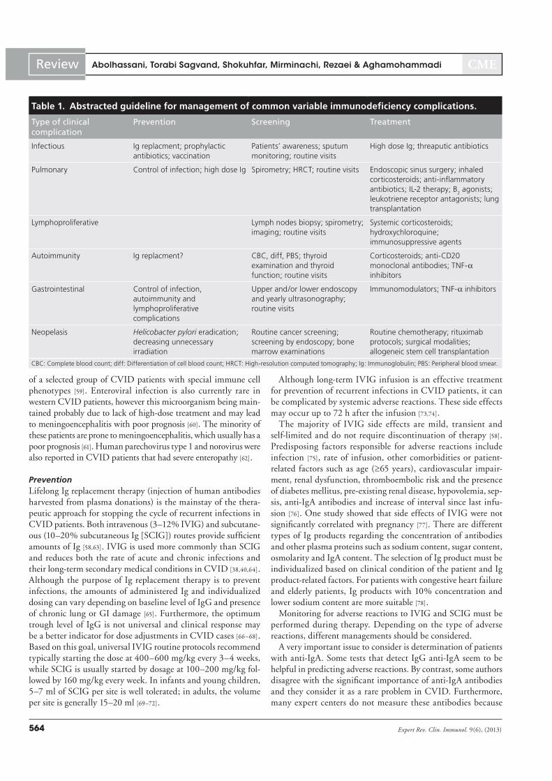
The clinical manifestations of CVID constitute six major categories including: infections, pulmonary complications, granulo matous or polyclonal lymphocytic infiltrative diseases, autoimmunity, GI diseases and neoplasias (TABLE 1). These can be established in different periods of life, from childhood to late adulthood, with a bimodal age distribution, demonstrating two peaks between 1 and 5 years and 18 and 25 years [3,4,47].
Management of infectious complications
Virtually all CVID patients encounter chronic or recurrent infec-tions, particularly sinusitis, otitis, bronchitis and pneumonia [47–49]. Approximately 90% of CVID patients have encountered at least one episode of chronic sinusitis and 70% have suffered recurrent otitis media before diagnosis [50–52]. Previously, there had been a history of at least one episode of pneumonia before diagno-sis in 75–85% of the CVID patients as well as multiple episodes in many others [38,41]. The majority of morbidities and mortalities of CVID is due to long-term sequelae of recurrent respiratory tract infections including chronic sinusitis, hearing loss (due to tym-panic membrane perforation) and bronchiectasis [4]. Encapsulated (Haemophylus influenzae, Streptococcus pneumoniae) or atypical (Mycoplasma spp.) bacteria are the major causative agents for recur-rent infections of both the upper and the lower respiratory tract [4,35,53]. Failure of antigen-specific IgG production, which increases susceptibility to encapsulated bacteria, seems to be responsible for recurrent rhinoviral infections in CVID [51].
The GI tract is the second organ that is involved in infections in 10–40% of the CVID cases. Various pathogens can cause GI infections in CVID patients including: Giardia lamblia, Campylobacter jejuni, Salmonella spp., Cryptosporidium parvum,
CMV, Clostridium difficile, Helicobacter pylori, HBV, HCV and so on. Giardiasis is the most prevalent GI infection, especially in those with undetectable serum IgA levels [54]. H. pylori is an important pathogen in CVID, resulting in chronic active gastritis involving antrum and corpus, achlorhydria, gastric adenocarcinoma and gastric lymphoma [55,56].
The CNS (meningoencephalitis is especially caused by Enterovirus spp.), joints, bones (especially due to Mycoplasma spp.), skin and eyes are also affected by CVID and their involve-ment might be the first and sole presentation of CVID [57]. Fibrotic bladder can develop due to recurrent urinary tract infections due to Ureaplasma urealyticum [25,36].
Viral hepatitis (especially HCV transmitted by intravenous Ig [IVIG] administration) and severe Herpes zoster infection have been reported in large numbers of patients [4]. Because of recent devel-opments in Ig preparation including careful selection of donors, plasma antibody screening and effective procedures of viral inacti-vation, the rate of these infections are now very rare in the western world [58]. CMV is involved in many inflammatory complications
Figure 1. Differential diagnosis for common variable immunodeficiency with definite single defect gene. CVID: Common variable immunodeficiency; HIGM: Hyper IgM syndrome; ICOS: Inducible T-cell costimulator; SCID: Severe combined immunodeficiency; XLA: X-linked agammaglobulinemia; XLP: X-linked lymphoproliferative disease.
CVID
CD19, CD21, CD81
XLP
ICOS
CD20
HIGM
XLA
SCID
LRBA1
Management of common variable immunodeficiency
CME
Expert Rev. Clin. Immunol. 9(6), (2013)564
Review
of a selected group of CVID patients with special immune cell phenotypes [59]. Enteroviral infection is also currently rare in western CVID patients, however this microorganism being main-tained probably due to lack of high-dose treatment and may lead to meningoencephalitis with poor prognosis [60]. The minority of these patients are prone to meningoencephalitis, which usually has a poor prognosis [61]. Human parechovirus type 1 and norovirus were also reported in CVID patients that had severe enteropathy [62].
Prevention
Lifelong Ig replacement therapy (injection of human antibodies harvested from plasma donations) is the mainstay of the thera-peutic approach for stopping the cycle of recurrent infections in CVID patients. Both intravenous (3–12% IVIG) and subcutane-ous (10–20% subcutaneous Ig [SCIG]) routes provide sufficient amounts of Ig [58,63]. IVIG is used more commonly than SCIG and reduces both the rate of acute and chronic infections and their long-term secondary medical conditions in CVID [38,40,64]. Although the purpose of Ig replacement therapy is to prevent infections, the amounts of administered Ig and individualized dosing can vary depending on baseline level of IgG and presence of chronic lung or GI damage [65]. Furthermore, the optimum trough level of IgG is not universal and clinical response may be a better indicator for dose adjustments in CVID cases [66–68]. Based on this goal, universal IVIG routine protocols recommend typically starting the dose at 400–600 mg/kg every 3–4 weeks, while SCIG is usually started by dosage at 100–200 mg/kg fol-lowed by 160 mg/kg every week. In infants and young children, 5–7 ml of SCIG per site is well tolerated; in adults, the volume per site is generally 15–20 ml [69–72].
Although long-term IVIG infusion is an effective treatment for prevention of recurrent infections in CVID patients, it can be complicated by systemic adverse reactions. These side effects may occur up to 72 h after the infusion [73,74].
The majority of IVIG side effects are mild, transient and self-limited and do not require discontinuation of therapy [58]. Predisposing factors responsible for adverse reactions include infection [75], rate of infusion, other comorbidities or patient-related factors such as age ( 65 years), cardiovascular impair-ment, renal dysfunction, thromboembolic risk and the presence of diabetes mellitus, pre-existing renal disease, hypovolemia, sep-sis, anti-IgA antibodies and increase of interval since last infu-sion [76]. One study showed that side effects of IVIG were not significantly correlated with pregnancy [77]. There are different types of Ig products regarding the concentration of antibodies and other plasma proteins such as sodium content, sugar content, osmolarity and IgA content. The selection of Ig product must be individualized based on clinical condition of the patient and Ig product-related factors. For patients with congestive heart failure and elderly patients, Ig products with 10% concentration and lower sodium content are more suitable [78].
Monitoring for adverse reactions to IVIG and SCIG must be performed during therapy. Depending on the type of adverse reactions, different managements should be considered.
A very important issue to consider is determination of patients with anti-IgA. Some tests that detect IgG anti-IgA seem to be helpful in predicting adverse reactions. By contrast, some authors disagree with the significant importance of anti-IgA antibodies and they consider it as a rare problem in CVID. Furthermore, many expert centers do not measure these antibodies because
Table 1. Abstracted guideline for management of common variable immunodeficiency complications.
Type of clinical complication
Prevention Screening Treatment
Infectious Ig replacment; prophylactic
antibiotics; vaccination
Patients’ awareness; sputum
monitoring; routine visits
High dose Ig; threaputic antibiotics
Pulmonary Control of infection; high dose Ig Spirometry; HRCT; routine visits Endoscopic sinus surgery; inhaled
corticosteroids; anti-inflammatory
antibiotics; IL-2 therapy; B2 agonists;
leukotriene receptor antagonists; lung
transplantation
Lymphoproliferative Lymph nodes biopsy; spirometry;
imaging; routine visits
Systemic corticosteroids;
hydroxychloroquine;
immunosuppressive agents
Autoimmunity Ig replacment? CBC, diff, PBS; thyroid
examination and thyroid
function; routine visits
Corticosteroids; anti-CD20
monoclonal antibodies; TNF-α
inhibitors
Gastrointestinal Control of infection,
autoimmunity and
lymphoproliferative
complications
Upper and/or lower endoscopy
and yearly ultrasonography;
routine visits
Immunomodulators; TNF-α inhibitors
Neopelasis Helicobacter pylori eradication;
decreasing unnecessary
irradiation
Routine cancer screening;
screening by endoscopy; bone
marrow examinations
Routine chemotherapy; rituximab
protocols; surgical modalities;
allogeneic stem cell transplantation
CBC: Complete blood count; diff: Differentiation of cell blood count; HRCT: High-resolution computed tomography; Ig: Immunoglobulin; PBS: Peripheral blood smear.
Abolhassani, Torabi Sagvand, Shokuhfar, Mirminachi, Rezaei & Aghamohammadi

CME
565www.expert-reviews.com
Review
there is little correlation between levels and reactions. However, low-IgA preparations or SCIG should be used in CVID cases who generate anti-IgA antibodies against routine IVIG. After preven-tion of all predisposing factors for adverse reactions, premedication including paracetamol (500–1000 mg), oral diphenhydramine (25–50 mg), corticosteroids/acetaminophen/anti-inflammatory drugs should be considered prior to treatment for patients who experience adverse reaction following IVIG infusions. If persistent headache symptoms are observed, the patient can switch to SCIG to eliminate the problem and no further premedication is needed [79,80]. Despite this idea, many physicians do not agree with this strategy and they manage headache critically [76].
Most patients tolerate IVIG and SCIG well if administered by an expert nurse. Avoiding infusion during active infection and changing the preparation in those rare patients with repeated reac-tions usually solves the problem. Moreover, hydration of patient (before, after and during treatment), preparation of Ig product before infusion and using a heating pad or warm blanket because of chilling or local swelling are other strategies to facilitate intra-venous replacement therapy [81–83]. Besides preference of SCIG in patients with major adverse events compared with IVIG, this type of replacement reduces fluctuations in serum IgG concentrations, does not require venous access, prepares rapid infusions, decreases risk of fluid overload or hyperosmolarity, is prescribed home-based self-administration requiring minimal skills, improves quality of life and saves travel time, reduces utilization of the healthcare sys-tem and eliminates missing work or school. However, local reac-tions to SCIG are unacceptable for patients with general edema or lack of subcutaneous tissue. Discomfort associated with the needle stick of SCIG can be minimized with local anesthetic creams [58,71].
The second line in prevention of infection in CVID patients is antibiotic prophylaxis, especially in the cases with bronchi-ectasis, frequent infections (generally more than three per year) or disruptive infections (hospital admission, prolonged period off work, secondary complications such as empyema) [84]. However, this effect of prophylactic antibiotics in CVID patients has not been rigorously assessed [85]. Previous microbiology results, serial sputum testing and antibiotic sensitivity of cultured organisms determine the choice of antimicrobial prophylaxis [86]. Daily use of trimethoprim– sulfamethoxazole or macrolides, which provide substantial anti-inflammatory effects, provide more benefit than much greater doses of Ig therapy in patients with persistent lung diseases [14,87]. Other regimes for prophylaxis consist of azithro-mycin 250 mg three times/week, cotrimoxazole 960 mg three times/week, amoxicillin 500 mg two times/day and ciprofloxacin 250 mg two times/day [88–90]. By contrast, some authors suggest that the use of prophylactic antibiotics should be avoided because of an increased risk of infection with fungi or other resistant organisms. However, resistant organisms can be treated if they arise by changing of antibiotics according to an algorithm of alternative antibiotics [87].
Immunization by polysaccharide vaccines may be effective in selected CVID patients with normal class-switched memory B cells. Activated vaccine (MMR and varicella) may be neutralized
by antibodies of IVIG and are not recommended in CVID. In contrast, antibody against seasonal influenza virus is not presented in IVIG, therefore yearly recommendation for inactivated influ-enza vaccine administration is acceptable. Moreover, for indirect protection, close-contact relatives of patients should be vaccinated against influenza [91,92].
Follow-up
Pay attention to educate patients (and parents of children) about alarming signs of infections and the necessity of holding sputum pots themselves before starting antibiotics which is the main step for early detection of infections. Beside patients, raising the aware-ness of medical and social caregivers for appropriate approach to recurrent infection is critical. Clinical visits and physical examina-tion for early detection of infections in CVID cases that warrant immediate treatment should be carried out every 3–6 months. Sputum monitoring and analysis must be performed for all cases with productive cough. Chronic viral infection is often very dif-ficult to diagnose without a sophisticated virology laboratory. Although with current legislation on the control of Ig products and viral inactivation, no case of viral transmission by Ig admin-istration has been reported since 2000, the HCV viral genome (HCV RNA) must be checked for regularly in all patients already receiving IVIG. Very recently, detection of κ-deleting recombina-tion circles provides a tool for neonatal screening of B-cell forma-tion defects before presentation of infections, but the sensitivity and specificity of this modality should be evaluated in CVID cases [93,94].
Treatment
Early treatment of infections at the first signs and symptoms should be considered an integral part of the treatment of CVID to prevent secondary structural damage. In each type of infec-tion, samples must be collected before oral or intravenous anti-biotic therapy if possible. Although culture and sensitivity results help clinicians choose the most effective drugs, this should not lead to delayed empiric therapy. The effective empiric therapy for sinopulmonary infections in patients not taking prophylactic drugs (amoxicillin, macrolide or levofloxacin) differ from cases taking prophylactic drugs (amoxicillin clavulanate, macrolide or ciprofloxacin [90]). Initial treatment in GI microorganisms, the second most important group of infections, is determined on the basis of culture results and biopsy findings and usually includes antibiotics, restoration of nutrients and rehydration.
The use of antiviral agents, such as gancliclovir, pleconaril or ribavirin, appears to be safe and sometimes effective but should be evaluated in controlled clinical trials [60,95].
It should be noted that active infection plays a role in increas-ing the risk of adverse reaction to IVIG, therefore combination of antibiotic therapy and Ig replacement must be planned in these cases. A prolonged course of treatment should be consid-ered in cases with relapse of infections. Because of the nature of the disease, resistant common organisms and unusual organisms (Pseudomonas spp., Mycobacterium tuberculosis and opportunistic infections) should be reviewed when treatment fails.
Management of common variable immunodeficiency

CME
Expert Rev. Clin. Immunol. 9(6), (2013)566
Review
Management of pulmonary complications
As the most frequent manifestation of CVID, pulmonary diseases are present in most patients at the time of diagnosis and they significantly increase morbidity and mortality in CVID patients [47,51]. Recurrent pulmonary infections with pyogenic bacteria among CVID patients can culminate in permanent pulmonary disorders such as air-trapping, ground-glass attenuation, paren-chymal opacification, reticular fibrosis, bronchial wall thickening, atelectasis and bronchiectasis (obstructive and restrictive) [48,51]. These complications can develop in various periods of life but are slightly milder among pediatric patients than in adult cases [51]. Of the aforementioned complications, bronchiectasis subsequent to recurrent pneumonia is more prominent and develops in approxi-mately a third of patients [4,96]. Despite regular and adequate treatment in response to the immune dysregulation that occurs in CVID, affected patients may develop inflammatory diseases, which could result in more pulmonary complications, especially bronchiectasis, in this group of patients [47,97].
Prevention
Prophylaxis and treatment of upper and lower respiratory tract infec-tions with greater doses of Ig (600 mg/kg/month) and regular sup-pressive antimicrobials are the best means for controlling progression of lung disease, however, randomized clinical trials are needed for the exact proof [68,98]. Reducing contact with fungal sources should be noted by patients, especially Aspergillus spp.-infected areas [99].
Follow-up
Although there is no consensus for monitoring lung damage in CVID patients, pulmonary lung function tests (spirometery and carbon monoxide diffusion; at baseline and every 1–2 years) and high-resolution computed tomography (gold standard test; at baseline and every 4–5 years) are the best recommended exami-nations. Plain chest X-ray radiography is of limited value in CVID; however, it should be considered if the patient is febrile, has pleuritic pain and signs of consolidation, effusion or collapse. Moreover, because of the probability of radiosensitivity in some CVID cases, lower intervals with other X-ray procedures should be avoided as screening leads to excessive radiation exposure over time [48,51,100–102]. Biopsy and pathological investigation should be carried out if large or persistent nodules are found in the lungs, which may change our therapeutic strategy to treat for polyclonal lymphocytic infiltrative disease-like granulomatous infiltrates [53,103].
Treatment
Endoscopic sinus surgery may be required in cases of chronic sinusitis. Obstructive airway diseases of CVID patients can be managed with inhaled corticosteroids [99]. There are several studies that report methods for preserving and improving lung function in pulmonary complicated CVID cases, including postural drainage, inspiratory muscle training and pulmonary rehabilitation programs [104,105], advocating higher IgG trough levels (700–800 mg/dl) [27], using anti-inflammatory effects of macrolides with azithromycin [106–108], inhaled corticosteroids
fluticasone or nebulized gentamicin for reduction of sputum production [109,110], using an oral quinolone and aerosolized colimycin or tobramycin for aggressive eradication of colonized Pseudomonas spp. [111,112], NSAIDs [113] and mucolytics [114]. Of interest, patients with bronchiectasis required IVIG higher than 600 mg/dl to achieve the same IgG level compared with patients-without bronchiectasis [115,116]. Clinical response, lung function and sputum sampling should be performed for respiratory health monitoring after starting the treatment.
In CVID patients with pulmonary diseases, new therapeutic approaches focus on IL-2 therapy [117–120], short and long-acting inhaled B
2-agonists in bronchiectasis [121,122] and leukotriene
receptor antagonists [113]. Although most bronchiectatic CVID patients tend to have generalized lung damage, selected cases with highly localized disease may benefit from surgical procedure [123].
Lung insufficiency due to progressive intractable pulmonary disease necessitates continuous oxygen treatment and/or heart or lung transplantation, although overall outcomes of this modality are variable in CVID cases [53,124–126].
Management of polyclonal lymphocytic infiltrative
complications
Approximately 10–25% of CVID patients encounter various lympho proliferative and granulomatous diseases and the mean age at onset of these complications is 20–40 years. Granulomatous lesions can affect any organ in the progression of CVID, but the most common site is the lung, presenting with a pattern of interstitial lung disease (granulomatous lymphocytic interstitial lung disease [GLILD]) [48,103,127,128].
The frequency of multisystemic involvement is unknown and probably underestimated [129]. All CVID patients with pulmo-nary granulomatous diseases suffer from shortness of breath and persistent cough. Although physiological functions are normal in patients with pulmonary granulomas, it is not excluded that these patients would have eventually developed abnormal tests over time [48]. Lymphoid interstitial pneumonitis (LIP) is another involvement of the pulmonary system in lymphoproliferative dis-orders without evidence to be related to any known pulmonary infection and it is associated with other connective tissue disorders [36]. LIP and GLILD complications have a poor outcome and many unanswered questions about the pathogenesis and appropri-ate therapeutic intervention of both complications remain [130].
The lymphoproliferative and granulomatous diseases in CVID seem to be related to severe depletion of switched memory B cells and CD4+ native T cells [131]. Splenomegaly and malignancies are also more frequent in this subgroup of CVID patients [48]. Expansion of CD8+ cells and lack of CD4+ cells has already been shown to be associated with the granuloma formation in tis-sues [132–134]. Genetic predisposition may have a role in different aspects of granulomatous diseases in CVID patients.
Prevention
Ig replacement therapy has no effect on polyclonal lymphocytic infiltrative diseases as a preventive agent for inflammatory process that is made by an immune dysregulation mechanism [40,135].
Abolhassani, Torabi Sagvand, Shokuhfar, Mirminachi, Rezaei & Aghamohammadi

CME
567www.expert-reviews.com
Review
Follow-up
Biopsy via open lung or transbronchial surgery may be required for large and persistent lymph nodes. Signs of interstitial lung disease including dyspnea and reduced exercise tolerance should be consid-ered in regular visits. Restrictive ventilatory pattern in pulmonary function test and parenchymal nodules or ground glass opacities in high-resolution computed tomography may alert physicians to consider pulmonary lymphoproliferative diseases. However, other imaging modalities (e.g., MRI, endoscopy) for assessment of progression of granulomatous diseases may be indicated.
Treatment
The optimum therapeutic option for CVID-related lympho-proliferative diseases is not clear, and this complication remains a major clinical challenge. High doses of systemic corticosteroids (10 mg a day or 20 mg every other day or twice-daily inhaled beclomethasone) is the first choice for interstitial lung disease with restrictive ventilatory defect due to lymphoproliferative disease such as pulmonary granulomas and LIP, but their long-term use is limited because of the risk of infections [136,137]. For long-term therapy, hydroxychloroquine is prescribed with dosage of 200–400 mg a day (range: 3.5–6.5 mg/kg) [138–140]. Steroid-sparing immunosuppressive agents have also been rec-ommended in special situations when inflammation is predo-minantly pulmonary, including cyclosporine A (125 mg a day) [135], methotrexate [141], azathioprine, mycophenolate mofetil and 6-mercapto purine [136]. All mentioned therapeutic modali-ties are used in case reports or limited studies and their results for extrapolation to CVID should be tested in clinical trials. Monoclonal antibodies as TNF-α inhibitors, such as etanercept [132,142] and infliximab [143–146], can be used in the case of gen-eralized granulomatous lesions combined with autoimmunity, but no systematic clinical trials have investigated their efficacy and safety. Opportunistic infections like Pneumocystis jirovecii pneumonia may result when patients are under treatment for lymphoproliferative diseases [52,115].
Hepatomegaly and lymphadenopathy have also not been proven for treatment recommendations. Splenectomy should be avoided because its efficacy in the treatment of cytopenia is controversial and this surgery predisposes patients to severe infections [4,147,148].
Management of autoimmune complications
Autoimmune-associated disorders, reported in 20–25% of CVID patients, are caused by immune dysregulation due to failed or circumvented specific autoreactivity checkpoints during the B-cell development process [149]. This dysregulation cul-minates in production of multiple autoantibodies against vari-ous self-antigenic targets [150]. Autoimmune thrombocytopenic purpura (ITP) and autoimmune hemolytic anemia (AIHA) are the most common types of autoimmune consequences, occur-ring in 5–8% of the all registered CVID patients [4,151]. These disorders present in some patients before the diagnosis of CVID. Thus, CVID must be considered as a differential diagnosis in adult-onset ITP and AIHA [152,153]. Other autoimmune disorders
include the presence of anti-IgA antibodies [154], autoimmune neutropenia [36], pernicious anemia [140,150], juvenile rheuma-toid arthritis, systemic lupus erythematosus, psoriasis, auto-immune thyroiditis, autoimmune hepatitis, primary biliary cirrhosis, insulin dependent diabetes, sicca syndrome, vitiligo and vasculitis [25,36,155,156].
Prevention
No preventive modalities are presently available, however, IVIG treatment has led to reduction of incidence of the autoimmune diseases especially within cases with ITP and AIHA [153].
Follow-up
General medical vigilance and hematologic laboratory monitoring with intervals of 3–6 months is the way for monitoring of hema-tologic autoimmunity. Some clinical immunologists recommend annual thyroid examination and thyroid function testing as part of routine follow-up in CVID patients.
Treatment
Common treatment protocols for autoimmunity are used simi-larly in CVID cases. In case of hematologic autoimmunity, managements are based on intravenous corticosteroids (1 g of methylprednisolone in mild disease) or anti CD-20 monoclonal antibodies (rituximab in severe disorders) [36,157,158]. Persistent autoimmunity can be handled by high Ig immunomodulatory effects (1 g/kg/week) for a short time.
TNF-α inhibitors might be used in overlapped phenotypes of autoimmunity (e.g., inf liximab for Crohn’s disease and etanercept for rheumatoid arthritis) and polyclonal lymphocytic infiltration [142,159].
Management of GI complications & enteropathy
The GI tract constitutes the largest human immune organ and is therefore expected to be affected by CVID [55,160–162]. Virtually 60% of CVID patients present with diarrhea and 10% develop digestive complications such as idiopathic malabsorption associ-ated with weight loss [163]. Other gut symptoms such as discomfort and bloating are also common [25]. Inflammatory GI disorders are extremely frequent in CVID patients [164]. Approximately 20% of CVID patients have GI symptoms not related to infectious causes [4]. Classical Crohn’s disease is probably not seen in CVID, although it has a high incidence in X-linked agamamglobuline-mia. Celiac (gluten-sensitive) disease does occur but is very rare. CVID-related enteropathy is a sprue-like illness with villus atro-phy and crypt hyperplasia in biopsy but shows no improvement by gluten-free diet [165]. Inflammatory bowel disease (ulcerative colitis, ulcerative proctitis or Crohn’s disease), which can cause stricture, lymphocytic colitis and collagen enterocolitis, is more prevalent in CVID patients than the normal population [166]. Intestinal lymphangiectasia, nodular lymphoid hyperplasia and nonspecific malabsorption have also been reported [4]. Defective cellular immunity, rather than antibody deficiency alone, appears to predispose patients to such symptoms [164]. Up to 10% of CVID patients may suffer from liver dysfunctions (abnormal
Management of common variable immunodeficiency
CME
Expert Rev. Clin. Immunol. 9(6), (2013)568
Review
liver function tests, predominantly increased alkaline phosphatase and nodular regenerative hyperplasia) caused by infections (e.g., HBV and HCV), autoimmunity (e.g., autoimmune hepatitis, primary biliary cirrhosis) and granulomatous diseases [84,160,167]. Previous studies suggested that nodular regenerative hyperplasia has different etiology, including circulating immune complexes or light chain deposits in the sinusoidal walls [168]. Unexplained liver disease with portal hypertension occurs in 5–10% of the patients. Autoimmunity (69.2%) and lymphocyte abnormalities (78.6%) were observed more frequently in CVID patients with hepatic dysfunctions [169–173].
Prevention
No effective preventive modalities are presently available for GI complications. However, prevention of primary hepatic complica-tions (viral hepatitis, autoimmunity and granulomatous diseases) helps progression of CVID cases from regenerative hyperplasia leading to portal hypertension, cholestasis and hepatic dysfunc-tion. It remains to be proven whether fast treatment of intestinal infections improves outcomes for CVID patients [14].
Follow-up
Screening for GI complications is more unclear, unless attention is paid to the patient complaints (diarrhea refractory to antibiotic treatments) and clinical symptoms (especially weight loss) that are key alarm signs [14]. Biannual upper and/or lower endoscopy and yearly ultrasonograohy may help screening for GI symptomatic CVID cases.
Treatment
The management of severe inflammatory enteropathy in CVID is based on low-dose immunomodulators (azathioprine or 6-mercapto purine) and TNF-α blockers (infliximab or etanercept) [165,174]. Management in mild inflammatory bowel disease, how-ever, is the same as for immunocompetent patients. The use of long-term high-dose corticosteroids is controversial because of the increased probability of intestinal CMV infection [160,175].
Management of malignancy
CVID patients are at higher risk of neoplasias (hematological or solid tumors) compared with normal population (over ten-times the risk) [176]. The most common type of malignancy is non-Hodgkin’s lymphoma (NHL), which is more likely to be of B-cell origin [177]. Lymphoid malignancies (including mucosa-associ-ated lymphoid tissue lymphoma, marginal zone lymphoma and T-cell-rich B-cell EBV-associated lymphoma) are more prevalent in younger patients [5,34,178]; by contrast, CVID adults are more susceptible to GI tract malignancies, especially adenocarcinoma of the stomach [177]. It was estimated that 8.2% of CVID cases are susceptible for lymphoma, however, females and CVID cases with higher levels of serum IgM are more prone to this secondary com-plication [26]. Polyclonal lymphocytic infiltration is a clinical pre-dictor associated with an increased risk of lymphoid malignancy [51]. Furthermore, in CVID patients, clinical and family histories of neoplasia should be taken accurately along with consideration
of surveillance for malignancy, especially lymphoma and gastric cancer [48]. Risk of gastric cancer, especially gastric adenocarci-noma, may be increased by H. pylori infections and chromosomal radiosensitivity [177,179]. Chronic viral infections with CMV and human herpesvirus 8 may predispose CVID patients to develop NHL [176].
Prevention
H. pylori antigen screening in feces and appropriate endoscopy with examination for this microorganism and its eradication may prevent gastric cancer [180]. Moreover, decreasing unneces-sary exposure of CVID patients to irradiation can reduce risk of iatrogenic cancer due to chromosomal radiosensitivity [177].
Follow-up
First of all, age-appropriate cancer screening of general healthy population (colonoscopy, prostate examination, cervical smears and mammograms) should be programmed for all CVID patients more tenuously. CVID-specific screening by endoscopy for find-ing mucosal changes should be performed in symptomatic cases. Histopathological investigation of enlarged nodes via excision of the whole lymph node and periodic complete blood counts and differential white blood cell counts are important means for screening of lymphoid malignancy [181]. Bone marrow examina-tions for lymphoma screening, however, are not positive, except in the most advanced cases.
Treatment
Management of neoplasia in CVID is similar to routine chemo-therapy protocols for cancerous patients as well as standard rituxi-mab protocols. Surgical modalities such as total gastrectomy are lifesaving for early diagnosed cancers [177].
Because of the relation of presentation of malignancy and impaired T-cell immunity in CVID, allogeneic stem cell trans-plantation is now considered in these selected CVID cases. However, it should be noticed that these potentially curative approaches are experimental and should only be proposed for therapy-refractory life-threatening complications (late-onset combined immunodeficiency subgroup) with careful process for donor selection [182]. The best response of allogeneic stem cell transplantation is seen in the patients with NHL resolving all CVID-related consequences. Graft-versus-host disease should be monitored in these patients [183].
Prognosis & surveillance
Major indicators that affect poor CVID prognosis are structural pulmonary damages, GLILD, severe autoimmunity, malignancy and extent of end-organ damage, which all can be managed by therapeutic strategies [103]. Moreover, implementation of appropri-ate prevention, screening and treatment protocols during recent years improved mortality rate after 10 years of follow-up from 20–40% [4,33] to 5–10% [13,34]. Respiratory tract insufficiency, especially cor pulmonale, serves as the most frequent cause of death in CVID patients followed by lymphoma and liver fail-ure. Despite clinical manifestations, low levels of IgG, poor
Abolhassani, Torabi Sagvand, Shokuhfar, Mirminachi, Rezaei & Aghamohammadi

CME
569www.expert-reviews.com
Review
Key issues
Education of patients, immunoglobulin replacement therapy, prophylactic and therapeutic antibiotics and complementary vaccinations
are the main means for tackling infectious complications of common variable immunodeficiency (CVID) cases.
Pulmonary function tests and high-resolution computed tomography should be considered in all CVID patients to diagnose secondary
lung damages at the earliest time possible.
Annual ultrasonography and endoscopy every 2 years are the best screening methods for individuals with suspected gut complication.
Steroid-sparing immunosuppressive agents and TNF-α inhibitors may be useful therapeutic modalities in patients with
lymphoproliferative and autoimmune phenotypes.
Beside routine protocols of chemotherapy, allogeneic stem cell transplantation may have an appropriate result in malignant CVID
patients.
T-cell responses to antigens, and a low percentage of peripheral B cells are laboratory factors that can predict lower survival rates in CVID.
Expert commentary
Ig replacement therapy, either subcutaneous or intravenous, is the mainstay of therapy in patients with CVID, while specific treat-ment for certain complications associated with disease is needed. In addition to recurrent infections, in which antibiotic prophylaxis might be added to Ig replacement therapy, further pulmonary problems as well as polyclonal lymphocytic infiltrative complica-tions, enteropathy, autoimmune diseases and malignancies require
additional management, and higher doses of Ig replacement therapy can be recommended in certain conditions.
Five-year view
It is to be hoped that underlying genetic defect(s) and precise pathophysiology of CVID will be identified in the near future, considering multicenter international studies in this field, which could provide novel possibilities for treatment of this disease. Moreover, conducting some studies on safety and efficacy of cur-rent treatments can help clinicians come to a consensus on treat-ment of this disease, which definitely can improve the prognosis of the patients.
References
1 Notarangelo LD, Fischer A, Geha RS
et al. Primary immunodeficiencies: 2009
update. J. Allergy Clin. Immunol. 124(6),
1161–1178 (2009).
2 Notarangelo LD. Primary immuno-
deficiencies. J. Allergy Clin. Immunol.
125(2 Suppl. 2), S182–S194 (2010).
3 Aghamohammadi A, Farhoudi A,
Moin M et al. Clinical and immunologi-
cal features of 65 Iranian patients with
common variable immunodeficiency.
Clin. Diagn. Lab. Immunol. 12(7),
825–832 (2005).
4 Cunningham-Rundles C, Bodian C.
Common variable immunodeficiency:
clinical and immunological features of 248
patients. Clin. Immunol. 92(1), 34–48
(1999).
5 Cunningham-Rundles C, Siegal FP,
Cunningham-Rundles S, Lieberman P.
Incidence of cancer in 98 patients with
common varied immunodeficiency. J. Clin.
Immunol. 7(4), 294–299 (1987).
6 Ko J, Radigan L, Cunningham-Rundles C.
Immune competence and switched
memory B cells in common variable
immunodeficiency. Clin. Immunol. 116(1),
37–41 (2005).
7 Darragh PM. The prevalence and
prevention of severe mental handicap in
Northern Ireland. Ir. Med. J. 75(1), 16–19
(1982).
8 Fasth A. Primary immunodeficiency
disorders in Sweden: cases among children,
1974-1979. J. Clin. Immunol. 2(2), 86–92
(1982).
9 Hammarström L, Vorechovsky I, Webster
D. Selective IgA deficiency (SIgAD) and
common variable immunodeficiency
(CVID). Clin. Exp. Immunol. 120(2),
225–231 (2000).
10 Luzi G, Businco L, Aiuti F. Primary
immunodeficiency syndromes in Italy:
a report of the national register in children
and adults. J. Clin. Immunol. 3(4),
316–320 (1983).
11 Chapel H, Geha R, Rosen F; IUIS PID
(Primary Immunodeficiencies) Classifica-
tion committee. Primary immunodefi-
ciency diseases: an update. Clin. Exp.
Immunol. 132(1), 9–15 (2003).
12 Notarangelo L, Casanova JL, Fischer A
et al.; International Union of Immunologi-
cal Societies Primary Immunodeficiency
diseases classification committee. Primary
immunodeficiency diseases: an update.
J. Allergy Clin. Immunol. 114(3), 677–687
(2004).
13 Chapel H, Lucas M, Lee M et al. Common
variable immunodeficiency disorders:
division into distinct clinical phenotypes.
Blood 112(2), 277–286 (2008).
14 Cunningham-Rundles C. How I treat
common variable immune deficiency. Blood
116(1), 7–15 (2010).
15 Janeway CA, Apt L, Gitlin D. Agamma-
globulinemia. Trans. Assoc. Am. Physicians
66, 200–202 (1953).
16 Grimbacher B, Warnatz K, Peter HH. The
immunological synapse for B-cell memory:
the role of the ICOS and its ligand for the
longevity of humoral immunity. Curr.
Opin. Allergy Clin. Immunol. 3(6),
409–419 (2003).
17 Grimbacher B, Hutloff A, Schlesier M
et al. Homozygous loss of ICOS is
associated with adult-onset common
variable immunodeficiency. Nat. Immunol.
4(3), 261–268 (2003).
18 van Zelm MC, Reisli I, van der Burg M
et al. An antibody-deficiency syndrome
due to mutations in the CD19 gene.
N. Engl. J. Med. 354(18), 1901–1912
(2006).
19 Lopez-Herrera G, Tampella G,
Pan-Hammarström Q et al. Deleterious
mutations in LRBA are associated with a
syndrome of immune deficiency and
autoimmunity. Am. J. Hum. Genet. 90(6),
986–1001 (2012).
20 Kuijpers TW, Bende RJ, Baars PA et al.
CD20 deficiency in humans results in
impaired T cell-independent antibody
Management of common variable immunodeficiency

CME
Expert Rev. Clin. Immunol. 9(6), (2013)570
Review
responses. J. Clin. Invest. 120(1), 214–222
(2010).
21 van Zelm MC, Smet J, Adams B et al.
CD81 gene defect in humans disrupts
CD19 complex formation and leads to
antibody deficiency. J. Clin. Invest. 120(4),
1265–1274 (2010).
22 Frank MM. CD21 deficiency, complement,
and the development of common variable
immunodeficiency. J. Allergy Clin.
Immunol. 129(3), 811–813 (2012).
23 Thiel J, Kimmig L, Salzer U et al. Genetic
CD21 deficiency is associated with
hypogammaglobulinemia. J. Allergy Clin.
Immunol. 129(3), 801–810.e6 (2012).
24 Salzer U, Warnatz K, Peter HH. Common
variable immunodeficiency – an update.
Arthritis Res. Ther. 14(5), 223 (2012).
25 Yong PF, Tarzi M, Chua I, Grimbacher B,
Chee R. Common variable immunodefi-
ciency: an update on etiology and
management. Immunol. Allergy Clin. North
Am. 28(2), 367–386, ix (2008).
26 Resnick ES, Moshier EL, Godbold JH,
Cunningham-Rundles C. Morbidity and
mortality in common variable immune
deficiency over 4 decades. Blood 119(7),
1650–1657 (2012).
27 Fried AJ, Bonilla FA. Pathogenesis,
diagnosis, and management of primary
antibody deficiencies and infections. Clin.
Microbiol. Rev. 22(3), 396–414 (2009).
28 Aghamohammadi A, Parvaneh N, Rezaei
N. Common variable immunodeficiency:
a heterogeneous group needs further
subclassification. Expert Rev. Clin.
Immunol. 5(6), 629–631 (2009).
29 Aghamohammadi A, Kanegane H, Moein
M et al. Identification of an SH2D1A
mutation in a hypogammaglobulinemic
male patient with a diagnosis of common
variable immunodeficiency. Int. J. Hematol.
78(1), 45–47 (2003).
30 Oraei M, Aghamohammadi A, Rezaei N
et al. Naive CD4+ T cells and recent thymic
emigrants in common variable immunode-
ficiency. J. Investig. Allergol. Clin. Immunol.
22(3), 160–167 (2012).
31 Rezaei N, Aghamohammadi A,
Nourizadeh M et al. Cytokine production
by activated T cells in common variable
immunodeficiency. J. Investig. Allergol.
Clin. Immunol. 20(3), 244–251 (2010).
32 Salzer U, Maul-Pavicic A,
Cunningham-Rundles C et al. ICOS
deficiency in patients with common
variable immunodeficiency. Clin. Immunol.
113(3), 234–240 (2004).
33 Hermaszewski RA, Webster AD. Primary
hypogammaglobulinaemia: a survey of
clinical manifestations and complications.
Q. J. Med. 86(1), 31–42 (1993).
34 Quinti I, Soresina A, Spadaro G et al.;
Italian Primary Immunodeficiency
Network. Long-term follow-up and
outcome of a large cohort of patients with
common variable immunodeficiency.
J. Clin. Immunol. 27(3), 308–316 (2007).
35 Bonilla FA, Bernstein IL, Khan DA et al.;
American Academy of Allergy, Asthma and
Immunology; American College of Allergy,
Asthma and Immunology; Joint Council of
Allergy, Asthma and Immunology. Practice
parameter for the diagnosis and manage-
ment of primary immunodeficiency. Ann.
Allergy Asthma Immunol. 94(5 Suppl. 1),
S1–S63 (2005).
36 Chapel H, Cunningham-Rundles C.
Update in understanding common variable
immunodeficiency disorders (CVIDs) and
the management of patients with these
conditions. Br. J. Haematol. 145(6),
709–727 (2009).
37 Abolhassani H, Aghamohammadi A,
Abolhassani F, Eftekhar H, Heidarnia M,
Rezaei N. Health policy for common
variable immunodeficiency: burden of the
disease. J. Investig. Allergol. Clin. Immunol.
21(6), 454–458 (2011).
38 Busse PJ, Razvi S, Cunningham-Rundles
C. Efficacy of intravenous immunoglobulin
in the prevention of pneumonia in patients
with common variable immunodeficiency.
J. Allergy Clin. Immunol. 109(6),
1001–1004 (2002).
39 Björkander J, Wadsworth C, Hanson LA.
1040 prophylactic infusions with an
unmodified intravenous immunoglobulin
product causing few side-effects in patients
with antibody deficiency syndromes.
Infection 13(3), 102–110 (1985).
40 de Gracia J, Vendrell M, Alvarez A et al.
Immunoglobulin therapy to control lung
damage in patients with common variable
immunodeficiency. Int. Immunopharmacol.
4(6), 745–753 (2004).
41 Pourpak Z, Aghamohammadi A,
Sedighipour L et al. Effect of regular
intravenous immunoglobulin therapy on
prevention of pneumonia in patients with
common variable immunodeficiency.
J. Microbiol. Immunol. Infect. 39(2),
114–120 (2006).
42 Warnatz K, Denz A, Dräger R et al. Severe
deficiency of switched memory B cells
(CD27(+)IgM(-)IgD(-)) in subgroups of
patients with common variable immunode-
ficiency: a new approach to classify a
heterogeneous disease. Blood 99(5),
1544–1551 (2002).
43 Wehr C, Kivioja T, Schmitt C et al. The
EUROclass trial: defining subgroups in
common variable immunodeficiency. Blood
111(1), 77–85 (2008).
44 Piqueras B, Lavenu-Bombled C, Galicier L
et al. Common variable immunodeficiency
patient classification based on impaired B
cell memory differentiation correlates with
clinical aspects. J. Clin. Immunol. 23(5),
385–400 (2003).
45 Giovannetti A, Pierdominici M, Mazzetta
F et al. Unravelling the complexity of T cell
abnormalities in common variable
immunodeficiency. J. Immunol. 178(6),
3932–3943 (2007).
46 Malphettes M, Gérard L, Carmagnat M
et al.; DEFI Study Group. Late-onset
combined immune deficiency: a subset of
common variable immunodeficiency with
severe T cell defect. Clin. Infect. Dis. 49(9),
1329–1338 (2009).
47 Aghamohammadi A, Abolhassani H,
Moazzami K, Parvaneh N, Rezaei N.
Correlation between common variable
immunodeficiency clinical phenotypes and
parental consanguinity in children and
adults. J. Investig. Allergol. Clin. Immunol.
20(5), 372–379 (2010).
48 Park MA, Li JT, Hagan JB, Maddox DE,
Abraham RS. Common variable immuno-
deficiency: a new look at an old disease.
Lancet 372(9637), 489–502 (2008).
49 Oksenhendler E, Gérard L, Fieschi C et al.;
DEFI Study Group. Infections in 252
patients with common variable immuno-
deficiency. Clin. Infect. Dis. 46(10),
1547–1554 (2008).
50 Aghamohammadi A, Moazzami K, Rezaei
N et al. ENT manifestations in Iranian
patients with primary antibody deficien-
cies. J. Laryngol. Otol. 122(4), 409–413
(2008).
51 Hampson FA, Chandra A, Screaton NJ
et al. Respiratory disease in common
variable immunodeficiency and other
primary immunodeficiency disorders. Clin.
Radiol. 67(6), 587–595 (2012).
52 Martínez García MA, de Rojas MD,
Nauffal Manzur MD et al. Respiratory
disorders in common variable
immunodeficiency. Respir. Med. 95(3),
191–195 (2001).
53 Cunningham-Rundles C. Common
variable immunodeficiency. Curr. Allergy
Asthma Rep. 1(5), 421–429 (2001).
54 Onbasi K, Günsar F, Sin AZ, Ardeniz O,
Kokuludag A, Sebik F. Common variable
Abolhassani, Torabi Sagvand, Shokuhfar, Mirminachi, Rezaei & Aghamohammadi

CME
571www.expert-reviews.com
Review
immunodeficiency (CVID) presenting
with malabsorption due to giardiasis.
Turk. J. Gastroenterol. 16(2), 111–113
(2005).
55 Kalha I, Sellin JH. Common variable
immunodeficiency and the gastrointestinal
tract. Curr. Gastroenterol. Rep. 6(5),
377–383 (2004).
56 Zullo A, Romiti A, Rinaldi V et al. Gastric
pathology in patients with common
variable immunodeficiency. Gut 45(1),
77–81 (1999).
57 Aghamohammadi A, Abolhassani H,
Hirbod-Mobarakeh A et al. The uncom-
mon combination of common variable
immunodeficiency, macrophage activation
syndrome, and cytomegalovirus retinitis.
Viral Immunol. 25(2), 161–165 (2012).
58 Rezaei N, Abolhassani H,
Aghamohammadi A, Ochs HD. Indications
and safety of intravenous and subcutaneous
immunoglobulin therapy. Expert Rev. Clin.
Immunol. 7(3), 301–316 (2011).
59 Marashi SM, Raeiszadeh M, Enright V
et al. Influence of cytomegalovirus
infection on immune cell phenotypes in
patients with common variable immunode-
ficiency. J. Allergy Clin. Immunol. 129(5),
1349–1356.e3 (2012).
60 Rotbart HA, Webster AD; Pleconaril
Treatment Registry Group. Treatment of
potentially life-threatening enterovirus
infections with pleconaril. Clin. Infect. Dis.
32(2), 228–235 (2001).
61 Halliday E, Winkelstein J, Webster AD.
Enteroviral infections in primary
immunodeficiency (PID): a survey of
morbidity and mortality. J. Infect. 46(1),
1–8 (2003).
62 van de Ven AA, Douma JW, Rademaker C
et al. Pleconaril-resistant chronic parecho-
virus-associated enteropathy in agamma-
globulinaemia. Antivir. Ther. 16(4),
611–614 (2011).
63 Weiler CR. Immunoglobulin therapy:
history, indications, and routes of
administration. Int. J. Dermatol. 43(3),
163–166 (2004).
64 Salehzadeh M, Aghamohammadi A, Rezaei
N. Evaluation of immunoglobulin levels
and infection rate in patients with common
variable immunodeficiency after immuno-
globulin replacement therapy. J. Microbiol.
Immunol. Infect. 43(1), 11–17 (2010).
65 Koleba T, Ensom MH. Pharmacokinetics
of intravenous immunoglobulin: a system-
atic review. Pharmacotherapy 26(6),
813–827 (2006).
66 Yong PL, Boyle J, Ballow M et al. Use of
intravenous immunoglobulin and
adjunctive therapies in the treatment of
primary immunodeficiencies: A working
group report of and study by the Primary
Immunodeficiency Committee of the
American Academy of Allergy Asthma and
Immunology. Clin. Immunol. 135(2),
255–263 (2010).
67 Orange JS, Hossny EM, Weiler CR et al.;
Primary Immunodeficiency Committee of
the American Academy of Allergy, Asthma
and Immunology. Use of intravenous
immunoglobulin in human disease:
a review of evidence by members of the
Primary Immunodeficiency Committee of
the American Academy of Allergy, Asthma
and Immunology. J. Allergy Clin. Immunol.
117(Suppl. 4), S525–S553 (2006).
68 Eijkhout HW, van Der Meer JW,
Kallenberg CG et al.; Inter-University
Working Party for the Study of Immune
Deficiencies. The effect of two different
dosages of intravenous immunoglobulin on
the incidence of recurrent infections in
patients with primary hypogammaglobu-
linemia. A randomized, double-blind,
multicenter crossover trial. Ann. Intern.
Med. 135(3), 165–174 (2001).
69 Moore ML, Quinn JM. Subcutaneous
immunoglobulin replacement therapy for
primary antibody deficiency: advancements
into the 21st century. Ann. Allergy Asthma
Immunol. 101(2), 114–121; quiz 122 (2008).
70 Chapel HM, Spickett GP, Ericson D, Engl
W, Eibl MM, Bjorkander J. The compari-
son of the efficacy and safety of intravenous
versus subcutaneous immunoglobulin
replacement therapy. J. Clin. Immunol.
20(2), 94–100 (2000).
71 Abolhassani H, Sadaghiani MS,
Aghamohammadi A, Ochs HD, Rezaei N.
Home-based subcutaneous immunoglobu-
lin versus hospital-based intravenous
immunoglobulin in treatment of primary
antibody deficiencies: systematic review
and meta analysis. J. Clin. Immunol. 32(6),
1180–1192 (2012).
72 Berger M; Flebogamma 5% DIF Investiga-
tors. A multicenter, prospective, open label,
historically controlled clinical trial to
evaluate efficacy and safety in primary
immunodeficiency diseases (PID) patients
of Flebogamma 5% DIF, the next
generation of Flebogamma. J. Clin.
Immunol. 27(6), 628–633 (2007).
73 Dashti-Khavidaki S, Aghamohammadi A,
Farshadi F et al. Adverse reactions of
prophylactic intravenous immunoglobulin;
a 13-year experience with 3004 infusions in
Iranian patients with primary immunodefi-
ciency diseases. J. Investig. Allergol. Clin.
Immunol. 19(2), 139–145 (2009).
74 Aghamohammadi A, Farhoudi A, Nikzad
M et al. Adverse reactions of prophylactic
intravenous immunoglobulin infusions in
Iranian patients with primary immunodefi-
ciency. Ann. Allergy Asthma Immunol.
92(1), 60–64 (2004).
75 Khan S, Abuzakouk M, Doré PC, Sewell
WA. Administering intravenous immuno-
globulin during infection is associated with
infusion reactions in selected patients. Ir. J.
Med. Sci. 180(1), 125–128 (2011).
76 Maarschalk-Ellerbroek LJ, Hoepelman IM,
Ellerbroek PM. Immunoglobulin treatment
in primary antibody deficiency. Int. J.
Antimicrob. Agents 37(5), 396–404 (2011).
77 Brinker KA, Silk HJ. Common variable
immune deficiency and treatment with
intravenous immunoglobulin during
pregnancy. Ann. Allergy Asthma Immunol.
108(6), 464–465 (2012).
78 Stein MR, Nelson RP, Church JA et al.;
IgPro10 in PID study group. Safety and
efficacy of Privigen, a novel 10% liquid
immunoglobulin preparation for intrave-
nous use, in patients with primary
immunodeficiencies. J. Clin. Immunol.
29(1), 137–144 (2009).
79 Brennan VM, Salomé-Bentley NJ, Chapel
HM; Immunology Nurses Study.
Prospective audit of adverse reactions
occurring in 459 primary antibody-defi-
cient patients receiving intravenous
immunoglobulin. Clin. Exp. Immunol.
133(2), 247–251 (2003).
80 de Albuquerque Campos R, Sato MN,
da Silva Duarte AJ. IgG anti-IgA subclasses
in common variable immunodeficiency and
association with severe adverse reactions to
intravenous immunoglobulin therapy.
J. Clin. Immunol. 20(1), 77–82 (2000).
81 Burks AW, Sampson HA, Buckley RH.
Anaphylactic reactions after gamma
globulin administration in patients with
hypogammaglobulinemia. Detection of
IgE antibodies to IgA. N. Engl. J. Med.
314(9), 560–564 (1986).
82 Sati HI, Ahya R, Watson HG. Incidence
and associations of acute renal failure
complicating high-dose intravenous
immunoglobulin therapy. Br. J. Haematol.
113(2), 556–557 (2001).
83 Saroukhani S, Aghamohammadi A,
Mahmoudi-Gharaei J et al. Behavior
abnormality following intravenous
immunoglobulin treatment in patients with
primary antibody deficiencies. Hum.
Psychopharmacol. 25(5), 419–422 (2010).
Management of common variable immunodeficiency

CME
Expert Rev. Clin. Immunol. 9(6), (2013)572
Review
84 Lindberg K, Gustafson R, Samuelson A,
Rynnel-Dagöö B. Impact of IgG replace-
ment therapy and antibiotic treatment on
the colonization of non-encapsulated
Haemophilus influenzae in the nasopharynx
in patients with hypogammaglobulinaemia.
Scand. J. Infect. Dis. 33(12), 904–908
(2001).
85 Stirling RG. Primary immunodeficiency:
a call for multidisciplinary care. Lancet
372(9653), 1877–1878; author reply 1878
(2008).
86 Sewell WA, Buckland M, Jolles SR.
Therapeutic strategies in common variable
immunodeficiency. Drugs 63(13),
1359–1371 (2003).
87 López-Boado YS, Rubin BK. Macrolides as
immunomodulatory medications for the
therapy of chronic lung diseases. Curr.
Opin. Pharmacol. 8(3), 286–291 (2008).
88 Luisi F, Gandolfi TD, Daudt AD, Sanvitto
JP, Pitrez PM, Pinto LA. Anti-inflammato-
ry effects of macrolides in childhood lung
diseases. J. Bras. Pneumol. 38(6), 786–796
(2012).
89 Langelot M, Cellerin L, Germaud P.
Anti-inflammatory effects of macrolides:
applications in lung disease. Rev. Pneumol.
Clin. 62(4), 215–222 (2006).
90 Resnick E, Cunningham-Rundles C.
Treatment of common variable immune
deficiency. Exp. Opin. Orphan Drugs 1(2),
157–166 (2013).
91 Kroger AT, Atkinson WL, Marcuse EK,
Pickering LK; Advisory Committee on
Immunization Practices (ACIP) Centers for
Disease Control and Prevention (CDC).
General recommendations on immuniza-
tion: recommendations of the Advisory
Committee on Immunization Practices
(ACIP). MMWR. Recomm. Rep.
55(RR-15), 1–48 (2006).
92 Atkinson WL, Pickering LK, Schwartz B,
Weniger BG, Iskander JK, Watson JC;
Centers for Disease Control and Preven-
tion. General recommendations on
immunization. Recommendations of the
Advisory Committee on Immunization
Practices (ACIP) and the American
Academy of Family Physicians (AAFP).
MMWR. Recomm. Rep. 51(RR-2), 1–35
(2002).
93 Serana F, Airò P, Chiarini M et al. Thymic
and bone marrow output in patients with
common variable immunodeficiency.
J. Clin. Immunol. 31(4), 540–549 (2011).
94 Nakagawa N, Imai K, Kanegane H et al.
Quantification of κ-deleting recombination
excision circles in Guthrie cards for the
identification of early B-cell maturation
defects. J. Allergy Clin. Immunol. 128(1),
223–225.e2 (2011).
95 Medlicott SA, Coderre S, Horne G,
Panaccione R. Multimodal immuno-
suppressant therapy in steroid-refractory
common variable immunodeficiency sprue:
a case report complicating cytomegalovirus
infection. Int. J. Surg. Pathol. 14(1),
101–106 (2006).
96 Sicherer SH, Winkelstein JA. Primary
immunodeficiency diseases in adults.
JAMA 279(1), 58–61 (1998).
97 Tanaka N, Kim JS, Bates CA et al. Lung
diseases in patients with common variable
immunodeficiency: chest radiographic, and
computed tomographic findings.
J. Comput. Assist. Tomogr. 30(5), 828–838
(2006).
98 Roifman CM, Levison H, Gelfand EW.
High-dose versus low-dose intravenous
immunoglobulin in hypogammaglobuli-
naemia and chronic lung disease. Lancet
1(8541), 1075–1077 (1987).
99 Quartier P, Debré M, De Blic J et al. Early
and prolonged intravenous immunoglobu-
lin replacement therapy in childhood
agammaglobulinemia: a retrospective
survey of 31 patients. J. Pediatr. 134(5),
589–596 (1999).
100 Aghamohammadi A, Moin M, Kouhi A
et al. Chromosomal radiosensitivity in
patients with common variable immunode-
ficiency. Immunobiology 213(5), 447–454
(2008).
101 Palanduz S, Palanduz A, Yalcin I et al.
In vitro chromosomal radiosensitivity in
common variable immune deficiency. Clin.
Immunol. Immunopathol. 86(2), 180–182
(1998).
102 Vorechovský I, Scott D, Haeney MR,
Webster DA. Chromosomal radiosensitivity
in common variable immune deficiency.
Mutat. Res. 290(2), 255–264 (1993).
103 Bates CA, Ellison MC, Lynch DA, Cool
CD, Brown KK, Routes JM. Granuloma-
tous-lymphocytic lung disease shortens
survival in common variable immunodefi-
ciency. J. Allergy Clin. Immunol. 114(2),
415–421 (2004).
104 Garrod R, Lasserson T. Role of physiother-
apy in the management of chronic lung
diseases: an overview of systematic reviews.
Respir. Med. 101(12), 2429–2436 (2007).
105 Bradley J, Moran F, Greenstone M.
Physical training for bronchiectasis.
Cochrane Database Syst. Rev. 3, CD002166
(2002).
106 Yalçin E, Kiper N, Ozçelik U et al. Effects
of claritromycin on inflammatory
parameters and clinical conditions in
children with bronchiectasis. J. Clin.
Pharm. Ther. 31(1), 49–55 (2006).
107 Koh YY, Lee MH, Sun YH, Sung KW,
Chae JH. Effect of roxithromycin on
airway responsiveness in children with
bronchiectasis: a double-blind, placebo-
controlled study. Eur. Respir. J. 10(5),
994–999 (1997).
108 Cymbala AA, Edmonds LC, Bauer MA
et al. The disease-modifying effects of
twice-weekly oral azithromycin in patients
with bronchiectasis. Treat. Respir. Med.
4(2), 117–122 (2005).
109 Lin HC, Cheng HF, Wang CH, Liu CY,
Yu CT, Kuo HP. Inhaled gentamicin
reduces airway neutrophil activity and
mucus secretion in bronchiectasis. Am. J.
Respir. Crit. Care Med. 155(6), 2024–2029
(1997).
110 Tsang KW, Tan KC, Ho PL et al. Inhaled
fluticasone in bronchiectasis: a 12 month
study. Thorax 60(3), 239–243 (2005).
111 Jensen T, Pedersen SS, Garne S, Heilmann
C, Høiby N, Koch C. Colistin inhalation
therapy in cystic fibrosis patients with
chronic Pseudomonas aeruginosa lung
infection. J. Antimicrob. Chemother. 19(6),
831–838 (1987).
112 Chuchalin A, Csiszér E, Gyurkovics K
et al. A formulation of aerosolized
tobramycin (Bramitob) in the treatment
of patients with cystic fibrosis and
Pseudomonas aeruginosa infection:
a double-blind, placebo-controlled,
multicenter study. Paediatr. Drugs
9(Suppl. 1), 21–31 (2007).
113 Corless JA, Warburton CJ. Surgery vs
non-surgical treatment for bronchiectasis.
Cochrane Database Syst. Rev. 4, CD002180
(2000).
114 O’Donnell AE, Barker AF, Ilowite JS,
Fick RB. Treatment of idiopathic
bronchiectasis with aerosolized recombi-
nant human DNase I. rhDNase Study
Group. Chest 113(5), 1329–1334 (1998).
115 Touw CM, van de Ven AA, de Jong PA
et al. Detection of pulmonary complica-
tions in common variable immunodefi-
ciency. Pediatr. Allergy Immunol. 21(5),
793–805 (2010).
116 Busse PJ, Farzan S, Cunningham-Rundles
C. Pulmonary complications of common
variable immunodeficiency. Ann. Allergy
Asthma Immunol. 98(1), 1–8; quiz 8
(2007).
117 Foruny JR, Bárcena R, Moreno A et al.
Benefit of pegylated interferon-alpha-2a/
ribavirin in a patient with common variable
Abolhassani, Torabi Sagvand, Shokuhfar, Mirminachi, Rezaei & Aghamohammadi

CME
573www.expert-reviews.com
Review
immunodeficiency and hepatitis C virus
cirrhosis after liver transplantation and
splenic embolization. Transplantation
82(2), 289–290 (2006).
118 Cunningham-Rundles C, Bodian C, Ochs
HD, Martin S, Reiter-Wong M, Zhuo Z.
Long-term low-dose IL-2 enhances
immune function in common variable
immunodeficiency. Clin. Immunol. 100(2),
181–190 (2001).
119 Rump JA, Jahreis A, Schlesier M, Stecher
S, Peter HH. A double-blind, placebo-
controlled, crossover therapy study with
natural human IL-2 (nhuIL-2) in
combination with regular intravenous
gammaglobulin (IVIG) infusions in 10
patients with common variable immunode-
ficiency (CVID). Clin. Exp. Immunol.
110(2), 167–173 (1997).
120 Ten RM, Anderson PM, Zein NN,
Temesgen Z, Clawson ML, Weiss W.
Interleukin-2 liposomes for primary
immune deficiency using the aerosol route.
Int. Immunopharmacol. 2(2–3), 333–344
(2002).
121 Franco F, Sheikh A, Greenstone M. Short
acting beta-2 agonists for bronchiectasis.
Cochrane Database Syst. Rev. 3, CD003572
(2003).
122 Sheikh A, Nolan D, Greenstone M.
Long-acting beta-2-agonists for bronchiec-
tasis. Cochrane Database Syst. Rev. 4,
CD002155 (2001).
123 Corless JA, Warburton CJ. Surgery vs
non-surgical treatment for bronchiectasis.
Cochrane Database Syst. Rev. 4, CD002180
(2000).
124 Bethune CA, Spickett GP. Common
variable immunodeficiency: an update on
therapeutic approaches. BioDrugs 13(4),
243–253 (2000).
125 Burton CM, Milman N, Andersen CB,
Marquart H, Iversen M. Common variable
immune deficiency and lung transplanta-
tion. Scand. J. Infect. Dis. 39(4), 362–367
(2007).
126 Hill AT, Thompson RA, Wallwork J,
Stableforth DE. Heart lung transplantation
in a patient with end stage lung disease due
to common variable immunodeficiency.
Thorax 53(7), 622–623 (1998).
127 Morimoto Y, Routes JM. Granulomatous
disease in common variable immunodefi-
ciency. Curr. Allergy Asthma Rep. 5(5),
370–375 (2005).
128 Chase NM, Verbsky JW, Hintermeyer MK
et al. Use of combination chemotherapy for
treatment of granulomatous and lympho-
cytic interstitial lung disease (GLILD) in
patients with common variable immunode-
ficiency (CVID). J. Clin. Immunol. 33(1),
30–39 (2013).
129 Aghamohammadi A, Abolhassani H, Rezaei
N et al. Cutaneous granulomas in common
variable immunodeficiency: case report and
review of literature. Acta Dermatovenerol.
Croat. 18(2), 107–113 (2010).
130 Park JH, Levinson AI. Granulomatous-
lymphocytic interstitial lung disease
(GLILD) in common variable immunode-
ficiency (CVID). Clin. Immunol. 134(2),
97–103 (2010).
131 Bouvry D, Mouthon L, Brillet PY et al.;
Groupe Sarcoïdose Francophone.
Granulomatosis-associated common
variable immunodeficiency disorder:
a case-control study versus sarcoidosis. Eur.
Respir. J. 41(1), 115–122 (2013).
132 Lin JH, Liebhaber M, Roberts RL, Dyer Z,
Stiehm ER. Etanercept treatment of
cutaneous granulomas in common variable
immunodeficiency. J. Allergy Clin.
Immunol. 117(4), 878–882 (2006).
133 Ardeniz O, Cunningham-Rundles C.
Granulomatous disease in common variable
immunodeficiency. Clin. Immunol. 133(2),
198–207 (2009).
134 North ME, Webster AD, Farrant J. Primary
defect in CD8+ lymphocytes in the antibody
deficiency disease (common variable
immunodeficiency): abnormalities in
intracellular production of interferon-gam-
ma (IFN-gamma) in CD28+ (‘cytotoxic’)
and CD28- (‘suppressor’) CD8+ subsets.
Clin. Exp. Immunol. 111(1), 70–75 (1998).
135 Davies CW, Juniper MC, Gray W, Gleeson
FV, Chapel HM, Davies RJ. Lymphoid
interstitial pneumonitis associated with
common variable hypogammaglobulinae-
mia treated with cyclosporin A. Thorax
55(1), 88–90 (2000).
136 Paramothayan S, Lasserson T. Treatments
for pulmonary sarcoidosis. Respir. Med.
102(1), 1–9 (2008).
137 Boursiquot JN, Gérard L, Malphettes M
et al.; DEFI study group. Granulomatous
disease in CVID: retrospective analysis of
clinical characteristics and treatment
efficacy in a cohort of 59 patients. J. Clin.
Immunol. 33(1), 84–95 (2013).
138 Fazzi P. Pharmacotherapeutic management
of pulmonary sarcoidosis. Am. J. Respir.
Med. 2(4), 311–320 (2003).
139 Kyburz D, Brentano F, Gay S. Mode of
action of hydroxychloroquine in RA –
evidence of an inhibitory effect on toll-like
receptor signaling. Nat. Clin. Pract.
Rheumatol. 2(9), 458–459 (2006).
140 Knight AK, Cunningham-Rundles C.
Inflammatory and autoimmune complica-
tions of common variable immune
deficiency. Autoimmun. Rev. 5(2), 156–159
(2006).
141 Paramothayan S, Lasserson TJ, Walters
EH. Immunosuppressive and cytotoxic
therapy for pulmonary sarcoidosis.
Cochrane Database Syst. Rev. 3, CD003536
(2006).
142 Smith KJ, Skelton H. Common variable
immunodeficiency treated with a recombi-
nant human IgG, tumour necrosis
factor-alpha receptor fusion protein. Br. J.
Dermatol. 144(3), 597–600 (2001).
143 Fernández-Ruiz M, Guerra-Vales JM,
Francisco-Javier CF et al. Fever of
unknown origin in a patient with common
variable immunodeficiency associated with
multisystemic granulomatous disease.
Intern. Med. 46(15), 1197–1202 (2007).
144 Hatab AZ, Ballas ZK. Caseating granu-
lomatous disease in common variable
immunodeficiency treated with infliximab.
J. Allergy Clin. Immunol. 116(5),
1161–1162 (2005).
145 Baughman RP, Drent M, Kavuru M et al.;
Sarcoidosis Investigators. Infliximab
therapy in patients with chronic sarcoidosis
and pulmonary involvement. Am. J. Respir.
Crit. Care Med. 174(7), 795–802 (2006).
146 Thatayatikom A, Thatayatikom S, White
AJ. Infliximab treatment for severe
granulomatous disease in common variable
immunodeficiency: a case report and
review of the literature. Ann. Allergy
Asthma Immunol. 95(3), 293–300 (2005).
147 Sève P, Bourdillon L, Sarrot-Reynauld F
et al.; DEF-I Study Group. Autoimmune
hemolytic anemia and common variable
immunodeficiency: a case-control study of
18 patients. Medicine (Baltimore). 87(3),
177–184 (2008).
148 Wong MS, Chan GC, Ha SY, Lau YL.
Clinical characteristics of chronic
idiopathic thrombocytopenia in Chinese
children. J. Pediatr. Hematol. Oncol. 24(8),
648–652 (2002).
149 Tsuiji M, Yurasov S, Velinzon K, Thomas
S, Nussenzweig MC, Wardemann H.
A checkpoint for autoreactivity in human
IgM+ memory B cell development. J. Exp.
Med. 203(2), 393–400 (2006).
150 Brandt D, Gershwin ME. Common
variable immune deficiency and auto-
immunity. Autoimmun. Rev. 5(7), 465–470
(2006).
151 Cunningham-Rundles C. Hematologic
complications of primary immune
Management of common variable immunodeficiency

CME
Expert Rev. Clin. Immunol. 9(6), (2013)574
Review
deficiencies. Blood Rev. 16(1), 61–64
(2002).
152 Ramyar A, Aghamohammadi A, Moazzami
K et al. Presence of idiopathic thrombocyto-
penic purpura and autoimmune hemolytic
anemia in the patients with common
variable immunodeficiency. Iran. J. Allergy.
Asthma. Immunol. 7(3), 169–175 (2008).
153 Michel M, Chanet V, Galicier L et al. Auto-
immune thrombocytopenic purpura and
common variable immunodeficiency:
analysis of 21 cases and review of the
literature. Medicine (Baltimore) 83(4),
254–263 (2004).
154 Horn J, Thon V, Bartonkova D et al.
Anti-IgA antibodies in common variable
immunodeficiency (CVID): diagnostic
workup and therapeutic strategy. Clin.
Immunol. 122(2), 156–162 (2007).
155 Aukrust P, Lien E, Kristoffersen AK et al.
Persistent activation of the tumor necrosis
factor system in a subgroup of patients with
common variable immunodeficiency –
possible immunologic and clinical
consequences. Blood 87(2), 674–681
(1996).
156 Cunningham-Rundles C, Lieberman P,
Hellman G, Chaganti RS. Non-Hodgkin
lymphoma in common variable immunodefi-
ciency. Am. J. Hematol. 37(2), 69–74 (1991).
157 Cunningham-Rundles C. Autoimmune
manifestations in common variable
immunodeficiency. J. Clin. Immunol.
28(Suppl. 1), S42–S45 (2008).
158 Wakim M, Shah A, Arndt PA et al.
Successful anti-CD20 monoclonal
antibody treatment of severe autoimmune
hemolytic anemia due to warm reactive
IgM autoantibody in a child with common
variable immunodeficiency. Am. J.
Hematol. 76(2), 152–155 (2004).
159 Nos P, Bastida G, Beltran B, Aguas M,
Ponce J. Crohn’s disease in common
variable immunodeficiency: treatment with
antitumor necrosis factor alpha. Am. J.
Gastroenterol. 101(9), 2165–2166 (2006).
160 Daniels JA, Lederman HM, Maitra A,
Montgomery EA. Gastrointestinal tract
pathology in patients with common
variable immunodeficiency (CVID):
a clinicopathologic study and review. Am. J.
Surg. Pathol. 31(12), 1800–1812 (2007).
161 Khodadad A, Aghamohammadi A,
Parvaneh N et al. Gastrointestinal
manifestations in patients with common
variable immunodeficiency. Dig. Dis. Sci.
52(11), 2977–2983 (2007).
162 Ebrahimi Daryani N, Aghamohammadi A,
Mousavi Mirkala MR et al. Gastrointesti-
nal complications in two patients with
common variable immunodeficiency. Iran.
J. Allergy. Asthma. Immunol. 3(3), 149–152
(2004).
163 Sneller MC. Common variable immunode-
ficiency. Am. J. Med. Sci. 321(1), 42–48
(2001).
164 Washington K, Stenzel TT, Buckley RH,
Gottfried MR. Gastrointestinal pathology
in patients with common variable
immunodeficiency and X-linked agamma-
globulinemia. Am. J. Surg. Pathol. 20(10),
1240–1252 (1996).
165 Chua I, Standish R, Lear S et al. Anti-
tumour necrosis factor-alpha therapy for
severe enteropathy in patients with
common variable immunodeficiency
(CVID). Clin. Exp. Immunol. 150(2),
306–311 (2007).
166 Conlong P, Rees W, Shaffer JL et al.
Primary antibody deficiency and Crohn’s
disease. Postgrad. Med. J. 75(881), 161–164
(1999).
167 Ravindran J, Gillis D, Rowland R, Heddle
R. Common variable immunodeficiency
associated with nodular regenerative
hyperplasia of the liver. Aust. N. Z. J. Med.
25(6), 741 (1995).
168 Malamut G, Ziol M, Suarez F et al.
Nodular regenerative hyperplasia: the main
liver disease in patients with primary
hypogammaglobulinemia and hepatic
abnormalities. J. Hepatol. 48(1), 74–82
(2008).
169 Smith MS, Webster AD, Dhillon AP et al.
Orthotopic liver transplantation for chronic
hepatitis in two patients with common
variable immunodeficiency.
Gastroenterology 108(3), 879–884 (1995).
170 Fuss IJ, Friend J, Yang Z et al. Nodular
regenerative hyperplasia in common
variable immunodeficiency. J. Clin.
Immunol. 33(4), 348–758 (2013).
171 Ward C, Lucas M, Piris J, Collier J, Chapel
H. Abnormal liver function in common
variable immunodeficiency disorders due to
nodular regenerative hyperplasia. Clin. Exp.
Immunol. 153(3), 331–337 (2008).
172 Mañas MD, Marchán E, Gijón J, Martín F.
Nodular regenerative hyperplasia of the
liver in a patient with common variable
immunodeficiency. An. Med. Interna 23(8),
395–397 (2006).
173 Arenillas Rocha L, Soriano Viladomiu A,
Smithson Amat A. Nodular regenerative
hyperplasia and common variable
immunodeficiency. Gastroenterol. Hepatol.
26(8), 524 (2003).
174 Agarwal S, Cunningham-Rundles C.
Autoimmunity in common variable
immunodeficiency. Curr. Allergy Asthma
Rep. 9(5), 347–352 (2009).
175 Raeiszadeh M, Kopycinski J, Paston SJ
et al. The T cell response to persistent
herpes virus infections in common variable
immunodeficiency. Clin. Exp. Immunol.
146(2), 234–242 (2006).
176 Chua I, Quinti I, Grimbacher B. Lym-
phoma in common variable immunodefi-
ciency: interplay between immune
dysregulation, infection and genetics. Curr.
Opin. Hematol. 15(4), 368–374 (2008).
177 Abolhassani H, Aghamohammadi A,
Imanzadeh A, Mohammadinejad P,
Sadeghi B, Rezaei N. Malignancy
phenotype in common variable immunode-
ficiency. J. Investig. Allergol. Clin. Immunol.
22(2), 133–134 (2012).
178 Kinlen LJ, Webster AD, Bird AG et al.
Prospective study of cancer in patients with
hypogammaglobulinaemia. Lancet
1(8423), 263–266 (1985).
179 Iglesias Alzueta J, Matamoros Florí N.
Common variable immunodeficiency.
Review. Allergol. Immunopathol. (Madr.)
29(3), 113–118 (2001).
180 Dhalla F, da Silva SP, Lucas M, Travis S,
Chapel H. Review of gastric cancer risk
factors in patients with common variable
immunodeficiency disorders, resulting in a
proposal for a surveillance programme.
Clin. Exp. Immunol. 165(1), 1–7 (2011).
181 Gompels MM, Hodges E, Lock RJ et al.;
Associated Study Group. Lymphoprolifera-
tive disease in antibody deficiency:
a multi-centre study. Clin. Exp. Immunol.
134(2), 314–320 (2003).
182 Ochtrop ML, Goldacker S, May AM et al.
T and B lymphocyte abnormalities in bone
marrow biopsies of common variable
immunodeficiency. Blood 118(2), 309–318
(2011).
183 Rizzi M, Neumann C, Fielding AK et al.
Outcome of allogeneic stem cell trans-
plantation in adults with common
variable immunodeficiency. J. Allergy
Clin. Immunol. 128(6), 1371–1374.e2
(2011).
Abolhassani, Torabi Sagvand, Shokuhfar, Mirminachi, Rezaei & Aghamohammadi

CME
575www.expert-reviews.com
Review
A review on guidelines for management and treatment of common variable immunodeficiency
To obtain credit, you should first read the journal article. After reading the article, you should be able to answer the following, related, multiple-choice questions. To complete the questions (with a minimum 70% passing score) and earn continuing medi-cal education (CME) credit, please go to www.medscape.org/journal/expertimmunology. Credit cannot be obtained for tests completed on paper, although you may use the worksheet below to keep a record of your answers. You must be a registered user on Medscape.org. If you are not registered on Medscape.org, please click on the New Users: Free Registration link on the left hand side of the website to register. Only one answer is correct for each question. Once you successfully answer all post-test questions you will be able to view and/or print your certificate. For ques-tions regarding the content of this activity, contact the accredited provider, [email protected]. For technical assistance, contact [email protected]. American Medical Association’s Physician’s Recognition Award (AMA PRA) credits are accepted in the US as evidence of participation in CME activities. For further information on this award, please refer to http://www.ama-assn.org/ama/pub/category/2922.html. The AMA has determined
that physicians not licensed in the US who participate in this CME activity are eligible for AMA PRA Category 1 Credits™. Through agreements that the AMA has made with agencies in some countries, AMA PRA credit may be acceptable as evidence of participation in CME activities. If you are not licensed in the US, please complete the questions online, print the AMA PRA CME credit certificate and present it to your national medical association for review.
Activity Evaluation Where 1 is strongly disagree and 5 is strongly agree
1 2 3 4 5
1. The activity supported the learning objectives.
2. The material was organized clearly for
learning to occur.
3. The content learned from this activity will
impact my practice.
4. The activity was presented objectively and
free of commercial bias.
1. You are seeing an 18-year-old young woman with recurrent upper respiratory and gastrointestinal infections. She was just diagnosed with common variable immunodeficiency (CVID). What can you tell her about this diagnosis?
A It may be diagnosed among children as young as 2 years of age
B The principal clinical presentation of CVID is recurrent infections
C Non-encapsulated bacteria promote most bacterial infections among patients with CVID
D Campylobacter species promote most gastrointestinal infections among patients with CVID
2. You decide to initiate immunoglobulin G (IgG) replacement therapy for this patient. What should you consider regarding this therapy?
A Only intravenous therapy provides consistently sufficient doses of IgG
B Subcutaneous doses of IgG should not exceed 100 mg/kg per week
C Subcutaneous administration of IgG provides a more stable serum level of IgG compared with intravenous administration
D Side effects are more common among adolescents vs adults over age 65
3. The patient continues to have a cough after 2 courses of antibiotic therapy. What should you consider regarding pulmonary complications of CVID?
A Younger age is associated with more severe pulmonary complications
B Her dosage of IgG may need to be increased
C Plain radiography remains the best means to evaluate chronic pulmonary symptoms among patients with CVID
D Corticosteroids are to be avoided in the management of pulmonary disease among patients with CVID
4. What else should you consider regarding the potential complications of CVID?
A Autoimmune thrombocytopenic purpura and autoimmune haemolytic anemia are the most common autoimmune complications of CVID
B CVID-related enteropathy usually improves after gluten is eliminated from the diet
C CVID is associated with a higher risk of hematological but not solid malignancy
D The most common malignancy associated with CVID is chronic leukocytic leukemia
Management of common variable immunodeficiency