For Peer Review - unimi.it
33
For Peer Review HDAC8 regulates canonical Wnt pathway to promote differentiation in skeletal muscles Journal: Journal of Cellular Physiology Manuscript ID Draft Wiley - Manuscript type: Original Research Article Date Submitted by the Author: n/a Complete List of Authors: Ferrari, Luca; Università degli Studi di Milano, Dipartimento di Biotecnologie Mediche e Medicina Traslazionale Bragato, Cinzia; Fondazione IRCCS Istituto Neurologico C. Besta, Milano, Italy; PhD program in Neuroscience, University of Milano-Bicocca Brioschi, Loredana; Università degli Studi di Milano, Dipartimento di Biotecnologie Mediche e Medicina Traslazionale Spreafico, Marco; Università degli Studi di Milano, Dipartimento di Biotecnologie Mediche e Medicina Traslazionale Esposito, Simona; Università degli Studi di Milano, Dipartimento di Biotecnologie Mediche e Medicina Traslazionale Pezzotta, Alex; Università degli Studi di Milano, Dipartimento di Biotecnologie Mediche e Medicina Traslazionale Pizzetti, Fabrizio; Department of Experimental, Diagnostic and Specialty Medicine, University of Bologna, Italy. Moreno Fortuny, Artal; Division of Cell Matrix Biology & Regenerative Medicine, FBMH, University of Manchester. UK.; Developmental Genetics, Department of Biomedicine, University of Basel, Basel, Switzerland. Giordano, Antonio; Temple University-Sbarro Institute for Cancer Research and Molcular Medicine, Biology Bellipanni, Gianfranco; College of Science and Technology Temple University, Sbarro Institute for Cancer Research and Molecular Medicine ; College of Science and Technology Temple University, Department of Biology Riva, Paola; Università degli Studi di Milano, Dipartimento di Biotecnologie Mediche e Medicina Traslazionale Frabetti, Flavia; Department of Experimental, Diagnostic and Specialty Medicine, University of Bologna, Italy. Viani, Paola; Università degli Studi di Milano, Dipartimento di Biotecnologie Mediche e Medicina Traslazionale Cossu, Giulio; Division of Cell Matrix Biology & Regenerative Medicine, FBMH, University of Manchester. UK. Mora, Marina; Fondazione IRCCS Istituto Neurologico C. Besta, Milano, Italy Marozzi, Anna; Università degli Studi di Milano, Dipartimento di Biotecnologie Mediche e Medicina Traslazionale Pistocchi, Anna; University of Milan, Medical Biotechnologies and Translational Medicine; University of Milan, Biosciences John Wiley & Sons, Inc. Journal of Cellular Physiology
Transcript of For Peer Review - unimi.it
For Peer Review
HDAC8 regulates canonical Wnt pathway to promote
differentiation in skeletal muscles
Journal: Journal of Cellular Physiology
Manuscript ID Draft
Wiley - Manuscript type: Original Research Article
Date Submitted by the Author: n/a
Complete List of Authors: Ferrari, Luca; Università degli Studi di Milano, Dipartimento di Biotecnologie Mediche e Medicina Traslazionale Bragato, Cinzia; Fondazione IRCCS Istituto Neurologico C. Besta, Milano, Italy; PhD program in Neuroscience, University of Milano-Bicocca Brioschi, Loredana; Università degli Studi di Milano, Dipartimento di Biotecnologie Mediche e Medicina Traslazionale Spreafico, Marco; Università degli Studi di Milano, Dipartimento di Biotecnologie Mediche e Medicina Traslazionale Esposito, Simona; Università degli Studi di Milano, Dipartimento di Biotecnologie Mediche e Medicina Traslazionale Pezzotta, Alex; Università degli Studi di Milano, Dipartimento di Biotecnologie Mediche e Medicina Traslazionale Pizzetti, Fabrizio; Department of Experimental, Diagnostic and Specialty Medicine, University of Bologna, Italy. Moreno Fortuny, Artal; Division of Cell Matrix Biology & Regenerative Medicine, FBMH, University of Manchester. UK.; Developmental Genetics, Department of Biomedicine, University of Basel, Basel, Switzerland. Giordano, Antonio; Temple University-Sbarro Institute for Cancer Research and Molcular Medicine, Biology Bellipanni, Gianfranco; College of Science and Technology Temple University, Sbarro Institute for Cancer Research and Molecular Medicine ; College of Science and Technology Temple University, Department of Biology Riva, Paola; Università degli Studi di Milano, Dipartimento di Biotecnologie Mediche e Medicina Traslazionale Frabetti, Flavia; Department of Experimental, Diagnostic and Specialty Medicine, University of Bologna, Italy. Viani, Paola; Università degli Studi di Milano, Dipartimento di Biotecnologie Mediche e Medicina Traslazionale Cossu, Giulio; Division of Cell Matrix Biology & Regenerative Medicine, FBMH, University of Manchester. UK. Mora, Marina; Fondazione IRCCS Istituto Neurologico C. Besta, Milano, Italy Marozzi, Anna; Università degli Studi di Milano, Dipartimento di Biotecnologie Mediche e Medicina Traslazionale Pistocchi, Anna; University of Milan, Medical Biotechnologies and Translational Medicine; University of Milan, Biosciences
John Wiley & Sons, Inc.
Journal of Cellular Physiology
For Peer Review
Key Words: HDAC8, zebrafish, WNT/beta catenin, skeletal muscle
Page 1 of 31
John Wiley & Sons, Inc.
Journal of Cellular Physiology
123456789101112131415161718192021222324252627282930313233343536373839404142434445464748495051525354555657585960
For Peer Review
1
HDAC8 regulates canonical Wnt pathway to promote differentiation in skeletal muscles 1
Luca Ferrari1*, Cinzia Bragato
2,5*, Loredana Brioschi
1*, Marco Spreafico
1, Simona Esposito
1, Alex 2
Pezzotta1, Fabrizio Pizzetti
3, Artal Moreno-Fortuny
4,6, Gianfranco Bellipanni
7,8, Antonio 3
Giordano7,8
, Paola Riva1, Flavia Frabetti
3, Paola Viani
1, Giulio Cossu
4, Marina Mora
2, Anna 4
Marozzi1, Anna Pistocchi
1# 5
6
1 Dipartimento di Biotecnologie Mediche e Medicina Traslazionale, Università degli Studi di 7
Milano, Italy. 8
2 Fondazione IRCCS Istituto Neurologico C. Besta, Milano, Italy. 9
3 Department of Experimental, Diagnostic and Specialty Medicine, University of Bologna, Italy. 10
4 Division of Cell Matrix Biology & Regenerative Medicine, FBMH, University of Manchester. 11
UK. 12
5 PhD program in Neuroscience, University of Milano-Bicocca. 13
6 Developmental Genetics, Department of Biomedicine, University of Basel, Basel, Switzerland. 14
7Department of Biology, College of Science and Technology, Temple University, Philadelphia, 15
Pennsylvania. 16
8Sbarro Institute for Cancer Research and Molecular Medicine, College of Science and Technology, 17
Temple University, Philadelphia, Pennsylvania. 18
19
*The authors equally contributed to this work 20
# Correspondence to: [email protected] 21
22
Page 2 of 31
John Wiley & Sons, Inc.
Journal of Cellular Physiology
123456789101112131415161718192021222324252627282930313233343536373839404142434445464748495051525354555657585960
For Peer Review
2
Running Title: HDAC8 role in skeletal muscle differentiation 1
2
Acknowledgements 3
We thank P.L. Lollini, University of Bologna, for providing rhabdomyosarcoma cell lines; Cotelli 4
F. and Mazzola M., University of Milan, for the priceless advices, practical help and useful 5
discussion of the zebrafish data. This work was supported by the AIRC, Associazione Italiana per la 6
Ricerca sul Cancro (MFAG#18714). The funders had no role in study design, data collection and 7
interpretation, or the decision to submit the work for publication. 8
9
Keywords: HDAC8, skeletal muscle, rhabdomyosarcoma, Wnt, zebrafish 10
11
12
Page 3 of 31
John Wiley & Sons, Inc.
Journal of Cellular Physiology
123456789101112131415161718192021222324252627282930313233343536373839404142434445464748495051525354555657585960
For Peer Review
3
Abstract 1
Histone deacetylase 8 (HDAC8) is a class 1 histone deacetylase and a member of the cohesin 2
complex. HDAC8 is expressed in smooth muscles but its expression in skeletal muscle has not been 3
described. We show for the first time that HDAC8 is expressed in human and zebrafish skeletal 4
muscles. Using RD/12 and RD/18 rhabdomyosarcoma cells with low and high differentiation 5
potency respectively, we highlight a specific correlation with HDAC8 expression and an advanced 6
stage of muscle differentiation. We inhibit HDAC8 activity trough the specific PCI-34051 inhibitor 7
in murine C2C12 myoblasts and zebrafish embryos and we observed skeletal muscles 8
differentiation impairment. We also found a positive regulation of the canonical Wnt signalling by 9
HDAC8 that might explain muscle differentiation defects. These findings suggest a novel 10
mechanism through which HDAC8 expression in a specific time window of skeletal muscle 11
development positively regulates canonical Wnt pathway that is necessary for muscle 12
differentiation. 13
14
Page 4 of 31
John Wiley & Sons, Inc.
Journal of Cellular Physiology
123456789101112131415161718192021222324252627282930313233343536373839404142434445464748495051525354555657585960
For Peer Review
4
Introduction 1
Skeletal muscle is necessary to accomplish fundamental functions such as the maintenance of the 2
body structure, motility and metabolism by storing and consuming energy. Skeletal muscle 3
development is a multistep process in which myogenic cells are committed to proliferating 4
myogenic precursors that then differentiate into myoblasts and myocytes that fuse to form a 5
multinucleated myotube. Several signals are essential for the regulation of skeletal muscle 6
differentiation involving transcription factors, signalling molecules, transduction pathways and 7
epigenetic modifications. Among these, the histone deacetylases (HDACs) are frequently part of the 8
regulatory elements of muscle genes (Sincennes, Brun, & Rudnicki, n.d.). The HDAC family 9
comprises at least 18 different enzymes classified in four classes in mammals, and has been 10
originally identified for histone deacetylation activity and nucleosome stability. Recent evidence 11
pinpoints their role in deacetylation also of non-histone targets such as p53 and alpha-tubulin (de 12
Leval et al., 2006) as well as in gene transcription (Grunstein, 1997; Megee, Morgan, Mittman, & 13
Smith, 1990). Skeletal muscle is necessary to accomplish fundamental functions such as the 14
maintenance of the body structure, motility and metabolism by storing and consuming energy. 15
Skeletal muscle development is a multistep process in which myogenic cells are committed to 16
proliferating myogenic precursors that then differentiate into myoblasts and myocytes that fuse to 17
form a multinucleated myotube. Several signals are essential for the regulation of skeletal muscle 18
differentiation involving transcription factors, signalling molecules, transduction pathways and 19
epigenetic modifications. Among these, the histone deacetylases (HDACs) are frequently part of the 20
regulatory elements of muscle genes (Sincennes, Brun, & Rudnicki, 2016). The HDAC family 21
comprises at least 18 different enzymes classified in four classes in mammals, and has been 22
originally identified for histone deacetylation activity and nucleosome stability. Recent evidence 23
pinpoints their role in deacetylation also of non-histone targets such as p53 and alpha-tubulin (de 24
Leval et al., 2006) as well as in gene transcription (Grunstein, 1997; Megee et al., 1990). 25
Page 5 of 31
John Wiley & Sons, Inc.
Journal of Cellular Physiology
123456789101112131415161718192021222324252627282930313233343536373839404142434445464748495051525354555657585960
For Peer Review
5
HDAC8 is the last cloned and characterized member of class I HDACs (Buggy et al., 2000; Van 1
den Wyngaert et al., 2000), it diverges from other class I enzymes as the C-terminal protein-binding 2
domain is not present, probably indicating a functional specialization during evolution (Gregoretti, 3
Lee, & Goodson, 2004),(Somoza et al., 2004). HDAC8 is ubiquitously expressed and can localize to 4
either the nucleus or the cytoplasm interacting with non-histone proteins such as the cohesin protein 5
SMC3, estrogen receptor a (ERRa), p53, inv(16) fusion protein (Deardorff et al., 2012; Durst, 6
Lutterbach, Kummalue, Friedman, & Hiebert, 2003; Wilson, Tremblay, Deblois, Sylvain-Drolet, & 7
Giguère, 2010; Wu et al., 2013). Moreover, in normal human tissues HDAC8 is expressed by 8
smooth muscle including vascular and visceral smooth muscle cells, myoepithelial cells, and 9
myofibroblasts (Durst et al., 2003; Wu et al., 2013) where interacts with cortical actin-binding 10
protein cortactin and Smooth Muscle Actin (SMA) and regulates smooth muscle contraction 11
(Buggy et al., 2000; J. Li et al., 2014; Olson et al., 2014). 12
In this study, we describe for the first time a specific HDAC8 expression in human and zebrafish 13
(Danio rerio) skeletal muscle and murine and human myogenic cells. In particular, we have 14
analyzed the time course of HDAC8 expression during skeletal muscle differentiation in murine 15
C2C12 myoblasts and zebrafish. We noticed that HDAC8 is mainly expressed when differentiation 16
is already started; moreover, in rhabdomyosarcoma derived cell lines RD/12 and RD/18 with low 17
and high differentiation potentcy respectively, the increment of HDAC8 expression during the 18
differentiation is prominent in RD/18 than in RD/12 cell line. We also demonstrate that HDAC8 19
promotes muscle differentiation in vitro and in vivo as the pharmacological block of its deacetylase 20
activity inhibits myogenesis in the C2C12 cellular model and in zebrafish. This function is 21
accomplished through the canonical Wnt pathway that is down-regulated when HDAC8 activity is 22
inhibited. Our results link for the first time the HDAC8 activity to broad aspects of skeletal muscle 23
development and open new possibility in the use of HDAC8 specific inhibitors (i.e. PCI-34051) 24
(Balasubramanian et al., 2008)) for therapeutic intervention on skeletal muscle diseases. 25
26
Page 6 of 31
John Wiley & Sons, Inc.
Journal of Cellular Physiology
123456789101112131415161718192021222324252627282930313233343536373839404142434445464748495051525354555657585960
For Peer Review
6
Material and Methods 1
2
Animals 3
Zebrafish (Danio rerio) embryos were raised and maintained under standard conditions and national 4
guidelines (Italian decree 4th March 2014, n.26). All experimental procedures were approved by 5
IACUC (Institutional Animal Care and Use Committee). Zebrafish AB strains obtained from the 6
Wilson lab, University College London, London, United Kingdom were maintained at 28°C on a 14 7
h light/10 h dark cycle. Embryos were collected by natural spawning, staged according to Kimmel 8
and colleagues (Kimmel, Ballard, Kimmel, Ullmann, & Schilling, 1995) and raised at 28°C in fish 9
water (Instant Ocean, 0,1% Methylene Blue in Petri dishes), according to established techniques. 10
We express the embryonic ages in hours post fertilization (hpf) and days post fertilization (dpf). 11
After 24 hpf, to prevent pigmentation 0,003% 1-phenyl-2-thiourea (Sigma-Aldrich, Saint Louis, 12
Missouri, USA) was added to the fish water. Embryos were washed, dechorionated and 13
anaesthetized, with 0.016% tricaine (Ethyl 3-aminobenzoate methanesulfonate salt; Sigma-Aldrich), 14
before observations and picture acquisitions. Embryos were fixed overnight in 4% 15
paraformaldehyde (Sigma-Aldrich) in PBS at 4 °C, then dehydrated stepwise to methanol and 16
stored at −20 °C. 17
18
C2C12 and rhabdomyosarcoma cells 19
C2C12 cells were maintained in growth medium Dulbecco’s modified Eagle medium (DMEM) 20
supplemented with 10% fetal bovine serum (FBS, Euroclone, Pero, Italy), 100 IU/mL penicillin and 21
100 µg/mL streptomycin in a humidified incubator at 37 °C with 5% CO2. After reaching 80–90% 22
confluence, cells were washed in phosphate-buffered saline (PBS) and differentiated in DMEM 23
medium with Horse Serum 2% (HS, Thermo Fisher Scientific, Waltham, MS, USA). The medium 24
was changed every 48 hours and cultured up to 9 days of differentiation. 25
Page 7 of 31
John Wiley & Sons, Inc.
Journal of Cellular Physiology
123456789101112131415161718192021222324252627282930313233343536373839404142434445464748495051525354555657585960
For Peer Review
7
RD/12 and RD/18 cell lines were two different clone originally isolated from the human embryonal 1
rhabdomyosarcoma cell lines RD by Lollini and colleagues (Lollini et al., 1991). Cells were 2
cultured in DMEM supplemented with 100 IU/mL penicillin, 100 µg/mL streptomycin and either 3
10% fetal bovine serum or 2% horse serum. The culture medium was renewed every 48-72 hours up 4
to 11 days of culture in differentiation medium. 5
6
RT-PCR and quantitative real time PCR (qPCR) 7
Total RNAs were isolated from C2C12, RD/12, RD/18 cells and zebrafish embryos at different 8
developmental stages using Trizol reagent (Life Technologies, Carlsbad, CA, USA) according to 9
the producer’s instructions. After treatment with DNase I RNase-free (Roche, Basel, Switzerland) 10
to avoid possible genomic contamination, 1µg of RNA was reverse-transcribed using the “ImProm-11
II™ Reverse Transcription System” (Promega, Madison, WI, USA) and a mixture of oligo(dT) and 12
random primers according to manufacturer’s instructions. qPCRs on C2C12 and 13
rhabdomyosarcoma RNAs were carried out in a total volume of 20 µl containing 1X SsoAdv 14
Universal SYBR Green Super Mix (Bio-Rad, Hercules, CA, USA), using proper amount of the RT 15
reaction. qPCRs were performed using the CFX-96 TM (Bio-Rad). Relative expression of HDAC8 16
was normalized with different reference genes, in particular TATA-box binding protein (TBP) and 17
glyceraldehyde-3-phosphate dehydrogenase (GAPDH) was used for C2C12 cell line while Actin 18
and beta-2-microglobulin (B2M) for rhabdomyosacoma cell lines. 19
qPCRs in zebrafish were carried out in a total volume of 20 µl containing 1X iQ SYBR Green 20
Super Mix (Promega), using proper amount of the RT reaction. PCRs were performed using the 21
BioRad iCycler iQ Real Time Detection System (BioRad). For normalization purposes, rpl8 22
expression levels were tested in parallel with the gene of interest. Primer list in Supplementary 23
Table 1. 24
25
In situ hybridization, histological analysis and immunohistochemistry 26
Page 8 of 31
John Wiley & Sons, Inc.
Journal of Cellular Physiology
123456789101112131415161718192021222324252627282930313233343536373839404142434445464748495051525354555657585960

For Peer Review
8
Whole mount in situ hybridization (WISH) experiments, were carried out as described by Thisse 1
and colleagues (Thisse & Thisse, 2008). Antisense riboprobes were previously in vitro labelled with 2
modified nucleotides (i.e. digoxigenin, fluorescein, Roche). hdac8 probe was cloned in our 3
laboratory. Primer list in Supplementary Table 1. WISH experiments were done at least in 3 batches 4
of embryos of (minimum 30 embryos for each category). Immunohistochemistry analysis was 5
carried out on 6 um-thick cryosections from human skeletal muscle biopsy. The muscle biopsy was 6
performed after informed consent, snap-frozen in isopentane/liquid nitrogen, and maintained in 7
liquid nitrogen. Cryosections were permeabilized in cold methanol (MetOH) 50% for 1 minute and 8
MetOH 100% for 1 minute. Cryosections were hydrated with PBS and then blocked for 30 min at 9
room temperature in Normal Goat Serum (NGS) 1X and incubated with primary and secondary 10
antibodies. Primary antibodies were anti-HDAC8 (1:100) (polyclonal clone (H-145): sc-11405, 11
Santa Cruz Biotechnology, Inc., Santa Cruz, CA, USA) and anti-Lamin B (1:100) (monoclonal 12
clone, Novocastra/YLEM, New Castle-upon-Tyne, UK). Secondary antibody were Alexa 488-13
conjugated goat anti-mouse IgG or Alexa 546-conjugated goat anti-rabbit IgG, (Invitrogen Life 14
Technologies, Carlsbad, CA, USA) both diluted 1:2000. As control, sections were incubated either 15
with isotype specific IgG or the primary antibody was omitted. Sections were examined either 16
under a Zeiss fluorescence microscope. Immunohistochemistry in zebrafish was carried out as 17
described in Pistocchi and colleagues (Pistocchi, Gaudenzi, et al., 2013). Primary antibody was 18
mouse anti-sarcomeric (MF20, DSHB, diluition 1:4). Secondary antibody was EnVision+ System- 19
HRP Labelled Polymer anti-mouse (Dako, Glostrup, Denmark). Images of embryos and sections 20
were acquired using a microscope equipped with a digital camera with LAS Leica imaging software 21
(Leica, Wetzlar, Germany). Images were processed using the Adobe Photoshop software and when 22
necessary, different focal images planes of the same image have been took separately and later 23
merged in a single image. 24
25
Injections 26
Page 9 of 31
John Wiley & Sons, Inc.
Journal of Cellular Physiology
123456789101112131415161718192021222324252627282930313233343536373839404142434445464748495051525354555657585960
For Peer Review
9
Injections were carried out on 1- to 2-cell stage embryos; the dye tracer rhodamine dextran was also 1
co-injected. To repress hdac8 mRNA translations, one morpholino was synthesized (Gene Tools 2
LLC, Philomath OR, USA) targeting hdac8-ATG. and used at the concentration of 1 pmole/embryo 3
in 1x Danieau buffer (pH 7,6). A standard control morpholino oligonucleotide (ctrl-MO) was 4
injected in parallel (Nasevicius & Ekker, 2000). ATG-hdac8-MO: 5’-5
CATTACTGTCGCTTTTTTCACTCAT-3’. 6
7
PCI-34051 treatment 8
For C2C12 cells, HDAC8 inhibitor PCI-34051 (PCI) (Cayman Chemical; Ann Arbor, MI, USA) 9
was administrated at 25 µM together with differentiating-medium; negative controls were treated 10
with the solvent Dimethyl-sulfoxide (DMSO). The PCI was changed every 24 hours until myogenic 11
differentiation. Zebrafish embryos after the shield developmental stage (6 hpf), were treated with 12
150 µM PCI added to the fish water at 28°C kept in dark. As a control DMSO was used at the same 13
concentration. The PCI was changed every 24 hours and the embryos are let grown until the desired 14
developmental stage. For dose-dependent assays in zebrafish, the PCI was administrated at 50, 100, 15
150, 250 µM 16
17
Western Blotting 18
Whole cell extracts from at least 30 zebrafish embryos were classically prepared in RIPA buffer (50 19
mM Tris-HCl pH 7.4, 1% NP-40, 150 mM NaCl, 0.25% sodium deoxycholate, 1mM EDTA, 1mM 20
PMSF, protease inhibitors Roche) (2 µl/embryo or 1µl/ tail). Yolk was previously removed from 21
embryos to avoid yolk protein contamination. The protein concentration was determined using a 22
Micro BCA protein assay kit according to the manufacturer's instructions (Euroclone). 30-40 µg of 23
each sample were loaded onto a 7.5% or 10% polyacrylamide gels and subjected to electrophoresis. 24
The proteins were then transferred onto PVDF membranes which were blocked using a blocking 25
Page 10 of 31
John Wiley & Sons, Inc.
Journal of Cellular Physiology
123456789101112131415161718192021222324252627282930313233343536373839404142434445464748495051525354555657585960

For Peer Review
10
solution at room temperature for 1 hour prior to incubation with the primary antibodies listed in 1
Supplementary Table 2. After incubation with the HRP-conjugated secondary antibodies for 1 h at 2
room temperature (Secondary antibodies are listed in Supplementary Table 2). The protein bands 3
were detected using ECL detection systems. Imaging acquisition has been done with the Alliance 4
MINI HD9 AUTO Western Blot Imaging System (UVItec Limited, Cambridge) and analysed with 5
the related software (Bellipanni, Murakami, & Weinberg, 2010). 6
7
8
Page 11 of 31
John Wiley & Sons, Inc.
Journal of Cellular Physiology
123456789101112131415161718192021222324252627282930313233343536373839404142434445464748495051525354555657585960
For Peer Review
11
Results 1
HDAC8 is expressed in skeletal muscle and its expression correlates with an advance differentiated 2
state of muscle cells. 3
Several expression profiles of HDAC8 suggested that it has a ubiquitous expression in human 4
tissues, with higher expression in particular organs such as brain, pancreas, kidney, prostate, liver 5
and smooth muscles. HDAC8 transcript and protein have been detected both in the nucleus and 6
cytosol, suggesting that HDAC8 might have a variable localization within the cell, depending on the 7
cell type and its post-translational modifications such as phosphorylation (Buggy et al., 2000; de 8
Ruijter, van Gennip, Caron, Kemp, & van Kuilenburg, 2003; Hu et al., 2000; Waltregny et al., 9
2004). Using immunofluorescence assays we detected for the first time an expression of HDAC8 in 10
normal human skeletal muscle with a predominant nuclear localization of the protein, as shown by 11
the co-localization of HDAC8 and Lamin B (Figure 1A-C). 12
In parallel, we cloned the zebrafish orthologue of human HDAC8 (Chr 7: 51,656,099-51,710,015), 13
and by whole mount in situ hybridization analyses (WISH) we confirmed the expression of hdac8 14
in skeletal muscle of zebrafish embryos at different developmental stages (Figure 1D-F’). In 15
zebrafish the expression of hdac8 varied among the developmental stages analysed (24, 36 and 48 16
hours post fertilization, hpf), and was increased at 36 hpf when the first myogenic wave have 17
already occurred (Stellabotte, Dobbs-McAuliffe, Fernandez, Feng, & Devoto, 2007) (Figure 1D-F’). 18
We therefore investigated a possible correlation between HDAC8 expression and skeletal muscle 19
differentiation progression. We first examined its expression in murine C2C12 skeletal myogenic 20
cells, which represent a highly suitable model for analysis of myogenic differentiation. C2C12 21
myoblasts proliferate in growth medium with high serum concentration (10% FBS) until they reach 22
confluence, while differentiation into multinucleated myotubes is triggered shifting to 23
differentiation medium with low serum concentration (2% horse serum). Hdac8 transcript, analysed 24
by qRT-PCR techniques, was present in C2C12 cells in growth medium and in differentiation 25
medium at 1 days but its expression was significantly increased at 7 and 9 days of differentiation 26
Page 12 of 31
John Wiley & Sons, Inc.
Journal of Cellular Physiology
123456789101112131415161718192021222324252627282930313233343536373839404142434445464748495051525354555657585960

For Peer Review
12
(Figure 1G). In zebrafish the expression of hdac8 analysed by qRT-PCR techniques confirmed the 1
results previously shown by WISH, as the transcript is increased after the first myogenic wave when 2
differentiation was accomplished with an expression peak at 36 hpf (Figure 1H). To further confirm 3
the correlation between HDAC8 expression and an advanced stage of differentiation, we choose two 4
different subclones of the rhabdomyosarcoma cell line RD which differ in the differentiation 5
potency: the RD/18 cells are able to reach a terminal differentiation while the RD/12 cells do not 6
fully differentiate (Lollini et al., 1991). The expression of HDAC8 was significantly increased at 11 7
days of differentiation with an increment of 4 fold in RD/18 and about 1 fold in RD/12 (Figure 1I). 8
HDAC8 activity regulates skeletal muscle differentiation in zebrafish and C2C12 myoblasts. 9
To investigate a possible function of HDAC8 in differentiating skeletal muscles we took advantage 10
of the well characterized PCI inhibitor that blocks HDAC8 deacetylase activity (Balasubramanian et 11
al., 2008). We administrated PCI to zebrafish embryos in-vivo and C2C12 cells in-vitro. Zebrafish 12
embryos were treated with a concentration of 150 µM of PCI from the 50% stage of epiboly, a 13
developmental stage in which the mesodermal layer, from which skeletal muscle derives, is 14
positioning in the gastrula. At 48 hpf, zebrafish embryos presented morphological defects in the 15
Central Nervous System and muscles, the regions where hdac8 transcript was more expressed as 16
shown in Figure 1. The PCI treated embryos could be divided in three phenotypical classes based 17
on the severity of the CNS and muscle phenotype: class I showed a phenotype comparable to the 18
control embryos treated with the solvent DMSO, class II presented a mild phenotype and class III 19
presented a severe phenotype (Figure 2A-D, class quantification in E). We performed a dose-20
response assay demonstrating that the observed phenotypes were correlated to the doses of PCI 21
treatment (Suppl. Figure S1). The sarcomeric myosins, that are expressed in differentiated and 22
functional muscle, were diminished in PCI-treated embryos in comparison to controls analysed by 23
immunohistochemistry and Western blot techniques (Figure 2 F-I). Interestingly, same 24
morphological defects and myosin reduction were obtained in zebrafish embryos injected with the 25
Page 13 of 31
John Wiley & Sons, Inc.
Journal of Cellular Physiology
123456789101112131415161718192021222324252627282930313233343536373839404142434445464748495051525354555657585960

For Peer Review
13
hdac8 morpholino (hdac8-MO) that blocks Hdac8 protein production. These data indicate that the 1
skeletal muscle differentiation impairment was specific due to Hdac8 loss-of-function (Suppl. 2
Figure S2). Moreover, at 24 hpf the embryos treated with PCI did not present myogenic impairment 3
confirming that Hdac8 activity is not necessary during early skeletal muscle differentiation (Suppl. 4
Figure S3). 5
Also in-vitro, PCI treatment blocked differentiation of C2C12 myoblasts in comparison to DMSO 6
treated cells. Under differentiating conditions, wild-type C2C12 cells fused into multinucleated 7
myotubes. By contrast, when challenged to differentiate in low-serum medium in presence of PCI, 8
C2C12 cells remained mononucleated and maintained an undifferentiated phenotype. We assessed 9
that the differentiation of PCI treated cells was impaired in comparison to DMSO treated cells as 10
the levels of sarcomeric myosins analysed by Western blot technique were diminished (Figure 2J-11
K). 12
13
HDAC8 regulates skeletal muscle differentiation through the activation of the canonical Wnt 14
pathway. 15
In order to gain mechanistic insights into how HDAC8 regulates skeletal muscle differentiation, we 16
hypothesized that it modulates the canonical Wnt pathway, a well-known regulator of skeletal 17
muscle development and differentiation (Rudnicki & Williams, 2015). Indeed, in a hepatocellular 18
model, it has been demonstrated that HDAC8 positively regulates the β-catenin/TCF signalling 19
acting in concert with EZH2 to epigenetically repress Wnt antagonists (Tian et al., 2015). 20
Therefore, we analysed the activation status of the canonical Wnt pathway in zebrafish embryos and 21
C2C12 myoblasts treated with PCI. The phosphorylated and active form of β-catenin was 22
diminished by Western blot analyses in PCI treated zebrafish embryos in comparison to controls 23
treated with the DMSO. By contrast, the levels of total β-catenin were even increased (Figure 3A, 24
quantification in B and C). This last result is not surprising since we have seen similar up-regulation 25
Page 14 of 31
John Wiley & Sons, Inc.
Journal of Cellular Physiology
123456789101112131415161718192021222324252627282930313233343536373839404142434445464748495051525354555657585960
For Peer Review
14
of β-Catenin in zebrafish embryos with impaired activity of the canonical Wnt pathway (Valenti et 1
al., 2015). To verify the efficiency of the PCI-mediated Hdac8 inhibition that is responsible of the 2
Wnt pathway down-regulation, we analysed the acetylation status of Smc3, a known Hdac8 target 3
(Deardorff et al., 2012). Acetylated Smc3 (Smc3ac) levels were increased following PCI treatment 4
of the embryos, confirming the block of Hdac8 activity (Figure 3D, quantification in E). Same 5
results were obtained in the C2C12 cells in differentiation medium treated with PCI: Western blot 6
analyses confirmed the lower expression of active β-catenin in comparison to total β-catenin 7
(Figure Figure 3F, quantification in G and H) and increased levels of Smc3ac following PCI 8
treatment (Figure 3I, quantification in J). 9
The Wnt pathway in zebrafish can be activated through chemical treatments such as LiCl 10
(Pistocchi, Fazio, et al., 2013). Therefore, to further demonstrate that skeletal muscle differentiation 11
impairment observed with PCI-mediated Hdac8 inhibition was specifically due to Wnt pathway 12
down-regulation, we re-activated the pathway adding LiCl in PCI-treated zebrafish embryos. The 13
morphological defects presented by PCI-treated embryos at 36 hpf (embryos with morphological 14
defects: 50/70) were partially rescued by LiCl addition (embryos with morphological defects: 15
20/70) (Figure 4A-C). Moreover, the levels of sarcomeric myosins analysed by Western blot 16
techniques were rescued in embryos treated with PCI+LiCl in comparison to embryos treated only 17
with PCI (Figure 4D, quantification in E). We also verify the efficiency of LiCl treatment by 18
measuring the active -catenin levels (Figure 4D, quantification in F). 19
20
Page 15 of 31
John Wiley & Sons, Inc.
Journal of Cellular Physiology
123456789101112131415161718192021222324252627282930313233343536373839404142434445464748495051525354555657585960

For Peer Review
15
Discussion 1
In previous works HDAC8 was shown to be expressed in smooth muscle cells in association with 2
SMA and cortactin (Jia Li et al., 2014)nd its silencing by RNA interference (RNAi) impairs the 3
contraction of smooth muscle cultured cells (Waltregny et al., 2005). However, the role and 4
mechanism of HDAC8 action in smooth muscle tissues are largely unknown. In this work, we 5
described for the first time the expression and role of HDAC8 in the skeletal muscle. Firstly, we 6
demonstrated that HDAC8 is expressed in human and zebrafish skeletal muscle; then we analysed 7
the expression of HDAC8 during muscle differentiation in the murine C2C12 skeletal muscle cells, 8
during zebrafish muscle development and in two types of rhabdomyosarcoma cells with various 9
degree of invasiveness correlating to their ability to differentiate (RD/12 and RD/18). We decided 10
to include these cells in the expression analyses as it has been reported that HDACi synergize with 11
current anticancer drugs to induce apoptosis in rhabdomyosarcoma although the authors observed a 12
switch to myogenic differentiation (Vleeshouwer-Neumann et al., 2015,,Di Pompo et al., 2015). 13
Interestingly, we correlate the expression of HDAC8 with an advanced differentiation state of 14
skeletal muscles. Indeed, both in C2C12 cells and zebrafish, HDAC8 expression is weak in the 15
initial phases and increases later during the muscle differentiation process. These data are even 16
more striking in the rhabdomyosarcoma cells, where the RD/18 cell line cultured in the 17
differentiation medium for 11 days shows a greater increase in the HDAC8 expression compared to 18
the RD/12 cell line maintained in the same conditions. This increase correlates with the 19
differentiation capacity of the two cell lines. 20
For functional analyses, we treated the C2C12 cells and the zebrafish embryos with the HDAC8 21
inhibitor PCI-34051. In zebrafish, we also performed loss-of-function studies by injecting the 22
oligonucleotide antisense morpholino designed against hdac8 to compare and confirm the results 23
obtained with the PCI-34051 treatment. Both in the cellular and zebrafish models with reduced 24
HDAC8 activity, we observed an impairment in muscle differentiation following the initial 25
myoblast commitment, in line with the kinetic of HDAC8 expression previously analysed. In the 26
Page 16 of 31
John Wiley & Sons, Inc.
Journal of Cellular Physiology
123456789101112131415161718192021222324252627282930313233343536373839404142434445464748495051525354555657585960
For Peer Review
16
C2C12 cells, myoblasts were formed but failed to fuse in myotubes and to express the markers of 1
differentiation; in zebrafish, the levels of functional myosins were reduced after 24 hpf but the 2
myogenic program started, as demonstrated by the proper expression of the MRFs MyoD and Myog 3
and by the presence of myosin proteins. Interestingly, it has been already shown that the levels of 4
myogenin were not affected by myoblast exposure to HDACi (Iezzi, Cossu, Nervi, Sartorelli, & 5
Puri, 2002), suggesting that HDACi selectively activate late muscle markers. It is also reported a 6
dual action for HDACi on muscle differentiation, depending on the stage of administration: 7
previous studies reported that HDACi have different effects by promoting or inhibiting myogenesis 8
(Steinbach, Wolffe, & Rupp, 1997) and this discrepancy might be explained by the stage-specific 9
effects of HDACi exposure. In zebrafish embryos, we performed the Hdac8 inhibition by adding the 10
PCI-34051 inhibitor after the shield stage (6 hpf) to prevent gross morphological defects in the 11
initial phase of gastrulation when mesoderm is defined. 12
The block on muscle differentiation observed following HDAC8 inhibition is correlated with the 13
down-regulation of the canonical Wnt pathway. Several works demonstrate that the formation of 14
skeletal muscle is tightly modulated by Wnt signalling for self-renewal and muscle differentiation 15
and its dysregulation leads to perturbation of muscle fibers. Chemical modulation of the Wnt/β-16
catenin pathway in differentiating myoblasts, using the activator LiCl, increases both the number 17
and size of C2C12 myotubes while inhibitors of Wnt/β-catenin signalling result in a significant 18
decrease in myotube length (Abraham, 2016). Indeed, the Wnt target β-catenin interacts directly 19
with MyoD, enhancing its binding to E box elements and its transcriptional activity of muscle 20
specific genes. This transactivation is inhibited when β-catenin is deficient or the interaction 21
between MyoD and β-catenin is disrupted (Kim, Mei 2008). We demonstrate that the reduction of 22
myosins observed in PCI-34051 treated embryos was caused by a decrease in activated β-catenin 23
levels. A mechanism by which HDAC8 regulates the canonical Wnt pathway has been recently 24
described in human NAFLD-associated hepatocellular carcinoma (HCC) by Tian and colleagues 25
Page 17 of 31
John Wiley & Sons, Inc.
Journal of Cellular Physiology
123456789101112131415161718192021222324252627282930313233343536373839404142434445464748495051525354555657585960

For Peer Review
17
(Tian, Mok, Yang, & Cheng, 2016). HDAC8 physically interacts with the polycomb protein 1
enhancer of zeste homolog 2 (EZH2) and contributes to the activation of Wnt/-catenin signalling. 2
Further analyses are necessary to demonstrate whether this mechanism is conserved also in skeletal 3
muscle and acetylome profile following PCI inhibition may uncover HDAC8-related targets. 4
Dis-regulation of canonical Wnt signalling has been reported in different muscle pathologies, such 5
as Duchenne Muscular Dystrophy (DMD) (Trensz, Haroun, Cloutier, Richter, & Grenier, 2010), 6
FascioScapuloHumeral Muscular Dystrophy (FSHD) (Block et al., 2013) and OculoPharyngeal 7
Muscular Dystrophy (OPMD) (Abu-Baker et al., 2013). Inhibition of canonical Wnt signalling by 8
Dkk in a mouse model for DMD (mdx), was shown to reduce fibrosis (Trensz et al., 2010). HDACi 9
are recently emerged as potential pharmacological strategies for cancer treatment, and several of 10
them are already approved by the international Drug Administration agencies. The increasing 11
interest and use of HDACi has led to the development of class-specific inhibitors, such as the PCI-12
34051, which helps us to uncover the functional role of HDAC8 in skeletal muscle differentiation 13
and, in the future, might ameliorate the phenotype in pathological conditions. Based on the 14
numerous beneficial effects of HDACi in skeletal muscle under pathological conditions, we believe 15
that they are promising therapeutics. 16
17
18
Page 18 of 31
John Wiley & Sons, Inc.
Journal of Cellular Physiology
123456789101112131415161718192021222324252627282930313233343536373839404142434445464748495051525354555657585960
For Peer Review
18
Competing interests 1
All authors declare that they have no conflict of interest. Declaration of interest: none. 2
3
Author contributions: conceived and designed the experiments: AP1, AM
1, PV
1. Performed the 4
experiments on human samples: CB2,5
, MM2. Performed the experiments in zebrafish: MS
1, SE
1, 5
LB1, AP
1, AP
1, LF
1. Performed the experiments in C2C12 cells: LF
1, AMF
4,6, FP
3, FF
3. Performed 6
the experiments in rhabdomyosarcoma cells: FP3, FF
3. Analyzed the data on human sample: CB
2,5, 7
MM2. Analysed the data in zebrafish: MS
1, SE
1, LB
1, AP
1, AP
1, LF
1, PR
1, AG
7,8, GB
7,8. Analysed 8
the data in C2C12 cells: LF1
, PR
1, FP
3, FF
3, FB, AMF
4,6, GC
4. Wrote the paper: AP
1. Supervised 9
paper drafting: AP1, AG
7,8, GB
7,8. Supervised the research project AP
1. 10
11
Page 19 of 31
John Wiley & Sons, Inc.
Journal of Cellular Physiology
123456789101112131415161718192021222324252627282930313233343536373839404142434445464748495051525354555657585960
For Peer Review
19
References 1
Abraham, S. T. (2016). A role for the Wnt3a/β-catenin signaling pathway in the myogenic program 2
of C2C12 cells. In Vitro Cellular and Developmental Biology - Animal, 52(9), 935–941. 3
https://doi.org/10.1007/s11626-016-0058-5 4
Abu-Baker, A., Laganiere, J., Gaudet, R., Rochefort, D., Brais, B., Neri, C., … Rouleau, G. A. 5
(2013). Lithium chloride attenuates cell death in oculopharyngeal muscular dystrophy by 6
perturbing Wnt/β-catenin pathway. Cell Death & Disease, 4(10), e821. 7
https://doi.org/10.1038/cddis.2013.342 8
Balasubramanian, S., Ramos, J., Luo, W., Sirisawad, M., Verner, E., & Buggy, J. J. (2008). A novel 9
histone deacetylase 8 (HDAC8)-specific inhibitor PCI-34051 induces apoptosis in T-cell 10
lymphomas. Leukemia, 22(5), 1026–1034. https://doi.org/10.1038/leu.2008.9 11
Bellipanni, G., Murakami, T., & Weinberg, E. S. (2010). Molecular dissection of Otx1 functional 12
domains in the zebrafish embryo. Journal of Cellular Physiology, 222(2), 286–293. 13
https://doi.org/10.1002/jcp.21944 14
Block, G. J., Narayanan, D., Amell, A. M., Petek, L. M., Davidson, K. C., Bird, T. D., … Miller, D. 15
G. (2013). Wnt/β-catenin signaling suppresses DUX4 expression and prevents apoptosis of 16
FSHD muscle cells. Human Molecular Genetics, 22(23), 4661–72. 17
https://doi.org/10.1093/hmg/ddt314 18
Buggy, J. J., Sideris, M. L., Mak, P., Lorimer, D. D., McIntosh, B., & Clark, J. M. (2000). Cloning 19
and characterization of a novel human histone deacetylase, HDAC8. The Biochemical Journal, 20
350 Pt 1, 199–205. https://doi.org/10.1042/BJ3500199 21
de Leval, L., Waltregny, D., Boniver, J., Young, R. H., Castronovo, V., & Oliva, E. (2006). Use of 22
histone deacetylase 8 (HDAC8), a new marker of smooth muscle differentiation, in the 23
classification of mesenchymal tumors of the uterus. The American Journal of Surgical 24
Pathology, 30(3), 319–27. https://doi.org/10.1097/01.pas.0000188029.63706.31 25
de Ruijter, A. J. M., van Gennip, A. H., Caron, H. N., Kemp, S., & van Kuilenburg, A. B. P. (2003). 26
Page 20 of 31
John Wiley & Sons, Inc.
Journal of Cellular Physiology
123456789101112131415161718192021222324252627282930313233343536373839404142434445464748495051525354555657585960

For Peer Review
20
Histone deacetylases (HDACs): characterization of the classical HDAC family. The 1
Biochemical Journal, 370(Pt 3), 737–49. https://doi.org/10.1042/BJ20021321 2
Deardorff, M. A., Bando, M., Nakato, R., Watrin, E., Itoh, T., Minamino, M., … Shirahige, K. 3
(2012). HDAC8 mutations in Cornelia de Lange syndrome affect the cohesin acetylation cycle. 4
Nature, 489(7415), 313–317. https://doi.org/10.1038/nature11316 5
Di Pompo, G., Salerno, M., Rotili, D., Valente, S., Zwergel, C., Avnet, S., … Mai, A. (2015). Novel 6
Histone Deacetylase Inhibitors Induce Growth Arrest, Apoptosis, and Differentiation in 7
Sarcoma Cancer Stem Cells. Journal of Medicinal Chemistry, 58(9), 4073–4079. 8
https://doi.org/10.1021/acs.jmedchem.5b00126 9
Durst, K. L., Lutterbach, B., Kummalue, T., Friedman, A. D., & Hiebert, S. W. (2003). The inv(16) 10
fusion protein associates with corepressors via a smooth muscle myosin heavy-chain domain. 11
Molecular and Cellular Biology, 23(2), 607–19. https://doi.org/10.1128/MCB.23.2.607–12
619.2003 13
Gregoretti, I. V., Lee, Y. M., & Goodson, H. V. (2004). Molecular evolution of the histone 14
deacetylase family: Functional implications of phylogenetic analysis. Journal of Molecular 15
Biology, 338(1), 17–31. https://doi.org/10.1016/j.jmb.2004.02.006 16
Grunstein, M. (1997). Histone acetylation in chromatin structure and transcription. Nature, 17
389(6649), 349–352. https://doi.org/10.1038/38664 18
Hu, E., Chen, Z., Fredrickson, T., Zhu, Y., Kirkpatrick, R., Zhang, G. F., … Winkler, J. (2000). 19
Cloning and characterization of a novel human class I histone deacetylase that functions as a 20
transcription repressor. Journal of Biological Chemistry, 275(20), 15254–15264. 21
https://doi.org/10.1074/jbc.M908988199 22
Iezzi, S., Cossu, G., Nervi, C., Sartorelli, V., & Puri, P. L. (2002). Stage-specific modulation of 23
skeletal myogenesis by inhibitors of nuclear deacetylases. Proceedings of the National 24
Academy of Sciences of the United States of America, 99(11), 7757–62. 25
https://doi.org/10.1073/pnas.112218599 26
Page 21 of 31
John Wiley & Sons, Inc.
Journal of Cellular Physiology
123456789101112131415161718192021222324252627282930313233343536373839404142434445464748495051525354555657585960

For Peer Review
21
Kimmel, C., Ballard, W., Kimmel, S., Ullmann, B., & Schilling, T. (1995). Stages of embryonic 1
development of the zebrafish. Developmental Dynamics, 203(3), 253–310. 2
https://doi.org/10.1002/aja.1002030302 3
Li, J., Chen, S., Cleary, R. A., Wang, R., Gannon, O. J., Seto, E., & Tang, D. D. (2014). Histone 4
deacetylase 8 regulates cortactin deacetylation and contraction in smooth muscle tissues. 5
American Journal of Physiology. Cell Physiology, 307(3), C288-95. 6
https://doi.org/10.1152/ajpcell.00102.2014 7
Li, J., Chen, S., Cleary, R. A., Wang, R., Gannon, O. J., Seto, E., & Tang, D. D. (2014). Histone 8
deacetylase 8 regulates cortactin deacetylation and contraction in smooth muscle tissues. AJP: 9
Cell Physiology, 307(3), C288–C295. https://doi.org/10.1152/ajpcell.00102.2014 10
Lollini, P. L., De Giovanni, C., Landuzzi, L., Nicoletti, G., Scotlandi, K., & Nanni, P. (1991). 11
Reduced metastatic ability of in vitro differentiated human rhabdomyosarcoma cells. Invasion 12
& Metastasis, 11(2), 116–24. Retrieved from http://www.ncbi.nlm.nih.gov/pubmed/1917385 13
Megee, P. C., Morgan, B. A., Mittman, B. A., & Smith, M. M. (1990). Genetic analysis of histone 14
H4: Essential role of lysines subject to reversible acetylation. Science, 247(4944), 841–845. 15
https://doi.org/10.1126/science.2106160 16
Nasevicius, a, & Ekker, S. C. (2000). Effective targeted gene “knockdown” in zebrafish. Nature 17
Genetics, 26(2), 216–20. https://doi.org/10.1038/79951 18
Olson, D. E., Udeshi, N. D., Wolfson, N. A., Pitcairn, C. A., Sullivan, E. D., Jaffe, J. D., … Holson, 19
E. B. (2014). An unbiased approach to identify endogenous substrates of “histone” deacetylase 20
8. ACS Chemical Biology, 9(10), 2210–2216. https://doi.org/10.1021/cb500492r 21
Pistocchi, A., Fazio, G., Cereda, A., Ferrari, L., Bettini, L. R., Messina, G., … Massa, V. (2013). 22
Cornelia de Lange Syndrome: NIPBL haploinsufficiency downregulates canonical Wnt 23
pathway in zebrafish embryos and patients fibroblasts. Cell Death and Disease, 4(10), e866. 24
https://doi.org/10.1038/cddis.2013.371 25
Pistocchi, A., Gaudenzi, G., Foglia, E., Monteverde, S., Moreno-Fortuny, A., Pianca, A., … 26
Page 22 of 31
John Wiley & Sons, Inc.
Journal of Cellular Physiology
123456789101112131415161718192021222324252627282930313233343536373839404142434445464748495051525354555657585960
For Peer Review
22
Messina, G. (2013). Conserved and divergent functions of Nfix in skeletal muscle 1
development during vertebrate evolution. Development (Cambridge), 140(7). 2
https://doi.org/10.1242/dev.076315 3
Rudnicki, M. A., & Williams, B. O. (2015). Wnt signaling in bone and muscle. Bone. 4
https://doi.org/10.1016/j.bone.2015.02.009 5
Sincennes, M.-C., Brun, C. E., & Rudnicki, M. A. (n.d.). Tissue-Specific Progenitor and Stem Cells 6
Concise Review: Epigenetic Regulation of Myogenesis in Health and Disease. 7
https://doi.org/10.5966/sctm.2015-0266 8
Sincennes, M.-C., Brun, C. E., & Rudnicki, M. A. (2016). Concise Review: Epigenetic Regulation 9
of Myogenesis in Health and Disease. STEM CELLS Translational Medicine, 5(3), 282–290. 10
https://doi.org/10.5966/sctm.2015-0266 11
Somoza, J. R., Skene, R. J., Katz, B. A., Mol, C., Ho, J. D., Jennings, A. J., … Tari, L. W. (2004). 12
Structural snapshots of human HDAC8 provide insights into the class I histone deacetylases. 13
Structure, 12(7), 1325–1334. https://doi.org/10.1016/j.str.2004.04.012 14
Steinbach, O. C., Wolffe, A. P., & Rupp, R. A. W. (1997). Somatic linker histones cause loss of 15
mesodermal competence in Xenopus. Nature, 389(6649), 395–399. 16
https://doi.org/10.1038/38755 17
Stellabotte, F., Dobbs-McAuliffe, B., Fernandez, D. A., Feng, X., & Devoto, S. H. (2007). Dynamic 18
somite cell rearrangements lead to distinct waves of myotome growth. Development, 134(7), 19
1253–1257. https://doi.org/10.1242/dev.000067 20
Thisse, C., & Thisse, B. (2008). High-resolution in situ hybridization to whole-mount zebrafish 21
embryos. Nature Protocols, 3(1), 59–69. https://doi.org/10.1038/nprot.2007.514 22
Tian, Y., Mok, M., Yang, P., & Cheng, A. (2016). Epigenetic Activation of Wnt/β-Catenin 23
Signaling in NAFLD-Associated Hepatocarcinogenesis. Cancers, 8(8), 76. 24
https://doi.org/10.3390/cancers8080076 25
Tian, Y., Wong, V. W. S., Wong, G. L. H., Yang, W., Sun, H., Shen, J., … Chan, H. L. Y. (2015). 26
Page 23 of 31
John Wiley & Sons, Inc.
Journal of Cellular Physiology
123456789101112131415161718192021222324252627282930313233343536373839404142434445464748495051525354555657585960

For Peer Review
23
Histone deacetylase HDAC8 promotes insulin resistance and β-catenin activation in NAFLD-1
associated hepatocellular carcinoma. Cancer Research, 75(22), 4803–4816. 2
https://doi.org/10.1158/0008-5472.CAN-14-3786 3
Trensz, F., Haroun, S., Cloutier, A., Richter, M. V., & Grenier, G. (2010). A muscle resident cell 4
population promotes fibrosis in hindlimb skeletal muscles of mdx mice through the Wnt 5
canonical pathway. American Journal of Physiology-Cell Physiology, 299(5), C939–C947. 6
https://doi.org/10.1152/ajpcell.00253.2010 7
Valenti, F., Ibetti, J., Komiya, Y., Baxter, M., Lucchese, A. M., Derstine, L., … Bellipanni, G. 8
(2015). The Increase in Maternal Expression of axin1 and axin2 Contribute to the Zebrafish 9
Mutant Ichabod Ventralized Phenotype. Journal of Cellular Biochemistry, 116(3), 418–430. 10
https://doi.org/10.1002/jcb.24993 11
Van den Wyngaert, I., de Vries, W., Kremer, a, Neefs, J., Verhasselt, P., Luyten, W. H., & Kass, S. 12
U. (2000). Cloning and characterization of human histone deacetylase 8. FEBS Letters, 478(1–13
2), 77–83. https://doi.org/10.1016/S0014-5793(00)01813-5 14
Vleeshouwer-Neumann, T., Phelps, M., Bammler, T. K., MacDonald, J. W., Jenkins, I., & Chen, E. 15
Y. (2015). Histone Deacetylase Inhibitors Antagonize Distinct Pathways to Suppress 16
Tumorigenesis of Embryonal Rhabdomyosarcoma. PloS One, 10(12), e0144320. 17
https://doi.org/10.1371/journal.pone.0144320 18
Waltregny, D., De Leval, L., Glénisson, W., Ly Tran, S., North, B. J., Bellahcène, A., … 19
Castronovo, V. (2004). Expression of histone deacetylase 8, a class I histone deacetylase, is 20
restricted to cells showing smooth muscle differentiation in normal human tissues. The 21
American Journal of Pathology, 165(2), 553–564. https://doi.org/10.1016/S0002-22
9440(10)63320-2 23
Waltregny, D., Glénisson, W., Tran, S. L., North, B. J., Verdin, E., Colige, A., & Castronovo, V. 24
(2005). Histone deacetylase HDAC8 associates with smooth muscle alpha-actin and is 25
essential for smooth muscle cell contractility. Faseb, 19(8), 966–8. 26
Page 24 of 31
John Wiley & Sons, Inc.
Journal of Cellular Physiology
123456789101112131415161718192021222324252627282930313233343536373839404142434445464748495051525354555657585960
For Peer Review
24
https://doi.org/10.1096/fj.04-2303fje 1
Wilson, B. J., Tremblay, A. M., Deblois, G., Sylvain-Drolet, G., & Giguère, V. (2010). An 2
acetylation switch modulates the transcriptional activity of estrogen-related receptor alpha. 3
Molecular Endocrinology, 24(7), 1349–1358. https://doi.org/10.1210/me.2009-0441 4
Wu, J., Du, C., Lv, Z., Ding, C., Cheng, J., Xie, H., … Zheng, S. (2013). The up-regulation of 5
histone deacetylase 8 promotes proliferation and inhibits apoptosis in hepatocellular 6
carcinoma. Digestive Diseases and Sciences, 58(12). https://doi.org/10.1007/s10620-013-7
2867-7 8
9
10
Page 25 of 31
John Wiley & Sons, Inc.
Journal of Cellular Physiology
123456789101112131415161718192021222324252627282930313233343536373839404142434445464748495051525354555657585960

For Peer Review
25
Figures legends 1
Figure 1: HDAC8 is expressed in human, murine and zebrafish skeletal muscle and its 2
expression correlates with differentiation potency. (A-C) HDAC8 protein expression in normal 3
human skeletal muscles. Immunofluorescence staining of HDAC8 (A), Lamin B (B) and merge of 4
the two signals (C). The localization of HDAC8 in human skeletal muscle is predominantly nuclear 5
as shown by the co-localization with the Lamin B protein. (D-F) hdac8 mRNA expression in 6
zebrafish. WISH analyses of hdac8 transcript localization in skeletal muscle of zebrafish embryos at 7
24 hpf (D), 36 hpf (E) and 48 hpf (F). Transverse histological sections of the previously hybridized 8
embryos show the localization of hdac8 transcript in the myotome (D’,E’,F’). (G) Hdac8 qRT-PCR 9
analyses on murine C2C12 myoblasts at different stages of differentiation. Hdac8 expression is 10
increased at 7-9 days after the induction of the differentiation when differentiation is accomplished. 11
(H) hdac8 qRT-PCR analyses on RNA from 24, 36 and 48 hpf zebrafish embryos. hdac8 expression 12
is increased at 36 hpf when the first myogenic wave is completed. (I) HDAC8 qRT-PCR analyses 13
on RD/12 and RD/18 rhabdomyosarcoma cells. At 11 days after the induction of differentiation, 14
HDAC8 is more expressed in RD/18 cells that are able to fully differentiate in comparison to RD/12 15
cells. Scale bar represents 50 µm in (A-C) and 100 µm (D-F’). Asterisks represent **p<0.01, 16
***p<0.001, Student’s t test. 17
18
Figure 2: Inhibition of HDAC8 activity reduces skeletal muscle differentiation in zebrafish 19
embryos and murine C2C12 myblasts. (A-E) In-vivo treatment of zebrafish embryos with DMSO 20
or PCI. Different phenotypical classes with increased severity (B-D; quantification in E) with PCI 21
treatment compared to the control embryos treated with the DMSO (A). (F-G) 22
Immunohistochemical staining (IHC) and (H-I) western blot analyses of sarcomeric myosins. 23
Sarcomeric myosins are reduced in PCI treated embryos at 48 hpf (G) in comparison to controls 24
(F). Western blot analyses (H; quantification in I) confirmed all MyHC reduction in PCI treated 25
embryos in comparison to controls. (J-K). Inhibition of HDAC8 activity impaired C2C12 26
Page 26 of 31
John Wiley & Sons, Inc.
Journal of Cellular Physiology
123456789101112131415161718192021222324252627282930313233343536373839404142434445464748495051525354555657585960

For Peer Review
26
differentiation. Western blot analyses (J; quantification in K) confirmed all MyHC reduction in PCI 1
treated C2C12 cells in comparison to DMSO treated. Scale bars indicates 100 µm in (A, F). 2
Asterisks represent *p<0.05, Student’s t test. 3
4
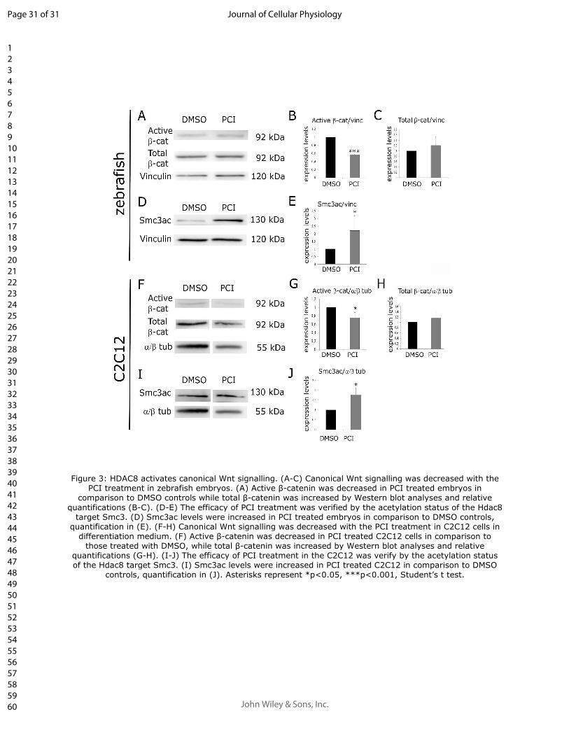
Figure 3: HDAC8 activates canonical Wnt signalling. (A-C) Canonical Wnt signalling was 5
decreased with the PCI treatment in zebrafish embryos. (A) Active β-catenin was decreased in PCI 6
treated embryos in comparison to DMSO controls while total β-catenin was increased by Western 7
blot analyses and relative quantifications (B-C). (D-E) The efficacy of PCI treatment was verified 8
by the acetylation status of the Hdac8 target Smc3. (D) Smc3ac levels were increased in PCI treated 9
embryos in comparison to DMSO controls, quantification in (E). (F-H) Canonical Wnt signalling 10
was decreased with the PCI treatment in C2C12 cells in differentiation medium. (F) Active β-11
catenin was decreased in PCI treated C2C12 cells in comparison to those treated with DMSO, while 12
total β-catenin was increased by Western blot analyses and relative quantifications (G-H). (I-J) The 13
efficacy of PCI treatment in the C2C12 was verify by the acetylation status of the Hdac8 target 14
Smc3. (I) Smc3ac levels were increased in PCI treated C2C12 in comparison to DMSO controls, 15
quantification in (J). Asterisks represent *p<0.05, ***p<0.001, Student’s t test. 16
17
Figure 4: The HDAC8-mediated positive regulation of Wnt signalling is responsible for 18
skeletal muscle differentiation. (A-C) Morphological defect presented by PCI-treated embryos 19
were rescued by LiCl addition. (D-F) Skeletal muscle differentiation was rescued when the Wnt 20
pathway was restored by LiCl in PCI treated zebrafish embryos. (D) Sarcomeric myosins, analysed 21
by Western blot techniques, decreased in PCI treated embryos and returned comparable to those 22
treated with DMSO when Wnt pathway was rescued adding LiCl (quantification in E). The efficacy 23
of LiCl treatment was verify measuring the active β-catenin by Western blot techniques 24
(quantification in F). Asterisks represent *p<0.05, Student’s t test. 25
Page 27 of 31
John Wiley & Sons, Inc.
Journal of Cellular Physiology
123456789101112131415161718192021222324252627282930313233343536373839404142434445464748495051525354555657585960
For Peer Review
27
1
2
Page 28 of 31
John Wiley & Sons, Inc.
Journal of Cellular Physiology
123456789101112131415161718192021222324252627282930313233343536373839404142434445464748495051525354555657585960

For Peer Review
Figure 1: HDAC8 is expressed in human, murine and zebrafish skeletal muscle and its expression correlates with differentiation potency. (A-C) HDAC8 protein expression in normal human skeletal muscles.
Immunofluorescence staining of HDAC8 (A), Lamin B (B) and merge of the two signals (C). The localization
of HDAC8 in human skeletal muscle is predominantly nuclear as shown by the co-localization with the Lamin B protein. (D-F) hdac8 mRNA expression in zebrafish. WISH analyses of hdac8 transcript localization in skeletal muscle of zebrafish embryos at 24 hpf (D), 36 hpf (E) and 48 hpf (F). Transverse histological sections of the previously hybridized embryos show the localization of hdac8 transcript in the myotome (D’,E’,F’). (G) Hdac8 qRT-PCR analyses on murine C2C12 myoblasts at different stages of differentiation. Hdac8 expression is increased at 7-9 days after the induction of the differentiation when differentiation is accomplished. (H) hdac8 qRT-PCR analyses on RNA from 24, 36 and 48 hpf zebrafish embryos. hdac8
expression is increased at 36 hpf when the first myogenic wave is completed. (I) HDAC8 qRT-PCR analyses on RD/12 and RD/18 rhabdomyosarcoma cells. At 11 days after the induction of differentiation, HDAC8 is more expressed in RD/18 cells that are able to fully differentiate in comparison to RD/12 cells. Scale bar represents 50 µm in (A-C) and 100 µm (D-F’). Asterisks represent **p<0.01, ***p<0.001, Student’s t test.
216x202mm (300 x 300 DPI)
Page 29 of 31
John Wiley & Sons, Inc.
Journal of Cellular Physiology
123456789101112131415161718192021222324252627282930313233343536373839404142434445464748495051525354555657585960

For Peer Review
Figure 2: Inhibition of HDAC8 activity reduces skeletal muscle differentiation in zebrafish embryos and murine C2C12 myblasts. (A-E) In-vivo treatment of zebrafish embryos with DMSO or PCI. Different
phenotypical classes with increased severity (B-D; quantification in E) with PCI treatment compared to the control embryos treated with the DMSO (A). (F-G) Immunohistochemical staining (IHC) and (H-I) western blot analyses of sarcomeric myosins. Sarcomeric myosins are reduced in PCI treated embryos at 48 hpf (G) in comparison to controls (F). Western blot analyses (H; quantification in I) confirmed all MyHC reduction in
PCI treated embryos in comparison to controls. (J-K). Inhibition of HDAC8 activity impaired C2C12 differentiation. Western blot analyses (J; quantification in K) confirmed all MyHC reduction in PCI treated
C2C12 cells in comparison to DMSO treated. Scale bars indicates 100 µm in (A, F). Asterisks represent *p<0.05, Student’s t test.
588x233mm (96 x 96 DPI)
Page 30 of 31
John Wiley & Sons, Inc.
Journal of Cellular Physiology
123456789101112131415161718192021222324252627282930313233343536373839404142434445464748495051525354555657585960
For Peer Review
Figure 3: HDAC8 activates canonical Wnt signalling. (A-C) Canonical Wnt signalling was decreased with the PCI treatment in zebrafish embryos. (A) Active β-catenin was decreased in PCI treated embryos in
comparison to DMSO controls while total β-catenin was increased by Western blot analyses and relative
quantifications (B-C). (D-E) The efficacy of PCI treatment was verified by the acetylation status of the Hdac8 target Smc3. (D) Smc3ac levels were increased in PCI treated embryos in comparison to DMSO controls,
quantification in (E). (F-H) Canonical Wnt signalling was decreased with the PCI treatment in C2C12 cells in differentiation medium. (F) Active β-catenin was decreased in PCI treated C2C12 cells in comparison to
those treated with DMSO, while total β-catenin was increased by Western blot analyses and relative quantifications (G-H). (I-J) The efficacy of PCI treatment in the C2C12 was verify by the acetylation status of the Hdac8 target Smc3. (I) Smc3ac levels were increased in PCI treated C2C12 in comparison to DMSO
controls, quantification in (J). Asterisks represent *p<0.05, ***p<0.001, Student’s t test.
Page 31 of 31
John Wiley & Sons, Inc.
Journal of Cellular Physiology
123456789101112131415161718192021222324252627282930313233343536373839404142434445464748495051525354555657585960

For Peer Review
Figure 4: The HDAC8-mediated positive regulation of Wnt signalling is responsible for skeletal muscle differentiation. (A-C) Morphological defect presented by PCI-treated embryos were rescued by LiCl addition. (D-F) Skeletal muscle differentiation was rescued when the Wnt pathway was restored by LiCl in PCI treated
zebrafish embryos. (D) Sarcomeric myosins, analysed by Western blot techniques, decreased in PCI treated embryos and returned comparable to those treated with DMSO when Wnt pathway was rescued adding LiCl (quantification in E). The efficacy of LiCl treatment was verify measuring the active β-catenin by Western
blot techniques (quantification in F). Asterisks represent *p<0.05, Student’s t test.
Page 32 of 31
John Wiley & Sons, Inc.
Journal of Cellular Physiology
123456789101112131415161718192021222324252627282930313233343536373839404142434445464748495051525354555657585960