TGS2 Estabilishing stability of an In Vitro diagnostic for ...

D³ DFA Chlamydiae Culture Confirmation Page 1 of 21
FOR IN VITRO DIAGNOSTIC USE
INTENDED USE The Diagnostic Hybrids, Inc. D³ DFA Chlamydiae Culture Confirmation Kit is intended for the qualitative identification of Chlamydiae lipopolysaccharide (LPS) in inoculated cell cultures by immunofluorescence using fluoresceinated monoclonal antibodies (MAbs). Performance has not been established with direct patient specimens.
SUMMARY AND EXPLANATION OF THE TEST The Chlamydiae are prokaryotic parasites of eukaryotic cells. These organisms contain enzymes and are able to synthesize proteins; however, they lack a mechanism to generate their own ATP (adenosine-triphosphate) and must rely upon the host cell for survival. The phylum Chlamydiae contains many species. Chlamydia trachomatis is primarily a human pathogen and is associated with endemic trachoma, inclusion conjunctivitis, lymphogranuloma venereum, nongonococcal urethritis epididymitis (male), cervicitis, salpingitis (female) and procitis in either sex. Chlamydophila psittaci is an avian Chlamydia which is transmitted to humans by infected birds resulting in pneumonia. Chlamydophila pneumoniae can cause pneumonia similar to Mycoplasma pneumoniae and it has been implicated in upper respiratory infections (i.e., cough and sore throat). Chlamydiae have a unique growth cycle exhibiting two life forms: the elementary body and the reticulocyte body. The elementary body, smaller in size, invades a susceptible cell through a phagocytic process. Once inside the cell, the elementary body surrounds itself with a protective vacuole and differentiates into an actively metabolizing reticulocyte body. The reticulocyte body divides through binary fission in the cell from approximately the 8th hour to 18 to 24 hours. During this time the Chlamydiae show their enhanced metabolic activity. At approximately 18 to 24 hours the reticulocyte bodies begin to differentiate into the smaller elementary bodies. The inclusions then consist of both reticulocyte bodies and (predominantly) elementary bodies. This entire process takes place in the vacuole. Between 48 to 72 hours, the vacuole will rupture, releasing the infectious elementary bodies and the growth cycle will continue. Cell culture isolation of Chlamydiae utilizes cell types such as McCoy, a mouse fibroblast cell line, or BGMK (Buffalo Green Monkey Kidney), an African Green Monkey cell line, in shell vial or multi-well plate formats. The cells are overlaid with a Chlamydia isolation medium containing cycloheximide to enhance Chlamydial growth prior to inoculation of the clinical specimen. The cultures are incubated for 48 to 72 hours to allow for the
For the qualitative identification of Chlamydiae lipopolysaccharide (LPS) in inoculated cell cultures by immunofluorescence using fluoresceinated monoclonal antibodies (MAbs).

D³ DFA Chlamydiae Culture Confirmation Page 2 of 21
formation of the vacuole described above. The presence of this elementary body containing vacuole can be determined by many methods, i.e. staining using iodine, Giemsa, or fluorescein-conjugated monoclonal antibodies. The use of iodine and Giemsa has been shown to be less sensitive than the use of fluorescein-conjugated monoclonal antibodies.1
PRINCIPLE OF THE PROCEDURE The D³ DFA Chlamydiae Culture Confirmation Kit uses a blend of two specific murine MAbs that are directly labeled with fluorescein isothiocyanate (FITC) for the qualitative detection of Chlamydiae in inoculated cell cultures. Cell culture isolation of Chlamydiae utilizes either McCoy cells, a mouse fibroblast cell line, or Buffalo Green Monkey Kidney (BGMK) cells in shell vial or multi-well plate formats. The cells are overlaid with a Chlamydia isolation medium containing cycloheximide to enhance Chlamydial growth prior to inoculation of the clinical specimen. The inoculated cells are centrifuged for 60 minutes and incubated at 35°C to 37°C for 48 to 72 hours. The cells are fixed in acetone and the Chlamydiae DFA Reagent is added to the cells to stain any Chlamydiae specific antigens that may be present following culture. After incubating for 15 to 30 minutes at 35°C to 37°C, the stained cells are washed with the supplied Phosphate Buffered Saline (PBS). A drop of the supplied Mounting Fluid is added to the multi-wells or to a clean microscope slide for mounting a coverslip. The cells are examined at 100 to 400X magnification using a microscope equipped with the correct filter combination for fluorescein. Chlamydiae infected cells will have bright, apple-green fluorescent inclusions while non-infected cells will fluoresce red due to the Evans Blue counter-stain.
REAGENTS AND MATERIALS PROVIDED The D³ DFA Chlamydiae Culture Confirmation Kit contains the following:
Chlamydiae DFA Reagent 5 mL One dropper bottle containing fluorescein labeled murine monoclonal antibodies directed against Chlamydiae LPS. The buffered, stabilized, aqueous solution contains Evans Blue as a counter-stain and 0.1% sodium azide as preservative
Chlamydia Antigen Control Slides 10 slides Five individually packaged control slides with wells containing cell culture-derived Chlamydia trachomatis positive cells and five individually packaged control slides with wells containing cell culture-derived negative cells. Each slide is intended to be stained only one time.
40X PBS Concentrate 25 mL One bottle containing a 40X concentrate consisting of 4% sodium azide (0.1% sodium azide after dilution to 1X using de-mineralized water) in PBS.
Mounting Fluid 7 mL One dropper bottle containing an aqueous, buffer-stabilized solution of glycerol, and 0.1% sodium azide.
MATERIALS REQUIRED BUT NOT PROVIDED Fluorescence microscope with the correct filter combination for FITC (excitation peak = 490 nm, emission
peak = 520 nm) Cell culture for Chlamydiae isolation for example; McCoy or BGMK cell lines (Both available from Quidel) Cover slips (22 x 50mm) for Antigen Control Slides and for specimen slides Universal Transport Medium (Available from Quidel) Chlamydia Isolation Medium (Eagle’s Minimum Essential Medium with 10% fetal bovine serum, 25mM
HEPES, 2.2 grams/liter sodium bicarbonate, cycloheximide, and antibiotics) (Available from Quidel)

D³ DFA Chlamydiae Culture Confirmation Page 3 of 21
Reagent grade acetone (>99% pure) chilled at 2°C to 8°C for fixation of shell vials NOTES: Keep the reagent grade acetone container tightly sealed to avoid hygroscopic absorption of water,
which may cause a hazy, nonspecific, background fluorescence. A mixture of 80% acetone/20% de-mineralized water is used for fixing cells in plastic multi-well plates.
Store at ambient temperature (20°C to 25°C). Sterile graduated pipettes: 10 mL, 5 mL, and 1 mL Sterile Pasteur pipettes or other “transfer”-type pipettes Fine-tipped forceps Wash bottle, 200 mL Bent-tip teasing needle (for removal of coverslip from a shell vial); fashion the teasing needle by bending
the tip of a syringe needle or similar object (i.e., mycology teasing needle) against a bench top or with a pair of forceps taking care to avoid injury.
Sodium hypochlorite solution, 0.5% (1:10 dilution of household bleach) Humid chamber (e.g., covered Petri dish with a damp paper towel placed in the bottom) Glass microscope slides Sterile nylon flock swab or polyester swab, non-inhibitory to Chlamydiae and tissue culture Incubator, 35°C to 37°C (CO2 or non-CO2 , depending on the cell culture format used) Centrifuge with free swinging bucket rotor De-mineralized water for dilution of PBS and for dilution of the reagent grade acetone for use in
polystyrene multi-well plates Chlamydiae stock cultures for use in monitoring the cell culture and staining procedures (Available from
Quidel) Aspirator Set-up: Vacuum aspirator with disinfectant trap containing sufficient household bleach (5%) that
the concentration is not decreased by more than 100 fold as it is diluted with discarded fluids Wash Container: Beaker, wash bottle or Coplin jar for washing slides Inverted Light Microscope: Used for examining the monolayers of cells prior to inoculation and
examination for toxicity (100X to 400X magnification capability)
WARNINGS AND PRECAUTIONS For in vitro diagnostic use only. For professional use only. Tissue culture cells may have some potential to be hazardous. Personnel working with these cultures must
be properly trained in safe handling techniques2,3,4 and have experience with tissue culture before attempting this procedure.
Follow Universal Precautions when handling the contents of this kit and patient samples. All procedures must be conducted in accordance with the CDC 5th edition Biosafety in Microbiological and
Biomedical Laboratories, 2007, and CLSI Approved Guideline M29-A, Protection of Laboratory Workers from Instrument Biohazards and Infectious Disease Transmitted by Blood, Body Fluids, and Tissue.
Sodium azide is included in the 40x PBS Concentrate at 4%, and in the other solutions in this kit at 0.1%. Aqueous solutions of sodium azide, when mixed with acids, may liberate toxic gas. Sodium azide may react with lead and copper plumbing to form highly explosive metal azides. Avoid disposal of these solutions down sanitary or industrial plumbing systems. Avoid release to the environment.
Sodium azide

D³ DFA Chlamydiae Culture Confirmation Page 4 of 21
Component Pictogram Hazard Statements Precautionary Statements
40X PBS Concentrate (Sodium azide, 4%)
Warning
H302 Harmful if swallowed.
H315 Causes skin irritation.
H320 Causes eye irritation.
H412 Harmful to aquatic life with long lasting effects.
P264 Wash hands thoroughly after handling.
P273 Avoid release to the environment. P280 Wear protective gloves and safety glasses.
P308+P313: If exposed or concerned – get medical advice/attention.
P305+P351+p338: IF IN EYES: Rinse cautiously with water for several minutes. Remove contact lenses, if present and easy to do. Continue rinsing.
P501 Avoid disposal of this material down sanitary or industrial plumbing systems.
Sodium azide is used as a preservative (0.1%) in the Chlamydiae DFA Reagent and Mounting Fluid. Sodium azide may react with lead and copper plumbing to form highly explosive metal azides. Avoid disposal of this material down sanitary or industrial plumbing systems. Aqueous solutions of sodium azide, when mixed with acids, may liberate a toxic gas. Use within a well-ventilated area.
Acetone, a reagent that is required for the test but not provided in the kit, is a flammable, volatile organic solvent. Use it in a well-ventilated area and keep away from flames and other sources of ignition.
Evans Blue counter-stain is a potential carcinogen. If eye or skin contact occurs, immediately flush with copious amounts of water.
Chlamydiae Antigen Control Slides are microscope slides onto which either cultured non-infected cells or Chlamydiae trachomatis-infected cells have been grown then fixed (killed) with acetone; a drying agent is included in the foil envelope to preserve antigen integrity; there is no residual acetone present.
Although Antigen Control Slides have been shown to be non-infectious, the same precautions taken in handling and disposing of other infectious materials should be employed in their use.
Each Chlamydiae Antigen Control Slide should be used only once. Do not re-use a Control Slide. Wear suitable protective clothing, gloves, and eye/face protection when handling the contents of this kit. The Chlamydiae DFA Reagent is supplied at working strength. Any dilution will decrease sensitivity. Do not expose Chlamydiae DFA Reagent to bright light during staining or storage. Microbial contamination of Chlamydiae DFA Reagent may cause a decrease in sensitivity. Reagents should be used prior to their expiration date. Store 1X PBS in a clean container to prevent contamination. All specimens and materials used to process them should be considered potentially infectious and handled
in a manner which prevents infection of laboratory personnel. Decontamination is most effectively accomplished using a 0.5% solution of sodium hypochlorite (1:10 dilution of household bleach).Never pipette reagents or clinical samples by mouth; avoid all contact of clinical samples with broken skin.
Avoid splashing and the generation of aerosols with clinical samples. Use aseptic technique and sterile equipment and materials for all tissue culture procedures. Reusable glassware must be washed and thoroughly rinsed free of all detergents. Use of other reagents than those specified with the components of this kit may lead to erroneous results. Testing should be performed in an area with adequate ventilation. Dispose of containers and unused contents in accordance with Federal, State and Local regulatory
requirements. Wash hands thoroughly after handling. For additional information on hazard symbols, safety, handling and disposal of the components within this
kit, please refer to the Safety Data Sheet (SDS) located at quidel.com.

D³ DFA Chlamydiae Culture Confirmation Page 5 of 21
Preparation of 1X PBS Solution After storage at 2°C to 8°C, some salts in the PBS Concentrate (40X) may have crystallized. Warm the
solution to ambient temperature (20°C to 25°C) to redissolve the crystals and mix. Add contents of the fully dissolved 25 mL PBS Concentrate to 975 mL of demineralized water. Label the 1X PBS with a 60 day expiration date after reconstitution and store at ambient temperature.
Storage
Table 1. Reagent Storage Conditions
Chlamydiae DFA Reagent Store at 2°C to 8°C in the dark.
Mounting Fluid
Chlamydia Antigen Control Slides Store at 2°C to 8°C.
40X PBS Concentrate NOTE: The Concentrate may crystallize when stored at 2°C to 8°C. The crystals will dissolve when the Concentrate is warmed to ambient temperature.
Store liquid at 2°C to 8°C prior to dilution.
1X PBS Store at ambient temperature (20°C to 25°C).
Stability Reagents and components will retain their full potency through the expiration date shown on the label of each bottle when stored at recommended temperatures. Light exposure of the Chlamydiae DFA Reagent should be kept to a minimum. Discard 1X PBS Solution if it becomes cloudy. SPECIMEN COLLECTION AND PREPARATION
Proper collection and handling of the patient specimen are the most important factors in successful Chlamydiae isolation. Specimen collection, processing, and cell culture isolation should be attempted only by laboratories trained in such procedures. Care should be taken during all specimen collection and handling to avoid generation of aerosols.
Specimen Collection5
Cervical Wipe the cervix prior to collection to remove WBC and mucous debris. Insert a sterile, large-tipped polyester swab into the endocervix, rotate and remove. Discard this swab. Insert a sterile, polyester swab into the cervical os to collect cells from the transitional zone. Rotate the
swab vigorously in firm contact with cervix surface to facilitate the collection of columnar epithelial cells. Withdraw swab without contacting vaginal surfaces.
NOTE: Recovery rates from females can be improved if the urethra is also sampled.
Urethral Insert a sterile, fine-tipped polyester swab 2 to 4 cm into the male urethra or 1 cm into the female urethra
and hold in place for 5 seconds. Rotate the swab several times to obtain columnar epithelial cells and withdraw.
NOTE: Patient should not have urinated within one hour of collection.

D³ DFA Chlamydiae Culture Confirmation Page 6 of 21
Eye Gently swab the lower conjunctiva with a sterile, fine-tipped polyester swab, collecting patient mucous
membrane cells.
Nasopharynx and Throat Gently insert a sterile nasopharyngeal fine-wire polyester swab into one or both anterior nares to the
posterior pharynx; rotate to collect mucous membrane cells and withdraw. Swab the posterior pharynx vigorously with a large-tipped, sterile polyester swab.
NOTE: Nasal aspirates collected by intubation are a superior source of the agent in infants with pneumonia.
Rectal Mucosa To collect cells from the mucosal surface, insert a sterile polyester swab 1-cm past the anal sphincter,
rotate in firm contact with the mucosal surface and withdraw. Several factors of specimen collection may affect the successful isolation of Chlamydiae. When swabs are used for specimen collection, cotton or polyester swabs should be used, as calcium alginate swabs have been shown to inhibit replication.4 Immerse swab immediately in appropriate transport medium. This will serve to stabilize Chlamydiae, if present, and will inhibit undesirable bacterial and fungal overgrowth. Place swab in transport tube, break off shaft, and tightly secure cap.
Specimen Transport and Storage All potentially infectious agents should be transported according to International Air Transport Association (IATA), International Civil Aviation Organization, (ICAO), Titles 42 and 49 of the U.S. Code of Federal Regulations, or other regulatory requirements, as may be applicable. Specimens should be transported on wet ice to the laboratory and processed and tested as soon as possible and then stored at 2°C to 8°C. Specimens should be stored at 2°C to 8°C for no longer than 48 hours before being tested. If longer storage is required, the specimens should be frozen at –70oC or lower.4 Freezing and thawing of specimens should be avoided since this will result in a loss of viability of Chlamydiae, leading to decreased sensitivity of the test.
PROCEDURE Preliminary Comments and Precautions Adhere to the recommended volumes and times in the following procedure to ensure that accurate results
are obtained. For specimen swabs received in transport medium with glass beads, vortex vigorously for about 15
seconds to dissociate adhered cells. For swabs not received in transport medium, transfer them to a tube of transfer medium containing glass beads and vortex vigorously for about 15 seconds to dissociate adhered cells.
When staining with fluorescent reagents and examining cells microscopically for fluorescence, it is very important to include controls, both positive and negative, to monitor the procedure and performance of the reagents. It is recommended that such controls be run with each batch of patient specimens.
The closed, humidified container for holding the slides during incubation should be kept in the incubator so it is at incubator temperature when the slides are placed in it. By doing this, the cells and antibody solution will come up to temperature rapidly, yielding the expected stain intensity.

D³ DFA Chlamydiae Culture Confirmation Page 7 of 21
It is recommended that the MAb reagents be brought to ambient temperature prior to use and immediately returned to 2°C to 8°C after use.
Regarding Cell Culture Testing Good Laboratory Practice dictates that positive and negative Chlamydia controls be run with each new
batch of cells to confirm their performance in culturing Chlamydiae. It is good practice to retain the medium removed from the positive monolayers until after staining results
have been obtained. If there is any question concerning the specimen results, the medium can be passed to another monolayer and incubated for the appropriate time period for repeat testing.
If using cell cultures in polystyrene, multi-well plates, the acetone fixative must be diluted with water to 80% by adding 20 mL of water to 80 mL of acetone.
Do not allow the monolayers to dry before fixing; this can lead to high background staining and decreased sensitivity.
Do not allow the antibody reagents to dry on the monolayers; this can lead to high background.
Regarding Immunofluorescence Microscopy It is good practice to examine the positive and negative controls before examining the test specimens. If
one of these fails to perform as expected, review the steps and conditions under which the test was performed to determine the cause(s). Do not report results until controls perform as expected.
There are three aspects of the fluorescence microscope that must be functioning properly and optimally in order to achieve maximum brightness of fluorescence: The activation light source has a finite life and as it ages, its output decreases, resulting in lower
fluorescence intensity from the DFAs. The light source is focused by a number of lenses and mirror(s). For maximum intensity, these must be
properly aligned. The filters used in the light path must be appropriate for the particular fluor, in this case, fluorescein.
There are several fluorescent artifacts that may be observed in the cell monolayers being examined: Cell debris, lint, etc. can nonspecifically adsorb DFAs, resulting in highly intense fluorescence. These can
be identified by their morphology, i.e., they don’t have the appearance of a complete cell and typically do not appear to be a part of the monolayer like the other cells.
A low grade, yellow-green fluorescence may sometimes be seen, particularly in areas that have piled cells or are near holes in the cell monolayer. In both cases, the diffusion of the entrapped DFAs is retarded during the wash step, resulting in the nonspecific fluorescence.
Intense fluorescence around the periphery of slide wells is indicative of drying of the DFA Reagent during incubation, suggesting that it was incubated too long or the humidity was not controlled.
Inadequate washing can lead to general, low grade fluorescence due to residual DFAs remaining on the monolayer of cells.
Bleaching or fading of the fluorescence of the stained cells may occur on exposure to light, particularly light of high intensity. This bleaching can occur when the microscope slide is viewed in a field for an extended period of time. Slides should be protected as much as possible during the assay.
Specimen Preparation Swabs containing specimen material should be handled with sterile forceps. The swab should be rotated in
transport medium and then pressed against the inside of the tube to allow excess fluid to drain back into the transport medium. Discard the swab into an appropriate disinfectant such as 0.5% sodium hypochlorite solution (1:10 dilution of household bleach). Decontaminate the forceps in between specimen disposal.
Disrupt cellular material in the transport medium by vortexing with sterile glass beads for 30 to 60 seconds, sonication at 10kc/sec for 30 to 60 seconds or by other methods determined by the laboratory to

D³ DFA Chlamydiae Culture Confirmation Page 8 of 21
be effective in disrupting cellular debris. This will enhance the release of cell-associated Chlamydiae into the medium.
To remove bacterial, fungal, and cellular debris, centrifuge the transport medium at 700xg for 10 minutes. Supernatant is then used as the inoculum. Heavily contaminated specimens, noted by a cloudy yellow coloration, may be further clarified by filtration through a sterile 0.45 micron membrane filter. The filtrate is then used as the inoculum. Since such procedures may reduce the number of Chlamydiae in a specimen, each laboratory should establish the efficacy of its specimen preparation procedure.
Cell Culture Testing – Shell Vial The laboratories that conducted studies for clearance of this assay (presented in section SPECIFIC
PERFORMANCE CHARACTERISTICS) inoculated duplicate shell vials and incubated for either 48 or 72 hours. Determine the number of vials needed based on the number of specimens and controls to be used. Examine the monolayers for proper morphology prior to inoculation. Aspirate maintenance medium from the monolayers and add 1 mL of Chlamydia Isolation Medium to each
shell vial. Add 0.2 to 0.4 mL of prepared specimen to each shell vial. Centrifuge the shell vials at 700xg for 1-hour at 20°C to 25°C. Place stoppered shell vials in an incubator at 35°C to 37°C for 48 to 72 hours. Remove the medium and add 1 mL of 1X PBS. Swirl to mix and then aspirate. Repeat this wash with another 1 mL of 1X PBS and then aspirate. Add 1 mL of chilled 100% acetone and allow it to stand for 5 to 10 minutes at 20°C to 25°C. Remove the fixative by aspiration. Add 0.5 mL of 1X PBS to wet the monolayer. Swirl and then aspirate completely. Add 4 drops of the Chlamydiae DFA Reagent to the fixed monolayers of patient and control samples, and
rock to ensure complete coverage of the monolayer by the Reagent. Place stoppered shell vials in a 35°C to 37°C incubator for 15 to 30 minutes. Aspirate the Chlamydiae DFA Reagent from the monolayers. Add 1 mL of the 1X PBS. Remove the 1X PBS by aspiration, repeat the wash step and again remove by aspiration. Add 1 mL of de-mineralized water. Remove the de-mineralized water by aspiration. Lift the coverslip from the bottom of the shell vial using a bent-tip needle on a syringe barrel, and, grasping
it with the fine tipped forceps, transfer it, monolayer-side down, to a small drop of Mounting Fluid on a standard microscope slide.
Examine the stained monolayers using a fluorescence microscope with magnifications between 100X and 400X. (See section Regarding Immunofluorescence Microscopy)
Refer to section INTERPRETATION OF RESULTS.
Cell Culture Testing – Multi-well Plate The laboratories that conducted studies for clearance of this assay (presented in the section SPECIFIC
PERFORMANCE CHARACTERISTICS) inoculated duplicate wells and incubated for either 48 or 72 hours. Determine the number of wells needed based on the number of specimens and controls to be used. Examine the monolayers for proper morphology prior to inoculation. Aspirate maintenance medium from the monolayers and add 1 mL of Chlamydia Isolation Medium to each
24-well multi-well plate monolayer; add 0.8 mL to each 48-well plate monolayer. Add 0.2 to 0.4 mL of prepared specimen to the appropriate well of a multi-well plate. Centrifuge the multi-well plates at 700xg for 1 hour at 20°C to 25°C.

D³ DFA Chlamydiae Culture Confirmation Page 9 of 21
Place the covered multi-well plates in a 35°C to 37°C incubator with a humidified, 5% CO2 atmosphere for 48 to 72 hours.
Remove the medium and add 1 mL of 1X PBS. Swirl to mix and then aspirate. Repeat this wash with another 1 mL of 1X PBS and then aspirate. Add 1 mL of 80% aqueous acetone and let stand 5 to 10 minutes at 20°C to 25°C.
NOTE: Do not allow the 80% acetone fixative to remain in the polystyrene wells longer than 10 minutes since it may craze and cloud the plastic, making it difficult to examine the monolayers.
Remove the fixative by aspiration. Add 0.5 mL of the 1X PBS to wet the monolayer. Swirl and then aspirate completely. Add 4 drops of the Chlamydiae DFA Reagent to the fixed monolayers of patient and control samples in
each 24-well multi-well plate; add 3 drops of the Chlamydiae DFA Reagent to the fixed monolayers of patient and control samples in each 48-well plate. Rock to ensure complete coverage of the monolayer by the Reagent.
Place the covered multi-well plate in a 35°C to 37°C, humidified incubator for 15 to 30 minutes. Aspirate the Chlamydiae DFA Reagent from the monolayers. Add 1 mL of the 1X PBS. Remove the 1X PBS by aspiration, repeat the wash step and again remove by aspiration. Add 1 mL of de-mineralized water. Remove the de-mineralized water by aspiration. Add 2 to 3 drops of Mounting Fluid to each monolayer, cover the plate. Examine the stained monolayers using a fluorescence microscope with magnifications between 100X and
400X. (See Regarding Immunofluorescence Microscopy) Refer to INTERPRETATION OF RESULTS.
Quality Control
Cell Culture To ensure sensitivity to Chlamydiae, a Chlamydia-inoculated control monolayer should be included each time a new lot of cell culture is used. Also, a non-inoculated monolayer from each lot should be kept and re-fed every 3 to 7 days and observed for normal cell growth; it may be used as a negative cell control when examining for CPE. Adverse storage conditions or handling procedures will also be reflected in the negative control. If control cultures fail to perform correctly, results are considered invalid.
Reagents A fresh set of Chlamydia Antigen Control Slides (1-positive and 1-negative slide) should be stained each time the staining procedure is performed to ensure proper test performance. The positive wells will show multiple infected cells with bright, apple-green fluorescent inclusions. Negative cells will fluoresce a dull red due to the included Evans Blue counter-stain. The negative well will contain only dull red fluorescing negative cells. Positive and negative controls must demonstrate appropriate fluorescence for specimen results to have validity. Antigen Control Slides may also aid in the interpretation of patient specimens.

D³ DFA Chlamydiae Culture Confirmation Page 10 of 21
INTERPRETATION OF RESULTS It is recommended that controls be examined first to ensure proper test performance before examination of the specimens. A positive reaction is one in which bright apple-green fluorescent inclusions are observed in the infected cells. Negative cells will fluoresce a dull red due to the Evans Blue counter-stain included in the Chlamydiae DFA Reagent. Technologists should not confuse cell clumps which may fluoresce due to entrapment of antibody with Chlamydiae-specific staining. Occasionally, dead, rounded cells due to specimen toxicity or improper cell storage may nonspecifically stain a dull olive green due to trapped antibody. Adequate washing between steps will help to eliminate this type of nonspecific staining. The entire monolayer of cells must be examined for fluorescent inclusions within the cells. If no fluorescent inclusions are found, the results of testing of the specimen should be reported as, “No Chlamydiae isolated by cell culture.” If fluorescent inclusions within the cells are found, it should be reported as “Chlamydiae isolated by cell culture.”
LIMITATIONS OF THE PROCEDURE Inappropriate specimen collection, storage, and transport may lead to false negative culture results.4 Incubation times or temperatures other than those cited in the test instructions may give erroneous
results. Isolation of Chlamydiae will vary greatly depending upon the specimen quality and subsequent handling. A
negative result does not exclude the possibility of Chlamydiae infection. Results of the test should be interpreted in conjunction with information available from epidemiological studies, clinical evaluation of the patient and other diagnostic procedures.
The monoclonal antibodies used in this kit are from hybridomas created using Chlamydophila pneumoniae infected cells as the immunogen. The specific antigen detected by the antibodies is the lipopolysaccharide molecule.
The monoclonal antibodies used in this kit are not species-specific and therefore cannot be used to differentiate among the different species of Chlamydiae.
A negative result in a cultured specimen does not rule out the presence of Chlamydiae infection. Performance of the kit can only be assured when components used in the assay are those supplied by
Quidel. Prolonged storage of the Chlamydiae DFA Reagent under bright light will decrease the staining intensity. Light background staining may occur with specimens contaminated with Staphylococcus aureus strains
containing large amounts of protein A. Protein A will bind the Fc portions of conjugated antibodies. Such binding can be distinguished from Chlamydial antigen binding on the basis of morphology, i.e., S. aureus-bound fluorescence appears as small (~1 micron diameter), bright dots. Results from cell cultures with bacterial contamination must, therefore, be interpreted with caution.
EXPECTED VALUES The clinical studies described in Section Specific Performance Characteristics were comprised of specimens collected and cultured for the presence of Chlamydiae. The specimen sources are described in Table 2 below.

D³ DFA Chlamydiae Culture Confirmation Page 11 of 21
Table 2. Specimen Sources
Site
Tota
l
Un
kno
wn
or
No
t In
dic
ate
d
Gen
ital
Pe
nis
Vag
inal
Cer
vica
l
Re
ctal
Pe
rin
eum
*
Eye
Ure
thra
l
Mo
uth
**
Re
spir
ato
ry‡
Intr
a ab
do
min
al
asp
irat
e
1 396 9 155 5 35 177 2 5 6 1 1
2 200 13 55 83 34 6 3 2 4
3 278 13 146 32 5 2 64 7 2 6 1
4 120 120 *perineum: groin, pubic, perianal, pelvic, endocervix **mouth: mouth, throat ‡respiratory: nasopharyngeal, nasal aspirate, tracheal aspirate, sputum
Both “fresh” (tested within 48 hours of receipt) or frozen (stored at –70oC or colder) specimens were used in the studies. The numbers of fresh and frozen specimens tested are summarized below (Table 3).
Table 3. Numbers of Specimens per Study Site
Sites Fresh
prospective Frozen
prospective Archived (frozen)
Site Total
1 156 240 0 396
2 90 84 26 200
3 23 68 187 278
4 0 120 0 120
SPECIFIC PERFORMANCE CHARACTERISTICS This study included nine hundred and ninety four (994) original specimens evaluated by D³ DFA Chlamydiae Culture Confirmation Kit and three currently marketed Culture Confirmation Kits (“Comparison” tests) for the presence of Chlamydiae. These evaluations were conducted at three external laboratory sites and one in-house laboratory: (1) A reference laboratory in the southeastern United States; (2) A hospital laboratory in the mid-west United States; (3) A hospital laboratory in the southwestern United States; and (4) Quidel’s in-house virology laboratory. The patient demographics are listed below (Table 4).

D³ DFA Chlamydiae Culture Confirmation Page 12 of 21
Table 4. Specimen Demographics
Values are # pos/Total Site 1 Site 2 Site 3 Site 4
Age F M F M F M F M
Totals 362 32 170 30 215 63 98 22
<1m 0/1 0/3 0/2 2/4 9/15 14/19 0 0
1m to 2y 0 0 0/4 0/2 4/28 11/22 0 0
2y to12y 0/2 0 1/28 1/13 6/13 0 0 0
12y to18y 0/17 0 31/117 5/10 79/143 14/20 0/7 0
18y to 21y 6/56 1/4 4/19 1/1 4/13 0/2 9/30 3/7
>21y 9/283 3/25 0 0 0/3 0 8/61 4/15
Age not reported 1/3 0 0 0 0 0 0 0
Age/gender not reported 0/2 0 0 0
Performance Characteristics For Each Study Site Percent Agreement between the D³ DFA Chlamydiae Culture Confirmation Kit and Comparison test was calculated for results from Sites 1, 2, and 3 using prospectively collected specimens, and is reported below (Table 5).
Table 5. Combined Percent Agreement Using Prospectively Collected Specimens
Comparison Test
+
D³ DFA Chlamydiae Culture Confirmation Kit
+ 42 4
2 613
Positive Percent Agreement* (PPA): 95.5% 95% CI**- PPA: 84.5% to 99.4%
Negative Percent Agreement***(NPA): 99.4% 95% CI – NPA: 98.3% to 99.8%
*“Positive Percent Agreement”, or “PPA”, values were calculated according to {[Total Number of Positive Results in Agreement by both Subject and Predicate Tests) divided by [(Total Number of Positive Results in Agreement by both Subject and Predicate Tests) plus (Number of Results Positive by Predicate but Negative by Subject)]} multiplied by 100%. **“95% CI” refers to 95% Confidence Intervals, which were calculated according to Exact method (Clopper, C. and S. Pearson, Biometrika 26:404-413, 1934). ***“Negative Percent Agreement”, or “NPA”, values were calculated according to {[Total Number of Negative Results in Agreement by both Subject and Predicate Tests) divided by [(Total Number of Negative Results in Agreement by both Subject and Predicate Tests) plus (Number of Results Negative by Predicate but Positive by Subject)]} multiplied by 100%.
Study Site 1 A total of 396 specimens were tested for the presence of Chlamydiae (156 fresh and 240 frozen) using D³ DFA Chlamydiae Culture Confirmation Kit and a comparison test. Briefly, 200 µL from the specimens were inoculated onto two 48/24-fill McCoy cluster plates. The inoculated cells were centrifuged at 700xg for 60
minutes, incubated at 35 to 37C for either 48 or 72 hours, then stained in accordance with the respective product insert procedures. The results of this testing site are summarized below (Table 6).

D³ DFA Chlamydiae Culture Confirmation Page 13 of 21
Table 6. Study Site 1 – D³ DFA Chlamydiae Culture Confirmation Kit and Comparison Device Results in Multi-Well Plate Culture
Fresh Frozen Comparison Test Comparison Test
+ +
D³ DFA Chlamydiae Culture Confirmation Kit
+ 0 0 + 18 3
0 156 1 218
Fresh Frozen
PPA: [>1 result required for calculation] 94.7% 95% CI – PPA: N/A 74.0% to 99.9%
NPA: 100% 98.6% 95% CI – NPA: 97.7% to 100% 96.1% to 99.7%
Study Site 2 A total of 200 specimens were tested for the presence of Chlamydiae using the same comparison test as Study Site 1 [90 fresh and 84 frozen (prospectively collected) and an additional 26 archival specimens.] Briefly, 200 µL from the specimens were inoculated into two McCoy shell vials. The inoculated cells were centrifuged at 700xg
for 60 minutes, incubated at 35 to 37C for either 48 or 72 hours, then stained in accordance with the respective product insert procedures. The results of this testing site are summarized below (Table 7).
Table 7. Study Site 2 – D³ DFA Chlamydiae Culture Confirmation Kit and Comparison Device Results in Shell-vial Culture
Fresh Frozen Comparison Test Comparison Test
+ +
D³ DFA Chlamydiae Culture Confirmation Kit
+ 11 0 + 8 1
0 79 1 74
Fresh Frozen PPA: 100% 88.9%
95% CI – PPA: 71.5% to 100% 51.8% to 99.7% NPA: 100% 98.7%
95% CI – NPA: 95.4% to 100% 92.8% to 100%
D³ DFA Chlamydiae Culture Confirmation Page 14 of 21
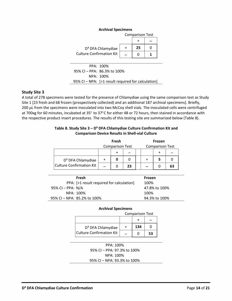
Archival Specimens
Comparison Test
+
D³ DFA Chlamydiae Culture Confirmation Kit
+ 25 0
0 1
PPA: 100% 95% CI – PPA: 86.3% to 100%
NPA: 100% 95% CI – NPA: [>1 result required for calculation]
Study Site 3 A total of 278 specimens were tested for the presence of Chlamydiae using the same comparison test as Study Site 1 [23 fresh and 68 frozen (prospectively collected) and an additional 187 archival specimens]. Briefly, 200 µL from the specimens were inoculated into two McCoy shell vials. The inoculated cells were centrifuged
at 700xg for 60 minutes, incubated at 35 to 37C for either 48 or 72 hours, then stained in accordance with the respective product insert procedures. The results of this testing site are summarized below (Table 8).
Table 8. Study Site 3 – D³ DFA Chlamydiae Culture Confirmation Kit and Comparison Device Results in Shell-vial Culture
Fresh Frozen Comparison Test Comparison Test
+ +
D³ DFA Chlamydiae Culture Confirmation Kit
+ 0 0 + 5 0
0 23 0 63
Fresh Frozen PPA: [>1 result required for calculation] 100%
95% CI – PPA: N/A 47.8% to 100% NPA: 100% 100%
95% CI – NPA: 85.2% to 100% 94.3% to 100%
Archival Specimens
Comparison Test
+
D³ DFA Chlamydiae Culture Confirmation Kit
+ 134 0
0 53
PPA: 100% 95% CI – PPA: 97.3% to 100%
NPA: 100% 95% CI – NPA: 93.3% to 100%

D³ DFA Chlamydiae Culture Confirmation Page 15 of 21
Study Site 4a A total of 120 frozen specimens were tested for the presence of Chlamydiae using a second comparison test. Briefly, 200 µL from the specimens were inoculated onto two 48/24-fill McCoy multi-well plates. The
inoculated cells were centrifuged at 700xg for 60 minutes, incubated at 35 to 37C for either 48 or 72 hours, then stained in accordance with the respective product insert procedures. The results of this testing site are summarized below (Table 9).
Table 9. Study Site 4a – D³ DFA Chlamydiae Culture Confirmation Kit and Comparison Device Results in Multi-well Plate Culture
Frozen Comparison Test
+
D³ DFA Chlamydiae Culture Confirmation Kit
+ 24 0
0 96
PPA: 100% 95% CI – PPA: 85.8% to 100%
NPA: 100% 95% CI – NPA: 96.2% to 100%
Study Site 4b The same 120 frozen specimens from above were tested for the presence of Chlamydiae using a third comparison test. Briefly, 200 µL from the specimens were inoculated onto two 48/24-fill McCoy multi-well
plates. The inoculated cells were centrifuged at 700xg for 60-minutes, incubated at 35 to 37C for either 48 or 72 hours, then stained in accordance with the respective product insert procedures. The results of this testing site are summarized below (Table 10).
Table 10. Study Site 4b – D³ DFA Chlamydiae Culture Confirmation Kit and Comparison Device Results in Multi-well Plate Culture
Frozen Comparison Test
+
D³ DFA Chlamydiae Culture Confirmation Kit
+ 23 1
0 96
PPA: 100% 95% CI – PPA: 85.2% to 100%
NPA: 99% 95% CI – NPA: 94.4% to 100%
Specificity Testing The D³ DFA Chlamydiae Culture Confirmation Kit was characterized for its ability to specifically detect Chlamydiae species. The FITC-conjugated MAbs used in the kit exhibited characteristic staining patterns on Chlamydiae infected cells, with fluorescent inclusions in infected cells. The conjugated MAbs reacted with Chlamydophila pneumoniae, Chlamydophila psittaci, as well as 15 serovars of Chlamydia trachomatis.

D³ DFA Chlamydiae Culture Confirmation Page 16 of 21
The D³ DFA Chlamydiae Culture Confirmation Kit was tested for cross-reactivity against a wide variety of cells and microorganisms. No cross-reactivity was observed for 20 host culture cell types or for 57 virus strains (cultured and processed for staining). Twenty-five (25) bacterial cultures, as well as one protozoan and one yeast specimen, were stained and examined for cross-reactivity. The protein-A produced by Staphylococcus aureus bound the Fc portion of the MAb’s and appeared as small points of fluorescence (see Limitations of Procedure) while all other bacterial cultures were negative. To test this product against Chlamydiae species, cell cultures (McCoy, BGMK, or HEp-2) were inoculated with approximately 1.5 x 10⁶ infective units of Chlamydia trachomatis, Chlamydophila psittaci, or Chlamydophila pneumoniae, and incubated for 2 days to yield a 3+ to 4+ infection. Cultures were processed and stained with the Chlamydiae DFA Reagent or specially prepared DFA reagents containing only one of the Chlamydia MAbs. Stringent conditions for cross-reactivity testing were achieved by using a high concentration Chlamydiae DFA Reagent and high titers of microorganisms. The DFA was prepared at 1.5X the concentration that is provided in the kit. Depending on the particular bacteria, the number of CFU tested ranged from 6.4x10⁴ to 2.9x10⁷. Depending on the virus, 150 to 2100 TCID50 viruses were inoculated into shell vial culture and incubated for 24 to 48 hours, to yield a 1+ to 3+ infection, processed and stained with the 1.5X DFA according to the procedure detailed in the product insert. Additionally, for some viruses and other microorganisms, commercial slides containing the particular agent were used to test for cross reactivity. Cell cultures were prepared in shell vial format. Confluent monolayers were stained with the 1.5X DFA Reagent according to the procedure as detailed in this product insert then examined for cross reactivity. Organisms and cell lines which were tested against the Chlamydiae DFA Reagent are listed below (Table 11).
Table 11. Organisms Tested for Reactivity with D³ DFA Chlamydiae Culture Confirmation Kit
MICROORGANISMS (BACTERIA, YEAST, PROTOZOA) Result (Reactive = +; Negative = – )
Chlamydiae Serovar Inoculum
(Infective units per culture)
Result (Reactive = +) (Negative = – )
Chlamydia trachomatis
A 1.5 x 10⁶ +
B 1.5 x 10⁶ +
C 1.5 x 10⁶ +
D 1.5 x 10⁶ +
E 1.5 x 10⁶ +
F 1.5 x 10⁶ +
G 1.5 x 10⁶ +
H 1.5 x 10⁶ +
I 1.5 x 10⁶ +
J 1.5 x 10⁶ +
K 1.5 x 10⁶ +
L1 1.5 x 10⁶ +
L2 1.5 x 10⁶ +
L3 1.5 x 10⁶ +
Ba 1.5 x 10⁶ +

D³ DFA Chlamydiae Culture Confirmation Page 17 of 21
MICROORGANISMS (BACTERIA, YEAST, PROTOZOA) Result (Reactive = +; Negative = – )
Chlamydiae (cont.) Inoculum Result
Chlamydophila pneumoniae 1.5 x 10⁶ +
Chlamydophila psittaci 1.5 x 10⁶ +
Other Bacteria CFU Tested Result
Acholeplasma laidlawi ~1.0 x 10⁷ Acinetobacter calcoaceticus 9.7 x 10⁵ Bordetella bronchiseptica 1.8 x 10⁵ Bordetella pertussis 4.7 x 10⁶ Corynebacterium diphtheriae 2.5 x 10⁶ Escherichia coli 2.6 x 10⁵ Gardnerella vaginalis 5.0 x 10⁵ Haemophilis influenzae type A 9.3 x 10⁵ Klebsiella pneumoniae 6.4 x 10⁶ Legionella pneumophila 6.5 x 10⁴ Moraxella cartarrhalis 6.4 x 10⁴ Mycoplasma hominis ~1.0 x 10⁴ Mycoplasma orale ~1.0 x 10⁴ Mycoplasma pneumoniae ~1.0 x 10⁴ Mycoplasma salivarium ~1.0 x 10⁷ Neisseria gonorrhoeae 1.3 x 10⁶ Proteus mirabilis 2.1 x 10⁶ Pseudomonas aeruginosa 1.0 x 10³ Salmonella enteriditis 2.5 x 10⁶
Salmonella typhimurium 1.8 x 10⁶ Staphylococcus aureus 1.0 x 10⁷ +
Streptococcus agalactiae 9.6 x 10⁶ Streptococcus pneumoniae 8.0 x 10⁵ Streptococcus pyogenes 2.9 x 10⁷ Ureaplasma urealyticum ~1.0 x 10⁴ Yeast CFU Tested Result
Candida glabrata 8.7 x 10⁶
Protozoan CFU Tested Result
Trichomonas vaginalis Commercially available
control slide*
*Test material is from commercially available prepared slides. Each positive well contains 10 to 50% reactive cells.
Virus Strains Inoculum (TCID50) Result
Adenovirus
Type 1 725 Type 3 725 Type 6 725 Type 7 725 Type 8 725
Type 10 725 Type 13 725 Type 14 725 Type 18 725

D³ DFA Chlamydiae Culture Confirmation Page 18 of 21
MICROORGANISMS (BACTERIA, YEAST, PROTOZOA) Result (Reactive = +; Negative = – )
Virus Strains (cont.) Inoculum (TCID50) Result
Adenovirus
Type 31 725 Type 40 725 Type 41 725
Influenza A
Aichi 2.1 x 10³
Malaya 2.1 x 10³ Hong Kong 2.1 x 10³
Denver 2.1 x 10³ Port Chalmers 2.1 x 10³
PR 2.1 x 10³ Victoria 2.1 x 10³
Influenza B
Hong Kong 2.1 x 10³ Maryland 2.1 x 10³
Mass 2.1 x 10³ Taiwan 2.1 x 10³
GL 2.1 x 10³ Russia 2.1 x 10³
RSV
Long 2.1 x 10³ Wash 2.1 x 10³ 9320 2.1 x 10³
Parainfluenza 1 C-35 Commercially available
control slide*
Parainfluenza 2 Greer Commercially available
control slide*
Parainfluenza 3 C 243 Commercially available
control slide*
HSV-1 1F 150
MacIntyre 150
HSV-2 MS 150
Strain G 150
CMV
Towne 700 AD169 700 Davis 700
VZV Ellen 500
Echovirus 4, 6, 9, 11, 30,
34 Commercially available
control slide*
Coxsackievirus B1, B2, B3, B4,
B5, B6 Commercially available
control slide*
Mumps Commercially available
control slide*
Measles (Rubeola)
Commercially available
control slide*
Poliovirus Types 1, 2, 3 Commercially available
control slide*
Epstein-Barr Commercially available
control slide*

D³ DFA Chlamydiae Culture Confirmation Page 19 of 21
MICROORGANISMS (BACTERIA, YEAST, PROTOZOA) Result (Reactive = +; Negative = – )
*Test material is from commercially available prepared slides. Each positive well contains 10 to 50% reactive cells.
HOST CELL CULTURE TYPES (shell vial monolayers were stained)
Cell Type Result Cell Type Result
A549 NCI-H292 BGMK pCMK HEp-2 pRhMK
HFF (Hs27) RD LLC-MK2 RhMK II McCoy RK (passage 1) MDCK R-Mix MRC-5 Vero MRHF Vero 76 Mv1Lu WI-38
WARRANTY STATEMENT These products are warranted to perform as described in their labeling and the Quidel literature when used in accordance with their instructions. THERE ARE NO WARRANTIES WHICH EXTEND BEYOND THIS EXPRESS WARRANTY AND QUIDEL DISCLAIMS ANY IMPLIED WARRANTY OF MERCHANTABILITY OR WARRANTY OF FITNESS FOR PARTICULAR PURPOSE. Quidel’s sole obligation and purchaser’s exclusive remedy for breach of this warranty shall be at the option of Quidel to repair or replace the products.
ASSISTANCE To place an order or for technical support, please contact a Quidel Representative at 800.874.1517 (in the U.S.) or 858.552.1100 (outside the U.S.), Monday through Friday, from 8:00 a.m. to 5:00 p.m., Eastern Time. Orders may also be placed by fax at (740) 592-9820. For e-mail support contact [email protected] or [email protected]. For services outside the U.S., please contact your local distributor. Additional information about Quidel, our products, and our distributors can be found on our website quidel.com. D³ is a registered trademark of Diagnostic Hybrids, Inc. in the United States and other countries.
REFERENCES 1. Eisenberg, Henry D. (1992). Clinical Microbiology Procedures Handbook, published by American Society for
Microbiology, Washington DC, section 10.6.9 2. Biosafety in Microbiological and Biomedical Laboratories (BMBL), 5th edition, 2007, CDC-NIH manual.
[http://www.cdc.gov/od/ohs/biosfty/bmbl5/bmbl5toc.htm] 3. Biosafety Manual, 3rd edition, 2004. World Health Organization [Manual may be available in additional
languages; refer to WHO web page.] [http://www.who.int/csr/resources/publications/biosafety/WHO_CDS_CSR_LYO_2004_11/en/]
4. Laboratory Biosafety Guidelines, 3rd edition, 2004. Published by authority of the Minister of Health, Population and Public Health Branch, Centre for Emergency Preparedness and Response [Guideline is available in French or English; refer to web page] [http://www.phac-aspc.gc.ca/publicat/lbg-ldmbl-04/index.htmL]
5. Eisenberg, Henry D. (1992). Clinical Microbiology Procedures Handbook, published by American Society for Microbiology, Washington DC, section 8.2.3

D³ DFA Chlamydiae Culture Confirmation Page 20 of 21
01-040000 – D³ DFA Chlamydiae Culture Confirmation Kit
MDSS GmbH Schiffgraben 41 30175 Hannover, Germany Diagnostic Hybrids, Inc. – a subsidiary of Quidel Corporation 2005 East State Street, Suite 100 Athens, OH 45701 USA quidel.com PI222000EN00 (05/16)

D³ DFA Chlamydiae Culture Confirmation Page 21 of 21