
Follicular Unit Extraction: Minimally Invasive Surgery for ... · Follicular Unit Extraction:...
9
© 2002 by the American Society for Dermatologic Surgery, Inc. • Published by Blackwell Publishing, Inc. ISSN: 1076-0512/02/$15.00/0 • Dermatol Surg 2002;28:720–728 Follicular Unit Extraction: Minimally Invasive Surgery for Hair Transplantation William R. Rassman, MD*, Robert M. Bernstein, MD* † , Robert McClellan, MD*, Roy Jones, MD,* Eugene Worton, MD* and Hendrik Uyttendaele, MD, PhD † *New Hair Institute Medical Group, A Professional Corporation, Los Angeles, California, and † Department of Dermatology, Columbia University, College of Physicians and Surgeons, New York, New York background. Follicular Unit Transplantation (FUT) is per- formed using large numbers of naturally occuring individual follicular units obtained by single-strip harvesting and stereo- microscopic dissection. Donor wound scarring from strip exci- sion, although an infrequent complication, still concerns enough patients that an alternative solution is warranted. objective. The purpose of this paper is to introduce Follicular Unit Extraction (The FOX Procedure), in which individual folli- cular units are removed directly from the donor region through very small punch excisions, and to describe a test (The FOX Test) that determines which patients are candidates for this pro- cedure. This paper explores the nuances, limitations, and practi- cal aspects of Follicular Unit Extraction (FUE). methods. FUE was performed using 1-mm punches to separate follicular units from the surrounding tissue down to the level of the mid dermis. This was followed by extraction of the follicular units with forceps. The FOX test was developed to determine which patients would be good candidates for the procedure. The test was performed on 200 patients. Representative patients who were FOX-positive and FOX-negative were studied histologically. results. The FOX Test can determine which patients are suit- able candidates for FUE. Approximately 25% of the patients biopsied were ideal candidates for FUE and 35% of the patients biopsied were good candidates for extraction. conclusion. FUE is a minimally invasive approach to hair transplantation that obviates the need for a linear donor inci- sion. This technique can serve as an important alternative to traditional hair transplantation in certain patients. W. R. RASSMAN, MD, R. M. BERNSTEIN, MD, R. MCCLELLAN, MD, R. JONES, MD, E. WORTON, MD AND H. UYTTENDAELE, MD, PHD HAVE INDICATED NO SIGNIFICANT INTEREST WITH COMMERCIAL SUPPORTERS. MODERN HAIR transplantation began in the 1950s with an open donor method of harvesting using the “standard” 4-mm punch based upon the pioneering work of Dr. Norman Orentreich. 1 The size of the punch was gradually reduced to improve the survival of hairs in the central part of the graft that had been subject to poor oxygenation (the donuting effect), and to make the hair transplant look more natural. Mini- micrografting—the use of large numbers of small grafts harvested with a multibladed knife rather than a punch, then cut to the size the physician needed—be- came the standard for hair transplantation in the early 1990s. 2 Follicular Unit Transplantation (FUT), a pro- cedure in which hair is transplanted in its naturally oc- curing individual follicular units, became state-of-the- art in the later half of the 1990s. 3–7 Punch harvesting was abandoned in favor of donor strip excision to maximize yield and minimize damage to the donor follicles. Punch harvesting became imprac- tical for smaller grafts, as small changes in the incident angle of the punch transected proportionally more hair as the diameter of the punch was reduced. Generating grafts from a donor strip harvested with a multibladed knife was superior to the punch graft technique because it allowed in-vitro visualization of the tissue during the dissection process, but still caused a significant amount of transection. Precise control of graft dissection through direct visualization of the tissue was maximized using single-strip harvesting and stereo-microscopic tech- niques. 3 These techniques allowed for the removal of intact individual follicular units from the donor strip and were the enabling technologies for FUT. The only significant disadvantage to single-strip harvesting is the resultant donor scar. Although the scar usually heals as a nearly undetectable fine line, such a scar can potentially present cosmetic problems in patients who choose to wear their hair very short or in the rare individual who heals with a widened scar. Additionally, single-strip harvesting can sometimes be problematic in patients with very tight scalps where a primary closure is difficult. To circumvent the necessity of producing a linear donor incision, we began exploring variations of the Address correspondence and reprint requests to: William R. Rassman, MD, New Hair Institute Medical Group, 9911 West Pico Boulevard, Suite 301, Los Angeles, CA 90035–2799.
Transcript of Follicular Unit Extraction: Minimally Invasive Surgery for ... · Follicular Unit Extraction:...

© 2002 by the American Society for Dermatologic Surgery, Inc. • Published by Blackwell Publishing, Inc.ISSN: 1076-0512/02/$15.00/0 • Dermatol Surg 2002;28:720–728
Follicular Unit Extraction: Minimally Invasive Surgery for Hair Transplantation
William R. Rassman, MD*, Robert M. Bernstein, MD*
†
, Robert McClellan, MD*, Roy Jones, MD,* Eugene Worton, MD* and Hendrik Uyttendaele, MD, PhD
†
*New Hair Institute Medical Group, A Professional Corporation, Los Angeles, California, and
†
Department of
Dermatology, Columbia University, College of Physicians and Surgeons, New York, New York
background.
Follicular Unit Transplantation (FUT) is per-formed using large numbers of naturally occuring individualfollicular units obtained by single-strip harvesting and stereo-microscopic dissection. Donor wound scarring from strip exci-sion, although an infrequent complication, still concerns enoughpatients that an alternative solution is warranted.
objective.
The purpose of this paper is to introduce FollicularUnit Extraction (The FOX Procedure), in which individual folli-cular units are removed directly from the donor region throughvery small punch excisions, and to describe a test (The FOXTest) that determines which patients are candidates for this pro-cedure. This paper explores the nuances, limitations, and practi-cal aspects of Follicular Unit Extraction (FUE).
methods.
FUE was performed using 1-mm punches to separate
follicular units from the surrounding tissue down to the level ofthe mid dermis. This was followed by extraction of the follicularunits with forceps. The FOX test was developed to determinewhich patients would be good candidates for the procedure. Thetest was performed on 200 patients. Representative patients whowere FOX-positive and FOX-negative were studied histologically.
results.
The FOX Test can determine which patients are suit-able candidates for FUE. Approximately 25% of the patientsbiopsied were ideal candidates for FUE and 35% of the patientsbiopsied were good candidates for extraction.
conclusion.
FUE is a minimally invasive approach to hairtransplantation that obviates the need for a linear donor inci-sion. This technique can serve as an important alternative totraditional hair transplantation in certain patients.
W. R. RASSMAN, MD, R. M. BERNSTEIN, MD, R. MCCLELLAN, MD, R. JONES, MD, E. WORTON, MD AND H. UYTTENDAELE, MD, PHD HAVE INDICATED NO SIGNIFICANT INTEREST WITH COMMERCIAL SUPPORTERS.
MODERN HAIR transplantation began in the 1950swith an open donor method of harvesting using the“standard” 4-mm punch based upon the pioneeringwork of Dr. Norman Orentreich.
1
The size of thepunch was gradually reduced to improve the survivalof hairs in the central part of the graft that had beensubject to poor oxygenation (the donuting effect), andto make the hair transplant look more natural. Mini-micrografting—the use of large numbers of smallgrafts harvested with a multibladed knife rather than apunch, then cut to the size the physician needed—be-came the standard for hair transplantation in the early1990s.
2
Follicular Unit Transplantation (FUT), a pro-cedure in which hair is transplanted in its naturally oc-curing individual follicular units, became state-of-the-art in the later half of the 1990s.
3–7
Punch harvesting was abandoned in favor of donorstrip excision to maximize yield and minimize damageto the donor follicles. Punch harvesting became imprac-
tical for smaller grafts, as small changes in the incidentangle of the punch transected proportionally more hairas the diameter of the punch was reduced. Generatinggrafts from a donor strip harvested with a multibladedknife was superior to the punch graft technique becauseit allowed in-vitro visualization of the tissue during thedissection process, but still caused a significant amountof transection. Precise control of graft dissection throughdirect visualization of the tissue was maximized usingsingle-strip harvesting and stereo-microscopic tech-niques.
3
These techniques allowed for the removal ofintact individual follicular units from the donor stripand were the enabling technologies for FUT.
The only significant disadvantage to single-stripharvesting is the resultant donor scar. Although thescar usually heals as a nearly undetectable fine line,such a scar can potentially present cosmetic problemsin patients who choose to wear their hair very short orin the rare individual who heals with a widened scar.Additionally, single-strip harvesting can sometimes beproblematic in patients with very tight scalps where aprimary closure is difficult.
To circumvent the necessity of producing a lineardonor incision, we began exploring variations of the
Address correspondence and reprint requests to: William R. Rassman,MD, New Hair Institute Medical Group, 9911 West Pico Boulevard,Suite 301, Los Angeles, CA 90035–2799.
Dermatol Surg 28:8:August 2002
rassman et al: follicular unit extraction
721
punch technique. However, instead of using thepunches to merely remove small pieces of hair bearingtissue, we have attempted to directly extract intact in-dividual follicular units from the donor area withoutsignificant follicular transection, in effect, creatinggrafts that are identical to those generated by single-strip harvesting and stereo-microscopic dissection.
There are several problems inherent in removing in-dividual follicular units with small punches. First, anysignificant variation between the incident angle of thepunch and the exiting hair can result in graft transec-tion. Keeping the punch parallel to the folliclesthroughout the entire length of the graft is difficult, asthe visual cues used to guide one’s hand are lost oncethe punch passes into the depths of the tissue. Second,it is difficult to keep the punch perfectly orientedalong a single axis when advancement into the tissuerequires a back and forth twisting motion between thefingers. Third, the anatomic characteristics of follicu-lar units are problematic for direct extraction.
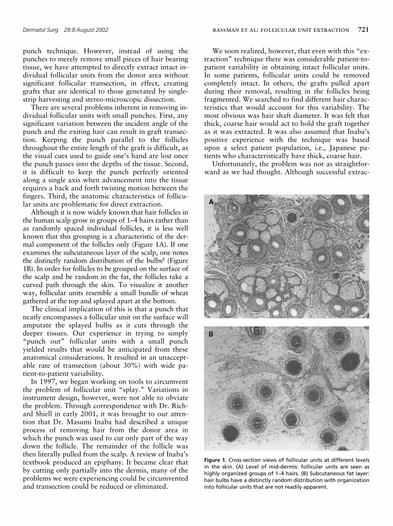
Although it is now widely known that hair follicles inthe human scalp grow in groups of 1–4 hairs rather thanas randomly spaced individual follicles, it is less wellknown that this grouping is a characteristic of the der-mal component of the follicles only (Figure 1A). If oneexamines the subcutaneous layer of the scalp, one notesthe distinctly random distribution of the bulbs
8
(Figure1B). In order for follicles to be grouped on the surface ofthe scalp and be random in the fat, the follicles take acurved path through the skin. To visualize it anotherway, follicular units resemble a small bundle of wheatgathered at the top and splayed apart at the bottom.
The clinical implication of this is that a punch thatneatly encompasses a follicular unit on the surface willamputate the splayed bulbs as it cuts through thedeeper tissues. Our experience in trying to simply“punch out” follicular units with a small punchyielded results that would be anticipated from theseanatomical considerations. It resulted in an unaccept-able rate of transection (about 30%) with wide pa-tient-to-patient variability.
In 1997, we began working on tools to circumventthe problem of follicular unit “splay.” Variations ininstrument design, however, were not able to obviatethe problem. Through correspondence with Dr. Rich-ard Shiell in early 2001, it was brought to our atten-tion that Dr. Masumi Inaba had described a uniqueprocess of removing hair from the donor area inwhich the punch was used to cut only part of the waydown the follicle. The remainder of the follicle wasthen literally pulled from the scalp. A review of Inaba’stextbook produced an epiphany. It became clear thatby cutting only partially into the dermis, many of theproblems we were experiencing could be circumventedand transection could be reduced or eliminated.
We soon realized, however, that even with this “ex-traction” technique there was considerable patient-to-patient variability in obtaining intact follicular units.In some patients, follicular units could be removedcompletely intact. In others, the grafts pulled apartduring their removal, resulting in the follicles beingfragmented. We searched to find different hair charac-teristics that would account for this variability. Themost obvious was hair shaft diameter. It was felt thatthick, coarse hair would act to hold the graft togetheras it was extracted. It was also assumed that Inaba’spositive experience with the technique was basedupon a select patient population, i.e., Japanese pa-tients who characteristically have thick, coarse hair.
Unfortunately, the problem was not as straightfor-ward as we had thought. Although successful extrac-
Figure 1. Cross-section views of follicular units at different levelsin the skin. (A) Level of mid-dermis: follicular units are seen ashighly organized groups of 1–4 hairs. (B) Subcutaneous fat layer:hair bulbs have a distinctly random distribution with organizationinto follicular units that are not readily apparent.
722
rassman et al: follicular unit extraction
Dermatol Surg 28:8:August 2002
tion occasionally did correlate positively with hairshaft diameter, we found a number of Asian patientswhose grafts fragmented during extraction and somefine-haired Caucasian patients in whom extractionwas relatively easy. It was apparent that other factorswere involved. It seemed that the dermis might be asimportant as the hair shaft in providing integrity tothe graft during extraction. In conjunction with thedepartment of dermatology of Columbia University,we began to study the donor tissue histologically tosee what specific factors contributed to the integrity ofthe graft and could account for differences in theirability to be extracted.
We have defined Follicular Unit Extraction (TheFOX Procedure™) as a technique of harvesting intactindividual follicular units directly from the donor areausing a small punch. In the procedure, a 1-mm punchis placed directly over an individual follicular unitand, following the angle of the emergent hairs, thepunch is passed partially through the dermis. Once theinitial advancement is made, the bulk of the follicle ispulled with forceps, literally “extracting” the follicu-lar unit from the scalp. It is important that the punchis small enough to leave an imperceptible donor scar.
The FOX Test™ allows the physician to determinewhich patients will be good candidates for the proce-dure. The FOX Test involves five or more small biop-sies taken from the back of the scalp in the same man-ner as in the FOX Procedure. The extracted grafts areexamined under a stereomicroscope for their integrityand/or sent for histologic examination.
This paper describes the FOX Procedure and theFOX Test, the indications for use, and the limitationsof the technique. It also attempts to explain why onlyselect patients are good candidates for the technique.
Materials and Methods
The FOX Test
During the period of study, 200 consecutive patients under-going their first follicular unit hair transplant procedurewere asked to voluntarily undergo a FOX Test immediatelyprior to donor strip harvesting. After informed consent wasobtained, biopsies were performed in the area where the do-nor strip was to be harvested.
The donor area was prepared for extraction by cuttingthe hair to a length of approximately 2 mm with an electricclipper. This length was long enough to ascertain the hairdirection, yet short enough that a 1-mm punch could fitneatly over the tuft of hair arising from the follicular unit.
Ring-block anesthesia was established using a mixture oflidocaine/bupivicaine/epinephrine. Local tumescence was es-tablished with a solution of Lactated Ringer’s. A 1-mmpunch was used for the biopsies. Between five and 10 biop-sies were taken from each patient. In select patients, a small
section of intact donor strip was removed along with thepunch biopsies and was submitted for histologic examina-tion.
Each punch was advanced into the dermis (approxi-mately 2 mm), with care not to enter into the subcutaneousspace. With experience, one can feel the increased resis-tance, indicating that the punch had passed through thepapillary dermis into the denser reticular dermis. If the resis-tance subsides, the punch has entered the subcutaneousspace and has cut too deep.
After the punch incision was made, rat-toothed forcepswere used to apply gentle pressure on the skin around thegraft, elevating it lightly to allow the top part of the graft tobe grasped. The amount of force (pull) needed to remove thegraft varied from graft to graft and from patient to patient,although there seemed to be greater variability between pa-tients than between grafts in the same patient. Care wastaken not to tear the graft from the donor site, but rather togently extract the graft. If the graft was grabbed too close tothe epidermal surface, pulled too hard, or too quickly, ittended to fragment. If the graft fragmented in spite of care-ful technique, it was scored as a negative test. Transectionoccurred if the surgeon was unable to keep the punch paral-lel to the upper portion of the follicular unit. The operatorwas usually able to adjust for this within the first few passes.If the problem persisted, it implied a negative test result.
The extracted grafts were examined under a dissectingmicroscope to determine if the entire shaft came out intactor if hair fragments were produced. They were then classi-fied and sorted according to size (the number of presumedhairs contained in the original follicular unit). Biopsies wereassessed on a scale of 1–5. A score of 1 was assigned whenall of the follicular units were extracted intact. Patients whoscored 2 had grafts that were extracted with most of theanatomy intact, but significant loss of the surrounding fataround the lower part of the follicle or some degree of am-putation (
�
20%) of the lower portion of the follicle. Pa-tients who scored 4 had most of the surrounding fat avulsedwith a significant number of distal follicles amputated. Ascore of 5 reflected significant damage in virtually all of thegrafts, with the upper portion of the follicles being avulsedfrom the lower segment in practically all the samples.
For clinical purposes, patients with positive FOX Testswere those classified as class 1 or 2, and were considered po-tential candidates for the FOX Procedure. Those classifiedas class 3 were considered neutral (these patients might beconsidered positive if there was a strong indication to do theprocedure). Those that were classified as class 4 or class 5were considered FOX-negative and not candidates for theFOX Procedure.
The FOX Procedure
The FOX Procedure was performed on patients who wereeither FOX Test Class 1 or Class 2. The procedure is essen-tially identical to that of the FOX Test except that the orga-nization of the larger procedure is more complex. The area

Dermatol Surg 28:8:August 2002
rassman et al: follicular unit extraction
723
needed for extraction is generally 8–10 times greater thanthe area needed for single-strip harvesting, with FOX Class2 patients in the upper part of the range and FOX Class 1patients in the lower range, since the extraction is more effi-cient.
As an example, for a 500-graft extraction procedure, adonor area of approximately 50 cm
2
would be required in aFOX Class 2 patient and 40 cm
2
in a patient classified asFox Class 1. With single-strip harvesting, the same numberof grafts would require 5 cm
2
(since the yield averages ap-proximately 100 follicular units/cm
2
). This same 50 cm
2
area would yield only 200–300 intact follicular units in aFOX Class 4 or Class 5 patient, due to the excessive rate oftransection. (Figure 2).
Results (Table 1)
Histological Examination
We investigated whether histological differences be-tween hair follicles of FOX-positive and FOX-nega-tive patients correlated with the observed clinical dif-ferences. Hematoxylin-eosin staining on scalp biopsies
Figure 2. (A) Donor area 3 weeks postop showing mild residual er-ythema. (B) Donor area 3 months postop.
Table 1.
FOX Test Biopsy Data
FOX Class # of patients % of total
1 53 26.5%2 72 36.0%3 23 11.5%4 20 10.0%5 32 16.0%Total 200 100%
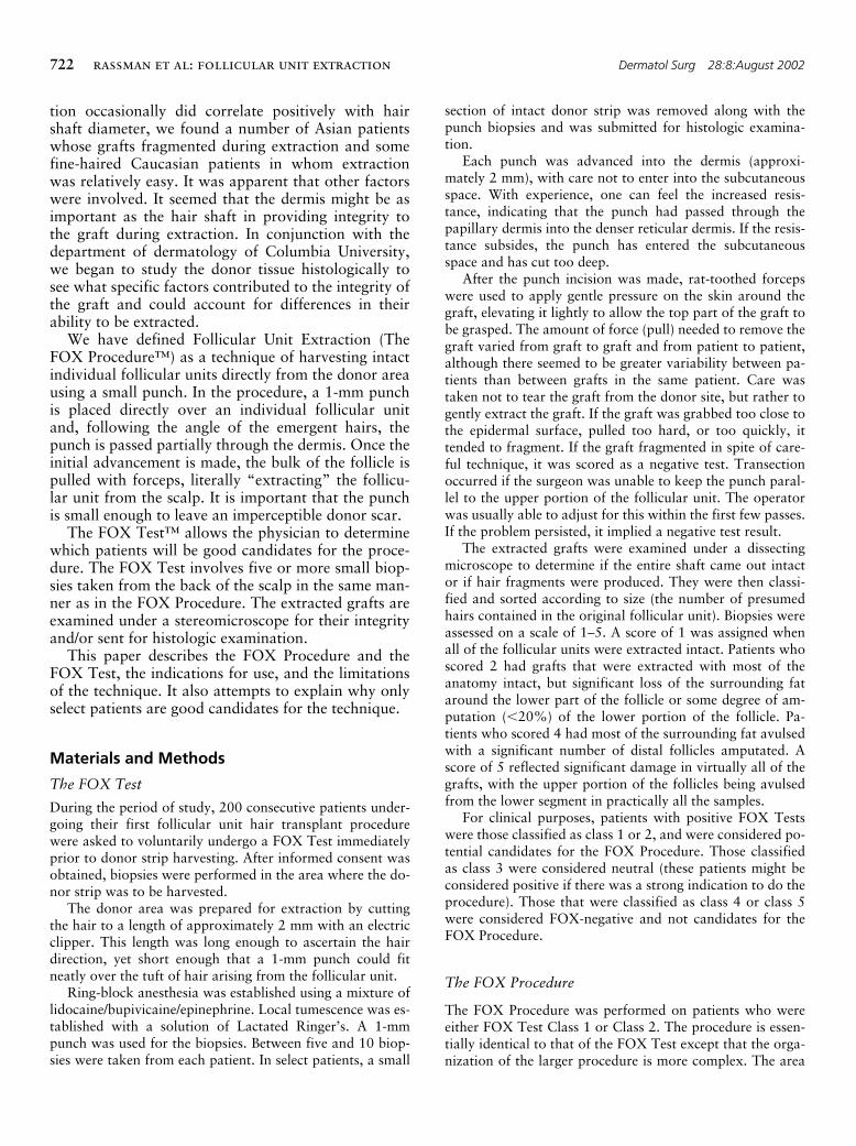
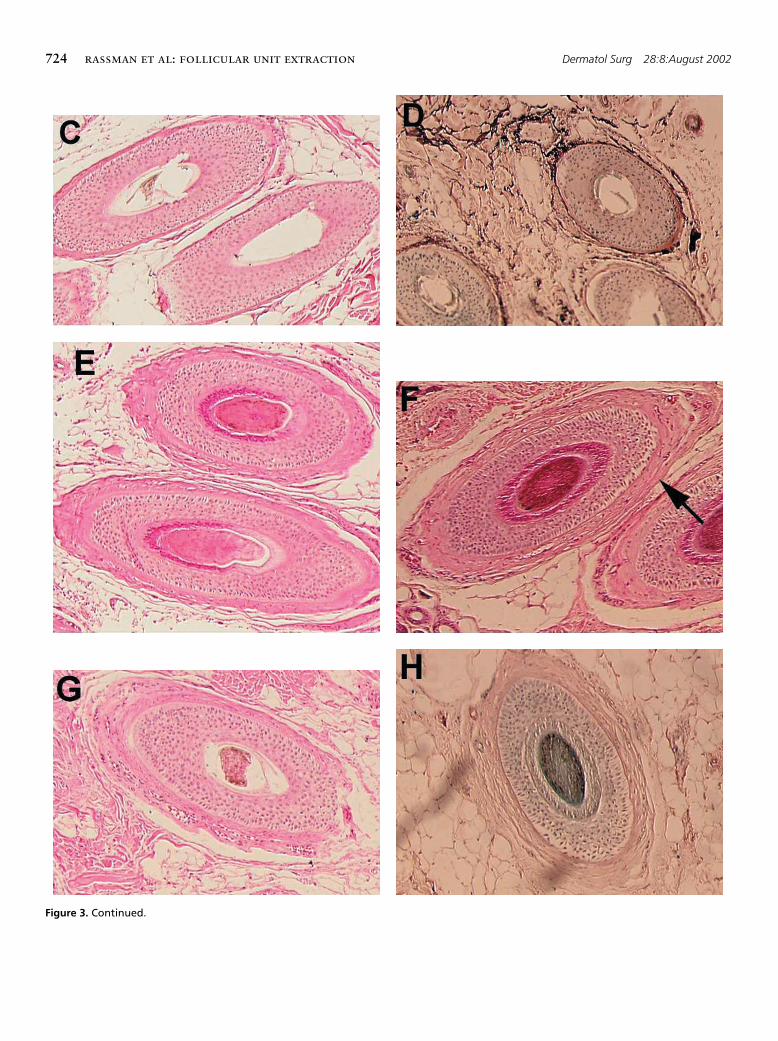
Figure 3. Hematoxylin-eosin staining of scalp biopsies of 6 differ-ent patients. Figure(A)-(C) were taken from FOX Class 1 patients.Figure(E)-(G) were taken from FOX Class 5 patients. The dermalsheath surrounding the hair follicle (see arrows) appears thinnerin FOX Class 1 patients as compared to FOX Class 5 patients. Or-cein staining to detect elastin bundles reveals stronger stainingwithin the dermal sheath of a FOX class 1 patient (D) as comparedto a FOX Class 5 patient (H). Original magnification � 250.
724
rassman et al: follicular unit extraction
Dermatol Surg 28:8:August 2002
Figure 3. Continued.

Dermatol Surg 28:8:August 2002
rassman et al: follicular unit extraction
725
of 10 patients was performed. Five of the patientstested were classified as FOX positive (class 1) andfive were classified as FOX negative (class 5).
Histological analysis did not reveal any differencesin the ratio of anagen vs. telogen hairs between FOX-negative and FOX-positive patients. However, a con-sistent difference in the thickness of the dermal sheath,a mesenchymal condensate that surrounds the hairfollicle, was observed between FOX-negative andFOX-positive patients. FOX-negative patients have athicker eosinophilic dermal sheath surrounding thehair follicle (Figures 3E–G), whereas 4 out of 5 FOX-positive patients have a thinner dermal sheath (Figures3A–C). For this analysis we compared hair follicles ofsimilar size and stage which were sectioned at a simi-lar dermal depth as well as at a similar angle, in orderto control for other factors that can influence thethickness of the dermal sheath.
To investigate whether qualitative differences withinthe dermal sheath may correlate with the observedclinical differences between FOX-negative and FOX-positive patients, we studied the elastin and smoothmuscle content of the dermal sheath and associated fi-broblasts. Orcein staining to detect elastin fibers re-veals stronger and more prominent staining within thedermal sheath of FOX-positive patients (Figure 2D)compared to FOX-negative patients (Figure 3H). Im-munohistochemistry using antismooth muscle actinantibodies did not reveal any significant differencesFOX-negative and FOX-positive patients.
Discussion
Donor Wound Healing
In Follicular Unit Extraction, healing occurs by sec-ondary intention, similar to the classic open donormethod reported by Sasagawa (1930),
9
Okuda (1939),
10
and Orentreich (1959).
1
The major difference is thewound size. It is only the very small 1 mm wounds ofFUE that provide for rapid healing, produce an imper-ceptible donor scar, and offer a distinct advantageover the strip method with respect to donor healing.
Traditional Punch Grafting vs. Extraction
There are two methods by which follicular units can beremoved directly from the donor area using a punch.In the traditional way, the punch is used to cut the sur-rounding tissue the full length of the follicle. This canproduce an unacceptable rate of transection due to themechanical difficulty in keeping the punch aligned par-allel with the hair follicles, and from the splay of indi-vidual follicles as they enter the subcutaneous fat.
Inaba suggested a second method. He noted that af-ter partial coring of the single-hair follicular unit, “the
free composite hair is lifted with a pincette and its rootis nipped with a very thin Pean’s forceps.”
11
With that,the hair can be plucked out of the donor area once thegraft has been freed at its base. Inaba’s insights wereingenious in that he identified that only the upper thirdof the hair shaft needed to be cored and that tractioncould be applied to the hair follicle as it was pulledfrom its home position. The fact that he needed to havethe root of the hair follicle “nipped” indicates that hedidn’t realize it was possible to pluck the hair com-pletely out of the donor area in some patients.
The ability to “pluck” out a follicular unit without“nipping the base” varies with each patient. This is par-tially reflected in the FOX Test results discussed above.In patients who tested FOX positive, the yield was gen-erally good and the surgery was efficient with the pulltechnique. In FOX-negative patients, on the other hand,FUE was far more difficult to perform, was generallyless efficient, and had a high miss or transection rate.
Histology
The role of the dermal sheath in hair follicle develop-ment, growth, and cycling is not well understood. It ispossible that its function is to provide structural sup-port to the hair follicle unit. We can only speculate ifthe observed differences in thickness and elastin con-tent between FOX-negative and FOX-positive patientsare related to the observed clinical differences.
Our findings—that the dermal sheath of FOX-posi-tive patients is thinner and more elastin-rich thanthose of FOX-negative patients—was surprising, aswe had predicted just the opposite. We expected tofind that FOX-positive patients had a thick dermalsheath surrounding the hair follicle that would act as asupport capsule and facilitate hair follicle unit integ-rity upon extraction.
The thicker dermal sheath actually observed inFOX-negative patients may serve to anchor the hairfollicle more tightly within the dermis and thus mayprevent the follicular unit from being easily extracted.However, it is our clinical impression that this expla-nation is incorrect, since the tissue of FOX-negativepatients appears more friable and tends to pull apartduring the extraction process, rather than causing in-creased resistance.
The hypothesis that the size of the dermal sheath isthe determining factor may be an oversimplification. Itis likely that the molecular composition of the dermalsheath may be of much greater significance. It is possi-ble that the higher elastin content, and consequentlyhigher elastin/collagen ratio, found in the thin dermalsheaths of FOX-positive patients may influence thepossible anchoring function and/or possible supportcapsule function of the dermal sheath.

726
rassman et al: follicular unit extraction
Dermatol Surg 28:8:August 2002
gion. In contrast, for eyebrow transplants, restoringtemples, or refining the frontal hairline, the surgeonmight select all 1- and 2-hair follicular units.
Donor Cosmesis
With follicular unit extraction, the prepped area pre-sents a significant postop cosmetic problem for tworeasons. First, the area to be accessed is much larger(8–10 times as large), and second, the clipped area isnot excised during the procedure, as it is in strip har-vesting. To deal with this problem, the male patient isadvised to have his hair either one of two lengths atthe time of surgery. The ideal way for the patient tohide the harvested zone would be to grow the hair inthe donor area long (longer than needed for strip har-vesting) so it would cover the wider shaved area. Analternative would be to cut all the patient’s hair veryshort so it would be essentially the same length as theharvested area. The wounds are generally not detect-able after one week. By this time, the donor areablends in well with the surrounding scalp. The short-
Patient Variability
We assessed a number of factors to explain patient-to-patient variability, including hair shaft diameter, hairdensity, scalp tightness, and race. There were no sta-tistically significant correlations with these factors,but there was a tendency for patients with more coarsehair to display a greater degree of FOX positivity. Asexpected, Asian patients were more likely to be FOX-positive than Caucasian patients. It was interesting,however, that some very coarse-haired Asian patientstested as low as a FOX Class 4.
It is our impression that with a greater sample size(
n
-value), a statistically significant correlation withsome clinically observable hair characteristics willarise, with hair shaft diameter the most likely. It is ob-vious, however, that multiple factors are involved. De-spite the somewhat ambiguous histologic results, wehave the strong clinical suspicion that dermal integrityplays a key role in FOX positivity, and we will con-tinue to pursue this area of research.
Not surprisingly, there was also variability withindifferent parts of the scalp in the same patient. Hairdirection can vary significantly within localized areas,as each follicular unit does not grow parallel to adja-cent ones, even when the scalp is stretched with trac-tion or tumescence. Because of this, the surgeon mustcontinually monitor hair direction throughout the ex-traction procedure.
Transection
Dr. Inaba is of the opinion that when a hair shaft istransected, many of the follicles survive to produce anew hair.
11
It is important to keep in mind that Inabafocused upon single hair follicles and had no statisticalscientific evidence to support his claims. If his assump-tions are correct, then complete removal of the hairshaft is not critical. On the other hand, Dr. Kim hasshown that although the amputated upper end of thehair shaft can regenerate a hair follicle (absent the dis-tal third), the new hair follicle does not grow to thefull diameter of its ancestor.
12
Therefore, it is reason-able to assume that if maximum fullness from a trans-plant is to be achieved, complete removal of the hairshaft and bulb is important in this new technique.
Follicular Unit Selection
Unlike strip harvesting where every follicular unit inan area is removed, in the FOX Procedure the surgeoncan select which units to extract. Since the averageCaucasian has approximately 30% 3- and 4-hair folli-cular units and 15% naturally occuring 1-hair units,one could select only the largest grafts when trying toachieve the greatest density, such as in the forelock re-
Figure 4. (A) 46-year-old Norwood Class 3 pattern with a persis-tant forelock. (B) Nine months after 742 follicular units (per-formed in two Fox procedure spaced 12 days apart) with hair cutvery short to show density/fullness comparison with forelock.
Dermatol Surg 28:8:August 2002
rassman et al: follicular unit extraction
727
hair approach offers a much wider potential donorarea for the FOX Procedure and may be a better choicefor larger sessions.
Indications
In its present state, FUE has the following indications:
♦
People with limited hair loss or those who requiresmall sessions. This group would include patients withandrogenetic alopecia in a Norwood Class 3 patternor those with small vertex balding areas (Figure 4)
♦
Limited cosmetic areas, such as widow’s peaks, eye-brows, eyelashes, mustaches
♦
Alopecia secondary to dermatologic conditions
♦
Scarring from dermatologic conditions, trauma, orneurosurgical procedures
♦
Individuals with low donor supplies, heavily scarreddonor areas, or very tight scalps
♦
Patients who tend to heal with wide scars
♦
Select repairs
♦
Those who wear their hair very short
♦
Athletes who must resume full activity soon afterthe procedure
With further experience with this technique, we expectthese indications will be modified and new indicationswill be found.
Conclusion
FUE is a minimally invasive surgical procedure thatcan benefit a limited subset of patients in a hair resto-
ration practice. The FOX Procedure involves the di-rect extraction of the follicular units from a patient’sdonor area using a small punch. At this time, approxi-mately 60% of patients are candidates for this proce-dure, and the procedure itself is practical in individu-als who require less than 600 grafts at a sitting.Healing is quick, scarring is virtually nonexistent, anddiscomfort in the donor area has been virtually elimi-nated. The authors believe that FUE can benefit selectpatients and should be available in every hair restora-tion practice for use in appropriate candidates.
References
1. Orentreich N. Autografts in alopecias and other selected dermato-logical conditions. Ann New York Acad Sciences 1959;83:463–79.
2. Rassman WR, Carson S. Micrografting in extensive quantities: theideal hair restoration procedure. Dermatol Surg 1995;21:306–11.
3. Limmer BL. Elliptical donor stereoscopically assisted micrograftingas an approach to further refinement in hair transplantation. Der-matol Surg 1994;20:789–93.
4. Bernstein RM, Rassman WR, Szaniawski W, Halperin A. Folliculartransplantation. Int J Aesthetic Restorative Surgery 1995;3:119–32.
5. Bernstein RM, Rassman WR. Follicular transplantation: patientevaluation and surgical planning. Dermatol Surg 1997;23:771–84.
6. Bernstein RM, Rassman WR. The aesthetics of follicular transplan-tation. Dermatol Surg 1997;23:785–99.
7. Bernstein RM, Rassman WR, Seager D, et al. Standardizing theclassification and description of follicular unit transplantation andmini-micrografting techniques. Dermatol Surg 1998;24:957–63.
8. Bernstein RM. A neighbor’s view of the “follicular family unit.”Hair Transplant Forum Intl 1998;8 (3):23–5.
9. Sasagawa M. Hair transplantation. Jpn J Dermatol 1930;30:493(In Japanese).
10. Okuda S. Clinical and experimental studies of transplantation ofliving hairs. Jpn J Dermatol Urol 1939;46:135–8 (In Japanese).
11. Inaba M. Androgenetic alopecia: modern concepts of pathogenesisand treatment. Tokyo: Springer-Verlag 1996, 238–45.
12. Kim JC, Choi YC. Regrowth of grafted human scalp hair after re-moval of the bulb. Dermatol Surg 1995;21:312–3.
Commentary
One of the most serious complications of hair transplanting inthe last decade has been the postoperative development of wideand cosmetically embarrassing donor area scars. There are mul-tiple possible potential causes for this complication. The twomost important ones, however, are probably the patient’s ge-netic susceptibility to poor healing and closing the donorwound with too much tension. The first is unavoidable but thesecond is usually the result of a misjudgment on the part of asurgeon who is attempting to get as much donor tissue as possi-ble in a single session. In particular, in my opinion, the “mega-session” of 2500, 3000, or even more grafts was a disaster foran undocumented number of patients, usually with tighter thanaverage scalps, who either pushed their surgeon into doing “asmuch as possible” in a single session or who were encouragedby their doctors to do so. Because some of these wide scars are
relatively resistant to successful revision, there is now a sizablegroup of patients who had no apparent solution to the problemexcept transplanting hair into the scar, if indeed donor hair waseven available. For these individuals, the thought of anotherstrip being excised, with the possibility of another unsightlyscar, was an appalling prospect. In the past few years they havemade their plight widely known, primarily through the Inter-net, both looking for a solution and warning others of the“dangers” of strip harvesting. An acceptable solution to someof the problems eventually showed up – also on the Internet in-stead of in medical journals. It consists of the excision of singlefollicular units directly from the donor area instead of from anexcised strip of donor tissue. The technique has now achievedalmost a cult status in Internet hair transplant “chat rooms”.Rassman
et al’
s article documents their method.
728
rassman et al: follicular unit extraction
Dermatol Surg 28:8:August 2002
It would appear that the main advantages of this techniqueare: a) Selectivity; follicular units can be chosen and excised onthe basis of how many hairs they contain. As Rassman haspointed out, one can facilitate the production of the greatestpossible density, for example, by selectively removing only FUwith three or more hairs, b) No sutures are required, and c) Alldonor sites rapidly shrink and heal with scars so small they aredifficult to find except on very close inspection. FUE, therefore,might be advantageous for patients with particularly tightscalps or an inherent tendency to heal with wide scars if stripsare excised. It may also be preferred by those who are fright-ened to try strip harvesting either because of past problemswith this technique or because they have seen, heard, or readabout people who have had such problems.
There are, however, a number of significant disadvantagesto FUE:
a) Donor Area Damage. The excision of FU via FUE in-volves a tremendous increase in the total length of inci-sions when compared to conventional strip harvesting,with as yet unknown consequence. For example, if onewere extracting 500 FU, 500 grafts with a 1 mm diame-ter would be excised. The perimeter of a circle is calcu-lated with the formula 2
D
R; in this case 2
�
3.14
�
.5
�
3.12 mm. 500 grafts would therefore involve incisions to-taling 1560 mm (500
�
3.12) or 156 cm or 62.4 inches(156 divided by 2.5) or 5.2 feet! If instead strip harvest-ing were employed, a strip that had a surface area of ap-proximately 500 mm, for example 6 mm
�
83 mm,could be expected to produce 500 FU. The perimeter ofsuch an area would be approximately 83
�
83 or 166mm, plus the length of the tapered ends. It is unlikely thatwould total much more than 200 mm, as opposed to the1560 mm of incisions created by FUE. It might be arguedthat the FUE method could be expected to result in lessdamage to deeper vessels than conventional strip excisionbecause the incisions only go as deep as the mid-dermis.However, any difference probably would not be that greatwhen compared to good strip harvesting that includes aneffort to limit the depth of incisions to high subcutaneoustissue. It seems quite likely that small vessel damage willbe similar, regardless of whether small vessels in the highsubcutaneous tissue are cut with a blade or torn as a graftis pulled out of its bed. FUE’s nearly eight-fold guaranteedincrease in incision length is therefore hard for me to ac-cept for all but special situations. Under the circum-stances, I also find it hard to accept the designation of thisapproach as “minimally invasive surgery.”
b) Cosmetic Factors. The authors point out that large areasof the donor area have to be clipped short in preparationfor FUE in order to find a significant number of FU thatare suitable for extraction, presenting “a significant post-op cosmetic problem.” For a 500-graft procedure, for ex-ample, Rassman found that 50 cm would have to beclipped for a FOX class II patient and 40 cm for a FOXclass I patient. (This compares with clipping an area justslightly larger than 5 cm for conventional strip harvest-ing.) There is also usually insufficient hair superior to theclipped donor area to comb over and thereby camouflage
it. This is obviously much more cosmetically embarrass-ing than conventional harvesting, where most of theclipped area is excised and the donor site can be easilycamouflaged, even with 1” (or less) length hair. Rassmansuggests that clipping all the rim hair to a 2- or 3-mm lengthminimizes the cosmetic problem, but of course this sug-gestion does not totally avoid it. As described, it will stillbe far more noticeable than conventional harvesting forat least the first postoperative week.
c) Follicle Damage. It is hard to know how many FU will belethally injured during extraction with FUE—even inFOX class I and II patients—and 40% of Rassman’s andBernstein’s patients were not class I or II. Almost cer-tainly, more will be damaged than can be expected withgood conventional harvesting. In addition, rather thanworking “blind” in the depths of the wound with FUE,FU prepared carefully from an excised strip on a well-lighted surface with whatever magnification is required—ironically, long championed by Rassman and Bernstein—is almost certain to result in less damage to follicles.When dealing with a limited number of follicles per pa-tient, anything that increases the risk to them is obviouslydisadvantageous.
d) Distortion of Follicle Direction. When the small donorsites heal, they will create scars that will distort the hairdirection or angle of adjacent FU. This will have the resultof making further harvesting by either FUE or conven-tional means more hazardous to the remaining follicles.
e) Increased Risk of Infection. If additional FUE proceduresare done one or a few days apart, in order to avoid d),there will almost certainly be a greater likelihood of bac-terial pathogens being present in the donor area.
f) Increased Operating Time. FUE is far more time-consum-ing than conventional harvesting.
For all of the above reasons, while FUE will find a role toplay in hair restoration surgery, I agree with the authors that itsrole should be limited. They have noted that “approximately60% of patients are candidates for this procedure,” but it isworthwhile to emphasize that they also believe it may only beof benefit in “a limited subset of patients.” They list a numberof possible indications for FUE. However, given the above-noted drawbacks, I would tend to reduce that list to two ofthem: a) Patients treating only specific limited areas, such aswidow’s peaks, eyebrows, eyelashes, or moustaches, for whichFU with fewer or more hairs and finer or coarser hairs wouldbe preferable, and b) Patients who have bad scarring of thescalp that is unamenable to adequate revision. Patients withoutsuch a history should be advised that the likelihood of develop-ing a wide scar from the excision of a 6 mm
�
83 mm strip, forexample, (to produce 500 FU) is next to zero in the vast major-ity of people. Despite the hype that may grow for a while, in myopinion FUE has considerably less chance of becoming the“state-of-the-art” in donor harvesting, for most patients/doc-tors, than FUT has of actually being the “state-of-the-art” inhair transplanting for most patients/doctors.
Walter Unger, MD
Toronto, Canada


![Minimally invasive non-surgical vs. surgical approach for ...dictable [12]. More recently, minimally invasive surgical therapy (MIST), modified minimally invasive surgical therapy](https://static.fdocuments.in/doc/165x107/5eddda76ad6a402d6669115c/minimally-invasive-non-surgical-vs-surgical-approach-for-dictable-12-more.jpg)

