
Folic acid supplement rescues ethanol-induced ...
10
1 © The Author(s) 2020. Published by Oxford University Press on behalf of the Institute of Biochemistry and Cell Biology, Shanghai Institutes for Biological Sciences, Chinese Academy of Sciences. All rights reserved. For permissions, please email: [email protected] Acta Biochim Biophys Sin, 2020, 00(00), 1–10 doi: 10.1093/abbs/gmaa030 Original Article Original Article Folic acid supplement rescues ethanol-induced developmental defects in the zebrafish embryos Qiu Jiang 2 , Ding Lu 3 , Feng Wang 1 , Yawen Zhang 1 , Li Cao 4 , Yonghao Gui 1, *, and Shuna Sun 1, * 1 Children’s Hospital, Fudan University, Shanghai 201102, China, 2 Shanghai Institute of Cardiovascular Diseases, Zhongshan Hospital and Institute of Biomedical Sciences, Fudan University, Shanghai 200032, China, 3 Department of Pediatrics, Shanghai Municipal Eighth People’s Hospital, Shanghai 200235, China, and 4 Department of Ultrasound, Obstetrics and Gynecology Hospital of Fudan University, Shanghai 200011,China ∗ Correspondence address. Tel: +86-21-64931990 Fax: +86-21-64931914; E-mail: [email protected] (S.S.) / E-mail: [email protected] (Y.G.) Received 14 June 2019; Editorial Decision 12 January 2020 Abstract Fetal alcohol syndrome (FASD) describes a range of birth defects. Mechanisms of FASD-associated defects are not well understood. It has great significance to investigate whether nutrient sup- plements like folic acid (FA) can effectively rescue ethanol-induced defects. Moreover, it is very important to determine the optimal time for FA supplementation when it can most effectively antagonize the teratogenic effects of ethanol during embryonic development. Our results indicated that ethanol exposure interrupted the development of zebrafish embryos and induced multiple defects in cardiac function, pharyngeal arch arteries, vessel, craniofacial cartilage, pharyngeal arches, brain, somite and hemoglobin formation. The expressions of critical genes that play important roles in above organs such as tbx1, flk-1, hand2, ngn1, huc, titin, gata-1 and c-myb were reduced, and the apoptosis was increased in ethanol-treated group. FA supplementation could reverse ethanol-induced defects, improve the decreased expressions of above genes and reduce the apoptosis. We also found that giving FA at 6–12 h post-fertilization (hpf), which is at the gastrula period (5.25–10 hpf), can obviously prevent the teratogenicity of ethanol. This research provides clues for elucidating the mechanism of fetal abnormalities caused by alcohol intake and for preventing FASD. Key words: folic acid, ethanol, developmental defects, zebrafish Introduction Fetal alcohol syndrome (FASD) is associated with maternal alco- hol consumption. Clinically, it is characterized by cardiovascular malformation, craniofacial abnormalities, central nervous system damage and body growth defects. Epidemiological studies showed that FASD incidence is higher in infants born to lower socioeco- nomic status alcoholic mothers compared to infants born to middle class alcoholics [1–3]. A lower socioeconomic population is at a greater risk for poor nutrition such as folic acid (FA). FASD is the most frequent preventable birth defect syndrome [4]. Epidemiologic studies showed that severe congenital heart defect prevalence was significantly reduced by prenatal FA fortification [5, 6]. FA is an essential vitamin that participates in nucleic acid syn- thesis and repair. FA also plays a crucial role as a co-factor in 1- carbon metabolism as tetrahydrofolate, which is needed in DNA and histone methyl transfer. FA deficiency and inhibition have been found to induce embryonic malformations [7–10]. Prenatal FA sup- plementation significantly reduces the risk of neural tube defects, congenital heart defects and cleft lip/palate [11], and thus, FA is a recommended dietary supplement for pregnant mothers [12]. Studies Downloaded from https://academic.oup.com/abbs/advance-article-abstract/doi/10.1093/abbs/gmaa030/5822774 by Beurlingbiblioteket user on 12 May 2020
Transcript of Folic acid supplement rescues ethanol-induced ...

1
© The Author(s) 2020. Published by Oxford University Press on behalf of the Institute of Biochemistry and Cell Biology, Shanghai Institutes for Biological Sciences, ChineseAcademy of Sciences. All rights reserved. For permissions, please email: [email protected]
Acta Biochim Biophys Sin, 2020, 00(00), 1–10doi: 10.1093/abbs/gmaa030
Original Article
Original Article
Folic acid supplement rescues ethanol-induced
developmental defects in the zebrafish embryos
Qiu Jiang2, Ding Lu3, Feng Wang1, Yawen Zhang1, Li Cao4, Yonghao
Gui1,*, and Shuna Sun1,*
1Children’s Hospital, Fudan University, Shanghai 201102, China, 2Shanghai Institute of Cardiovascular Diseases,Zhongshan Hospital and Institute of Biomedical Sciences, Fudan University, Shanghai 200032, China, 3Departmentof Pediatrics, Shanghai Municipal Eighth People’s Hospital, Shanghai 200235, China, and 4Department of Ultrasound,Obstetrics and Gynecology Hospital of Fudan University, Shanghai 200011,China∗Correspondence address. Tel: +86-21-64931990 Fax: +86-21-64931914; E-mail: [email protected] (S.S.) / E-mail:[email protected] (Y.G.)
Received 14 June 2019; Editorial Decision 12 January 2020
Abstract
Fetal alcohol syndrome (FASD) describes a range of birth defects. Mechanisms of FASD-associated
defects are not well understood. It has great significance to investigate whether nutrient sup-
plements like folic acid (FA) can effectively rescue ethanol-induced defects. Moreover, it is very
important to determine the optimal time for FA supplementation when it can most effectively
antagonize the teratogenic effects of ethanol during embryonic development. Our results indicated
that ethanol exposure interrupted the development of zebrafish embryos and induced multiple
defects in cardiac function, pharyngeal arch arteries, vessel, craniofacial cartilage, pharyngeal
arches, brain, somite and hemoglobin formation. The expressions of critical genes that play
important roles in above organs such as tbx1, flk-1, hand2, ngn1, huc, titin, gata-1 and c-myb
were reduced, and the apoptosis was increased in ethanol-treated group. FA supplementation
could reverse ethanol-induced defects, improve the decreased expressions of above genes and
reduce the apoptosis. We also found that giving FA at 6–12 h post-fertilization (hpf), which is at the
gastrula period (5.25–10 hpf), can obviously prevent the teratogenicity of ethanol. This research
provides clues for elucidating the mechanism of fetal abnormalities caused by alcohol intake and
for preventing FASD.
Key words: folic acid, ethanol, developmental defects, zebrafish
Introduction
Fetal alcohol syndrome (FASD) is associated with maternal alco-hol consumption. Clinically, it is characterized by cardiovascularmalformation, craniofacial abnormalities, central nervous systemdamage and body growth defects. Epidemiological studies showedthat FASD incidence is higher in infants born to lower socioeco-nomic status alcoholic mothers compared to infants born to middleclass alcoholics [1–3]. A lower socioeconomic population is at agreater risk for poor nutrition such as folic acid (FA). FASD is themost frequent preventable birth defect syndrome [4]. Epidemiologic
studies showed that severe congenital heart defect prevalence wassignificantly reduced by prenatal FA fortification [5,6].
FA is an essential vitamin that participates in nucleic acid syn-thesis and repair. FA also plays a crucial role as a co-factor in 1-carbon metabolism as tetrahydrofolate, which is needed in DNAand histone methyl transfer. FA deficiency and inhibition have beenfound to induce embryonic malformations [7–10]. Prenatal FA sup-plementation significantly reduces the risk of neural tube defects,congenital heart defects and cleft lip/palate [11], and thus, FA is arecommended dietary supplement for pregnant mothers [12]. Studies
Dow
nloaded from https://academ
ic.oup.com/abbs/advance-article-abstract/doi/10.1093/abbs/gm
aa030/5822774 by Beurlingbiblioteket user on 12 May 2020

2 Folic acid supplement rescues ethanol-induced developmental defects in the zebrafish embryos
showed that FA transport to the fetus is impaired in pregnancieswith chronic alcohol exposure. Embryonic ethanol exposure affectsFA metabolism, including reduced maternal-to-fetal folate transferand reduced expression of folate-metabolizing enzymes. Alcoholingestion can also inhibit folate-dependent DNA methylation [13].
A better understanding of the effects of ethanol in embryonicdevelopment in animal model may provide important clues for thestudy of the relationship between alcohol consumption and fetalanomaly in human. Exploring the best time and possible mecha-nism of FA antagonizing ethanol using animal model is of greatsignificance to effectively prevent ethanol-induced abnormal humanembryonic development.
Zebrafish shares high evolutionary conservation with human,and zebrafish offers several distinct advantages for genetic andembryological studies because of its external fertilization, rapiddevelopment, easy genetic manipulability, high fecundity and embryooptical clarity. Zebrafish embryo experiments showed that ethanolexposure interrupted divergent cardiac morphogenetic events caus-ing heart defects [14]. The transgenic zebrafish lines can help toclearly observe the target organ. Using the transgenic zebrafishline Tg(fli1:neGFP)y7 expressing green fluorescent protein (GFP) inendothelial and endocardial cells [15], we can better visualize thevessel and endothelial cells.
In this study, we used zebrafish to observe the teratogenic effectsof ethanol on embryonic development and detect the expressionlevels of related genes as well as apoptosis. We also explored thebest time for FA administration during embryonic development whenit can most effectively antagonize the teratogenic effects of ethanol.This research provides clues for elucidating the mechanism of fetalabnormalities caused by alcohol intake in humans. Our investigationis also expected to provide clues for the design of better therapeuticsand preventive measures for FASD.
Materials and Methods
Zebrafish strains and maintenance
Wild-type (AB strain) zebrafish, Tg(fli1:neGFP)y7 transgeniczebrafish lines were raised and housed under standard laboratoryconditions [16]. The breeding facility was purchased from AquaticHabitats Corporation (Apopka, USA). Embryos were obtained fromnatural spawning, raised at 28.5○C in embryo medium (EM) andstaged according to the standard protocol [17]. Embryos used for insitu hybridization were treated with 0.003% PTU (phenylthiourea)to prevent pigmentation at 24 h post-fertilization (hpf). Embryoswere fixed in 4% paraformaldehyde (PFA)–phosphate-bufferedsaline (PBS) at 4○C and stored in 100% methanol at −20○C prior toin situ hybridization.
Ethanol was diluted into egg water, and zebrafish embryos weretreated with different concentrations of ethanol at 2–48 hpf. Ethanolwith a concentration of 400 mM was chosen as the optimal concen-tration for treatment because under this concentration the majorityof embryos developed obvious malformations and the embryos couldsurvive up to 96 h after fertilization. FA (Sigma, St Louis, USA) stocksolution was freshly prepared prior to use and diluted with embryomedium. Different concentrations of FA were tested, and 75 μM waschosen as the optimal concentration for FA treatment to minimizethe toxic effects of ethanol (400 mM) and achieve maximum rescuephenotypes.
In the ethanol treatment experiment, embryos were transferredto EM containing 400 mM ethanol at 2–48 hpf. Eggs were coveredduring incubation to prevent evaporation of ethanol. In the FA rescue
experiment, the ethanol-treated embryos were divided into eightgroups. FA was added to the ethanol-treated group with a finalconcentration of 75 μM at different stages: 0–6 hpf, 6–12 hpf, 12–18 hpf, 18–24 hpf, 24–30 hpf, 30–36 hpf, 36–42 hpf and 42–48 hpf.
At 48 hpf, the ethanol-treated embryos and the FA+ ethanol-treated embryos were transferred back to EM. Embryos treatedwith 75 μM FA at 6–12 hpf were defined as FA-treated group andwere transferred back to EM at 12 hpf. Each group contained 100embryos.
Microangiography
To monitor the development of the pharyngeal arch arteries andvessels, fluorescein (2–5 nl) was microinjected into the hearts of livingzebrafish embryos at 60 hpf [9,10]. About 2–3 min later, images ofthe pharyngeal arch arteries and vessels were observed under a BX61fluorescent microscope (Olympus, Tokyo, Japan) and captured witha DP70 digital camera (Olympus).
Measurement of heart rate and analyses of ventricular
contractility
The hearts of zebrafish embryos were easily observed with a micro-scope. Heart rate was measured for 1 min. Cardiac contractions wererecorded with a video camera (TK-C1381; JVC, Yokohama, Japan)as previously described [9,10]. The diastolic and systolic lengths ofventricles were measured to calculate the ventricular shortening frac-tion (VSF) using the following formula: VSF=(ventricular length atdiastole−ventricular length at systole)/ventricular length at diastole.
Histological analysis
At 60 hpf, controls and ethanol-treated larvae were fixed with 4%paraformaldehyde in 0.1 M phosphate buffer (pH=7.4) overnight.Larvae were then washed in phosphate-buffered saline (pH=7.4),dehydrated through a graded series of ethanol and embedded in JB-4 embedding medium. Serial sections were made, each containing10 larvae. Sections were mounted and dried on glass slides andstained with hematoxylin and eosin (H&E; Sangon Biotech, Shang-hai, China) according to the manufacturer’s instruction.
Cartilage staining
Embryos at 96 hpf were cartilage stained with Alcian Blue (SangonBiotech) as described previously [18]. Briefly, embryos were fixed in4% paraformaldehyde overnight at 4○C. After several washes withPBS, embryos were rinsed with acid alcohol (70% ethanol and 0.37%HCl). Embryos were then stained with 0.1% Alcian Blue in acidalcohol for 4 h at room temperature and destained in acid alcoholovernight. After rehydration with PBS, embryos were digested with1% trypsin for 1 h at 37○C. Embryos were then washed with PBSseveral times and stored in 70% glycerol.
O-dianisidine staining for globin
At 48 hpf, embryos were dechorionated and fixed in 4% paraformalde-hyde overnight at 4○C, then rinsed with PBS before O-dianisidinestaining. Fixed embryos were incubated in the staining buffer for15 min in the dark. The staining buffer consisted of 0.6 mg/mlO-dianisidine (Sigma), 10 mM sodium acetate, 0.65% hydrogenperoxide and 40% (v/v) ethanol [19].
Dow
nloaded from https://academ
ic.oup.com/abbs/advance-article-abstract/doi/10.1093/abbs/gm
aa030/5822774 by Beurlingbiblioteket user on 12 May 2020
Folic acid supplement rescues ethanol-induced developmental defects in the zebrafish embryos 3
Whole-mount terminal deoxynucleotide TUNEL
staining
The transferase-mediated dUTP nick-end labeling (TUNEL) stainingwas performed using an in situ Cell Death Detection kit (Roche,Indianapolis, USA) according to the manufacturer’s instructions.Briefly, for whole-mount TUNEL staining, embryos at 10 hpf and20 hpf were fixed with 4% paraformaldehyde overnight at 48○C,rinsed with PBS, incubated with a 1:10 TUNEL working solution ina dark environment at 37○C for 1 h and washed three times withPBS. Then, embryos were stained in the dark at room temperaturefor 5 min and then washed three times with PBS.
Whole-mount in situ antisense RNA hybridization
Whole-mount in situ hybridization of zebrafish embryos was per-formed as previously described [20]. Plasmids encoding zebrafishtbx1, flk-1, hand2, ngn1, huc, titin, gata-1and c-myb were linearizedby restriction enzymes before transcription with T7 or SP6 RNApolymerase (Roche). Digoxigenin-labeled probes were synthesizedusing DIG RNA Labeling Kit (Roche). Whole-mount RNA in situhybridization was carried out using DIG-labeled antisense RNAprobes according to standard procedures as described. The num-ber of embryos with decreased expression of genes was analyzed.Stained embryos were examined with BX61 and SZX12 microscopes(Olympus), and images were captured with a DP70 digital camera(Olympus).
RNA isolation and Q-PCR
Total RNA was extracted from embryos using TRIzol RNA isolationreagent (Invitrogen, Carlsbad, USA). First-strand cDNA was reversetranscribed using oligo-dT primers and MMLV reverse transcriptase(Promega, Madison, USA). The cDNA was analyzed immediately orstored at −20○C until use.
Quantitative real-time polymerase chain reaction (Q-PCR) wasperformed using the 7300 Real-Time PCR System (Applied Biosys-tems, Carlsbad, USA). The sequence-specific primers were designedusing Primer Express 2.0 software (Applied Biosystems) and listedin Table 1. The SYBR Green method was used to quantify cDNA. β-actin was used as the internal control. The value of expression level ofeach gene was divided by the amount of β-actin in individual cDNAsamples. The specificity of each reaction was controlled by meltingcurve analysis. Results were analyzed using the 7300 Real Time PCRSystem software (Applied Biosystems).
Statistical analysis
Data were presented as the mean±standard deviation (SD). Compar-isons between groups were analyzed by ANOVA (t-test with Bonfer-roni correction). P<0.05 was considered statistically significant.
Results
Ethanol exposure induced the pharyngeal arch arteries
and vessels defects, impaired the cardiac function and
reduced the expressions of tbx1 and flk-1
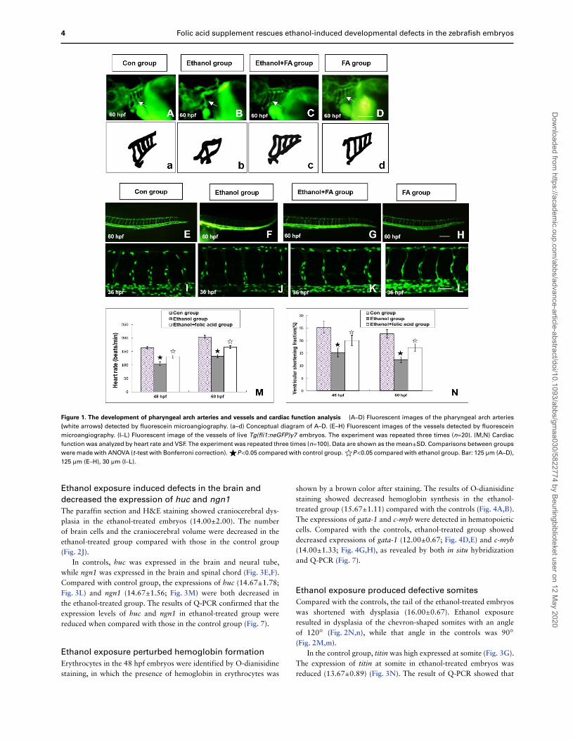
In controls, the pharyngeal arch arteries (Fig. 1A,a) and intersomiticvessels (Fig. 1E) could be clearly observed at 60 hpf by microangiog-raphy. Compared with controls, ethanol-treated embryos showedthe dysplastic pharyngeal arch arteries (15.67±2.22; Fig. 1B,b) and
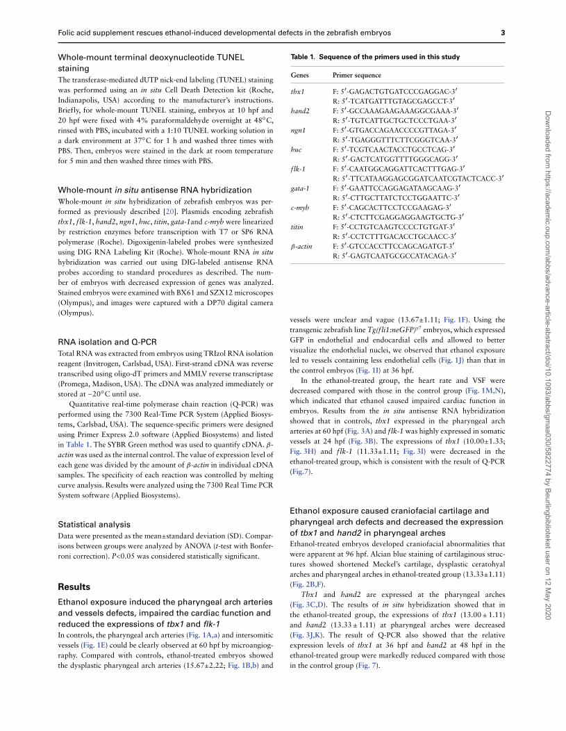
Table 1. Sequence of the primers used in this study
Genes Primer sequence
tbx1 F: 5′-GAGACTGTGATCCCGAGGAC-3′
R: 5′-TCATGATTTGTAGCGAGCCT-3′
hand2 F: 5′-GCCAAAGAAGAAAGGCGAAA-3′
R: 5′-TGTCATTGCTGCTCCCTGAA-3′
ngn1 F: 5′-GTGACCAGAACCCCGTTAGA-3′
R: 5′-TGAGGGTTTCTTCGGGTCAA-3′
huc F: 5′-TCGTCAACTACCTGCCTCAG-3′
R: 5′-GACTCATGGTTTTGGGCAGG-3′
flk-1 F: 5′-CAATGGCAGGATTCACTTTGAG-3′
R: 5′-TTCATAAGGAGCGGATCAATCGTACTCACC-3′
gata-1 F: 5′-GAATTCCAGGAGATAAGCAAG-3′
R: 5′-CTTGCTTATCTCCTGGAATTC-3′
c-myb F: 5′-CAGCACTTCCTCCGAAGAG-3′
R: 5′-CTCTTCGAGGAGGAAGTGCTG-3′
titin F: 5′-CCTGTCAAGTCCCCTGTGAT-3′
R: 5′-CCTCTTTGACACCTGCAACC-3′
β-actin F: 5′-GTCCACCTTCCAGCAGATGT-3′
R: 5′-GAGTCAATGCGCCATACAGA-3′
vessels were unclear and vague (13.67±1.11; Fig. 1F). Using thetransgenic zebrafish line Tg(fli1:neGFP)y7 embryos, which expressedGFP in endothelial and endocardial cells and allowed to bettervisualize the endothelial nuclei, we observed that ethanol exposureled to vessels containing less endothelial cells (Fig. 1J) than that inthe control embryos (Fig. 1I) at 36 hpf.
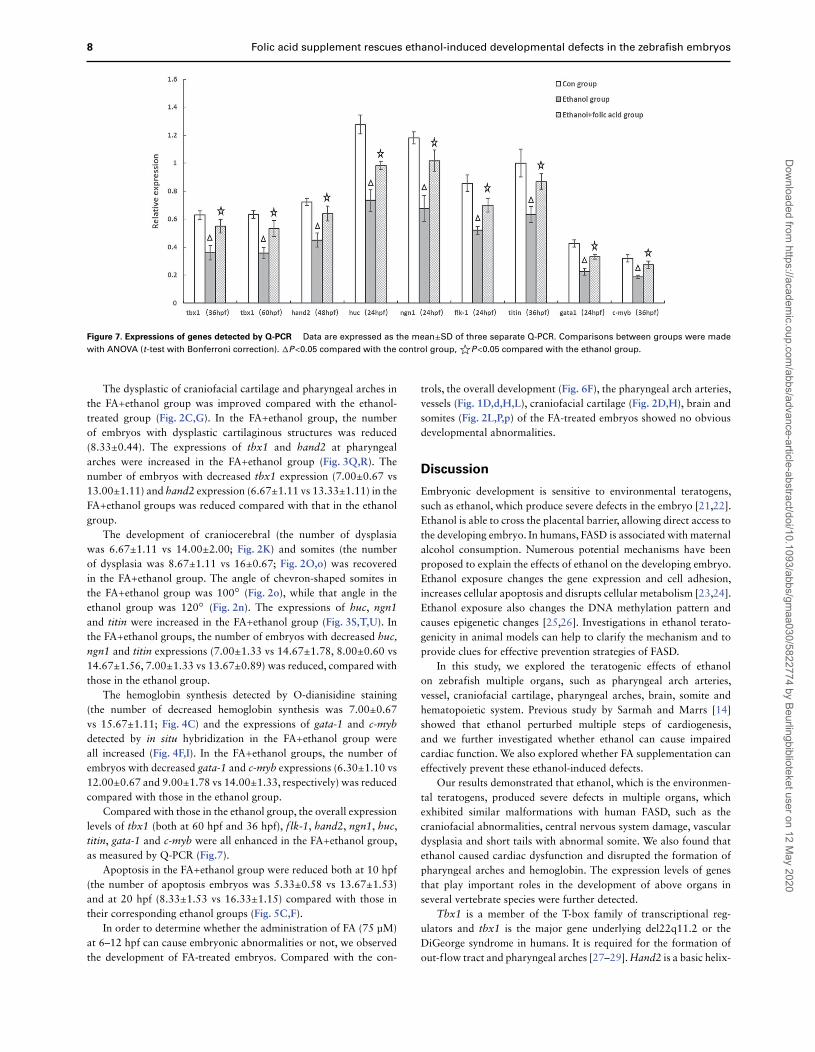
In the ethanol-treated group, the heart rate and VSF weredecreased compared with those in the control group (Fig. 1M,N),which indicated that ethanol caused impaired cardiac function inembryos. Results from the in situ antisense RNA hybridizationshowed that in controls, tbx1 expressed in the pharyngeal archarteries at 60 hpf (Fig. 3A) and flk-1 was highly expressed in somaticvessels at 24 hpf (Fig. 3B). The expressions of tbx1 (10.00±1.33;Fig. 3H) and flk-1 (11.33±1.11; Fig. 3I) were decreased in theethanol-treated group, which is consistent with the result of Q-PCR(Fig.7).
Ethanol exposure caused craniofacial cartilage and
pharyngeal arch defects and decreased the expression
of tbx1 and hand2 in pharyngeal arches
Ethanol-treated embryos developed craniofacial abnormalities thatwere apparent at 96 hpf. Alcian blue staining of cartilaginous struc-tures showed shortened Meckel’s cartilage, dysplastic ceratohyalarches and pharyngeal arches in ethanol-treated group (13.33±1.11)(Fig. 2B,F).
Tbx1 and hand2 are expressed at the pharyngeal arches(Fig. 3C,D). The results of in situ hybridization showed that inthe ethanol-treated group, the expressions of tbx1 (13.00±1.11)and hand2 (13.33±1.11) at pharyngeal arches were decreased(Fig. 3J,K). The result of Q-PCR also showed that the relativeexpression levels of tbx1 at 36 hpf and hand2 at 48 hpf in theethanol-treated group were markedly reduced compared with thosein the control group (Fig. 7).
Dow
nloaded from https://academ
ic.oup.com/abbs/advance-article-abstract/doi/10.1093/abbs/gm
aa030/5822774 by Beurlingbiblioteket user on 12 May 2020
4 Folic acid supplement rescues ethanol-induced developmental defects in the zebrafish embryos
Figure 1. The development of pharyngeal arch arteries and vessels and cardiac function analysis (A–D) Fluorescent images of the pharyngeal arch arteries
(white arrows) detected by fluorescein microangiography. (a–d) Conceptual diagram of A–D. (E–H) Fluorescent images of the vessels detected by fluorescein
microangiography. (I–L) Fluorescent image of the vessels of live Tg(fli1:neGFP)y7 embryos. The experiment was repeated three times (n=20). (M,N) Cardiac
function was analyzed by heart rate and VSF. The experiment was repeated three times (n=100). Data are shown as the mean±SD. Comparisons between groups
were made with ANOVA (t-test with Bonferroni correction).☀P<0.05 compared with control group.☆P<0.05 compared with ethanol group. Bar: 125 μm (A–D),
125 μm (E–H), 30 μm (I–L).
Ethanol exposure induced defects in the brain and
decreased the expression of huc and ngn1
The paraffin section and H&E staining showed craniocerebral dys-plasia in the ethanol-treated embryos (14.00±2.00). The numberof brain cells and the craniocerebral volume were decreased in theethanol-treated group compared with those in the control group(Fig. 2J).
In controls, huc was expressed in the brain and neural tube,while ngn1 was expressed in the brain and spinal chord (Fig. 3E,F).Compared with control group, the expressions of huc (14.67±1.78;Fig. 3L) and ngn1 (14.67±1.56; Fig. 3M) were both decreased inthe ethanol-treated group. The results of Q-PCR confirmed that theexpression levels of huc and ngn1 in ethanol-treated group werereduced when compared with those in the control group (Fig. 7).
Ethanol exposure perturbed hemoglobin formation
Erythrocytes in the 48 hpf embryos were identified by O-dianisidinestaining, in which the presence of hemoglobin in erythrocytes was
shown by a brown color after staining. The results of O-dianisidinestaining showed decreased hemoglobin synthesis in the ethanol-treated group (15.67±1.11) compared with the controls (Fig. 4A,B).The expressions of gata-1 and c-myb were detected in hematopoieticcells. Compared with the controls, ethanol-treated group showeddecreased expressions of gata-1 (12.00±0.67; Fig. 4D,E) and c-myb(14.00±1.33; Fig. 4G,H), as revealed by both in situ hybridizationand Q-PCR (Fig. 7).
Ethanol exposure produced defective somites
Compared with the controls, the tail of the ethanol-treated embryoswas shortened with dysplasia (16.00±0.67). Ethanol exposureresulted in dysplasia of the chevron-shaped somites with an angleof 120○ (Fig. 2N,n), while that angle in the controls was 90○
(Fig. 2M,m).In the control group, titin was high expressed at somite (Fig. 3G).
The expression of titin at somite in ethanol-treated embryos wasreduced (13.67±0.89) (Fig. 3N). The result of Q-PCR showed that
Dow
nloaded from https://academ
ic.oup.com/abbs/advance-article-abstract/doi/10.1093/abbs/gm
aa030/5822774 by Beurlingbiblioteket user on 12 May 2020

Folic acid supplement rescues ethanol-induced developmental defects in the zebrafish embryos 5
Figure 2. The development of craniofacial cartilages, pharyngeal arches, brain and somites (A–H) Craniofacial cartilages and pharyngeal arches were shown
by Alcian blue staining. mk: Meckel’s cartilage, ch: ceratohyal arch, ph: pharyngeal arches. (I–L) Craniocerebral were shown by paraffin section and H&E staining.
The white arrow indicates the brain cells. (m–p) The enlarged images of black rectangles in M–P. (A–D) Ventral views, heads to the top. (E–H) Left-lateral views
with heads to the top. (I–L) Dorsal views, heads to the top. (M–P) Left-lateral views, head to the left. All experiments were repeated three times (n=20). Bar:
125 μm (A–H), 25 μm (I-L), 250 μm (M-P).
the expression level of titin at 36 hpf was also decreased in theethanol-treated group compared with that in the control group(Fig. 7).
Ethanol exposure increased apoptosis
Compared with the controls (Fig. 5A,D), ethanol-treated embryosexhibited the increased apoptosis in the upper part of the body at10 hpf (13.67±1.53) and in the entire body at 20 hpf (16.33±1.15;Fig. 5B,E).
FA supplementation at 6–12 hpf can best rescue the
teratogenic effects of ethanol
The number of dysplasia embryos at 72 hpf and the number ofdead individuals at fifth day after fertilization were counted inall groups. Comparing with the ethanol control, five groups withFA supplementation at 0–6 hpf, 6–12 hpf, 12–18 hpf, 18–24 hpfand 24–30 hpf showed obviously decreased number of malforma-tion and death (Fig. 6A,B). These results demonstrated that FAsupplementation before 30 hpf can rescue the teratogenic effectsof ethanol. Among these five groups, the number of malforma-
Dow
nloaded from https://academ
ic.oup.com/abbs/advance-article-abstract/doi/10.1093/abbs/gm
aa030/5822774 by Beurlingbiblioteket user on 12 May 2020

6 Folic acid supplement rescues ethanol-induced developmental defects in the zebrafish embryos
Figure 3. Expressions of tbx1, flk-1, hand2, huc, ngn1 and titin The expression of genes was detected by whole-mount in situ hybridization. (A,H,O) The
expression of tbx1 in pharyngeal arch arteries (black arrows). (B,I,P) The expression of flk-1 in vessels (black arrows). (C,J,Q) The expressions of tbx1 in pharyngeal
arches (black arrows) and ears (white arrows). (D,K,R) The expressions of hand2 in pharyngeal arches (black arrows) and fins (white arrows). (E,L,S) The
expressions of huc in brain (black arrows) and neural tube (white arrows). (F,M,T) The expressions of ngn1 in brain (black arrows) and spinal chord (white
arrows). (G,N,U) The expressions of titin in somites (black arrows). All experiments were repeated three times (n=20). Bar: 125 μm (A,H,O), 165 μm (B–D,I–K,P–R), 250 μm (E,F,L,M,S,T), 225 μm (G,N,U).
Figure 4. Hematopoiesis and expressions of gata-1 and c-myb (A–C) O-dianisdine staining. The hemoglobin in erythrocytes was shown by brown color after
staining (black arrows). (D–F) The expressions of gata1 in erythroid progenitor cell (black arrows). (G–I) The expressions of c-myb in hematopoietic stem cell
(black arrows). All experiments were repeated three times (n=20). Bar: 250 μm (A–C), 125 μm (D–F,G–I).
tion and death was reduced most evidently when FA was sup-plemented at 6–12 hpf (Fig. 6A,B). These results indicated thatFA supplementation at 6–12 hpf can best rescue the teratogeniceffects of ethanol, then ethanol-treated embryos with FA supple-mentation at 6–12 hpf was defined as ethanol+FA group. Ethanol-treated embryos developed obvious dysplasia (Fig. 6D), and themalformation of ethanol+FA embryos was improved (Fig. 6E). FA-treated embryos showed no obvious developmental abnormalities(Fig. 6F).
FA supplementation prevented ethanol-induced
defects and apoptosis
Compared with that in the ethanol group, the number of embryoswith malformations of the pharyngeal arch arteries (8.00±0.67;Fig. 1C,c) and intersomitic vessels (9.67±1.11; Fig. 1G,K) in theFA+ethanol group was reduced, and the heart rate and VSF wereincreased (Fig. 1M,N), which indicated that the abnormal cardiacfunction and the malformation of the pharyngeal arch arteries andintersomitic vessels were obviously improved. The expressions of
Dow
nloaded from https://academ
ic.oup.com/abbs/advance-article-abstract/doi/10.1093/abbs/gm
aa030/5822774 by Beurlingbiblioteket user on 12 May 2020

Folic acid supplement rescues ethanol-induced developmental defects in the zebrafish embryos 7
Figure 5. Cell apoptosis detected by whole-mount TUNEL staining (A,D) Control group at 10 hpf and 20 hpf. (B) Ethanol group at 10 hpf, the apoptosis was
increased in upper part of the body (black arrow). (E) Ethanol group at 20 hpf, enhanced apoptosis was detected at the head (black arrow), body (white arrow)
and tail (blue arrow). (C,F) Ethanol+FA group at 10 hpf and 20 hpf, apoptosis was decreased compared with the ethanol group. All experiments were repeated
three times (n=20). Bar: 125 μm.
Figure 6. FA supplementation rescued the teratogenic effects of ethanol Ethanol group was treated with 400 mM ethanol at 2–48 hpf. FA rescue groups were
given FA (75 μM) to ethanol group at 0–6 hpf, 6–12 hpf, 12–18 hpf, 18–24 hpf, 24–30 hpf, 30–36 hpf, 36–42 hpf and 42–48 hpf. (A) Analysis of the number of
dysplasia embryos in the ethanol-treated group and FA rescue groups at 72 hpf. (B) Analysis of the number of dead embryos in the ethanol-treated group
and FA rescue groups on Day 5 after fertilization. Data are shown as the mean±SD. (n=100, the experiment was repeated three times). Comparisons between
groups were made with ANOVA (t-test with Bonferroni correction). �P<0.05 compared between ethanol group and FA rescue groups. ☆P>0.05 compared
between ethanol group and FA rescue groups. FA rescue group (6–12 hpf) were compared with other FA rescue groups (0–6 hpf, 12–18 hpf, 18–24 hpf,
24–30 hpf), respectively, P<0.05. (C) Control group. (D) Ethanol group. (E) Ethanol+FA (6–12 hpf) group. (F) FA group (treated with 75 μM FA at 6–12 hpf).
Bar: 250 μm.
tbx1 in thepharyngealarcharteriesat 60 hpf (Fig. 3O) and the expres-sions of flk-1 (Fig. 3P) at 24 hpf were increased in the FA+ethanolgroup compared with that in the ethanol group. The number of
embryos with decreased tbx1 expression (7.00±0.67 vs 10.00±1.33)and flk-1 expression (6.67±1.11 vs 11.33±1.11) in the FA+ethanolgroups was reduced compared to those in the ethanol group.
Dow
nloaded from https://academ
ic.oup.com/abbs/advance-article-abstract/doi/10.1093/abbs/gm
aa030/5822774 by Beurlingbiblioteket user on 12 May 2020
8 Folic acid supplement rescues ethanol-induced developmental defects in the zebrafish embryos
Figure 7. Expressions of genes detected by Q-PCR Data are expressed as the mean±SD of three separate Q-PCR. Comparisons between groups were made
with ANOVA (t-test with Bonferroni correction). �P<0.05 compared with the control group,☆P<0.05 compared with the ethanol group.
The dysplastic of craniofacial cartilage and pharyngeal arches inthe FA+ethanol group was improved compared with the ethanol-treated group (Fig. 2C,G). In the FA+ethanol group, the numberof embryos with dysplastic cartilaginous structures was reduced(8.33±0.44). The expressions of tbx1 and hand2 at pharyngealarches were increased in the FA+ethanol group (Fig. 3Q,R). Thenumber of embryos with decreased tbx1 expression (7.00±0.67 vs13.00±1.11) and hand2 expression (6.67±1.11 vs 13.33±1.11) in theFA+ethanol groups was reduced compared with that in the ethanolgroup.
The development of craniocerebral (the number of dysplasiawas 6.67±1.11 vs 14.00±2.00; Fig. 2K) and somites (the numberof dysplasia was 8.67±1.11 vs 16±0.67; Fig. 2O,o) was recoveredin the FA+ethanol group. The angle of chevron-shaped somites inthe FA+ethanol group was 100○ (Fig. 2o), while that angle in theethanol group was 120○ (Fig. 2n). The expressions of huc, ngn1and titin were increased in the FA+ethanol group (Fig. 3S,T,U). Inthe FA+ethanol groups, the number of embryos with decreased huc,ngn1 and titin expressions (7.00±1.33 vs 14.67±1.78, 8.00±0.60 vs14.67±1.56, 7.00±1.33 vs 13.67±0.89) was reduced, compared withthose in the ethanol group.
The hemoglobin synthesis detected by O-dianisidine staining(the number of decreased hemoglobin synthesis was 7.00±0.67vs 15.67±1.11; Fig. 4C) and the expressions of gata-1 and c-mybdetected by in situ hybridization in the FA+ethanol group wereall increased (Fig. 4F,I). In the FA+ethanol groups, the number ofembryos with decreased gata-1 and c-myb expressions (6.30±1.10 vs12.00±0.67 and 9.00±1.78 vs 14.00±1.33, respectively) was reducedcompared with those in the ethanol group.
Compared with those in the ethanol group, the overall expressionlevels of tbx1 (both at 60 hpf and 36 hpf), flk-1, hand2, ngn1, huc,titin, gata-1 and c-myb were all enhanced in the FA+ethanol group,as measured by Q-PCR (Fig.7).
Apoptosis in the FA+ethanol group were reduced both at 10 hpf(the number of apoptosis embryos was 5.33±0.58 vs 13.67±1.53)and at 20 hpf (8.33±1.53 vs 16.33±1.15) compared with those intheir corresponding ethanol groups (Fig. 5C,F).
In order to determine whether the administration of FA (75 μM)at 6–12 hpf can cause embryonic abnormalities or not, we observedthe development of FA-treated embryos. Compared with the con-
trols, the overall development (Fig. 6F), the pharyngeal arch arteries,vessels (Fig. 1D,d,H,L), craniofacial cartilage (Fig. 2D,H), brain andsomites (Fig. 2L,P,p) of the FA-treated embryos showed no obviousdevelopmental abnormalities.
Discussion
Embryonic development is sensitive to environmental teratogens,such as ethanol, which produce severe defects in the embryo [21,22].Ethanol is able to cross the placental barrier, allowing direct access tothe developing embryo. In humans, FASD is associated with maternalalcohol consumption. Numerous potential mechanisms have beenproposed to explain the effects of ethanol on the developing embryo.Ethanol exposure changes the gene expression and cell adhesion,increases cellular apoptosis and disrupts cellular metabolism [23,24].Ethanol exposure also changes the DNA methylation pattern andcauses epigenetic changes [25,26]. Investigations in ethanol terato-genicity in animal models can help to clarify the mechanism and toprovide clues for effective prevention strategies of FASD.
In this study, we explored the teratogenic effects of ethanolon zebrafish multiple organs, such as pharyngeal arch arteries,vessel, craniofacial cartilage, pharyngeal arches, brain, somite andhematopoietic system. Previous study by Sarmah and Marrs [14]showed that ethanol perturbed multiple steps of cardiogenesis,and we further investigated whether ethanol can cause impairedcardiac function. We also explored whether FA supplementation caneffectively prevent these ethanol-induced defects.
Our results demonstrated that ethanol, which is the environmen-tal teratogens, produced severe defects in multiple organs, whichexhibited similar malformations with human FASD, such as thecraniofacial abnormalities, central nervous system damage, vasculardysplasia and short tails with abnormal somite. We also found thatethanol caused cardiac dysfunction and disrupted the formation ofpharyngeal arches and hemoglobin. The expression levels of genesthat play important roles in the development of above organs inseveral vertebrate species were further detected.
Tbx1 is a member of the T-box family of transcriptional reg-ulators and tbx1 is the major gene underlying del22q11.2 or theDiGeorge syndrome in humans. It is required for the formation ofout-flow tract and pharyngeal arches [27–29]. Hand2 is a basic helix-
Dow
nloaded from https://academ
ic.oup.com/abbs/advance-article-abstract/doi/10.1093/abbs/gm
aa030/5822774 by Beurlingbiblioteket user on 12 May 2020

Folic acid supplement rescues ethanol-induced developmental defects in the zebrafish embryos 9
loop-helix transcription factor, and it is expressed in the neural crestderivative tissues, lateral plate mesoderm, pharyngeal arches andheart [30,31]. Ngn1 (neurogenin1) is also the basic helix-loop-helixtranscription factor and is expressed in specific regions within thedeveloping brain and spinal cord. Ngn1 directs neuronal differen-tiation of progenitor cells during development [32–34]. Huc is theimportant neuronal marker, which is necessary for nervous systemdevelopment. Huc belongs to the family of RNA-binding proteinsimplicated in neuronal differentiation and maintenance [35,36]. Flk-1 is the receptor of VEGF and one of the earliest markers forangioblasts, which is indispensable for the migration of angioblastsfrom ventral mesoderm to midline [37]. Flk-1 is crucial for furtherformation of the vascular system [38]. Gata-1 is essential for ery-thropoiesis and erythroid development. The dysfunction of gata-1is related to aplastic anemia [39,40]. C-myb plays critical roles inthe differentiation of hematopoietic progenitor cells [41,42]. Titinacts as a scaffold for signaling proteins in muscle and is responsiblefor establishing and maintaining the structure and elasticity of sar-comeres in striated muscle [43,44]. In this study, we found that theexpression levels of tbx1, flk-1, hand2, ngn1, huc, titin, gata-1 andc-myb were reduced in ethanol-treated group.
Ethanol exposure may result in DNA instability and reducedcell proliferation. We detected apoptosis by using TUNEL assay,which is a method for detecting DNA breakage by labeling theterminal end of nucleic acids. In this study, excessive apoptosis weredetected in ethanol-treated embryos, which indicated that ethanolcould promote apoptosis.
Our results showed that the expression levels of tbx1, flk-1,hand2, ngn1, huc, titin, gata-1 and c-myb were reduced and theapoptosis was increased in ethanol-treated group, and the disrupteddevelopment in the ethanol-treated embryos was related to thereduced transcript levels of these genes and increased apoptosis.
The effects of ethanol on development may be influenced bycomorbid environmental and nutritional factors. FA is an essentialvitamin that participates in nucleic acid synthesis and repair. FAis also required for the production of methyl groups, which aresubsequently used to methylate DNA during epigenetic events. FAalso has antioxidant properties [45,46]. Exogenous FA may preventabnormal epigenetic processes and may prevent disruptions gener-ated by ethanol exposure. In the present study, we found that thediverse teratogenic effects of ethanol on zebrafish embryos could beextensive rescued by FA supplementation. Moreover, after FA sup-plementation to the ethanol-treated group, the reduced expression ofgenes in the ethanol-treated embryos was improved and the increasedapoptosis in the ethanol-treated group was reduced. It is possible thatthe mechanism of FA rescuing teratogenic effects of ethanol maybe related to the increased expressions of these genes and reducedapoptosis.
Although the specific molecular mechanisms remain to bedefined, our investigation confirmed that FA protects the embryosfrom ethanol-induced defects. We also explored at which stage FAsupplementation can most effectively rescue the teratogenic effectsof ethanol. The results showed that FA supplementation at 6–12 hpfcan best prevent ethanol’s teratogenicity. In zebrafish, 6–12 hpf isat the gastrula period (5.25–10 hpf), which is the early embryonicand crucial developmental period. In the gastrula period, the onsetof gastrulation occurs at 50%-epiboly (5.25 hpf). Moreover, thegastrula period ends (10 hpf) when epiboly is complete and the tailbud is formed. In the gastrula period, genes are strongly expressedand the morphogenetic cell movements of involution, convergenceand extension occur, producing the primary germ layers and the
embryonic axis. In this period, the embryo is very sensitive to theenvironmental factors. Moreover, this period is at the early embry-onic development. Our study indicated that embryos with ethanolexposure should take FA supplementation at early stage of embryonicdevelopment to best rescue the teratogenic effect of ethanol.
Our study also demonstrated that ethanol exposure inducedmultiple defects in zebrafish embryos and these defects are similarto FASD in human. We found that the gene expression levels werereduced and the apoptosis was increased in ethanol-treated group.Our findings of ethanol-induced malformations in zebrafish may bean important step to understand the underlying molecular causes forFASD. Our future investigations will focus on the molecular mecha-nism underlying ethanol-induced abnormalities to better understandethanol-induced deficits with FASD.
Our study also showed that multiple defects caused by ethanolexposure in zebrafish could be rescued by FA supplementation.We suggest that FA should be given at early stage of embryonicdevelopment to best attenuate the teratogenic effect of ethanol tofetus. This study may provide clues to the design of preventivemeasures for FASD.
The diverse effects of ethanol on zebrafish embryo developmentand the extensive rescue by FA supplementation suggest that a globaldevelopment regulatory mechanism affects various developmentalevents. Our further research will help us to elucidate the teratogenicmechanism of ethanol and to find suitable therapeutic strategies forFASD, which is a frequent and devastating disorder in humans.
Acknowledgement
We would like to thank the members of the Prof. Yunzeng Zoulaboratory (Shanghai Institute of Cardiovascular Diseases, Zhong-shan Hospital and Institute of Biomedical Sciences, Fudan University,Shanghai, China) for their advice and support.
Funding
This work was supported by the grant from the Natural ScienceFund of Shanghai Science and Technology Commission (No.19ZR1406300).
References
1. G1 R, Cimmino L, Grasso GM. Alcohol consumption, pregnancy and fetalalcohol syndrome: implications in public health and preventive strategies.Ann Ig 2006, 18: 391–406.
2. Abel EL, Hannigan JH. Maternal risk factors in fetal alcohol syndrome:provocative and permissive influences. Neurotoxicol Teratol 1995, 17:445–462.
3. May PA1, Gossage JP, Marais AS, Hendricks LS, Snell CL, TabachnickBG, Stellavato C, et al. Maternal risk factors for fetal alcohol syndromeand partial fetal alcohol syndrome in South Africa: a third study. AlcoholClin Exp Res 2008, 32: 738–753.
4. May PA, Gossage JP, Kalberg WO, Robinson LK, Buckley D, Manning M,Hoyme HE. Prevalence and epidemiologic characteristics of FASD fromvarious research methods with an emphasis on recent in-school studies.Dev Disabil Res Rev 2009, 15: 176–192.
5. Bailey LB, Berry RJ. Folic acid supplementation and the occurrence ofcongenital heart defects, orofacial clefts, multiple births, and miscarriage.Am J Clin Nutr 2005, 81: 1213S–1217S.
6. Ionescu-Ittu R, Marelli AJ, Mackie AS, Pilote L. Prevalence of severecongenital heart disease after folic acid fortification of grain products:time trend analysis in Quebec, Canada. BMJ 2009, 338: b1673.
Dow
nloaded from https://academ
ic.oup.com/abbs/advance-article-abstract/doi/10.1093/abbs/gm
aa030/5822774 by Beurlingbiblioteket user on 12 May 2020

10 Folic acid supplement rescues ethanol-induced developmental defects in the zebrafish embryos
7. Meijer WM, de Walle HE, Kerstjens-Frederikse WS, de Jong-van denBerg LT. Folic acid sensitive birth defects in association with intrauter-ine exposure to folic acid antagonists. Reprod Toxicol 2005, 20:203–207.
8. Sun S, Gui Y, Wang Y, Qian L, Liu X, Jiang Q, Song H. Effects ofmethotrexate on the development of heart and vessel in zebrafish. ActaBiochim Biophys Sin 2009, 41: 86–96.
9. Sun S, Gui Y, Jiang Q, Song H. Dihydrofolate reductase is required for thedevelopment of the heart and the outflow tract in zebrafish. Acta BiochimBiophys Sin 2011, 43: 957–969.
10. Sun SN, Gui YH, Wang YX, Qian LX, Jiang Q, Liu D, Song HY. Effectof dihydrofolate reductase gene knock-down on the expression of heartand neural crest derivatives expressed transcript 2 in zebrafish cardiacdevelopment. Chin Med J (Engl) 2007, 120: 1166–1171.
11. Ballard MS, Sun M, Ko J. Vitamin A, folate, and choline as a possiblepreventive intervention to fetal alcohol syndrome. Med Hypotheses 2012,78: 489–493.
12. Wilson RD, Committee G, Wilson RD, Audibert F, Brock JA, Carroll J,Cartier L, et al. Pre-conception folic acid and mMultivitamin supplemen-tation for the primary and secondary prevention of neural tube defects andother folic acid-sensitive congenital anomalies. J Obstet Gynaecol Can2015, 37: 534–552.
13. Hutson JR, Stade B, Lehotay DC, Collier CP, Kapur BM. Folic acidtransport to the human fetus is decreased in pregnancies with chronicalcohol exposure. PLoS One 2012, 7: e38057.
14. Sarmah S, Marrs JA. Complex cardiac defects after ethanol exposureduring discrete cardiogenic events in zebrafish: prevention with folic acid.Dev Dyn 2013, 242: 1184–1201.
15. Jiang Q, Lagos-Quintana M, Liu D, Shi Y, Helker C, Herzog W, le NobleF. miR-30a acts as a positive regulator of tip cell differentiation and vesselbranching by targeting dll4. Hypertension 2013, 62: 592–598.
16. Westerfield M. The Zebrafish Book: A Guide for the Laboratory Use ofZebrafish (Danio rerio). Eugene, OR: University of Oregon Press, 1993,3.1–3.26
17. Kimmel CB, Ballard WW, Kimmel SR, Ullmann B, Schilling TF. Stages ofembryonic development of the zebrafish. Dev Dyn 1995, 203: 253–310.
18. Jiang Q, Liu D, Gong Y, Wang Y, Sun S, Gui Y, Song H. Yap is requiredfor the development of brain, eyes, and neural crest in zebrafish. BiochemBiophys Res Commun 2009, 384: 114–119.
19. Hu J, Sun S, Jiang Q, Sun S, Wang W, Gui Y, Song H. Yes-associatedprotein (yap) is required for early embryonic development in zebrafish(danio rerio). Int J Biol Sci 2013, 9: 267–278.
20. Thisse C, Thisse B. High-resolution insitu hybridization to whole-mountzebrafish embryos. Nat Protoc 2008, 3: 59–69.
21. Gilbert-Barness E. Teratogenic causes of malformations. Ann Clin Lab Sci2010, 40: 99e114.
22. Lipinski RJ, Hammond P, O’Leary-Moore SK, Ament JJ, Pecevich SJ,Jiang Y, et al. Ethanol-induced face-brain dysmorphology patterns arecorrelative and exposure-stage dependent. PLoS One 2012, 7: e43067.
23. Yelin R, Kot H, Yelin D, Fainsod A. Early molecular effects of ethanolduring vertebrate embryogenesis. Differentiation 2007, 75: 393–403.
24. Muralidharan P, Sarmah S, Zhou FC, Marrs JA. Fetal alcohol spec-trumdisorder (FASD) associated neural defects: complex mechanisms andpotential therapeutic targets. Brain Sci 2013, 3: 964–991.
25. Mandal C, Halder D, Jung KH, Chai YG. Gestational alcohol exposurealtered DNA methylation status in the developing fetus. Int J Mol Sci2017, 18: E1386.
26. Kaminen-Ahola N, Ahola A, Maga M, Mallitt KA, Fahey P, Cox TC,Whitelaw E, et al. Maternal ethanol consumption alters the epigenotypeand the phenotype of offspring in a mouse model. PLoS Genet 2010, 6:e1000811.
27. Xu H, Morishima M, Wylie JN, Schwartz RJ, Bruneau BG, Lindsay EA,Baldini A. Tbx1 has a dual role in the morphogenesis of the cardiacoutflow tract. Development 2004, 31: 3217–3227.
28. Kelly RG, Papaioannou VE. Visualization of outflow tract developmentin the absence of Tbx1 using an FgF10 enhancer trap transgene. Dev Dyn2007, 236: 821–828.
29. Morishima M, Yanagisawa H, Yanagisawa M, Baldini A. Ece1 and Tbx1define distinct pathways to aortic arch morphogenesis. Dev Dyn 2003,228: 95–104.
30. Zhao Y, Samal E, Srivastava D. Serum response factor regulates a muscle-specific microRNA that targets Hand2 during cardiogenesis. Nature2005, 436: 214–220.
31. Laurent F, Girdziusaite A, Gamart J, Barozzi I, Osterwalder M, AkiyamaJA, Lincoln J, et al. HAND2 target gene regulatory networks controlatrioventricular canal and cardiac valve development. Cell Rep 2017, 19:1602–1613.
32. McIntosh R, Norris J, Clarke JD, Alexandre P. Spatial distribution andcharacterization of non-apical progenitors in the zebrafish embryo centralnervous system. Open Biol 2017, 7: 160312.
33. Nakada Y, Parab P, Simmons A, Omer-Abdalla A, Johnson JE. Separableenhancer sequences regulate the expression of the neural bHLH transcrip-tion factor neurogenin 1. Dev Biol 2004, 271: 479–487.
34. Wang L, Zhang ZG, Zhang RL, Jiao ZX, Wang Y, Pourabdollah-NejadDS, LeTourneau Y, et al. Neurogenin 1 mediates erythropoietin enhanceddifferentiation of adult neural progenitor cells. J Cereb Blood Flow Metab2006, 26: 556–564.
35. Hinman MN, Lou H. Diverse molecular functions of Hu proteins. CellMol Life Sci 2008, 65: 3168–3181.
36. Diks SH, Bink RJ, van de Water S, Joore J, van Rooijen C, Verbeek FJ, denHertog J, et al. The novel gene asb11: a regulator of the size of the neuralprogenitor compartment. J Cell Biol 2006, 174: 581–592.
37. Liang D, Chang JR, Chin AJ. The role of vascular endothelial growthfactor (VEGF) in vasculogenesis, angiogenesis, and hematopoiesis inzebrafish development. Mech Dev 2001, 108: 29–43.
38. Zhong TP, Childs S, Leu JP. Gridlock signalling pathway fashions the firstembryonic artery. Nature 2001, 414: 216–220.
39. Dore LC, Amigo JD, Dos Santos CO, Zhang Z, Gai X, Tobias JW, Yu D,et al. A GATA-1- regulated microRNA locus essential for erythropoiesis.Proc Natl Acad Sci U S A 2008, 105: 3333–3338.
40. Shimizu R, Trainor CD, Nishikawa K, Kobayashi M, Ohneda K,Yamamoto M. GATA-1 self-association controls erythroid developmentin vivo. J Biol Chem 2007, 282: 15862–15871.
41. Greig KT, Carotta S, Nutt SL. Critical roles for c-Myb in hematopoieticprogenitor cells. Semin Immunol 2008, 20: 247–256.
42. Lidonnici MR, Corradini F, Waldron T, Bender TP, Calabretta B.Requirement of c-Myb for p210(BCR/ABL)-dependent transformationof hematopoietic progenitors and leukemogenesis. Blood 2008, 111:4771–4779.
43. Camara-Pereira ES, Campos LM, Vannier-Santos MA, Mermelstein CS,Costa ML. Distribution of cytoskeletal and adhesion proteins in adultzebrafish skeletal muscle. Histol Histopathol 2009, 24: 187–196.
44. Steffen LS, Guyon JR, Vogel ED, Howell MH, Zhou Y, Weber GJ, Zon LI,et al. The zebrafish runzel muscular dystrophy is linked to the titin gene.Dev Biol 2007, 309: 180–192.
45. Ibrahim W, Tousson E, El-Masry T, Arafa N, Akela M. The effectof folic acid as an antioxidant on the hypothalamic monoamines inexperimentally induced hypothyroid rat. Toxicol Ind Health 2012, 28:253–261.
46. Joshi R, Adhikari S, Patro BS, Chattopadhyay S, Mukherjee T. Freeradical scavenging behavior of folic acid: evidence for possible antioxidantactivity. Free Radic Biol Med 2001, 30: 1390–1399.
Dow
nloaded from https://academ
ic.oup.com/abbs/advance-article-abstract/doi/10.1093/abbs/gm
aa030/5822774 by Beurlingbiblioteket user on 12 May 2020