
FOCUS ON EARLY LYMPHOCYTE DEVELOPMENT …fbae.org/2009/FBAE/website/images/s/stem cells/Bone...
14
© 2006 Nature Publishing Group *Ludwig Institute for Cancer Research, Lausanne Branch, University of Lausanne, Chemin des Boveresses 155, 1066 Epalinges, Switzerland. ‡ Genetics and Stem Cell Laboratory, Swiss Institute for Experimental Cancer Research (ISREC) and School of Life Sciences, Ecole Polytechnique Federale de Lausanne (EPFL), Chemin des Boveresses 155, 1066 Epalinges, Switzerland. Correspondence to A.T. e-mail: [email protected] doi:10.1038/nri1779 Self-renewal The capacity of a stem cell to divide in such a way that one or both daughter cells retain the stem-cell fate. Steel-Dickie mice (Sl/Sl d ). A spontaneous mouse mutant with a defect in the production of membrane- bound stem-cell factor (SCF), although secreted SCF is produced normally Adult stem cells are present in most self-renewing tis- sues, including the skin, the intestinal epithelium and the haematopoietic system. On a single-cell basis, they have the capacity both to produce more stem cells of the same type (that is, to self-renew) and to give rise to a defined set of mature differentiated progeny to maintain or repair their host tissue 1–3 . The best-characterized adult stem cell is the haematopoietic stem cell (HSC) 4,5 . Since HSCs were first identified 6 , advances in technology have made it possible to purify adult mouse HSCs close to homogeneity. Several groups have achieved long-term reconstitution of the haematopoietic system of a lethally irradiated mouse by transplantation of a single purified bone-marrow HSC, providing functional proof of the existence of adult HSCs 2,7–9 . Maintenance of HSCs and regulation of their self-renewal and differentiation in vivo is thought to depend on their specific microenvironment, which has been historically called the haematopoietic- inductive microenvironment 10 or ‘stem-cell niche’ 11 . The crucial role of the microenvironment for HSC function has long been recognized because a mutation in the gene encoding membrane-bound stem-cell factor (SCF; also known as KIT ligand) that is present in Sl/Sl d mice (steel-Dickie mice) causes changes in the HSC niche and leads to the failure of bone-marrow HSC maintenance in vivo 12–14 . Nevertheless, the structure and localization, as well as the molecular and cellular basis for niche activity, have long remained a ‘black box’. It is only recently that the concept of a stem-cell niche has been supported by data on the molecules and cell types that are involved in its formation, first in invertebrates and more recently in mammals 1,15–17 . Many of the different types of signals that are exchanged between stem cells and niche cells, as well as some of the signalling pathways that control stem- cell maintenance, self-renewal and differentiation, have recently been identified. In this Review, we discuss models for the different types of bone-marrow HSC niches that might exist, particularly focusing on the molecules that are known to coordinate HSC function in vivo. The adult HSC Murine HSCs were initially identified on the basis of their ability to form colonies in the spleens of lethally irradiated mice following bone-marrow transfer 6,18 . Subsequently, a number of assays have been developed to monitor HSC activity in vivo and in vitro (BOX 1). The most widely accepted assay is the capacity of HSCs to provide lifelong reconstitution of all blood-cell lineages after transplantation into lethally irradiated recipients. The strictest version of this long-term repopulating (LTR) assay, known as serial transplantation, requires that HSC-containing donor bone marrow can be re-transplanted into secondary, and even tertiary, recipients while retaining both self-renewal and multi- lineage differentiation capacity 19 . These functional assays have been used to establish the cell-surface phenotype of mouse HSCs, allowing their prospective isolation by fluorescence-activated cell sorting (FACS) (BOX 1). All functional HSCs are found in the population of bone-marrow cells that does not express the cell-surface Bone-marrow haematopoietic- stem-cell niches Anne Wilson* and Andreas Trumpp ‡ Abstract | Adult stem cells hold many promises for future clinical applications and regenerative medicine. The haematopoietic stem cell (HSC) is the best-characterized somatic stem cell so far, but in vitro expansion has been unsuccessful, limiting the future therapeutic potential of these cells. Here we review recent progress in characterizing the composition of the HSC bone-marrow microenvironment, known as the HSC niche. During homeostasis, HSCs, and therefore putative bone-marrow HSC niches, are located near bone surfaces or are associated with the sinusoidal endothelium. The molecular crosstalk between HSCs and the cellular constituents of these niches is thought to control the balance between HSC self-renewal and differentiation, indicating that future successful expansion of HSCs for therapeutic use will require three-dimensional reconstruction of a stem-cell–niche unit. NATURE REVIEWS | IMMUNOLOGY VOLUME 6 | FEBRUARY 2006 | 93 REVIEWS FOCUS ON EARLY LYMPHOCYTE DEVELOPMENT
-
Upload
truongtruc -
Category
Documents
-
view
215 -
download
0
Transcript of FOCUS ON EARLY LYMPHOCYTE DEVELOPMENT …fbae.org/2009/FBAE/website/images/s/stem cells/Bone...

© 2006 Nature Publishing Group
*Ludwig Institute for Cancer Research, Lausanne Branch, University of Lausanne, Chemin des Boveresses 155, 1066 Epalinges, Switzerland.‡Genetics and Stem Cell Laboratory, Swiss Institute for Experimental Cancer Research (ISREC) and School of Life Sciences, Ecole Polytechnique Federale de Lausanne (EPFL), Chemin des Boveresses 155, 1066 Epalinges, Switzerland.Correspondence to A.T. e-mail: [email protected]:10.1038/nri1779
Self-renewalThe capacity of a stem cell to divide in such a way that one or both daughter cells retain the stem-cell fate.
Steel-Dickie mice (Sl/Sld). A spontaneous mouse mutant with a defect in the production of membrane-bound stem-cell factor (SCF), although secreted SCF is produced normally
Adult stem cells are present in most self-renewing tis-sues, including the skin, the intestinal epithelium and the haematopoietic system. On a single-cell basis, they have the capacity both to produce more stem cells of the same type (that is, to self-renew) and to give rise to a defined set of mature differentiated progeny to maintain or repair their host tissue1–3. The best-characterized adult stem cell is the haematopoietic stem cell (HSC)4,5. Since HSCs were first identified6, advances in technology have made it possible to purify adult mouse HSCs close to homogeneity. Several groups have achieved long-term reconstitution of the haematopoietic system of a lethally irradiated mouse by transplantation of a single purified bone-marrow HSC, providing functional proof of the existence of adult HSCs2,7–9. Maintenance of HSCs and regulation of their self-renewal and differentiation in vivo is thought to depend on their specific microenvironment, which has been historically called the haematopoietic-inductive microenvironment10 or ‘stem-cell niche’11. The crucial role of the microenvironment for HSC function has long been recognized because a mutation in the gene encoding membrane-bound stem-cell factor (SCF; also known as KIT ligand) that is present in Sl/Sld mice (steel-Dickie mice) causes changes in the HSC niche and leads to the failure of bone-marrow HSC maintenance in vivo12–14. Nevertheless, the structure and localization, as well as the molecular and cellular basis for niche activity, have long remained a ‘black box’. It is only recently that the concept of a stem-cell niche has been supported by data on the molecules and cell types that are involved in
its formation, first in invertebrates and more recently in mammals1,15–17. Many of the different types of signals that are exchanged between stem cells and niche cells, as well as some of the signalling pathways that control stem-cell maintenance, self-renewal and differentiation, have recently been identified. In this Review, we discuss models for the different types of bone-marrow HSC niches that might exist, particularly focusing on the molecules that are known to coordinate HSC function in vivo.
The adult HSCMurine HSCs were initially identified on the basis of their ability to form colonies in the spleens of lethally irradiated mice following bone-marrow transfer6,18. Subsequently, a number of assays have been developed to monitor HSC activity in vivo and in vitro (BOX 1). The most widely accepted assay is the capacity of HSCs to provide lifelong reconstitution of all blood-cell lineages after transplantation into lethally irradiated recipients. The strictest version of this long-term repopulating (LTR) assay, known as serial transplantation, requires that HSC-containing donor bone marrow can be re-transplanted into secondary, and even tertiary, recipients while retaining both self-renewal and multi-lineage differentiation capacity19. These functional assays have been used to establish the cell-surface phenotype of mouse HSCs, allowing their prospective isolation by fluorescence-activated cell sorting (FACS) (BOX 1).
All functional HSCs are found in the population of bone-marrow cells that does not express the cell-surface
Bone-marrow haematopoietic-stem-cell nichesAnne Wilson* and Andreas Trumpp‡
Abstract | Adult stem cells hold many promises for future clinical applications and regenerative medicine. The haematopoietic stem cell (HSC) is the best-characterized somatic stem cell so far, but in vitro expansion has been unsuccessful, limiting the future therapeutic potential of these cells. Here we review recent progress in characterizing the composition of the HSC bone-marrow microenvironment, known as the HSC niche. During homeostasis, HSCs, and therefore putative bone-marrow HSC niches, are located near bone surfaces or are associated with the sinusoidal endothelium. The molecular crosstalk between HSCs and the cellular constituents of these niches is thought to control the balance between HSC self-renewal and differentiation, indicating that future successful expansion of HSCs for therapeutic use will require three-dimensional reconstruction of a stem-cell–niche unit.
NATURE REVIEWS | IMMUNOLOGY VOLUME 6 | FEBRUARY 2006 | 93
REVIEWS F O C U S O N E A R LY LY M P H O C Y T E D E V E LO P M E N T

© 2006 Nature Publishing Group
Lin–
SCA1+
KIT+
Thy1.1low
FLT3–
Long-termself-renewalpotentialLT-HSC
SP+
N-cad+
TIE2+
CD38+
CD150+
Endoglin+
MYClow
Rholow
Lin–
SCA1+
KIT+
Thy1.1low
FLT3–
ST-HSC
CD34+
CD11blow
Lin–
SCA1+
KIT+
Thy1.1–
FLT3+
MPP
CD34+
CD11blow
CD48+
MYChi
Rhohi
CD4low
markers normally present on lineage (Lin)-committed haematopoietic cells but does express high levels of stem-cell antigen 1 (SCA1) and KIT. Therefore, this HSC-containing subset of bone-marrow cells is known as the LSK (Lin–SCA1+KIT+) subset. Because only some phenotypic LSK HSCs have LTR activity, they can be further subdivided into long-term (LT)-HSCs, which are CD34– fms-related tyrosine kinase 3 (FLT3)–CD150+ and have LTR activity, and short-term (ST)-HSCs, which are CD34+FLT3– and have only limited self-renewal activity9,20–22 (BOX 1). Although it has been shown that 100 LSK HSCs can provide protection from lethal irradiation23, several groups have succeeded in reconstituting all haematopoietic lineages from a single, purified HSC (BOX 1). These data clearly show that at the
clonal-level HSCs fulfill the characteristics of true adult stem cells — multi-lineage reconstitution and long-term self-renewal. Recent gene-profiling studies have begun to establish a transcriptional signature of purified HSCs, which is the first step to elucidating the molecu-lar mechanisms of HSC function9,24–27. Furthermore, the number of functional HSCs in vivo is altered in a large number of mutant mice (see Supplementary information S1 (table)), implicating several of these gene products in the regulation of self-renewal and differentiation of stem cells.
Asymmetric self-renewing division in stem cellsThe vast majority of cell divisions are symmetrical, producing identical daughter cells and leading (in the absence of apoptosis) to increased numbers of cells. This process is readily observed for cells in culture and also occurs during organogenesis, where substantial cellular expansion (including stem cells) occurs during embryo-genesis. By contrast, under homeostatic conditions in the adult, the number of tissue stem cells in a particular organ remains relatively constant, despite the fact that they proliferate, because they not only self-renew but also produce differentiated progeny.
This balance could be achieved if the number of stem cells dividing symmetrically to generate two identical daughter cells with stem-cell function was equivalent to the number of stem cells giving rise to two differentiated daughter cells. However, because this mechanism does not function at the single-cell level, and would require close coordination of two separate stem-cell populations, it is commonly assumed that an individual stem cell can give rise to two non-identical daughter cells, one maintaining stem-cell identity and the other becoming a differenti-ated cell. There are two mechanisms by which this asym-metry can be achieved, depending on whether it occurs pre- (divisional asymmetry), or post- (environmental asymmetry) cell division (FIG. 1).
Divisional asymmetry. In divisional asymmetry, specific cell-fate determinants in the cytoplasm (mRNA and/or proteins) redistribute unequally before the onset of cell division. During mitosis, the cleavage plane is oriented such that only one daughter cell receives the determinants. Therefore, two non-identical daughter cells are produced, one retaining the stem-cell fate while the other initiates differentiation (FIG. 1a).
In invertebrate model systems, the establishment of asymmetry by this mechanism is crucial for vari-ous developmental processes and the molecular basis for it has been well documented28. Asymmetrically localized proteins in Drosophila melanogaster include members of the partitioning defective (PAR) family of proteins, such as Inscuteable (INSC) and Partner of Inscuteable (PINS, the homologue of which is LGN in mammals), as well as NUMB, a negative modulator of Notch signalling29. However, only a few examples of divisional asymmetry have been documented in higher vertebrates28,30. For example, in the mamma-lian fetal epidermis, basal cells not only divide sym-metrically to allow a two-dimensional expansion of the
Box 1 | Characteristics of haematopoietic stem cells
Haematopoietic stem cells (HSCs) are defined functionally by their ability to mediate long-term repopulation of all blood-cell lineages (known as long-term repopulating (LTR) activity) and to form colony forming units in the spleen after transfer to lethally irradiated recipients. Assays to assess HSC activity in vitro include LTC-IC (long-term culture-initiating cell) and CAFC (cobblestone area-forming cell) assays131.
All LTR HSCs are contained in the lineage-negative (Lin)– stem-cell antigen 1 (SCA1)+KIT+ (LSK) subset that comprises ~0.5% of bone marrow132. 100 LSK cells are sufficient for multi-lineage LTR activity23. Additional markers to further subdivide the LSK population into long-term HSCs (LT-HSCs) and short-term HSCs (ST-HSCs), which have limited self-renewal activity, have been identified and are summarized in the figure. LTR activity is also enriched in the population of bone-marrow cells with low-level staining of rhodamine 123 (Rho)133. In addition, functional adult LTR HSCs can also be isolated by their ability to actively efflux the DNA-binding dye Hoechst 33342. This characteristic is designated as side-population (SP) ability134,135.
Single-cell reconstitution studies have indicated the following frequencies for multi-lineage reconstitution and long-term engraftment:• LSKThy1.1low cells (18%)2,7
• SP+RholowLin– cells (40%)136
• LSKCD150+CD48–CD41– cells (47%)9
• LSKSP+CD34– cells (35%)137 and (96%)8
LT-HSCs divide infrequently because (by DNA content) only ~5% are in the S or G2/M phases of the cell cycle51,138, and 60-70% of LSK cells are shown to be in G0 by Ki67 staining52. Studies using bromodeoxyuridine (BrdU) uptake have calculated that LSK HSCs divide every 30–60 days51,138. 3.8% of LSK CD150+ HSCs are in the S or G2/M phases of the cell cycle9. The low cycling status of HSCs might explain their significant resistance to cytotoxic drugs in vivo40.
Label-retaining cells (LRCs) are defined by their capacity to retain the DNA label BrdU long-term (for 70 days). Lin–KIT+ LRCs are enriched for phenotypic HSCs, but due to the nature of the assay, functional LTR activity cannot be assessed.
LT-HSCs, ST-HSCs and haematopoietic progenitor cells show substantially different gene expression patterns9,24–27.
FLT3, fms-related tyrosine kinase 3; MPP, multipotential progenitor; N-cad, N-cadherin; TIE2, tyrosine kinase receptor 2.
R E V I E W S
94 | FEBRUARY 2006 | VOLUME 6 www.nature.com/reviews/immunol
R E V I E W S

© 2006 Nature Publishing Group
aStem cell
Terminallydifferentiatedcell
Terminallydifferentiatedcell
bStem cell
Niche
Externalenvironment
BrdU labelling Incorporation of bromodeoxyuridine (BrdU) into newly synthesized DNA permits indirect detection of proliferating cells using fluorescently labelled BrdU-specific antibodies by either flow cytometry or fluorescence microscopy.
Trabecular bone Also known as cancellous bone, this is found in areas of rapid turnover such as the ends of the long bones.
epidermis, but also divide asymmetrically to promote stratification and differentiation of the skin. In this case, a protein complex that includes PAR3, LGN and a distant mouse homologue of D. melanogaster INSC (mINSC), forms an apical crescent that dictates the polarity of the ensuing cell division30.
Although such a mechanism has not been shown in any vertebrate stem-cell type in vivo, a number of in vitro studies indicate that HSCs might undergo some type of asymmetric division. In an analysis of the ability of either of the two daughter cells derived from a single cultured HSC to long-term reconstitute lethally irradiated recipients, it was shown that ~20% of HSCs produced non-identical daughter cells31–33. However, these studies neither provide a mechanism for the observed asymmetry, nor show if it occurs pre- or post-cell-division. Moreover, whether these in vitro studies reflect the situation of bone-marrow HSCs remains unclear. Future studies will need to take advantage of recently developed tools to monitor asymmetric determinants such as mINSC and LGN30 to determine whether, and to what extent, divisional asym-metry occurs in HSCs in vitro, and more importantly if it occurs in self-renewing HSCs in their niche.
Environmental asymmetry and the stem-cell niche con-cept. An alternative way to achieve asymmetry is by expo-sure of the two daughter stem cells to different extrinsic signals provided by distinct local micro environments (FIG. 1b). Therefore, a stem cell would first undergo a sym-metric self-renewing division, producing two identical daughter cells. While one daughter cell would remain in the niche microenvironment, conserving its stem-cell fate, the other would contact (passively or actively) a different microenvironment that would no longer preserve its stem-cell phenotype but would instead produce signals initiating differentiation16,17. Therefore, as with divisional asymmetry, the final product would be two non-identicaldaughter cells but achieved post-cell-division and not pre-cell-division (FIG. 1b).
Although the influence of the niche for stem-cell maintenance has been well documented, it has not been possible to monitor the division of vertebrate stem cells in vivo. However, recent studies of the mammalian epi-dermis indicate that the molecular mechanism for divi-sional asymmetry is conserved between invertebrates and vertebrates, raising the possibility that this mechanism might also mediate divisional asymmetry in mammalian stem cells (including HSCs). Therefore, it is possible that HSCs could undergo both divisional and environmen-tal asymmetric divisions; therefore both mechanisms could be used in parallel by independent HSCs to direct non-stem-cell daughters to distinct cell fates.
Stem-cell-niche functionA stem-cell niche can be defined as a spatial structure in which stem cells are housed and maintained by allowing self-renewal in the absence of differentiation. Although the concept of the stem-cell niche was initially proposed in vertebrates10,11, the D. melanogaster ovarian and testic-ular niches controlling germline stem-cell maintenance and differentiation were the first to be characterized34,35.
In higher organisms, the analysis of stem-cell–niche interactions has been hampered by their unknown loca-tion. However, during the past few years, substantial progress has been made in localizing adult stem cells in situ. Many studies have indicated that most adult tissue stem cells (such as HSCs, or epidermal stem cells (ESCs) in the skin) divide infrequently and can be quiescent for weeks or even months36–39. In support of this notion, the adult-stem-cell pool is largely resistant to classical chemo-therapeutic agents that target cycling cells40. In addition, HSCs that efficiently engraft after transplantation are mainly quiescent41,42 and considered to be metabolically inactive43. Moreover, when the DNA of adult stem cells is labelled during cellular proliferation by nucleotide analogues (such as 3H-thymidine or bromodeoxy uridine (BrdU labelling)), or by the histone H2B–enhanced-green-fluorescent-protein fusion protein (H2B–EGFP), the DNA label can be retained for months and has consequently been used to locate quiescent stem cells in situ37–39,44,45. For example, such label-retaining cells (LRCs) were initially identified using BrdU in the hair-follicle bulge in the skin, leading to the suggestion that ESCs were present in this structure37,46. However, the nature of this assay precludes a functional assessment of stem-cell activity post-identification, because to iden-tify BrdU+ cells, the cells must be fixed. Subsequently, H2B–EGFP was used to show that LRCs in the bulge are indeed functional ESCs38.
In the bone marrow, only BrdU+ LRCs have been identified in trabecular bone39,45. Nevertheless, in analogy to ESCs, BrdU+ LRCs in the bone marrow are probably highly enriched for functional HSCs, particularly if they also fail to express differentiation markers, although this
Figure 1 | A model of asymmetric cell division. a | During divisional asymmetry, cell-fate determinants are asymmetrically localized to only one of the two daughter cells, which retains stem-cell fate, while the second daughter cell differentiates. b | During environmental asymmetry, after division, one of two identical daughter cells remains in the self-renewing niche microenvironment while the other relocates outside the niche to a different, differentiation-promoting microenvironment.
R E V I E W S
NATURE REVIEWS | IMMUNOLOGY VOLUME 6 | FEBRUARY 2006 | 95
F O C U S O N E A R LY LY M P H O C Y T E D E V E LO P M E N T

© 2006 Nature Publishing Group
Non-niche microenvironmenta
Specialized niche cell
b
Cell-fate B
Cell-fate A
Specialized niche cell
Stem cellin G0
Self-renewal
Non-niche microenvironmentprovides signals that inducestem-cell differentiation anddivision
Non-specialized stromal cell
Non-specialized stromal cell
Niche
Stemcellin G0
Non-niche microenvironment Niche
Interface betweenniche and non-nichemicroenvironment
Niche microenvioronment providessignals that repress stem-celldifferentiation and division
Myeloablative agents Used to completely or partially eliminate the haematopoietic system. These agents include the use of whole-body irradiation or cytotoxic drugs such as 5-fluorouracil.
remains to be shown definitively. If so, these long-term quiescent HSCs are unlikely to contribute substantially to the normal homeostasis of the haematopoietic system with its high turnover rate. Instead, they might serve as a reserve pool that can be reactivated in response to stress or injury and might even be stored in a sepa-rate ‘quiescent-storage’ niche16 (FIG. 2a). In response to myeloablative agents, HSCs are released into the circu-lation (a process known as mobilization, as discussed later), enter the cell cycle to re-establish haematopoiesis and migrate to putative HSC niches in the spleen and liver. After repair, they return to their bone-marrow niches and become quiescent again27,47,48.
Although most adult stem cells are considered to be quiescent, this is not a requirement for all stem cells. For example, embryonic stem cells have enormous proliferative potential but retain their stem-cell fate, and fetal-liver HSCs, although highly proliferative, very efficiently reconstitute irradiated adult hosts49,50. Therefore, cell-cycle status might only reflect differ-ences between fetal (that is, clonally expanding) HSCs and adult (that is, steady-state) HSCs rather than be a measure of stem-cell fate. Moreover, even during homeo-stasis, a proportion of stem cells are expected to divide at least occasionally (particularly in highly regenerative
tissues such as the haematopoietic system), to maintain a constant flow of short-lived progenitors that can generate enough cells to replace those that are con-stantly lost during normal turnover. Indeed, continuous BrdU labelling has revealed a considerable number of cycling HSCs51,52.
It is currently unclear whether all postulated stem-cell-niche functions (storage of quiescent stem cells, self-renewal and inhibition of differentiation) can be provided by a single niche, or whether different types of niches coexist. The main function of a self-renewing niche (FIG. 2b) would be to guarantee that (by environ-mental and/or divisional asymmetry) one of the two daughters of a dividing stem cell maintains the stem-cell fate while the other produces differentiating pro-genitors53. Such a self-renewing stem-cell niche would be more complex than a quiescent-storage niche but would be the essential unit that maintains normal tissue homeostasis. In this type of niche, one can propose that quiescent stem cells would be anchored in the centre of the niche, whereas self-renewing stem cells would be located close to the border separating the niche from the non-niche microenvironment, which could provide signals that would induce differentiation and/or cell division (FIG. 2b).
Figure 2 | Different types of niche. a | Quiescent-storage niche. Resting (G0) stem cells are stored in ‘quiescent’ niches. Specialized niche cells generate a differentiation- and/or division-repressive environment. Under conditions of stress these might be mobilized to generate mature cells as required, and might then return to empty niches for storage or self-renewal. b | Self-renewing niche. Quiescent stem cells would be anchored in the centre of the niche, whereas self-renewing stem cells would be located close to the border separating the niche from the non-niche microenvironment (NNM). At this interface, niche signals (differentiation and/or division repression) and NNM signals (differentiation and/or division induction) intermingle to form a signalling centre. The appearance of a stem cell at the niche edge would expose it to proliferative and anti-adhesive signals emanating from the NNM. At the onset of cell division, one daughter cell would transit the interface towards the NNM to initiate differentiation, while the other would remain in the niche as a self-renewing stem cell, thereby achieving asymmetric division by environmental asymmetry53. Alternatively, signals from the periphery might induce asymmetric division of a formerly quiescent stem cell by polarization of determinants (right)30. Attachment of stem cells to niche cells (and possibly signals exchanged between them) would maintain stem-cell fate, while the budding daughter cell would initiate differentiation. Both mechanisms of asymmetrical division might occur in parallel, therefore allowing initiation of differentiation of stem cells to distinct cell fates (A and B).
R E V I E W S
96 | FEBRUARY 2006 | VOLUME 6 www.nature.com/reviews/immunol
R E V I E W S

© 2006 Nature Publishing Group
Osteoblasts Mesenchymal cells that produce bone matrix that forms bone after mineralization.
Osteoclasts Large, multi-nucleated cells derived from macrophages that resorb bone. The activity of osteoblasts and osteoclasts form an equilibrium that maintains bone during homeostasis and remodelling.
Endosteum The cellular lining separating bone from bone marrow. It comprises different cell types including osteoblasts, osteoclasts and stromal fibroblasts.
Bone-marrow sinusoids Low-pressure vascular channels surrounded by a single layer of fenestrated endothelium.
The bone-marrow HSC nicheThe link between bone-marrow formation (haema-topoiesis) and bone development (osteogenesis) was first recognized in the 1970s in studies showing that first bone and then vascularized bone marrow developed after subcutaneous transfer of total, unma-nipulated bone marrow54,55. The term ‘niche’ for the specific HSC bone-marrow microenvironment was first coined by Schofield, who proposed that HSCs are in intimate contact with bone, and that cell–cell contact was responsible for the apparently unlimited proliferative capacity and inhibition of maturation of HSCs11.
More recently, several mutant mice in which haema-topoiesis is defective as a consequence of primary defects in bone development or remodelling, have implicated osteoblasts and/or osteoclasts in the formation and function of the bone-marrow HSC environment or niche (TABLE 1). For example, mice lacking core binding factor α1 (CBFα1; also known as RUNX2), which is one of the earliest osteoblast-specific transcription factors, have defective bone-marrow haematopoiesis and exten-sive extramedullary haematopoiesis, owing to defects in osteoblast differentiation and the consequent failure to form bone56,57 (see Supplementary information S1 (table)). However, whether the haematopoietic deficiency is a secondary effect caused by the absence of a suitable bone-marrow cavity or whether the deficiency in CBFα1 directly affects haematopoiesis remains unclear. In this respect, several other mouse mutants with defects in bone development and/or remodelling have been described58 but potential effects on haematopoiesis have not been documented.
The physical location of HSCs close to the bone surface was first shown in 1975 (REF. 59). Morphological evidence for the presence of HSC niches in close association with the endosteum was provided more recently when HSC or haematopoietic progenitor activity and/or phenotype (TABLE 2) were shown to localize close to the endosteal lin-ing of bone-marrow cavities in trabecular regions of long
bones, whereas more differentiated haematopoietic pro-genitors were found mainly in the central bone-marrow region9,39,52,59–63. For example, 89% of CD45+Lin– LRCs were shown to be attached to the endosteal surface, and only 11% of these cells were in the centre of the bone marrow39. However, in another study, although 57% of a bone-marrow population highly enriched in LTR activ-ity and defined as CD150+CD48–CD41–Lin– (denoted CD150+ HSCs) were located in trabecular bone, only 14% were found at the endosteum and the rest were found at bone-marrow sinusoids (as discussed later)9.
The discrepancy between these two studies9,39 might be a reflection of the different criteria used to identify HSC subsets in situ, which could mean that the popula-tions contain different proportions of functional, or qui-escent versus self-renewing, HSCs (TABLE 2). Nevertheless, HSCs are likely to be located in close proximity to bone surfaces, supporting the concept of an endosteal niche.
The endosteal bone-marrow HSC niche. The first direct evidence for cells involved in bone formation having stem-cell-supporting activity was provided by studies in which both mouse and human osteoblast cell lines were shown to secrete a large number of cytokines that pro-mote the proliferation of haematopoietic cells in culture64. Furthermore, long-term bone-marrow cultures contain osteoblasts, and many, but not all, stromal cell lines that have been shown to maintain HSCs in vitro also show bone-formation activity64–66.
A direct role for the involvement of osteoblasts in HSC regulation and/or maintenance in vivo has recently been obtained from two studies in which osteoblast numbers were experimentally increased or decreased. In the first study67, osteoblast-specific expression of a constitutively active form of parathyroid hormone (PTH) or the PTH/PTH-related protein receptor (PPR), which is an important regulator of calcium homeostasis, and therefore bone formation and resorption, was achieved using the type 1 collagen α1 (Col1a1) promoter. This
Table 1 | Mouse models affecting bone development and haematopoiesis
Model Further details of model In vivo effects Refs
Decreased number of HSCs
Sl/Sld (steel-Dickie) mice Spontaneous mutant mouse strain lacking membrane-bound SCF but not soluble SCF
Decreased osteoblast development, compromised haematopoiesis and HSC self-renewal
13,14,117
CBFα1-deficient mice Knockout mice No osteoblasts, no bone or bone-marrow development and no adult haematopoiesis
56,139,140
Col1α1–thymidine-kinase transgenic mice
Osteoblast-specific expression of thymidine kinase allows conditional ablation of osteoblasts following administration of ganciclovir (Cytovene, Roche)
Reversible depletion of osteoblasts and haematopoietic progenitors or HSCs
69,70
Increased number of HSCs
Intravenous administration of PTH
N/A Increased number of osteoblasts and HSCs but not other haematopoietic-cell lineages
67
Conditional BMPR1A-deficient mice
Inducible deletion of Bmpr1a in bone-marrow stroma
Increased number of osteoblasts and HSCs 39
Col1α1–constitutively-active-PPR transgenic mice
Osteoblast-specific expression of constitutively active PPR
Increased number of osteoblasts and HSCs 67
BMPR1A, bone morphogenetic protein receptor 1A; CBFα1, core binding factor α1; Col1α1, type 1 collagen α1; HSC, haematopoietic stem cell; N/A, not applicapble; PRR, PTH/PTH-related protein receptor; PTH, parathyroid hormone; SCF, stem-cell factor.
R E V I E W S
NATURE REVIEWS | IMMUNOLOGY VOLUME 6 | FEBRUARY 2006 | 97
F O C U S O N E A R LY LY M P H O C Y T E D E V E LO P M E N T

© 2006 Nature Publishing Group
Bone morphogenetic protein Induces the formation of bone and cartilage, and is a member of the transforming growth factor-β (TGFβ) superfamily.
Stromal fibroblasts Part of the endosteal lining separating bone and bone marrow.
resulted in a simultaneous increase in the number of both osteoblasts and HSCs in the bone marrow. Moreover, the maintenance of HSCs in vitro was more efficient when supported by stromal cells that were isolated from these transgenic mice, presumably because of an increase in the proportion of osteoblasts in the stromal-cell population compared to stromal cells from wild-type mice67.
In a second study39, mice lacking bone morphogenetic protein (BMP)68 receptor 1A (BMPR1A, which is nor-mally expressed on osteoblasts lining the endosteum) in the bone-marrow stroma showed a simultaneous increase in the number of both osteoblasts and repopu-lating HSCs, although the number of more differentiated cells remained unchanged39. Moreover, Lin– LRCs and osteoblasts were shown to be in direct contact through homotypic N-cadherin interactions (TABLE 2). Therefore, specialized spindle-shaped N-cadherin-expressing osteo-blasts (SNOs) located in the endosteum were postulated to be essential components of the HSC bone-marrow niche (FIG. 3). Both studies39,67 show that an increase in the number of osteoblasts directly correlates with the number of functional LTR HSCs, indicating that osteoblasts (or a subset of these cells) are an essential part of the niche and are limiting for niche size and/or activity. This concept is supported by experiments in which osteoblasts were con-ditionally ablated by targeting the expression of thymidine kinase (which induces cell death in response to gancic-lovir (Cytovene, Roche), to the osteoblast lineage69,70. In these mice, progressive bone loss is accompanied by a decrease in bone-marrow cellularity, including a decrease in the number of LSK HSCs. Importantly, in response to
the loss of osteoblasts, progenitor cells (and presumably HSCs) are now found in the liver, spleen and peripheral blood. This type of extramedullary haematopoiesis is a typical response to bone-marrow stress. Interestingly, osteoblast depletion due to thymidine-kinase activity is reversible following the removal of ganciclovir and is accompanied by a corresponding re-emergence of bone-marrow haematopoiesis. By contrast, genetic ablation of osteoblasts using the osteocalcin promoter (which is active at a later stage of osteoblastogenesis than the Col1a1 promoter) to drive thymidine-kinase expression has no effect on haematopoiesis71, indicating that niches comprise immature osteoblasts. Although N-cadherin-expressing PPR+BMPR1A+ osteoblasts seem to be neces-sary and rate-limiting for niche function, it is probable that other cell types, such as osteoclasts, stromal fibroblasts and endothelial cells, also contribute to niche formation, activity or architecture.
The vascular bone-marrow HSC niche. The presence of a second specialized HSC microenvironment in the bone marrow has recently been postulated, as a large proportion of CD150+ HSCs were observed to be attached to the fenestrated endothelium of bone-marrow sinusoids9 (TABLE 2). A close interaction between HSCs and endothelial cells is not unexpected because both lineages arise from a common embryonic precursor, the haemangioblast72. Moreover, cell lines or purified primary endothelial cells that are derived from the yolk sac or the aorta–gonad–mesonephros promote the maintenance, or even clonal expansion,
Table 2 | Localization of HSCs or haematopoietic progenitor cells in the bone marrow
Cell population Source* Assay Result Refs
Femoral bone marrow Endogenous CFUs • 3-fold increase in CFU activity in endosteal bone marrow compared with central bone marrow
59
Endosteal bone-marrow cells identified as Lin–Sudan-Black-staining, and with lymphoid morphology
Endogenous Scanning electron microscopy and histology of opened rat bone. CFUs of endosteal bone marrow
• Lin– Sudan-Black-staining cells morphologically resembling lymphocytes located close to endosteum• High frequency of CFUs from endosteal bone marrow
60
Lin– bone marrow Transplanted In situ localization of CFSE-labelled cells transferred into sub-lethally irradiated hosts
• 15h post-transplantation CFSE-labelled cells attached to BMPR1A+OPN+N-cad+ osteoblasts at endosteum
52
Lin–Rholow WGAlow/int
bone marrowTransplanted In situ localization of CFSE-
labelled cells 10–15h post transfer into non-ablated recipients
• >60% CFSE-labelled cells at endosteum post-transplantation
63
LSK HSCs mixed with Lin–SCA1+ and Lin–KIT+ (HSPCs)
Transplanted Intravital bone-marrow imaging (calvarial bone) of transferred DiR-labelled cells
• Attachment of HSPCs to CXCL12+ vascular microdomains in the perivascular space 2h after transfer• No quantitation
62
Lin–CD45+ LRCs (BrdU+). At least 50% are also SCA1+KIT+
Endogenous In situ localization by immuno-histochemistry
• 6-fold increase of BrdU+Lin–CD45+ LRCs at endosteum compared with bone marrow centre• LRCs attached to N-cad+ osteoblasts
39
Lin–SCA1+CD41–CD45+ CD48–
CD150+ bone marrow or mobilized spleen
Endogenous In situ localization by immuno-histochemistry
• 57% in trabecular bone, 14% at endosteum• 60% associated with MECA-32+ sinusoidal epithelium in bone marrow• 100% in contact with or close to MECA-32+ sinusoidal epithelium in mobilized spleen
9
*In situ detection of endogenous or transplanted HSCs or haematopoietic progenitor cells. BMPR1A, bone morphogenetic protein receptor 1A; BrdU, bromodeoxyuridine; CFSE, 5(6)-carboxyfluorescein diacetate succinimidyl ester; CFUs, colony-forming units in the spleen; CXCL12, CXC-chemokine ligand 12; DiR, dialkylcarbocyanine; HSPCs, haematopoietic stem and progenitor populations; Lin–, lineage negative; LRCs, label-retaining cells; LSK, Lin–SCA1+KIT+ cells; MECA-32, a pan-endothelial marker; N-cad, N-cadherin; OPN, osteopontin; Rho, rhodamine 123; SCA1, stem-cell antigen 1; WGA, wheat germ agglutinin.
R E V I E W S
98 | FEBRUARY 2006 | VOLUME 6 www.nature.com/reviews/immunol
R E V I E W S

© 2006 Nature Publishing Group
Bone Bone marrowEndostealquiescent-storage niche
Endosteal self-renewing niche
Vascular niche
HSC in G0
Injury
SNO
Divisionalasymmetry
Non-specializedstromal cell
Environmentalasymmetry
Long-termself-renewal?
Long-termself-renewal
Sinusoidalendothelial cell
Migration to thethymus anddifferentiationinto thymocytes
Differentiation into B cellsthat migrate to stromalcells in the bone marrowexpressing CXCL12
Sinusoid
MPP
HSC
HSC
Differentiation intomegakaryocytes,erythrocytes andmyeloid cells
Mobilization The efflux of haematopoietic stem cells from the bone marrow into the vasculature in response to bone-marrow stress or injury, or after treatment with cytokines such as granulocyte colony-stimulating factor (G-CSF).
of adult LSK HSCs in vitro73,74. By contrast, vascular endothelial cells that are isolated from various adult non-haematopoietic organs have little or no ability to maintain HSCs in vitro75. Therefore, bone-marrow sinusoidal endothelial cells (BMECs) are functionally and phenotypically distinct from microvasculature-endothelial cells of other organs76. Indeed BMECs con-stitutively express cyto kines such as CXC-chemokine ligand 12 (CXCL12) and adhesion molecules such as endothelial-cell (E)-selectin and vascular cell-adhesion molecule 1 (VCAM1) that are important for HSC mobilization, homing and engraftment62,77,78 (FIG. 4).
A vascular bone-marrow HSC niche has previ-ously been predicted to form during HSC mobiliza-tion after myeloablation. Quiescent HSCs detach from the endosteal niche and migrate towards the centre of the bone marrow to the vascular zone from where they re-establish haematopoiesis76,77,79. The recent finding that CD150+ HSCs are attached to the sinu-soidal endothelium now raises the possibility that a vascular bone-marrow HSC niche might also exist during homeostasis9. Why have two apparently dis-tinct HSC niches in the bone marrow? Putative HSCs that have been identified by LRC assays are almost exclusively located in the endosteal niche39, indicating that this niche might contain the most dormant HSCs and therefore serve as a quiescent-storage niche, or a self-renewing niche comprising both quiescent and self-renewing HSCs. In contrast to label-retaining HSCs that have not divided for many weeks, the CD150+ HSC population comprises both long-term quiescent and self-renewing HSCs, because 3.8% of the cells are proliferating at any given time9. Because many of the proliferating cells are in contact with BMECs, it is probable that the vascular bone-marrow HSC niche contains self-renewing, rather than long-term dormant, HSCs. The location of CD150+ HSCs — in close proximity to sinusoids — would enable them to constantly monitor the concentration of blood-borne factors that reflect the status of the haematopoietic sys-tem. Under haematological stress, a rapid and robust response could be mounted, and if necessary more HSCs could be recruited from endosteal niches (FIG. 3).
BMECs are known to support the survival, prolifera-tion and differentiation of myeloid and megakaryocyte progenitors76,77,80, whereas primary bone-marrow stromal cells release factors that inhibit megakaryocyte matura-tion81. These data indicate that megakaryocyte lineage development (and possibly the development of other myeloid-cell types) might be predominantly initiated at the vascular niche76 (FIG. 3). It is probable that the pool of HSCs located in the vascular and self-renewing endosteal niches are freely exchanged to maintain homeostasis in a constantly changing haematopoietic environment. In addition, HSCs that are located in the self-renewing endosteal niche produce multipotential progenitors (MPPs) by divisional and/or environmental asymmetry (FIG. 3). These cells give rise to myeloid-cell lineages as well as lymphocyte precursors. B-cell progenitors are uniformly distributed throughout the bone marrow attached to CXCL12-expressing fibroblasts (in the B-cell niche)82,83. Because deletion of osteoblasts results in extramedullary haematopoiesis70, the vascular bone-marrow HSC niche alone might not be sufficient to maintain long-term haematopoiesis. This indicates that in the bone marrow the vascular niche might be a secondary niche, requiring an influx of HSCs from the primary endosteal niches (FIG. 3). Collectively, the vascular and endosteal niches strongly cooperate to control HSC quiescence and self-renewing activity (and therefore HSC number), as well as the produc-tion of early progenitors to maintain homeostasis or re-establish it after injury.
Figure 3 | Model of bone-marrow HSC niches. Endosteal bone surfaces are lined with stromal cells. Spindle-shaped N-cadherin-expressing osteoblasts (SNOs) serve as niche cells to maintain quiescence and prevent differentiation of attached haematopoietic stem cells (HSCs). The quiescent endosteal niche would maintain dormant HSCs long-term. In response to injury, quiescent HSCs might be activated and recruited to the vascular niche. The self-renewing niche would contain quiescent HSCs intermingled with dividing HSCs. Self-renewing HSCs produce multipotential progenitors (MPPs) either by divisional or environmental asymmetry. More HSCs can be generated by symmetrical divisions which might provide the vascular niche with new HSCs. Whether HSCs long-term self-renew in the vascular niche remains to be determined, and it is probable that influx of HSCs from endosteal niches is necessary to ensure prolonged haematopoietic-cell production at the vascular niche. HSCs in the vascular niche promote differentiation and expansion along megakaryocytic and other myeloid-cell lineages, particularly in response to injury. MPPs can give rise to all haematopoietic lineages, including B-cell precursors attached to randomly distributed CXC-chemokine ligand 12 (CXCL12)-expressing stromal cells that constitute a B-cell niche82. Unidentified T-cell precursors migrate to the thymus where they enter a microenvironment, promoting T-cell maturation.
R E V I E W S
NATURE REVIEWS | IMMUNOLOGY VOLUME 6 | FEBRUARY 2006 | 99
F O C U S O N E A R LY LY M P H O C Y T E D E V E LO P M E N T
© 2006 Nature Publishing Group
Trabecularbone
Bone-marrow cords
Microvasculature
Non-specializedstromal cell
Endothelial cell
Specializednicheosteoblast
Mobilization:5-FU, G-CSF orcylcophosphamide:neutrophil proteases
Lodging:CXCL12–CXCR4, membrane-bound SCF,CD26, CD44, OPN, RAC1 and RAC2
Liver
HSC
Circulation
Homing:E- and P-selectin,PSGL1, VLA4, VLA5,LFA1 and CD26
Spleen
White pulp
Red pulp
Sinusoidalendothelialcell
HSC
Homing The specific movement or migration of haematopoietic stem cells through the vasculature to the bone marrow.
Engraftment The production of more haematopoietic stem cells by symmetrical divisions and production of a large number of progenitors and differentiated cell types.
LRC assay(Label retaining cell assay). Identifies long-lived quiescent cells such as adult stem cells. They can be visualized in situ by pulse labelling of their DNA with BrdU (or 3H-thymidine or a histone H2B–EGFP transgene) followed by a chase period of a month or more. Detection of BrdU+ cells requires fixation, precluding subsequent functional analysis.
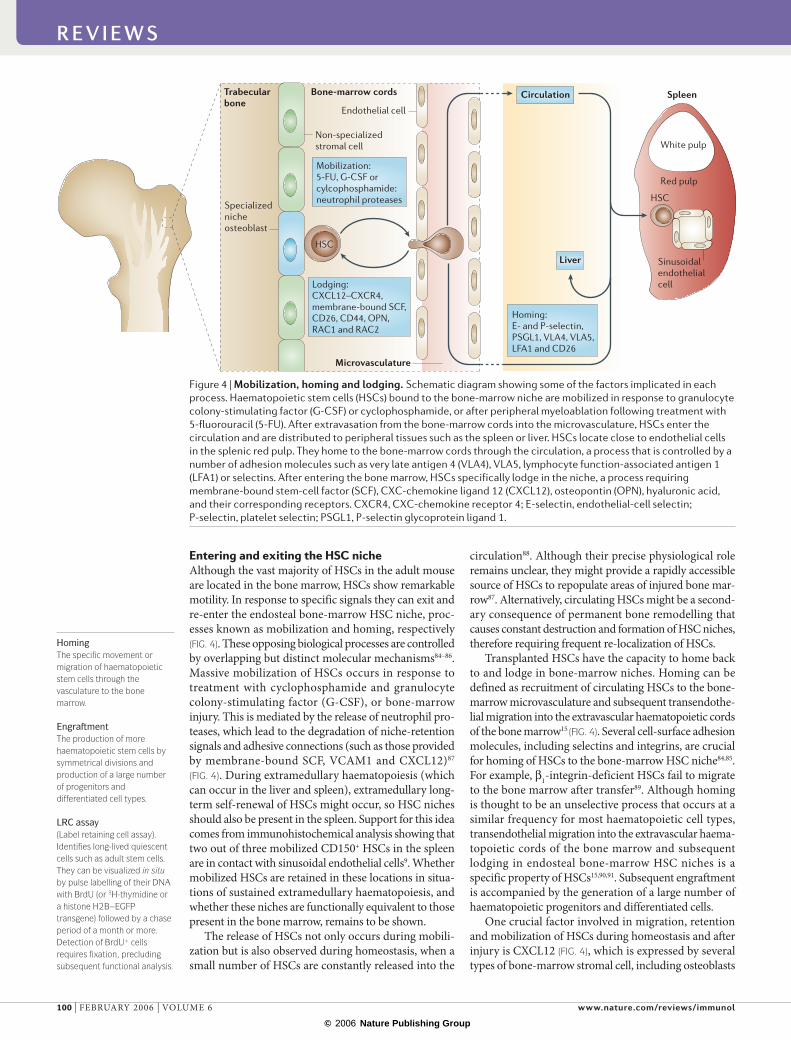
Entering and exiting the HSC nicheAlthough the vast majority of HSCs in the adult mouse are located in the bone marrow, HSCs show remarkable motility. In response to specific signals they can exit and re-enter the endosteal bone-marrow HSC niche, proc-esses known as mobilization and homing, respectively (FIG. 4). These opposing biological processes are controlled by overlapping but distinct molecular mechanisms84–86. Massive mobilization of HSCs occurs in response to treatment with cyclophosphamide and granulocyte colony-stimulating factor (G-CSF), or bone-marrow injury. This is mediated by the release of neutrophil pro-teases, which lead to the degradation of niche-retention signals and adhesive connections (such as those provided by membrane-bound SCF, VCAM1 and CXCL12)87
(FIG. 4). During extramedullary haematopoiesis (which can occur in the liver and spleen), extramedullary long-term self-renewal of HSCs might occur, so HSC niches should also be present in the spleen. Support for this idea comes from immunohistochemical analysis showing that two out of three mobilized CD150+ HSCs in the spleen are in contact with sinusoidal endothelial cells9. Whether mobilized HSCs are retained in these locations in situa-tions of sustained extramedullary haematopoiesis, and whether these niches are functionally equivalent to those present in the bone marrow, remains to be shown.
The release of HSCs not only occurs during mobili-zation but is also observed during homeostasis, when a small number of HSCs are constantly released into the
circulation88. Although their precise physiological role remains unclear, they might provide a rapidly accessible source of HSCs to repopulate areas of injured bone mar-row87. Alternatively, circulating HSCs might be a second-ary consequence of permanent bone remodelling that causes constant destruction and formation of HSC niches, therefore requiring frequent re-localization of HSCs.
Transplanted HSCs have the capacity to home back to and lodge in bone-marrow niches. Homing can be defined as recruitment of circulating HSCs to the bone-marrow microvasculature and subsequent transendothe-lial migration into the extravascular haematopoietic cords of the bone marrow15 (FIG. 4). Several cell-surface adhesion molecules, including selectins and integrins, are crucial for homing of HSCs to the bone-marrow HSC niche84,85. For example, β1-integrin-deficient HSCs fail to migrate to the bone marrow after transfer89. Although homing is thought to be an unselective process that occurs at a similar frequency for most haematopoietic cell types, trans endothelial migration into the extravascular haema-topoietic cords of the bone marrow and subsequent lodging in endosteal bone-marrow HSC niches is a specific property of HSCs15,90,91. Subsequent engraftment is accompanied by the generation of a large number of haematopoietic progenitors and differentiated cells.
One crucial factor involved in migration, retention and mobilization of HSCs during homeostasis and after injury is CXCL12 (FIG. 4), which is expressed by several types of bone-marrow stromal cell, including osteoblasts
Figure 4 | Mobilization, homing and lodging. Schematic diagram showing some of the factors implicated in each process. Haematopoietic stem cells (HSCs) bound to the bone-marrow niche are mobilized in response to granulocyte colony-stimulating factor (G-CSF) or cyclophosphamide, or after peripheral myeloablation following treatment with 5-fluorouracil (5-FU). After extravasation from the bone-marrow cords into the microvasculature, HSCs enter the circulation and are distributed to peripheral tissues such as the spleen or liver. HSCs locate close to endothelial cells in the splenic red pulp. They home to the bone-marrow cords through the circulation, a process that is controlled by a number of adhesion molecules such as very late antigen 4 (VLA4), VLA5, lymphocyte function-associated antigen 1 (LFA1) or selectins. After entering the bone marrow, HSCs specifically lodge in the niche, a process requiring membrane-bound stem-cell factor (SCF), CXC-chemokine ligand 12 (CXCL12), osteopontin (OPN), hyaluronic acid, and their corresponding receptors. CXCR4, CXC-chemokine receptor 4; E-selectin, endothelial-cell selectin; P-selectin, platelet selectin; PSGL1, P-selectin glycoprotein ligand 1.
R E V I E W S
100 | FEBRUARY 2006 | VOLUME 6 www.nature.com/reviews/immunol
R E V I E W S

© 2006 Nature Publishing Group
Angiogenic factorsThese factors (which include angiopoietin-1) promote the development of blood vessels, and are particularly important in embryonic and fetal development.
and vascular endothelial cells92,93. Similar to SCF, CXCL12 expression and secretion is induced in response to haema-topoietic-cell loss due to irradiation, chemotherapy or hypoxia, and purified HSCs migrate specifically towards CXCL12 but not towards any other single chemokine94. The biological effects of CXCL12 are mediated by its capacity to induce motility, chemotaxis and adhesion, as well as to induce secretion of matrix metalloproteinases (MMPs) and angiogenic factors (such as vascular endothe-lial growth factor (VEGF)) by cells expressing its recep-tor, CXC-chemokine receptor 4 (CXCR4). Mice lacking either CXCL12 or CXCR4 show similar embryonic lethal defects, including impaired myeloid- and B-cell haema-topoiesis87,95,96 (see Supplementary information S1 (table)). Importantly, CXCL12 is not essential for HSC generation or expansion in the fetal liver but is crucial for the colonization of bone marrow during late fetal devel-opment. Collectively, the genetic and functional data indicate that the CXCL12–CXCR4 pathway is crucial for retention and maintenance of adult HSCs.
The cytoskeleton also cooperates with cell-surface adhesion molecules to regulate migration and adhe-sion, and is essential for homing and mobilization. For example, Lin–KIT+ cells lacking the RHO family GTPase RAC1 not only fail to engraft97, but also have reduced homing efficiency to the bone marrow and endosteum. Moreover, the deletion of both RAC1 and RAC2 causes massive defects in HSC or haematopoietic-progenitor-cell proliferation, survival, adhesion to very late antigen 4 (VLA4) and/or VLA5, and migration towards CXCL12 in vitro. Deletion of both RAC1 and RAC2 in engrafted HSCs in vivo leads to a massive mobilization of HSC or haematopoietic progenitor cells to the peripheral blood. Together, these data indicate that RAC1 and RAC2 have essential roles in homing, lodging and retention of HSCs in the endosteal bone-marrow HSC niche86,97 (see Supplementary information S1 (table)). In summary, a complex combination of migration, adhesion, proteolysis and signalling occurs at the interface between HSCs and the endosteal bone-marrow niche (FIGS 4,5), and signals originating from the periphery can influence HSC hom-ing, retention and mobilization, therefore determining whether a niche is silent or whether HSCs exit the niche in response to stress.
Molecular crosstalk in the endosteal nicheAlthough little is known about the signals that are exchanged between HSCs and osteoblasts in situ, several receptors, membrane-anchored proteins and secreted factors are expressed by both cell types65. Comparative gene-expression profiling has recently been performed on HSC-supporting and non-supporting stromal cell lines, identifying a number of new molecules that might regulate endosteal bone-marrow HSC-niche activ-ity. These include various interleukins, oncostatin-M, ciliary neurotrophic factor and the membrane protein mKirre98. Here, however, we will focus on the role of molecules for which genetic or functional evidence has been shown in vivo for the regulation of HSC func-tion and/or niche activity (FIG. 5) (see Supplementary information S1 (table)).
Notch signalling. Signalling through Notch receptors is involved in many cell-fate decisions and is thought to have a role in the maintenance of stem cells in a variety of tissues99,100. Moreover, several Notch receptors and Notch-receptor ligands are expressed in the bone mar-row101, leading to the suggestion that Notch signalling has a role in HSC self-renewal and/or clonal expan-sion. Support for this hypothesis has been provided by in vitro culture of purified HSCs on various stromal cell lines102,103. In addition, overexpression of Notch1 in recombination-activating gene 1 (RAG1)-deficient Lin–
SCA1+ progenitors resulted in an increase in the number of HSCs or haematopoietic progenitor cells in vitro and in vivo104. Moreover, as expression of the Notch ligand Jagged-1 is upregulated on osteoblasts that are exposed to PTH, the concomitant increase in HSCs has been postulated to be caused by increased Notch signalling67. However, in contrast to studies leading to over-activation of Notch signalling, loss-of-function studies have failed to show any requirement for Notch signalling in HSCs. Conditional knockout mice for Jagged-1, Notch-1 and Notch-2, or CSL (the common mediator of all signal-ling through Notch receptors) have all been shown to be dispensable for HSC and niche function in vivo105–108. Together, these data indicate that signalling events occur-ring between HSCs and osteoblasts are more complex than has been previously assumed and involve factors other than Notch signals.
Osteopontin. One mechanism by which osteoblasts might regulate the number of HSCs in the bone marrow is through secretion of osteopontin (OPN), an acidic glycoprotein, into the bone matrix109. OPN-deficient mice have a two-fold increase in HSCs and, because the same effect was observed by transplanting wild-type HSCs into lethally irradiated OPN mutant recipients, OPN production by osteoblasts has a negative effect on HSC number91,110. Because cultured Lin–SCA1+ bone-marrow cells are induced to undergo apoptosis when exposed to soluble OPN, the increase in the number of HSCs in OPN-deficient mice has been postulated to be a result of decreased apoptosis110. In addition, OPN has been postulated to act as a negative regulator of HSCs by actively maintaining their quiescence91.
Membrane-bound SCF. The steel (Sl) locus encodes both membrane-bound SCF and secreted SCF. The lat-ter is produced by alternative splicing followed by pro-teolytic cleavage of membrane-bound SCF111. SCF binds and activates KIT, which is expressed at high levels by all LTR HSCs as well as other stem cells. Mutations at either of these loci affect migration and differentiation of primordial germ cells, neural-crest-derived melano-blasts, and haematopoietic cells112 (see Supplementary information S1 (table)). Analysis of the different SCF and KIT mutant mice showed that although not essential for the generation and initial clonal expansion of HSCs in the embryo and fetal liver, they are crucial for long-term maintenance and self-renewal of adult HSCs, raising the possibility that the SCF–KIT pathway mediates endosteal bone-marrow HSC niche activity (FIG. 5).
R E V I E W S
NATURE REVIEWS | IMMUNOLOGY VOLUME 6 | FEBRUARY 2006 | 101
F O C U S O N E A R LY LY M P H O C Y T E D E V E LO P M E N T

© 2006 Nature Publishing Group
Fibronectin
HSC
Motility,self-renewal,survival
Specializedniche osteoblast
Nucleus
LFA1 VLA
4
VLA
5
CD44
Hyaluronicacid
CD44
Frizzled
Integrin
?
Nucleus
OPNWNT
RAC
Notch TIE2SCA1
CXCR4
CXCL12
KIT
Membrane-bound SCF
ANG1
PTH
LRP5or LRP6
BMP
?
PLC, PKCSMADS
Jagged BMPR1A
RAC PI3K?FAK,MAPK
FAK,MAPK
PPR
N–c
adhe
rin N
–cad
heri
n
β-catenin
Non-canonicalWNT signallingpathway
MYBp18p27
CSL
Self-renewal
Adhesion,motility
Adhesion,motility
Adhesion
MYC
???
p21
β-catenin
BMI1HOXB4
ICAM1 VCAM1
Quiescence
Importantly, membrane-bound SCF is expressed by osteoblasts and has a higher and more sustained capacity to activate KIT on the cell surface of HSCs than secreted SCF112,113. In addition, membrane-bound SCF is a potent stimulator of adhesion of HSCs or haematopoietic pro-genitor cells to stromal cells114 because it can activate VLA4 and VLA5, indicating that membrane-bound SCF can affect the adhesive properties of the endosteal niche by modifying the functional state of specific integrins115. Transplantation of normal bone marrow into Sl/Sld mice results in impaired lodging and engraftment of the trans-planted HSCs12,116. In addition, the bone marrow of young Sl/Sld mice has normal LTR activity when transplanted into lethally irradiated recipients, whereas bone marrow from old Sl/Sld mice has greatly reduced LTR activity, indicating a progressive loss of HSC activity over time, potentially due to ceasing niche activity13,14. Collectively,
these data indicate that membrane-bound SCF is an essential component of the endosteal bone-marrow HSC niche that maintains long-term HSC activity in adult bone marrow. However, membrane-bound SCF is also required for osteoblast proliferation and activity in vivo, as shown by the development of osteopaenia in Sl/Sld mice117 (TABLE 1). Therefore, further research is required to clarify whether the effect of membrane-bound SCF is direct (due to its capacity to provide sustained activa-tion of KIT expressed by HSCs), or whether it is indirect (owing to its essential role in the maintenance of niche osteoblasts).
N-cadherin: a central HSC anchor? N-cadherin is expressed by both SNOs and a subset of LSK HSCs39,52. In addition, N-cadherin expression by HSCs localizes asymmetrically to the side of their attachment to SNOs39.
Figure 5 | A model of the endosteal niche–stem-cell synapse. Schematic diagram of the endosteal niche–stem-cell synapse showing putative ligand–receptor interactions and adhesion molecules, as well as some of the intracellular pathways that are activated following signalling. ANG1, angiopoietin-1; BMI1, polycomb repressor; BMP, bone morphogenetic protein; BMPR1A, BMP receptor 1A; CSL, CBF1 suppressor of Hairless and LAG1; CXCL12, CXC-chemokine ligand 12; CXCR4, CXC-chemokine receptor 4; FAK, focal adhesion kinase; HOXB4, homeobox B4; HSC, haematopoietic stem cell; ICAM1, intercellular adhesion molecule 1; LFA1, lymphocyte function-associated antigen 1; LRP, low-density-lipoprotein-receptor-related protein; MAPK, mitogen-activated protein kinase; OPN, osteopontin; PI3K, phosphatidylinositol-3 kinase; PLC, phospholipase C; PKC, protein kinase C; PPR, PTH/PTH-related protein receptor; PTH, parathyroid hormone; SCF, stem-cell factor; SMADS, mothers against decapentaplegic-related homologue; SNO, spindle-shaped N-cadherin-expressing osteoblast; TIE2, tyrosine kinase receptor 2; VCAM1, vascular cell-adhesion molecule 1; VLA4, very late antigen 4; ‘?’ denotes molecules and/or interactions for which only indirect or contradictory evidence is available.
R E V I E W S
102 | FEBRUARY 2006 | VOLUME 6 www.nature.com/reviews/immunol
R E V I E W S

© 2006 Nature Publishing Group
OP9 stromal cells A bone-marrow-derived cell line that can support the expansion of haematopoietic-cell lineages in culture.
Therefore, homotypic N-cadherin interactions have been postulated to be an important component of the anchor that links HSCs to SNOs in the endosteal niche.
In support of this hypothesis, ectopic expression of N-cadherin by OP9 stromal cells substantially increases their ability to maintain mouse HSCs in vitro45. However, genetic evidence of an essential role for N-cadherin in HSC–osteoblast adhesion and/or signalling is still lack-ing, as N-cadherin-mutant embryos fail to develop past mid-gestation118. Moreover, whether functional HSCs are enriched in N-cadherin-expressing LSK HSCs, com-pared to those not expressing this adhesion receptor, has not been shown. Nevertheless, indirect support for the importance of N-cadherin has been obtained from studies showing that MYC and tyrosine kinase recep-tor 2 (TIE2) control N-cadherin expression by HSCs in an antagonistic manner. The effects of MYC and TIE2 on HSCs and on N-cadherin expression correlate with a key function for N-cadherin in the retention of HSCs in the endosteal niche43,45,52,53.
The cell-adhesion signalling network. Genetic evidence for the requirement of TIE2 in HSC–niche interactions has been obtained from chimeric mice comprised of wild-type and TIE1–/–TIE2–/– morulae119. Although TIE1 and TIE2 are not required for the development and dif-ferentiation of fetal HSCs, HSCs lacking both TIE1 and TIE2 fail to be maintained in the adult microenviron-ment. In adult bone marrow, TIE2 (which is expressed specifically by LT-HSCs) is activated by angiopoietin-1 (ANG1), which is secreted by osteoblasts, leading to upregulation of N-cadherin expression by HSCs, pro-viding the first example of a secreted factor promoting HSC–osteoblast adhesion. Interestingly, the ANG1–TIE2 signalling pathway prevents HSC division and main-tains HSC quiescence, both in vitro and in vivo36,41,45,120. Collectively, these data strongly support the hypothesis that N-cadherin-expressing ANG1+ osteoblasts form a niche that maintains quiescence and prevents self-renewal or differentiation through TIE2 signalling (FIG. 5).
TIE2-mediated quiescence is potentially caused by positively regulating the cyclin-dependent-kinase inhibitor p21 (also known as CIP1 and WAF1). HSCs express high levels of p21, and mice lacking p21 show increased HSC proliferation at the expense of long-term self-renewal, indicating that p21 is essen-tial for maintenance of quiescence in HSCs120,121 (see Supplementary information S1 (table)). In contrast to TIE2, transcription of the gene encoding p21 is negatively regulated by MYC, which is expressed at low levels by HSCs but increases during initiation of HSC differentiation in a converse expression pattern to that of p21 (REFS 52,121,122). Interestingly, MYC-deficient LSKFLT3– HSCs overexpress N-cadherin and integrins such as lymphocyte function-associated antigen-1 (LFA1) and VLA5, and contact SNOs52. Although mutant LSKFLT3– HSCs self-renew normally, they have a severe niche-dependent differentiation defect and accumulate in situ. Conversely, enforced MYC activity in HSCs represses the expression of N-cadherin, as well as the expression of several integrins, by LSK HSCs.
Most importantly, MYC overexpressing HSCs are lost over time because of differentiation, presumably owing to their failure to be retained in the niche52. These data indicate that the balance between self-renewal and dif-ferentiation might be controlled by MYC-dependent retention or exit of HSCs from the niche52,53.
N-cadherin and WNT signalling. Intriguingly, it has recently been shown that the transmembrane metallo-proteinase ADAM10 (a disintegrin and metalloprotein-ase-10) is able to cleave N-cadherin that is expressed at the cell surface of fibroblasts and neuronal cells. This leads to the redistribution of β-catenin (which is associated with the intracellular portion of N-cadherin) from the cell surface to the cytoplasmic β-catenin pool, thereby decreasing the signalling threshold required for the expression of target genes of the canonical WNT sig-nalling pathway (which is mediated through β-catenin signal transduction cascades), such as the genes encod-ing cyclin D1 and MYC123. A similar re-distribution of β-catenin has also been reported after E-cadherin cleavage124, indicating that high levels of expression of cadherins, as observed for HSCs, might decrease cyto-plasmic β-catenin levels and therefore negatively regulate expression of β-catenin target genes.
This contrasts with studies in which activation of the WNT signalling pathway in cultured HSCs promotes symmetrical self-renewal in the absence of differentia-tion125,126. However, the importance of canonical WNT signalling during haematopoiesis has recently been questioned because β-catenin is dispensable for HSC function127. Although it is probable that the WNT sig-nalling pathway has an important role in HSC or haema-topoietic progenitor cell function100, the question remains whether WNT promotes self-renewal of LT-HSCs in vivo, or whether it is only important for the expansion and differentiation of non-HSC haematopoietic progenitor cells. The latter is in agreement with the expression pat-tern of the β-catenin target gene Myc, which is induced in LSKFLT3+ progenitor cells leading to downregulation of N-cadherin and integrin expression52. In this context, it is intriguing that the N-cadherin, TIE2, MYC, p21 and β-catenin pathways are apparently interconnected, leading to the suggestion that they might cooperatively control quiescence, self-renewal and initiation of HSC differentiation through interaction with the niche53 (FIG. 5 and Supplementary information S1 (table)).
The stem-cell–niche synapseThe picture emerging from accumulating genetic and functional data indicates that molecular crosstalk between HSCs and niche cells (particularly osteo blasts) involves a large number of molecules (cadherins, integrins, chemokines, cytokines, signalling molecules and receptors) that mediate at least two types of inter-action (FIG. 5). First, adhesive cell–extracellular-matrix (ECM) interactions such as CD44 binding to OPN or hyaluronic acid, and cell–cell interactions, such as those mediated by heterotypic VLA4–VCAM1 interactions and homotypic N-cadherin interactions. The main function of these interactions would be to maintain HSCs in close
R E V I E W S
NATURE REVIEWS | IMMUNOLOGY VOLUME 6 | FEBRUARY 2006 | 103
F O C U S O N E A R LY LY M P H O C Y T E D E V E LO P M E N T

© 2006 Nature Publishing Group
proximity to cells in the endosteal bone-marrow niche. In addition, most adhesion receptors are also linked to intracellular signalling cascades and actively participate in the signalling network controlling HSC maintenance (FIG. 5). Second, ligand–receptor interactions, through which intracellular signalling pathways are activated after ligand binding to receptors that are expressed by HSCs or SNOs (FIG. 5).
Most secreted signalling molecules are bound to the cell surface or ECM, and consequently do not diffuse far. Therefore, the tight adhesion and juxta position of HSCs to niche osteoblasts is essential for the for-mation of an intercellular space in which efficient ligand–receptor interaction can occur. Some osteo-blast-derived signals might be crucial to maintain HSCs in an undifferentiated state and these include the ligand–receptor pairs membrane-bound SCF–KIT, and ANG1–TIE2–MYC (see Supplementary information S1 (table)). Conversely, other ligand–receptor pairs, such as BMP–BMPR1A are important for the number and/or activity of niche osteoblasts. Therefore, in analogy to the neuronal and immunological synapses128, we propose the term ‘stem-cell–niche synapse’ for this adhesion and signalling unit (FIG. 5).
Concluding remarksDuring the past few years, the theoretical concept of a specific stem-cell microenvironment (that is, a stem-cell niche) that was proposed in the 1960s and 1970s, has finally received greater attention129. Substantial progress in localizing the bone-marrow HSC niche(s), as well as its characterization at the molecular and cel-lular levels, has been made. Nevertheless, important questions remain. These include, how many different types of haematopoietic niches exist in the bone mar-row and the periphery, how many HSCs each niche contains, and the exact role each niche unit has during homeostasis and in response to bone-marrow stress. It also remains unclear whether HSC–niche interac-tions are stable or dynamic. Furthermore, although osteoblasts have been shown to be rate-limiting for HSC number, very little is known about the specific differentiation stage of these cells. Are they the same mesenchymal-stem-cell-derived osteoblasts that continue to differentiate into osteocytes, or have they branched off to generate a distinct ‘niche-osteoblast’?
If the latter is the case, do they differentiate in response to signals that are derived from an attaching HSC? The recently identified vascular niche9 opens another chap-ter on HSC–bone-marrow-niche interactions, and the molecular events governing adhesion and signalling of BMECs with HSCs will be an area of intense future research and will move the endothelial-cell field to one of the centre stages of adult-stem-cell research.
Finally, whether long-term self-renewal occurs in sites of extramedullary haematopoiesis, such as the spleen and liver, and therefore maintains blood forma-tion during acute and chronic bone-marrow injury, remains unclear. First attempts to address this question indicate that mobilized splenic HSCs are found close to the vasculature (peripheral vascular niche)9. Are these areas active niches or do they only transiently maintain HSCs? Are they always present or do they form only after injury, and what are the equivalent niche structures in the liver? At the moment there are more questions than answers, but a better understanding of the differ-ent niches will also unearth similarities between them, which should facilitate the eventual reconstruction of active niches in vitro.
Collectively, the impressive progress in the HSC-niche field clearly indicates that substantial clonal expansion of HSCs in vitro unquestionably requires more than just a cytokine cocktail, and instead requires a three-dimensional reconstruction of the niche, includ-ing the appropriate cells and ECM to allow the genera-tion of a stable ‘stem-cell–niche synapse’. This requires not only further progress from the cell–and-molecular-biology end, but is in urgent need of input from matrix- and tissue-engineering fields. Future perspectives have never been more promising, and a breakthrough in the in vitro expansion field will eliminate one of the main obstacles for future regenerative medicine using adult stem cells130.
Note added in proofA recent report shows that HSCs that are deficient for the calcium-sensing receptor show decreased homing to the endosteal niche accompanied by diminished adhesion to collagen type I. These data indicate that local calcium gradients, as are observed around areas of bone remodel-ling, might be involved in engraftment and/or retention of HSCs to the endosteal niche141.
1. Fuchs, E., Tumbar, T. & Guasch, G. Socializing with the neighbors: stem cells and their niche. Cell 116, 769–778 (2004).
2. Osawa, M., Hanada, K., Hamada, H. & Nakauchi, H. Long-term lymphohematopoietic reconstitution by a single CD34–low/negative hematopoietic stem cell. Science 273, 242–245 (1996).
3. Watt, F. M. & Hogan, B. L. Out of Eden: stem cells and their niches. Science 287, 1427–1430 (2000).
4. Weissman, I. L. Stem cells: units of development, units of regeneration, and units in evolution. Cell 100, 157–168 (2000).
5. Kondo, M. et al. Biology of hematopoietic stem cells and progenitors: implications for clinical application. Annu. Rev. Immunol. 21, 759–806 (2003).
6. Till, J. E. & McCulloch, C. E. A direct measurement of the radiation sensitivity of normal mouse bone marrow cells. Radiat. Res. 14, 213–222 (1961).
7. Wagers, A. J., Sherwood, R. I., Christensen, J. L. & Weissman, I. L. Little evidence for developmental plasticity of adult hematopoietic stem cells. Science 297, 2256–2259 (2002).
8. Matsuzaki, Y., Kinjo, K., Mulligan, R. C. & Okano, H. Unexpectedly efficient homing capacity of purified murine hematopoietic stem cells. Immunity 20, 87–93 (2004).
9. Kiel, M. J., Yilmaz, O. H., Iwashita, T., Terhorst, C. & Morrison, S. J. SLAM family receptors distinguish hematopoietic stem and progenitor cells and reveal endothelial niches for stem cells. Cell 121, 1109–1121 (2005).This study shows that CD150, one of the signalling lymphocytic activation molecules (SLAMs) is expressed by LTR HSCs. CD150+ HSCs were localized near sinusoids in normal bone marrow, indicating that in addition to
the endosteal niche a second vascular niche might exist.
10. Curry, J. L., Trentin, J. J. & Wolf, N. Hemopoietic spleen colony studies. II. Erythropoiesis. J. Exp. Med. 125, 703–720 (1967).
11. Schofield, R. The relationship between the spleen colony-forming cell and the haemopoietic stem cell. Blood Cells 4, 7–25 (1978).This paper proposes that HSCs are associated with other cell types, and that these allow self-renewal but prevent cell maturation. Schofield termed this microenvironment the ‘stem-cell niche’.
12. McCulloch, E. A., Siminovitch, L., Till, J. E., Russell, E. S. & Bernstein, S. E. The cellular basis of the genetically determined hemopoietic defect in anemic mice of genotype Sl-Sld. Blood 26, 399–410 (1965).
R E V I E W S
104 | FEBRUARY 2006 | VOLUME 6 www.nature.com/reviews/immunol
R E V I E W S
© 2006 Nature Publishing Group
This paper (together with references 13 and 14) shows that normal bone marrow fails to engraft in Sl/Sld mice. This is probably the first study demonstrating that the microenvironment is essential for bone-marrow HSC function and/or maintenance.
13. Barker, J. E. Sl/Sld hematopoietic progenitors are deficient in situ. Exp. Hematol. 22, 174–177 (1994).
14. Barker, J. E. Early transplantation to a normal microenvironment prevents the development of Steel hematopoietic stem cell defects. Exp. Hematol. 25, 542–547 (1997).
15. Nilsson, S. K. & Simmons, P. J. Transplantable stem cells: home to specific niches. Curr. Opin. Hematol. 11, 102–106 (2004).
16. Ohlstein, B., Kai, T., Decotto, E. & Spradling, A. The stem cell niche: theme and variations. Curr. Opin. Cell Biol. 16, 693–699 (2004).
17. Spradling, A., Drummond-Barbosa, D. & Kai, T. Stem cells find their niche. Nature 414, 98–104 (2001).
18. Wu, A. M., Siminovitch, L., Till, J. E. & McCulloch, E. A. Evidence for a relationship between mouse hemopoietic stem cells and cells forming colonies in culture. Proc. Natl Acad. Sci. USA 59, 1209–1215 (1968).
19. Domen, J. & Weissman, I. L. Self-renewal, differentiation or death: regulation and manipulation of hematopoietic stem cell fate. Mol. Med. Today 5, 201–208 (1999).
20. Yang, L. et al. Identification of Lin–Sca1+kit +
CD34+Flt3– short-term hematopoietic stem cells capable of rapidly reconstituting and rescuing myeloablated transplant recipients. Blood 105, 2717–2723 (2005).
21. Adolfsson, J. et al. Identification of Flt3+lympho-myeloid stem cells lacking erythro-megakaryocytic potential a revised road map for adult blood lineage commitment. Cell 121, 295–306 (2005).
22. Christensen, J. L. & Weissman, I. L. Flk-2 is a marker in hematopoietic stem cell differentiation: a simple method to isolate long-term stem cells. Proc. Natl Acad. Sci. USA 98, 14541–14546 (2001).
23. Jordan, C. T. & Lemischka, I. R. Clonal and systemic analysis of long-term hematopoiesis in the mouse. Genes Dev. 4, 220–232 (1990).
24. Ramalho-Santos, M., Yoon, S., Matsuzaki, Y., Mulligan, R. C. & Melton, D. A. ‘Stemness’: transcriptional profiling of embryonic and adult stem cells. Science 298, 597–600 (2002).
25. Ivanova, N. B. et al. A stem cell molecular signature. Science 298, 601–604 (2002).
26. Akashi, K. et al. Transcriptional accessibility for genes of multiple tissues and hematopoietic lineages is hierarchically controlled during early hematopoiesis. Blood 101, 383–389 (2003).
27. Venezia, T. A. et al. Molecular signatures of proliferation and quiescence in hematopoietic stem cells. PLoS Biol. 2, e301 (2004).
28. Roegiers, F. & Jan, Y. N. Asymmetric cell division. Curr. Opin. Cell Biol. 16, 195–205 (2004).
29. Betschinger, J. & Knoblich, J. A. Dare to be different: asymmetric cell division in Drosophila, C. elegans and vertebrates. Curr. Biol. 14, R674–R685 (2004).
30. Lechler, T. & Fuchs, E. Asymmetric cell divisions promote stratification and differentiation of mammalian skin. Nature 437, 275–280 (2005).This paper shows that some basal cells of the skin epidermis divide in an asymmetric manner (divisional asymmetry), and that this correlates with the unequal distribution of determinants including PAR3, LGN and mINSC in the two daughter cells.
31. Suda, J., Suda, T. & Ogawa, M. Analysis of differentiation of mouse hemopoietic stem cells in culture by sequential replating of paired progenitors. Blood 64, 393–399 (1984).
32. Takano, H., Ema, H., Sudo, K. & Nakauchi, H. Asymmetric division and lineage commitment at the level of hematopoietic stem cells: inference from differentiation in daughter cell and granddaughter cell pairs. J. Exp. Med. 199, 295–302 (2004).
33. Ho, A. D. Kinetics and symmetry of divisions of hematopoietic stem cells. Exp. Hematol. 33, 1–8 (2005).
34. Xie, T. & Spradling, A. C. A niche maintaining germ line stem cells in the Drosophila ovary. Science 290, 328–330 (2000).
35. Kiger, A. A., White-Cooper, H. & Fuller, M. T. Somatic support cells restrict germline stem cell self-renewal and promote differentiation. Nature 407, 750–754 (2000).
36. Fleming, W. H. et al. Functional heterogeneity is associated with the cell cycle status of murine hematopoietic stem cells. J. Cell Biol. 122, 897–902 (1993).
37. Cotsarelis, G., Sun, T. T. & Lavker, R. M. Label-retaining cells reside in the bulge area of pilosebaceous unit: implications for follicular stem cells, hair cycle, and skin carcinogenesis. Cell 61, 1329–1337 (1990).
38. Tumbar, T. et al. Defining the epithelial stem cell niche in skin. Science 303, 359–363 (2004).In this paper, transgenic expression of H2B–EGFP is used to show that slow-cycling skin epidermal stem cells are primarily located in the hair follicle bulge. This technique, which identifies and allows purification of viable LRCs, should be adaptable for analysis of other adult stem cells and their associated niches.
39. Zhang, J. et al. Identification of the haematopoietic stem cell niche and control of the niche size. Nature 425, 836–841 (2003).N-cadherin was proposed as a critical molecule that anchors HSCs to osteoblasts through homotypic interactions. This study (together with reference 67), also shows that osteoblastic cells are a critical component of the endosteal bone-marrow niche.
40. Lerner, C. & Harrison, D. E. 5-Fluorouracil spares hemopoietic stem cells responsible for long-term repopulation. Exp. Hematol. 18, 114–118 (1990).
41. Spangrude, G. J. & Johnson, G. R. Resting and activated subsets of mouse multipotent hematopoietic stem cells. Proc. Natl Acad. Sci. USA 87, 7433–7437 (1990).
42. Uchida, N. et al. The unexpected G0/G1 cell cycle status of mobilized hematopoietic stem cells from peripheral blood. Blood 89, 465–472 (1997).
43. Suda, T., Arai, F. & Hirao, A. Hematopoietic stem cells and their niche. Trends Immunol. 26, 426–433 (2005).
44. Potten, C. S. & Loeffler, M. Stem cells: attributes, cycles, spirals, pitfalls and uncertainties. Lessons for and from the crypt. Development 110, 1001–1020 (1990).
45. Arai, F. et al. Tie2/angiopoietin-1 signalling regulates hematopoietic stem cell quiescence in the bone marrow niche. Cell 118, 149–161 (2004).This paper shows that the ANG1 produced by niche osteoblasts activates TIE2, which maintains HSC quiescence and increases N-cadherin expression, and therefore increases adhesion to the endosteal niche.
46. Oshima, H., Rochat, A., Kedzia, C., Kobayashi, K. & Barrandon, Y. Morphogenesis and renewal of hair follicles from adult multipotent stem cells. Cell 104, 233–245 (2001).
47. Van Zant, G. Studies of hematopoietic stem cells spared by 5-fluorouracil. J. Exp. Med. 159, 679–690 (1984).
48. Randall, T. D. & Weissman, I. L. Phenotypic and functional changes induced at the clonal level in hematopoietic stem cells after 5-fluorouracil treatment. Blood 89, 3596–3606 (1997).
49. Uchida, N. et al. ABC transporter activities of murine hematopoietic stem cells vary according to their developmental and activation status. Blood 103, 4487–4495 (2004).
50. Chambers, I. & Smith, A. Self-renewal of teratocarcinoma and embryonic stem cells. Oncogene 23, 7150–7160 (2004).
51. Cheshier, S. H., Morrison, S. J., Liao, X. & Weissman, I. L. In vivo proliferation and cell cycle kinetics of long-term self- renewing hematopoietic stem cells. Proc. Natl Acad. Sci. USA 96, 3120–3125 (1999).
52. Wilson, A. et al. c-Myc controls the balance between hematopoietic stem cell self-renewal and differentiation. Genes Dev. 18, 2747–2763 (2004).This paper provides genetic evidence that differential expression of MYC controls the balance between stem-cell self-renewal and differentiation by regulating entry and exit of HSCs from the endosteal niche.
53. Murphy, M. J., Wilson, A. & Trumpp, A. More than just proliferation: Myc function in stem cells. Trends Cell Biol. 15, 128–137 (2005).
54. Patt, H. M. & Maloney, M. A. Bone formation and resorption as a requirement for marrow development. Proc. Soc. Exp. Biol. Med. 140, 205–207 (1972).
55. Maloney, M. A. & Patt, H. M. On the origin of hematopoietic stem cells after local marrow extirpation. Proc. Soc. Exp. Biol. Med. 149, 94–97 (1975).
56. Deguchi, K. et al. Excessive extramedullary hematopoiesis in Cbfa1-deficient mice with a congenital lack of bone marrow. Biochem. Biophys. Res. Commun. 255, 352–359 (1999).
57. Ducy, P., Schinke, T. & Karsenty, G. The osteoblast: a sophisticated fibroblast under central surveillance. Science 289, 1501–1504 (2000).
58. Karsenty, G. & Wagner, E. F. Reaching a genetic and molecular understanding of skeletal development. Dev. Cell 2, 389–406 (2002).
59. Lord, B. I., Testa, N. G. & Hendry, J. H. The relative spatial distributions of CFUs and CFUc in the normal mouse femur. Blood 46, 65–72 (1975).
60. Gong, J. K. Endosteal marrow: a rich source of hematopoietic stem cells. Science 199, 1443–1445 (1978).This study provides the first functional evidence for the location of HSCs and progenitor cells close to endosteal surfaces of rat bone.
61. Askenasy, N. & Farkas, D. L. In vivo imaging studies of the effect of recipient conditioning, donor cell phenotype and antigen disparity on homing of haematopoietic cells to the bone marrow. Br. J. Haematol. 120, 505–515 (2003).
62. Sipkins, D. A. et al. In vivo imaging of specialized bone marrow endothelial microdomains for tumour engraftment. Nature 435, 969–973 (2005).
63. Nilsson, S. K., Johnston, H. M. & Coverdale, J. A. Spatial localization of transplanted hemopoietic stem cells: inferences for the localization of stem cell niches. Blood 97, 2293–2299 (2001).
64. Taichman, R. S. & Emerson, S. G. The role of osteoblasts in the hematopoietic microenvironment. Stem Cells 16, 7–15 (1998).
65. Taichman, R. S. Blood and bone: two tissues whose fates are intertwined to create the hematopoietic stem-cell niche. Blood 105, 2631–2639 (2005).
66. Oostendorp, R. A. et al. Stromal cell lines from mouse aorta-gonads-mesonephros subregions are potent supporters of hematopoietic stem cell activity. Blood 99, 1183–1189 (2002).
67. Calvi, L. M. et al. Osteoblastic cells regulate the haematopoietic stem cell niche. Nature 425, 841–846 (2003).
68. Chen, D., Zhao, M. & Mundy, G. R. Bone morphogenetic proteins. Growth Factors 22, 233–241 (2004).
69. Visnjic, D. et al. Conditional ablation of the osteoblast lineage in Col2.3δk transgenic mice. J. Bone Miner. Res. 16, 2222–2231 (2001).
70. Visnjic, D. et al. Hematopoiesis is severely altered in mice with an induced osteoblast deficiency. Blood 103, 3258–3264 (2004).This paper shows that conditional ablation of osteoblasts results in a reversible decrease of bone-marrow HSCs and extramedullary haematopoiesis, indicating that osteoblasts are not only required for maintenance of bone-marrow haematopoiesis, but are also an essential component of the niche.
71. Corral, D. A. et al. Dissociation between bone resorption and bone formation in osteopenic transgenic mice. Proc. Natl Acad. Sci. USA 95, 13835–13840 (1998).
72. Huber, T. L., Kouskoff, V., Fehling, H. J., Palis, J. & Keller, G. Haemangioblast commitment is initiated in the primitive streak of the mouse embryo. Nature 432, 625–630 (2004).
73. Ohneda, O. et al. Hematopoietic stem cell maintenance and differentiation are supported by embryonic aorta-gonad-mesonephros region-derived endothelium. Blood 92, 908–919 (1998).
74. Li, W. et al. Primary endothelial cells isolated from the yolk sac and para-aortic splanchnopleura support the expansion of adult marrow stem cells in vitro. Blood 102, 4345–4353 (2003).
75. Li, W., Johnson, S. A., Shelley, W. C. & Yoder, M. C. Hematopoietic stem cell repopulating ability can be maintained in vitro by some primary endothelial cells. Exp. Hematol. 32, 1226–1237 (2004).
76. Kopp, H. G., Avecilla, S. T., Hooper, A. T. & Rafii, S. The bone marrow vascular niche: home of HSC differentiation and mobilization. Physiology (Bethesda Md.) 20, 349–356 (2005).
77. Avecilla, S. T. et al. Chemokine-mediated interaction of hematopoietic progenitors with the bone marrow vascular niche is required for thrombopoiesis. Nature Med. 10, 64–71 (2004).
78. Rafii, S., Mohle, R., Shapiro, F., Frey, B. M. & Moore, M. A. Regulation of hematopoiesis by microvascular endothelium. Leuk. Lymphoma 27, 375–386 (1997).
R E V I E W S
NATURE REVIEWS | IMMUNOLOGY VOLUME 6 | FEBRUARY 2006 | 105
F O C U S O N E A R LY LY M P H O C Y T E D E V E LO P M E N T

© 2006 Nature Publishing Group
79. Heissig, B. et al. Recruitment of stem and progenitor cells from the bone marrow niche requires MMP-9 mediated release of kit-ligand. Cell 109, 625–637 (2002).
80. Rafii, S. et al. Human bone marrow microvascular endothelial cells support long-term proliferation and differentiation of myeloid and megakaryocytic progenitors. Blood 86, 3353–3363 (1995).
81. Delehanty, L. L. et al. Stromal inhibition of megakaryocytic differentiation is associated with blockade of sustained Rap1 activation. Blood 101, 1744–1751 (2003).
82. Tokoyoda, K., Egawa, T., Sugiyama, T., Choi, B. I. & Nagasawa, T. Cellular niches controlling B lymphocyte behaviour within bone marrow during development. Immunity 20, 707–718 (2004).
83. Hirose, J. et al. A developing picture of lymphopoiesis in bone marrow. Immunol. Rev. 189, 28–40 (2002).
84. Papayannopoulou, T. Bone marrow homing: the players, the playfield, and their evolving roles. Curr. Opin. Hematol. 10, 214–219 (2003).
85. Lapidot, T., Dar, A. & Kollet, O. How do stem cells find their way home? Blood 106, 1901–1910 (2005).
86. Cancelas, J. A. et al. Rac GTPases differentially integrate signals regulating hematopoietic stem cell localization. Nature Med. 11, 886–891 (2005).
87. Lapidot, T. & Petit, I. Current understanding of stem cell mobilization: the roles of chemokines, proteolytic enzymes, adhesion molecules, cytokines, and stromal cells. Exp. Hematol. 30, 973–981 (2002).
88. Wright, D. E., Wagers, A. J., Gulati, A. P., Johnson, F. L. & Weissman, I. L. Physiological migration of hematopoietic stem and progenitor cells. Science 294, 1933–1936 (2001).
89. Potocnik, A. J., Brakebusch, C. & Fassler, R. Fetal and adult hematopoietic stem cells require β1 integrin function for colonizing fetal liver, spleen, and bone marrow. Immunity 12, 653–663 (2000).
90. Nilsson, S. K. et al. Hyaluronan is synthesized by primitive hemopoietic cells, participates in their lodgment at the endosteum following transplantation, and is involved in the regulation of their proliferation and differentiation in vitro. Blood 101, 856–862 (2003).
91. Nilsson, S. K. et al. Osteopontin, a key component of the hematopoietic stem cell niche and regulator of primitive hematopoietic progenitor cells. Blood 106, 1232–1239 (2005).
92. Ara, T. et al. Long-term hematopoietic stem cells require stromal cell-derived factor-1 for colonizing bone marrow during ontogeny. Immunity 19, 257–267 (2003).The authors show that CXCL12 has an important role in colonization of the bone marrow by HSCs.
93. Ponomaryov, T. et al. Induction of the chemokine stromal-derived factor-1 following DNA damage improves human stem cell function. J. Clin. Invest. 106, 1331–1339 (2000).
94. Wright, D. E., Bowman, E. P., Wagers, A. J., Butcher, E. C. & Weissman, I. L. Hematopoietic stem cells are uniquely selective in their migratory response to chemokines. J. Exp. Med. 195, 1145–1154 (2002).
95. Nagasawa, T. et al. Defects of B-cell lymphopoiesis and bone-marrow myelopoiesis in mice lacking the CXC chemokine PBSF/SDF-1. Nature 382, 635–638 (1996).
96. Zou, Y. R., Kottmann, A. H., Kuroda, M., Taniuchi, I. & Littman, D. R. Function of the chemokine receptor CXCR4 in haematopoiesis and in cerebellar development. Nature 393, 595–599 (1998).
97. Gu, Y. et al. Hematopoietic cell regulation by Rac1 and Rac2 guanosine triphosphatases. Science 302, 445–449 (2003).
98. Moore, K. A. Recent advances in defining the hematopoietic stem cell niche. Curr. Opin. Hematol. 11, 107–111 (2004).
99. Artavanis-Tsakonas, S., Rand, M. D. & Lake, R. J. Notch signalling: cell fate control and signal integration in development. Science 284, 770–776 (1999).
100. Reya, T. & Clevers, H. Wnt signalling in stem cells and cancer. Nature 434, 843–850 (2005).
101. Radtke, F., Wilson, A., Mancini, S. J. & MacDonald, H. R. Notch regulation of lymphocyte development and function. Nature Immunol. 5, 247–253 (2004).
102. Duncan, A. W. et al. Integration of Notch and Wnt signalling in hematopoietic stem cell maintenance. Nature Immunol. 6, 314–322 (2005).
103. Varnum-Finney, B. et al. Pluripotent, cytokine-dependent, hematopoietic stem cells are immortalized by constitutive Notch1 signalling. Nature Med. 6, 1278–1281 (2000).
104. Stier, S., Cheng, T., Dombkowski, D., Carlesso, N. & Scadden, D. T. Notch1 activation increases hematopoietic stem cell self-renewal in vivo and favours lymphoid over myeloid lineage outcome. Blood 99, 2369–2378 (2002).
105. Han, H. et al. Inducible gene knockout of transcription factor recombination signal binding protein-J reveals its essential role in T versus B lineage decision. Int. Immunol. 14, 637–645 (2002).
106. Mancini, S. J. et al. Jagged1-dependent Notch signalling is dispensable for hematopoietic stem cell self-renewal and differentiation. Blood 105, 2340–2342 (2005).
107. Radtke, F. et al. Deficient T cell fate specification in mice with an induced inactivation of Notch1. Immunity 10, 547–558 (1999).
108. Saito, T. et al. Notch2 is preferentially expressed in mature B cells and indispensable for marginal zone B lineage development. Immunity 18, 675–685 (2003).
109. Denhardt, D. T. & Guo, X. Osteopontin: a protein with diverse functions. FASEB J. 7, 1475–1482 (1993).
110. Stier, S. et al. Osteopontin is a hematopoietic stem cell niche component that negatively regulates stem cell pool size. J. Exp. Med. 201, 1781–1791 (2005).
111. Flanagan, J. G., Chan, D. C. & Leder, P. Transmembrane form of the kit ligand growth factor is determined by alternative splicing and is missing in the Sld mutant. Cell 64, 1025–1035 (1991).
112. Lyman, S. D. & Jacobsen, S. E. c-kit ligand and Flt3 ligand: stem/progenitor cell factors with overlapping yet distinct activities. Blood 91, 1101–1134 (1998).
113. Miyazawa, K. et al. Membrane-bound Steel factor induces more persistent tyrosine kinase activation and longer life span of c-kit gene-encoded protein than its soluble form. Blood 85, 641–649 (1995).
114. Kinashi, T. & Springer, T. A. Steel factor and c-kit regulate cell-matrix adhesion. Blood 83, 1033–1038 (1994).
115. Kovach, N. L., Lin, N., Yednock, T., Harlan, J. M. & Broudy, V. C. Stem cell factor modulates avidity of α4 β1 and α5 β1 integrins expressed on hematopoietic cell lines. Blood 85, 159–167 (1995).
116. Driessen, R. L., Johnston, H. M. & Nilsson, S. K. Membrane-bound stem cell factor is a key regulator in the initial lodgment of stem cells within the endosteal marrow region. Exp. Hematol. 31, 1284–1291 (2003).
117. Lotinun, S., Evans, G. L., Turner, R. T. & Oursler, M. J. Deletion of membrane-bound steel factor results in osteopenia in mice. J. Bone Miner. Res. 20, 644–652 (2005).
118. Radice, G. L. et al. Developmental defects in mouse embryos lacking N-cadherin. Dev. Biol. 181, 64–78 (1997).
119. Puri, M. C. & Bernstein, A. Requirement for the TIE family of receptor tyrosine kinases in adult but not fetal hematopoiesis. Proc. Natl Acad. Sci. USA 100, 12753–12758 (2003).
120. Cheng, T. et al. Hematopoietic stem cell quiescence maintained by p21cip1/waf1. Science 287, 1804–1808 (2000).
121. Cheng, T., Shen, H., Rodrigues, N., Stier, S. & Scadden, D. T. Transforming growth factor β1 mediates cell-cycle arrest of primitive hematopoietic cells independent of p21(Cip1/Waf1) or p27(Kip1). Blood 98, 3643–3649 (2001).
122. Wu, S. et al. Myc represses differentiation-induced p21CIP1 expression via Miz-1-dependent interaction with the p21 core promoter. Oncogene 22, 351–360 (2003).
123. Reiss, K. et al. ADAM10 cleavage of N-cadherin and regulation of cell–cell adhesion and β-catenin nuclear signalling. EMBO J. 24, 742–752 (2005).
124. Ito, K. et al. Calcium influx triggers the sequential proteolysis of extracellular and cytoplasmic domains of E-cadherin, leading to loss of β-catenin from cell–cell contacts. Oncogene 18, 7080–7090 (1999).
125. Willert, K. et al. Wnt proteins are lipid-modified and can act as stem cell growth factors. Nature 423, 448–452 (2003).This paper, together with reference 126, provides evidence of a positive role for WNT signalling in
HSC self-renewal, whereas differentiation (at least in vitro) is inhibited.
126. Reya, T. et al. A role for Wnt signalling in self-renewal of haematopoietic stem cells. Nature 423, 409–414 (2003).
127. Cobas, M. et al. β-catenin is dispensable for hematopoiesis and lymphopoiesis. J. Exp. Med. 199, 221–229 (2004).
128. Friedl, P. & Storim, J. Diversity in immune-cell interactions: states and functions of the immunological synapse. Trends Cell Biol. 14, 557–567 (2004).
129. Powell, K. It’s the ecology, stupid. Nature 435, 268–270 (2005).
130. Sorrentino, B. P. Clinical strategies for expansion of haematopoietic stem cells. Nature Rev. Immunol. 4, 878–888 (2004).
131. Ploemacher, R. E. Stem cells: characterization and measurement. Baillieres Clin. Haematol. 10, 429–444 (1997).
132. Morrison, S. J., Uchida, N. & Weissman, I. L. The biology of hematopoietic stem cells. Annu. Rev. Cell Dev. Biol. 11, 35–71 (1995).
133. Visser, J. W., Bauman, J. G., Mulder, A. H., Eliason, J. F. & de Leeuw, A. M. Isolation of murine pluripotent hemopoietic stem cells. J. Exp. Med. 159, 1576–1590 (1984).
134. Bunting, K. D. ABC Transporters as phenotypic markers and functional regulators of stem cells. Stem Cells 20, 11–20 (2002).
135. Goodell, M. A., McKinney-Freeman, S. & Camargo, F. D. Isolation and characterization of side population cells. Methods Mol. Biol. 290, 343–352 (2005).
136. Uchida, N., Dykstra, B., Lyons, K. J., Leung, F. Y. & Eaves, C. J. Different in vivo repopulating activities of purified hematopoietic stem cells before and after being stimulated to divide in vitro with the same kinetics. Exp. Hematol. 31, 1338–1347 (2003).
137. Camargo, F. D., Chambers, S. M., Drew, E., McNagny, K. M. & Goodell, M. A. Hematopoietic stem cells do not engraft with absolute efficiencies. Blood 107, 501–507 (2006).
138. Bradford, G. B., Williams, B., Rossi, R. & Bertoncello, I. Quiescence, cycling, and turnover in the primitive hematopoietic stem cell compartment. Exp. Hematol. 25, 445–453 (1997).
139. Komori, T. et al. Targeted disruption of Cbfa1 results in a complete lack of bone formation owing to maturational arrest of osteoblasts. Cell 89, 755–764 (1997).
140. Otto, F. et al. Cbfa1, a candidate gene for cleidocranial dysplasia syndrome, is essential for osteoblast differentiation and bone development. Cell 89, 765–771 (1997).
141. Adams, G. D. et al. Stem cell engraftment at the endosteal niche is specified by the calcium-sensing receptor. Nature. Dec 28, 2005 (doi:10.1038/nature04247 16382241).
AcknowledgementsWe would l ike to thank F. Radtke, R. MacDonald, M. Murphy and G. Oser for their critical reading of the manuscript, and members of the Trumpp Laboratory for helpful discussions. We apologize to colleagues whose work could not be cited due to space limitations. This work was in part supported by grants to A.T. from the Swiss National Science Foundation, the Swiss Cancer League and the UBS Optimus Foundation. A.T. is member of the EMBO Young Investigator Program.
Competing interests statementThe authors declare no competing financial interests.
DATABASESThe following terms in this article are linked online to:Entrez Gene: http://www.ncbi.nlm.nih.gov/entrez/query.fcgi?db=geneCD34 | CD150 | CXCL12 | FLT3 | KIT | N-cadherin | SCA1 | SCF
FURTHER INFORMATIONAndreas Trumpp’s homepage: http://www.isrec.ch/research/groups/research_groups_detail_eid_1668_lid_2.htm
SUPPLEMENTARY INFORMATIONSee online article: S1 (table) Access to this links box is available online.
R E V I E W S
106 | FEBRUARY 2006 | VOLUME 6 www.nature.com/reviews/immunol
R E V I E W S


