Focal Parenchymal Atrophy and Fat Replacement Are Clues ...
9
Tohoku J. Exp. Med., 2020, 252, 63-71 63 Received June 8, 2020; revised and accepted August 20, 2020. Published online September 3, 2020; doi: 10.1620/tjem.252.63. Correspondence: Atsushi Masamune, M.D., Ph.D., Division of Gastroenterology, Tohoku University Graduate School of Medicine, 1-1 Seiryo-machi, Aoba-ku, Sendai, Miyagi 980-8574, Japan. e-mail: amasamune@med.tohoku.ac.jp ©2020 Tohoku University Medical Press. This is an open-access article distributed under the terms of the Creative Commons Attribution-NonCommercial-NoDerivatives 4.0 International License (CC-BY-NC-ND 4.0). Anyone may download, reuse, copy, reprint, or distribute the article without modifications or adaptations for non-profit purposes if they cite the original authors and source properly. https://creativecommons.org/licenses/by-nc-nd/4.0/ Focal Parenchymal Atrophy and Fat Replacement Are Clues for Early Diagnosis of Pancreatic Cancer with Abnormalities of the Main Pancreatic Duct Shin Miura, 1 Kiyoshi Kume, 1 Kazuhiro Kikuta, 1 Shin Hamada, 1 Tetsuya Takikawa, 1 Naoki Yoshida, 1 Seiji Hongo, 1 Yu Tanaka, 1 Ryotaro Matsumoto, 1 Takanori Sano, 1 Mio Ikeda, 1 Toru Furukawa, 2 Masahiro Iseki, 3 Michiaki Unno 3 and Atsushi Masamune 1 1 Division of Gastroenterology, Tohoku University Graduate School of Medicine, Sendai, Miyagi, Japan 2 Department of Investigative Pathology, Tohoku University Graduate School of Medicine, Sendai, Miyagi, Japan 3 Department of Surgery, Tohoku University Graduate School of Medicine, Sendai, Miyagi, Japan Pancreatic cancer is one of the most dangerous solid tumors, but its early diagnosis is difficult. The abnormality of the main pancreatic duct (MPD), such as a single localized stricture and upstream dilatation, might be useful in the early detection of pancreatic cancer. However, these findings are often observed in benign inflammatory cases. This study aimed to clarify whether early pancreatic cancer presenting MPD abnormalities has characteristic features different from those of benign cases. This is a single-center, retrospective study. We analyzed 20 patients who underwent pancreatectomy presenting with a single, localized MPD stricture without identifiable masses on imaging: 10 patients with pancreatic ductal adenocarcinoma (cancer group; 6 with stage 0 and 4 with stage I) and 10 patients with benign strictures (benign group; 8 with inflammation and 2 with low-grade pancreatic intraepithelial neoplasms). Pancreatectomy was performed in these benign cases because high-grade intraepithelial neoplasm was suspected. Although the proportion of patients with diabetes mellitus tended to be higher in the cancer group (6/10) than that in the benign group (1/10) (P = 0.058), other clinical characteristics were not different between the groups. Preoperative cytological malignancies were detected in four patients in the cancer group (4/10) but not in the benign group (P = 0.09). Focal parenchymal atrophy and fat replacement were more frequently detected on computed tomography in the cancer group (7/10) than in the benign group (1/10) (P = 0.02). In conclusion, focal parenchymal atrophy and fat replacement may provide clues for the early diagnosis of pancreatic cancer. Keywords: early pancreatic cancer; endoscopic ultrasound; pancreatic intraepithelial neoplasia; pancreatitis; serial pancreatic juice aspiration cytological examination Tohoku J. Exp. Med., 2020 September, 252 (1), 63-71. Introduction Pancreatic cancer is one of the most dangerous solid cancers (Kamisawa et al. 2016; Pereira et al. 2020). The American Cancer Society estimates that 57,600 individuals (30,400 men and 27,200 women) will be diagnosed with pancreatic cancer, and pancreatic cancer will cause approxi- mately 47,050 deaths (24,640 men and 22,410 women) in 2020 (https://www.cancer.org/cancer/pancreatic-cancer/ about/key-statistics.html, accessed on May 11, 2020). In Japan, pancreatic cancer is the fourth (fourth in men and third in women) leading cause of cancer-related death, and the mortality rate was 29.7 per 100,000 men and 27.4 per 100,000 women in 2018 (https://ganjoho.jp/reg_stat/statis- tics/dl/index.html#mortality, accessed on May 11, 2020). The 5-year relative survival rate is as low as 5.9% in men and 8.1% in women (https://ganjoho.jp/reg_stat/statistics/ dl/index.html#survival, accessed on May 11, 2020). Pancreatic cancer is predicted to become the second leading cause of cancer-related deaths by 2030 in the United States
Transcript of Focal Parenchymal Atrophy and Fat Replacement Are Clues ...

Early Diagnosis of Pancreatic Cancer 63Tohoku J. Exp. Med., 2020, 252, 63-71
63
Received June 8, 2020; revised and accepted August 20, 2020. Published online September 3, 2020; doi: 10.1620/tjem.252.63.Correspondence: Atsushi Masamune, M.D., Ph.D., Division of Gastroenterology, Tohoku University Graduate School of Medicine, 1-1
Seiryo-machi, Aoba-ku, Sendai, Miyagi 980-8574, Japan.e-mail: [email protected]
©2020 Tohoku University Medical Press. This is an open-access article distributed under the terms of the Creative Commons Attribution-NonCommercial-NoDerivatives 4.0 International License (CC-BY-NC-ND 4.0). Anyone may download, reuse, copy, reprint, or distribute the article without modifications or adaptations for non-profit purposes if they cite the original authors and source properly.https://creativecommons.org/licenses/by-nc-nd/4.0/
Focal Parenchymal Atrophy and Fat Replacement Are Clues for Early Diagnosis of Pancreatic Cancer with Abnormalities of the Main Pancreatic Duct
Shin Miura,1 Kiyoshi Kume,1 Kazuhiro Kikuta,1 Shin Hamada,1 Tetsuya Takikawa,1 Naoki Yoshida,1 Seiji Hongo,1 Yu Tanaka,1 Ryotaro Matsumoto,1 Takanori Sano,1 Mio Ikeda,1 Toru Furukawa,2 Masahiro Iseki,3 Michiaki Unno3 and Atsushi Masamune1
1Division of Gastroenterology, Tohoku University Graduate School of Medicine, Sendai, Miyagi, Japan2Department of Investigative Pathology, Tohoku University Graduate School of Medicine, Sendai, Miyagi, Japan3Department of Surgery, Tohoku University Graduate School of Medicine, Sendai, Miyagi, Japan
Pancreatic cancer is one of the most dangerous solid tumors, but its early diagnosis is difficult. The abnormality of the main pancreatic duct (MPD), such as a single localized stricture and upstream dilatation, might be useful in the early detection of pancreatic cancer. However, these findings are often observed in benign inflammatory cases. This study aimed to clarify whether early pancreatic cancer presenting MPD abnormalities has characteristic features different from those of benign cases. This is a single-center, retrospective study. We analyzed 20 patients who underwent pancreatectomy presenting with a single, localized MPD stricture without identifiable masses on imaging: 10 patients with pancreatic ductal adenocarcinoma (cancer group; 6 with stage 0 and 4 with stage I) and 10 patients with benign strictures (benign group; 8 with inflammation and 2 with low-grade pancreatic intraepithelial neoplasms). Pancreatectomy was performed in these benign cases because high-grade intraepithelial neoplasm was suspected. Although the proportion of patients with diabetes mellitus tended to be higher in the cancer group (6/10) than that in the benign group (1/10) (P = 0.058), other clinical characteristics were not different between the groups. Preoperative cytological malignancies were detected in four patients in the cancer group (4/10) but not in the benign group (P = 0.09). Focal parenchymal atrophy and fat replacement were more frequently detected on computed tomography in the cancer group (7/10) than in the benign group (1/10) (P = 0.02). In conclusion, focal parenchymal atrophy and fat replacement may provide clues for the early diagnosis of pancreatic cancer.
Keywords: early pancreatic cancer; endoscopic ultrasound; pancreatic intraepithelial neoplasia; pancreatitis; serial pancreatic juice aspiration cytological examinationTohoku J. Exp. Med., 2020 September, 252 (1), 63-71.
IntroductionPancreatic cancer is one of the most dangerous solid
cancers (Kamisawa et al. 2016; Pereira et al. 2020). The American Cancer Society estimates that 57,600 individuals (30,400 men and 27,200 women) will be diagnosed with pancreatic cancer, and pancreatic cancer will cause approxi-mately 47,050 deaths (24,640 men and 22,410 women) in 2020 (https://www.cancer.org/cancer/pancreatic-cancer/about/key-statistics.html, accessed on May 11, 2020). In
Japan, pancreatic cancer is the fourth (fourth in men and third in women) leading cause of cancer-related death, and the mortality rate was 29.7 per 100,000 men and 27.4 per 100,000 women in 2018 (https://ganjoho.jp/reg_stat/statis-tics/dl/index.html#mortality, accessed on May 11, 2020). The 5-year relative survival rate is as low as 5.9% in men and 8.1% in women (https://ganjoho.jp/reg_stat/statistics/dl/index.html#survival, accessed on May 11, 2020). Pancreatic cancer is predicted to become the second leading cause of cancer-related deaths by 2030 in the United States

S. Miura et al.64
(Rahib et al. 2014). Pancreatic ductal adenocarcinoma (PDAC) is the most common type of pancreatic cancer.
Early diagnosis is essential in improving the prognosis of pancreatic cancer, but it is challenging (Singhi et al. 2019; Vasen et al. 2019; Pereira et al. 2020). The Pancreatic Cancer Registry in Japan revealed that the 5-year survival rates of patients with the Union for International Cancer Control stage 0 (high-grade pancreatic intraepithelial neo-plasia [PanIN]/ carcinoma in situ [CIS]), stage IA (tumor size < 2 cm with no lymph node metastasis and no distant metastasis), and stage IB (tumor size > 2 cm but no more than 4 cm, with no lymph node metastasis and no distant metastasis) were 85.8%, 68.7%, and 59.7%, respectively, but accounted for only 1.7%, 4.1%, and 6.3%, respectively, in all registered patients (Egawa et al. 2012). The diagnosis of early pancreatic cancer, including stage 0, is extremely difficult because the tumor is usually unidentifiable by imaging modalities and only indirect findings caused by the tumor might provide a clue. Main pancreatic duct (MPD) abnormalities, such as localized stricture/cutoff and upstream dilatation, might be the key secondary findings for early detection of pancreatic cancer in the absence of tumor identification (Ahn et al. 2009; Chu et al. 2017; Kanno et al. 2018; Kanno et al. 2019; Elbanna et al. 2020). In the retrospective review, these MPD findings were found in 50% of patients with PDAC at 18 months before the actual diagnosis of PDAC (Gangi et al. 2004). Pancreatic cancer arising from the pancreatic duct epithelium forms a pancreatic duct stricture without forming a tumor at the stage of high-grade PanIN/CIS (Hruban et al. 2001). However, these findings are not specific to pancreatic can-cer and often observed in benign inflammatory cases. It is not rare that the cases with MPD abnormalities undergo pancreatectomy for suspected pancreatic cancer in the absence of positive cytology, and postoperative pathologi-cal examination revealed no malignancy. If we could dif-ferentiate MPD abnormalities associated with early pancre-atic cancer from benign ones, invasive pancreatectomy would have been avoided. This study aimed to clarify whether early pancreatic cancer presenting with MPD abnormalities has characteristic features different from those of benign cases.
Materials and MethodsStudy design
This is a single-center, retrospective observational study. This study was performed in accordance with the principles of the Declaration of Helsinki, and approved by the Ethics Committee of Tohoku University Graduate School of Medicine (article#: 2019-1-920; 2019-1-921). Informed consent was waived because of the retrospective nature of the study. Clinicopathological information was obtained from the medical records.
SubjectsWe analyzed patients presenting with single, localized
stricture and upstream dilatation of MPD without identifi-able masses on multi-detector row computed tomography (MD-CT), endoscopic ultrasound (EUS), and magnetic res-onance imaging/ magnetic resonance cholangiopancreatog-raphy (MRCP) who underwent pancreatectomy for sus-pected pancreatic cancer at Tohoku University Hospital between January 2011 and February 2020. Patients with diffuse or multiple MPD strictures diagnosed as autoim-mune pancreatitis or chronic pancreatitis, tumor-forming pancreatitis, intraductal papillary mucinous neoplasms, and intraductal papillary mucinous neoplasm-associated adeno-carcinoma as the postoperative pathology, and pancreatic cancers other than PDAC, such as acinar cell carcinoma, were excluded. EUS was performed using radial-array echoendoscopes (GF-UM2000, GF-UE260; Olympus, Tokyo, Japan) by experienced endoscopists with > 1,000 cases. Cytological examination was performed by brushing cytology, serial pancreatic juice aspiration cytological examination (SPACE), or pancreatic juice cytology during endoscopic retrograde pancreatography (ERP). If the cath-eter passed through the MPD stricture, endoscopic nasopan-creatic drainage was performed for SPACE, and pancreatic juice was obtained for cytological examination at least 3 times. If the brush passed through the stricture, brushing cytology was performed. If both procedures failed, casual pancreatic juice was collected for cytological examination. Class IV or V in cytology was defined as malignant accord-ing to the Papanicolaou classification (Papanicolaou 1954). We used a single- (PR-418Q; Olympus) or double-lumen catheter (Uneven cannula; Piolax Medical Devices, Yokohama, Japan) for pancreatic juice collection.
Pathological evaluation of fat infiltrationPathological fat infiltration was evaluated in resected
pancreas specimen using a 3-tiered scoring system as previ-ously described (Matsuda et al. 2017). To differentiate pan-creatic fat infiltration from peripancreatic soft tissues including adipose tissue, we focused on and evaluated inter-lobular and intralobular fat infiltration. Interlobular fat infiltration was defined as the presence of adipocytes within the interlobular space and was scored as follows: 0, no fat infiltration; 1, some adipocytes present; 2, numerous adipo-cytes separating the lobules. Intralobular fat infiltration was scored as follows: 0, no or rare adipocytes present in some lobules; 1, scattered adipocytes present in the majority of lobules; 2, numerous adipocytes forming clusters of more than 10 cells present in the majority of lobules.
Statistical analysisTo compare proportions, we used Fisher’s exact test.
Continuous variables were presented as mean ± standard deviation (SD) and compared using the unpaired Student’s t-test. Analyses were performed using JMP Pro 14. A two-sided P value < 0.05 was considered significant.

Early Diagnosis of Pancreatic Cancer 65
ResultsTwenty patients presented with single, localized stricture and upstream dilatation of MPD with no identifiable masses
During the study period, 360 patients underwent pan-createctomy with a postoperative pathological diagnosis of PDAC. According to the Union for International Cancer Control classification (8th edition), PDAC was Stage 0 in 6 patients (1.6%), Stage IA in 45 patients (12.1%), Stage IB in 21 patients (5.7%), Stage IIA in 132 patients (35.7%), Stage IIB in 83 patients (22.4%), Stage III in 23 patients (6.2%), and Stage IV in 50 patients (13.5%). In cases with Stages II, III, and IV, tumors were detected on preoperative
imaging in all except one patient. This patient had no MPD stricture and was excluded from the study. Six patients with Stage 0 (high-grade PanIN/CIS) and four with Stage IA presented with single, localized stricture and upstream dilatation of MPD with no identifiable masses on MD-CT, MRCP, and EUS. All 10 patients were histologically diag-nosed with PDAC (Fig. 1). The tumor sizes in Stage IA cases were 1.5 mm, 8 mm, 9 mm, and 9 mm in diameter. The localization of PDAC did not always correspond to MPD stricture and was occasionally located in the nearby branched pancreatic ducts. On the other hand, 10 benign (8 with inflammatory stricture and 2 with low-grade PanIN) cases presented with single, localized stricture and upstream
Fig.1. Case of carcinoma in situ. (A) MD-CT image. White arrow indicates focal parenchymal atrophy and fat replacement. (B) MRCP image. Local-
ized stricture (white arrow) and dilatation of the upstream MPD are observed. (C) EUS image. An MPD stricture (white arrow) was observed, but no tumor was detected. (D) ERP image. Localized stricture (white arrow) and up-stream dilatation of the MPD were observed. (E) Loupe image of the resected pancreas (H & E staining). Black arrow indicates the lesion of carcinoma in situ and asterisks indicate fat replacement. (F) Enlarged view of the lesion of high-grade pancreatic intraepithelial neoplasia/carcinoma in situ. Magnification: 200 ×.

S. Miura et al.66
dilatation of MPD with no identifiable masses (Fig. 2). Pancreatectomy was performed in these 10 benign cases with sufficient informed consent because high-grade PanIN/CIS was suspected. These 20 patients (10 in the cancer group and 10 in the benign group) were enrolled in this study.
Comparison of clinical characteristics between the early cancer and benign groups
Table 1 shows the clinical characteristics of the 20 patients stratified by group. The clinical characteristics did not have a significant difference between the two groups. The proportion of patients with diabetes mellitus (DM) tended to be higher in the cancer group (6/10, 60%) than that in the benign group (1/10, 10%), but the difference was not statistically significant (P = 0.058). Seven (70%) can-cer cases were asymptomatic and detected during the fol-low-up for other diseases (n = 3), deterioration of DM (n =
2), and medical checkup (n = 2). The MPD stricture was most frequently located in the pancreatic body and tail (9 [90%] cases in the cancer group and 10 [100%] cases in the benign group). Distal pancreatectomy, which is less inva-sive than pancreatoduodenectomy, was performed in all of the 10 benign cases. Serum pancreatic enzyme (mostly lipase) levels were elevated in 5 (50%) cancer and 4 (40%) benign cases, probably reflecting retention of pancreatic juice.
Focal parenchymal atrophy and fat replacement were associated with early pancreatic cancer
Table 2 shows the imaging findings. All imaging modalities could detect MPD abnormalities including local-ized stricture and upstream dilatation of MPD. MPD find-ings, such as the location, length of stenosis, diameter of the upstream duct, and severity of the stenosis, were not different between the groups (data not shown). Focal
Fig. 2. Case of inflammatory MPD stricture. (A) MD-CT image. Dilatation of the MPD is detected in the pancreatic tail. (B) MRCP image. Localized stricture (white
arrow) and dilatation of the upstream MPD are detected. (C) EUS image. MPD stricture (white arrow) and dilatation of MPD are detected. (D) ERP image. Localized stricture (white arrow) and upstream dilatation of the MPD are detected in the pancreatic tail. (E) Loupe image of the resected pancreas (H & E staining). The white arrow indicates the MPD stricture. Parenchymal fat replacement or atrophy is not observed. (F) Enlarged view. No atypical epithelium is ob-served. Magnification: 200 ×.
Early Diagnosis of Pancreatic Cancer 67
parenchymal atrophy with a constricted neck and concomi-tant fat replacement were more frequently detected on MD-CT in the cancer group (7/10, 70%) than in the benign group (1/10, 10%) (P = 0.02) (Fig. 3). EUS and magnetic resonance imaging failed to detect focal fatty changes (data not shown). In all except for one benign case, focal paren-chymal atrophy and fat replacement were not evident on imaging modalities (Fig. 2E).
Postoperative pathological examination revealed extensive fat infiltration and atrophy in the pancreatic parenchyma near the MPD stricture in the malignant cases (Fig. 1E). The strictures of MPD were due to intraluminal proliferation of epithelial cells accompanied by fibrosis in duct wall and periductal parenchyma. In the case with can-cer in branch duct, there was invasion surrounding the
branch duct, and MPD was narrowed by the fibrosis associ-ated with invasion and inflammation. In 3 malignant cases, parenchymal atrophy, fat replacement, and fibrosis were observed only in limited areas, which could not be detected on preoperative imaging modalities. Histological diagnosis of the cysts detected in 6 malignant cases was ductal dilata-tion in 5 cases and pseudocyst in one case. In the benign cases, the MPD strictures were due to fibrosis and inflam-mation in the duct wall and/or periductal parenchyma. Some of them were apparently restricted to the periductal region, which might be caused by acute pancreatitis in the limited area. The other cases showed more extensive parenchymal fibrosis, which might be caused by chronic pancreatitis. Histological diagnosis of the cysts detected in 4 benign cases was pseudocyst in 3 cases and intraductal
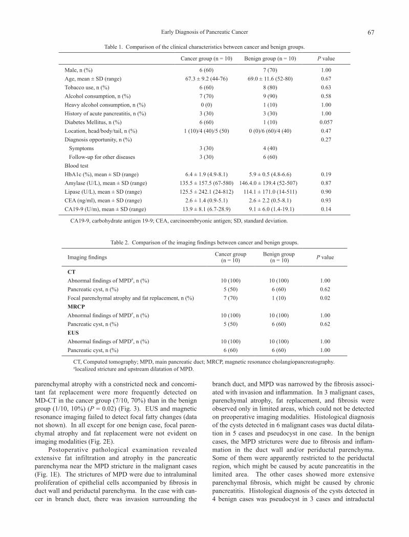
Table 1. Comparison of the clinical characteristics between cancer and benign groups.
Cancer group (n = 10) Benign group (n = 10) P value
Male, n (%) 6 (60) 7 (70) 1.00Age, mean ± SD (range) 67.3 ± 9.2 (44-76) 69.0 ± 11.6 (52-80) 0.67Tobacco use, n (%) 6 (60) 8 (80) 0.63Alcohol consumption, n (%) 7 (70) 9 (90) 0.58Heavy alcohol consumption, n (%) 0 (0) 1 (10) 1.00History of acute pancreatitis, n (%) 3 (30) 3 (30) 1.00Diabetes Mellitus, n (%) 6 (60) 1 (10) 0.057Location, head/body/tail, n (%) 1 (10)/4 (40)/5 (50) 0 (0)/6 (60)/4 (40) 0.47Diagnosis opportunity, n (%) 0.27 Symptoms 3 (30) 4 (40) Follow-up for other diseases 3 (30) 6 (60)Blood testHbA1c (%), mean ± SD (range) 6.4 ± 1.9 (4.9-8.1) 5.9 ± 0.5 (4.8-6.6) 0.19Amylase (U/L), mean ± SD (range) 135.5 ± 157.5 (67-580) 146.4.0 ± 139.4 (52-507) 0.87Lipase (U/L), mean ± SD (range) 125.5 ± 242.1 (24-812) 114.1 ± 171.0 (14-511) 0.90CEA (ng/ml), mean ± SD (range) 2.6 ± 1.4 (0.9-5.1) 2.6 ± 2.2 (0.5-8.1) 0.93CA19-9 (U/m), mean ± SD (range) 13.9 ± 8.1 (6.7-28.9) 9.1 ± 6.0 (1.4-19.1) 0.14
CA19-9, carbohydrate antigen 19-9; CEA, carcinoembryonic antigen; SD, standard deviation.
Table 2. Comparison of the imaging findings between cancer and benign groups.
Imaging findings Cancer group (n = 10)
Benign group (n = 10) P value
CTAbnormal findings of MPD#, n (%) 10 (100) 10 (100) 1.00Pancreatic cyst, n (%) 5 (50) 6 (60) 0.62Focal parenchymal atrophy and fat replacement, n (%) 7 (70) 1 (10) 0.02MRCPAbnormal findings of MPD#, n (%) 10 (100) 10 (100) 1.00Pancreatic cyst, n (%) 5 (50) 6 (60) 0.62EUSAbnormal findings of MPD#, n (%) 10 (100) 10 (100) 1.00Pancreatic cyst, n (%) 6 (60) 6 (60) 1.00
CT, Computed tomography; MPD, main pancreatic duct; MRCP, magnetic resonance cholangiopancreatography.#localized stricture and upstream dilatation of MPD.

S. Miura et al.68
papillary mucinous neoplasm in one case. Histological diagnosis of the cysts could not be made in two benign cases because the cysts were located outside of the resected areas.
The longitudinal diameter of MPD was not different between the malignant cases (1,607 ± 570 µm) and the benign cases (1,229 ± 196 µm) (P = 0.08). In addition, we evaluated fat infiltration using a 3-tiered scoring system (Matsuda et al. 2017). Interlobular fat infiltration was more evident in the malignant cases (1.90 ± 0.31) compared to the benign cases (0.88 ± 0.78) (P < 0.01). Similarly, intra-lobular fat infiltration was more evident in the malignant cases (1.80 ± 0.63) compared to the benign cases (0.22 ± 0.44) (P < 0.01). These results suggest the association of fat replacement with early pancreatic cancer.
Detection of cytological malignancy only in less than half of cancer cases
In this study, EUS-fine needle aspiration was not pos-sible because no imaging findings directly revealed pancre-atic tumor. Therefore, cytological examination was per-formed by brushing cytology, SPACE, or casual pancreatic juice cytology during ERP. In 10 patients with cancer, brushing cytology was performed in 7, SPACE in 8, and casual pancreatic juice cytology in 2. Three cases were positive for malignancy in brushing cytology and SPACE, and one case was positive for malignancy in SPACE. Cytology was positive in 4/8 (50%) patients who underwent
SPACE. Malignancy was detected in none of the casual pancreatic juice cytology cases. Altogether, malignancy was noted in 4 (40%) cases. In patients with benign cases, brushing cytology was performed in 6, SPACE in 6, and casual pancreatic juice cytology in 2 cases. Both brushing cytology and SPACE were performed in 4 patients. Malignancy was detected in none of the 10 benign cases. Preoperative cytological malignancy was specific for pan-creatic cancer, but the proportion of patients with positive malignancy was not different between the cancer and benign groups (P = 0.09).
Favorable prognosis in patients with early pancreatic cancer
All 6 patients with Stage 0 cancer had recurrence-free survival with an average observation period of 28 months (range, 2-54 months) postoperatively. In patients with Stage IA, 3 patients had recurrence-free survival for an average observation period of 38 (range, 21-68) months, but one patient had recurrence of PDAC in the remnant pancreas at 40 months. The patient had no distant metasta-ses but was unable to undergo additional surgery because he developed leukemia. He died 6 months after diagnosis. Ten patients with benign stricture underwent observation for an average of 24 months (range, 5-63 months) after sur-gery and did not develop acute pancreatitis or pancreatic cancer.
Fig. 3. MD-CT images of the cancer cases presenting with focal parenchymal atrophy. (A-C) MD-CT images of early pancreatic cancer cases presenting with focal parenchymal atrophy (white arrows). (D)
In a case of benign MPD stricture, parenchymal atrophy is not evident (white arrowhead). A cyst is detected (asterisk).

Early Diagnosis of Pancreatic Cancer 69
DiscussionIn this study, we analyzed 20 patients presenting with
single, localized MPD strictures without identifiable masses on imaging modalities and compared the characteristics between the early pancreatic cancer and benign groups. Although this study has limitations, such as a retrospective, single-center study design with a limited number of enrolled patients, it has a strength that compared patients with early cancer with benign cases whose diagnosis was proven by histopathological examination of the resected pancreas. Although the proportion of patients with DM tended to be higher in the cancer group (P = 0.058), clinical characteris-tics and MPD findings on imaging modalities had no signif-icant difference between the two groups. Preoperative cytological malignancies were specific to the cancer group but positive in less than half (4/10) of cases. Of note, focal parenchymal atrophy and fat replacement were more fre-quently observed in the cancer group than in the benign group. These results suggest the difficulty in differentiating cancer cases from benign cases solely based on clinical characteristics and MPD findings, but the presence of focal parenchymal atrophy and fat replacement detected on CT might be useful in the diagnosis of early pancreatic cancer. In a multicenter study of early pancreatic cancer in Japan (Kanno et al. 2018), focal fat replacement was detected in 21/50 (42.0%) Stage 0 and 61/146 (41.8%) Stage I cases. A case of acute pancreatitis-onset high-grade PanIN/CIS with focal fat replacement has been reported (Satoh et al. 2017). Fat replacement might not only result from but also be a risk factor for pancreatic carcinogenesis. Pancreatic fat infiltration is involved in PDAC development in animals and humans (Takahashi et al. 2018). The molecular mecha-nisms responsible for focal fatty changes remain unclear but are likely to involve interactions between cancer cells and the microenvironment (Erkan et al. 2012; Masamune and Shimosegawa 2013).
Although not specific to pancreatic cancer, it is increasingly recognized that abnormal MPD findings on imaging modalities are important in the diagnosis of early pancreatic cancer (Tanaka et al. 2010; Yoon et al. 2011; Yokode et al. 2018; Elbanna et al. 2020). These findings are occasionally found in asymptomatic patients during medical checkup or surveillance of other diseases (Kanno et al. 2018). A retrospective review of prediagnostic CT images revealed the presence of MPD abnormalities, such as dilatation and stricture/cutoff at 2-6 months prior to the diagnosis of pancreatic cancer in nearly half of cases (Gangi et al. 2004). Early pancreatic cancer, not by itself but con-comitant with inflammation and fibrosis, might play a role in the development of MPD changes. Yokode et al. (2018) examined the association between PanIN and radiologically detectable abnormalities. They showed that high-grade PanIN affecting MPD may present with localized duct stric-ture, and thus unexplained duct stenosis might become a diagnostic clue for premalignant lesions and early pancre-
atic cancer. PanIN itself was unlikely to cause duct stenosis because it was almost flat with no intraductal proliferation. MPD strictures are more likely to be associated with inflam-mation and fibrosis. To increase the diagnostic ability, Chen et al. (2020) developed algorithms to predict the risk of pancreatic cancer in patients with ductal abnormalities detected in routine clinical care based on clinical character-istics, such as DM and cross-sectional imaging findings. In this study, 2 patients with cancer, but none of the patients with benign cases, were diagnosed with deterioration of DM. Although the difference was not statistically signifi-cant (P = 0.058), the proportion of patients with DM tended to be higher in the cancer group in this study. DM is a known risk factor for pancreatic cancer and, has attracted attention, especially new-onset DM, as a diagnostic target of early pancreatic cancer (Singhi et al. 2019). New-onset DM after 50 years of age confers a 6-8-fold increased risk of PDAC, and approximately 1% of patients will be diag-nosed with PDAC within 3 years (Sharma et al. 2018). The Enriching New-Onset Diabetes for Pancreatic Cancer score system has been developed to stratify the risk of PDAC in patients with new-onset DM (Sharma et al. 2018).
In this study, one of the three patients with Stage IA developed recurrence of PDAC in the remnant pancreas at 40 months after surgery. Generally, recurrence of pancre-atic cancer is not so common because the majority of cases are diagnosed at advanced stages and the follow-up period is not extremely long. Miyazaki et al. (2014) reported that only 11 of 284 (3.9%) patients developed recurrence in the remnant pancreas postoperatively. Unlike pancreatic cancer in general, the prognosis of early pancreatic cancer is favor-able, and recurrence in the remnant pancreas is an important issue (Ikemoto et al. 2018). Resection of recurrent pancre-atic cancer in the remnant pancreas can offer a favorable outcome (Miyazaki et al. 2014). The development of new lesions was reported in 31/200 (15.5%) cases in a multi-center study in Japan (Kanno et al. 2018). In a prospective study of 30 patients with Stage 0 or IA, 9 patients devel-oped recurrence (8 in the remnant pancreas and 1 in the liver) during the median follow-up period of 54 months. Discrimination of local recurrence from multicentric cancer is difficult in the absence of genetic analysis (Gotoh et al. 2019).
Mikata et al. (2013) reported that SPACE was a feasi-ble method in the diagnosis of pancreatic high-grade PanIN/CIS. In the current study, malignancy was detected in 4 of 8 patients with cancer who underwent SPACE, and the sen-sitivity of cytology was insufficiently high. Six malignant and 10 benign cases underwent pancreatectomy in the absence of positive cytology. In clinical practice, there is no consensus on whether to proceed with pancreatectomy or follow-up in the case of negative cytology. The risk of cancer progression and degree of invasion in surgery should be balanced. In the case of 10 benign patients, the location of the MPD stricture in the pancreatic body and tail might have affected the decision because pancreatoduodenectomy

S. Miura et al.70
was not required and less invasive distal pancreatectomy was performed. A new modality and biomarker that can detect early pancreatic cancer with high sensitivity and specificity are urgently needed to manage this intractable disease.
AcknowledgmentsThis study was supported in part by a Grant-in-Aid from
KUROKAWA CANCER RESEARCH FOUNDATION (to Kazuhiro Kikuta).
Conflict of InterestThe authors declare no conflict of interest.
ReferencesAhn, S.S., Kim, M.J., Choi, J.Y., Hong, H.S., Chung, Y.E. & Lim,
J.S. (2009) Indicative findings of pancreatic cancer in predi-agnostic CT. Eur. Radiol., 19, 2448-2455.
Chen, W., Butler, R.K., Zhou, Y., Parker, R.A., Jeon, C.Y. & Wu, B.U. (2020) Prediction of pancreatic cancer based on imaging features in patients with duct abnormalities. Pancreas, 49, 413-419.
Chu, L.C., Goggins, M.G. & Fishman, E.K. (2017) Diagnosis and detection of pancreatic cancer. Cancer J., 23, 333-342.
Egawa, S., Toma, H., Ohigashi, H., Okusaka, T., Nakao, A., Hatori, T., Maguchi, H., Yanagisawa, A. & Tanaka, M. (2012) Japan Pancreatic Cancer Registry; 30th year anniversary: Japan Pancreas Society. Pancreas, 41, 985-992.
Elbanna, K.Y., Jang, H.J. & Kim, T.K. (2020) Imaging diagnosis and staging of pancreatic ductal adenocarcinoma: a compre-hensive review. Insights Imaging, 11, 58.
Erkan, M., Adler, G., Apte, M.V., Bachem, M.G., Buchholz, M., Detlefsen, S., Esposito, I., Friess, H., Gress, T.M., Habisch, H.J., Hwang, R.F., Jaster, R., Kleeff, J., Klöppel, G., Kordes, C., et al. (2012) StellaTUM: current consensus and discussion on pancreatic stellate cell research. Gut, 61, 172-178.
Gangi, S., Fletcher, J.G., Nathan, M.A., Christensen, J.A., Harmsen, W.S., Crownhart, B.S. & Chari, S.T. (2004) Time interval between abnormalities seen on CT and the clinical diagnosis of pancreatic cancer: retrospective review of CT scans obtained before diagnosis. AJR Am. J. Roentgenol., 182, 897-903.
Gotoh, Y., Ohtsuka, T., Nakamura, S., Shindo, K., Ohuchida, K., Miyasaka, Y., Mori, Y., Mochidome, N., Oda, Y. & Nakamura, M. (2019) Genetic assessment of recurrent pancreatic high-risk lesions in the remnant pancreas: metachronous multifocal lesion or local recurrence? Surgery, 165, 767-774.
Hruban, R.H., Adsay, N.V., Albores-Saavedra, J., Compton, C., Garrett, E.S., Goodman, S.N., Kern, S.E., Klimstra, D.S., Klöppel, G., Longnecker, D.S., Luttges, J. & Offerhaus, G.J. (2001) Pancreatic intraepithelial neoplasia: a new nomencla-ture and classification system for pancreatic duct lesions. Am. J. Surg. Pathol., 25, 579-586.
Ikemoto, J., Hanada, K., Minami, T., Okazaki, A., Abe, T., Amano, H. & Yonehara, S. (2018) Prospective follow-up study of the recurrence of pancreatic cancer diagnosed at an early stage: the value of endoscopic ultrasonography for early diagnosis of recurrence in the remnant pancreas. Pancreas, 47, 482-488.
Kamisawa, T., Wood, L.D., Itoi, T. & Takaori, K. (2016) Pancre-atic cancer. Lancet, 388, 73-85.
Kanno, A., Masamune, A., Hanada, K., Maguchi, H., Shimizu, Y., Ueki, T., Hasebe, O., Ohtsuka, T., Nakamura, M., Takenaka, M., Kitano, M., Kikuyama, M., Gabata, T., Yoshida, K., Sasaki, T., et al. (2018) Multicenter study of early pancreatic cancer in Japan. Pancreatology, 18, 61-67.
Kanno, Y., Koshita, S., Ogawa, T., Kusunose, H., Masu, K., Sakai, T., Yonamine, K., Kawakami, Y., Fujii, Y., Miyamoto, K., Murabayashi, T., Kozakai, F., Horaguchi, J., Noda, Y., Oikawa, M., et al. (2019) Predictive value of localized stenosis of the main pancreatic duct for early detection of pancreatic cancer. Clin. Endosc., 52, 588-597.
Masamune, A. & Shimosegawa, T. (2013) Pancreatic stellate cells: multi-functional cells in the pancreas. Pancreatology, 13, 102-105.
Matsuda, Y., Furukawa, T., Yachida, S., Nishimura, M., Seki, A., Nonaka, K., Aida, J., Takubo, K., Ishiwata, T., Kimura, W., Arai, T. & Mino-Kenudson, M. (2017) The prevalence and clinicopathological characteristics of high-grade pancreatic intraepithelial neoplasia: autopsy study evaluating the entire pancreatic parenchyma. Pancreas, 46, 658-664.
Mikata, R., Ishihara, T., Tada, M., Tawada, K., Saito, M., Kuro-sawa, J., Sugiyama, H., Sakai, Y., Tsuyuguchi, T., Miyazaki, M. & Yokosuka, O. (2013) Clinical usefulness of repeated pancreatic juice cytology via endoscopic naso-pancreatic drainage tube in patients with pancreatic cancer. J. Gastroen-terol., 48, 866-873.
Miyazaki, M., Yoshitomi, H., Shimizu, H., Ohtsuka, M., Yoshi-dome, H., Furukawa, K., Takayasiki, T., Kuboki, S., Okamura, D., Suzuki, D. & Nakajima, M. (2014) Repeat pancreatec-tomy for pancreatic ductal cancer recurrence in the remnant pancreas after initial pancreatectomy: is it worthwhile? Surgery, 155, 58-66.
Papanicolaou, G.N. (1954) Cytological evaluation of smears prepared by the tampon method for the detection of carcinoma of the uterine cervix. Cancer, 7, 1185-1190.
Pereira, S.P., Oldfield, L., Ney, A., Hart, P.A., Keane, M.G., Pandol, S.J., Li, D., Greenhalf, W., Jeon, C.Y., Koay, E.J., Almario, C.V., Halloran, C., Lennon, A.M. & Costello, E. (2020) Early detection of pancreatic cancer. Lancet Gastro-enterol. Hepatol., 5, 698-710.
Rahib, L., Smith, B.D., Aizenberg, R., Rosenzweig, A.B., Fleshman, J.M. & Matrisian, L.M. (2014) Projecting cancer incidence and deaths to 2030: the unexpected burden of thyroid, liver, and pancreas cancers in the United States. Cancer Res., 74, 2913-2921.
Satoh, T., Kikuyama, M., Kawaguchi, S., Kanemoto, H., Muro, H. & Hanada, K. (2017) Acute pancreatitis-onset carcinoma in situ of the pancreas with focal fat replacement diagnosed using serial pancreatic-juice aspiration cytologic examination (SPACE). Clin. J. Gastroenterol., 10, 541-545.
Sharma, A., Kandlakunta, H., Nagpal, S.J.S., Feng, Z., Hoos, W., Petersen, G.M. & Chari, S.T. (2018) Model to determine risk of pancreatic cancer in patients with new-onset diabetes. Gastroenterology, 155, 730-739. e733.
Singhi, A.D., Koay, E.J., Chari, S.T. & Maitra, A. (2019) Early detection of pancreatic cancer: opportunities and challenges. Gastroenterology, 156, 2024-2040.
Takahashi, M., Hori, M., Ishigamori, R., Mutoh, M., Imai, T. & Nakagama, H. (2018) Fatty pancreas: a possible risk factor for pancreatic cancer in animals and humans. Cancer Sci., 109, 3013-3023.
Tanaka, S., Nakao, M., Ioka, T., Takakura, R., Takano, Y., Tsukuma, H., Uehara, H., Suzuki, R. & Fukuda, J. (2010) Slight dilatation of the main pancreatic duct and presence of pancreatic cysts as predictive signs of pancreatic cancer: a prospective study. Radiology, 254, 965-972.
Vasen, H.F.A., Boekestijn, B., Ibrahim, I.S., Inderson, A., Bonsing, B.A., de Vos Tot Nederveen Cappel, W.H., Feshtali, S. & Wasser, M.N. (2019) Dilatation of the main pancreatic duct as first manifestation of small pancreatic ductal adenocarcinomas detected in a hereditary pancreatic cancer surveillance program. HPB (Oxford), 21, 1371-1375.
Yokode, M., Akita, M., Fujikura, K., Kim, M.J., Morinaga, Y., Yoshikawa, S., Terada, T., Matsukiyo, H., Tajiri, T., Abe-

Early Diagnosis of Pancreatic Cancer 71
Suzuki, S., Itoh, T., Hong, S.M. & Zen, Y. (2018) High-grade PanIN presenting with localised stricture of the main pancre-atic duct: a clinicopathological and molecular study of 10 cases suggests a clue for the early detection of pancreatic cancer. Histopathology, 73, 247-258.
Yoon, S.H., Lee, J.M., Cho, J.Y., Lee, K.B., Kim, J.E., Moon, S.K.,
Kim, S.J., Baek, J.H., Kim, S.H., Kim, S.H., Lee, J.Y., Han, J.K. & Choi, B.I. (2011) Small (</= 20 mm) pancreatic adenocarcinomas: analysis of enhancement patterns and secondary signs with multiphasic multidetector CT. Radi-ology, 259, 442-452.