fMRI Study of Cognitive Interference Processing in Females...
12
fMRI Study of Cognitive Interference Processing in Females with Fragile X Syndrome Leanne Tamm, Vinod Menon, Cindy K. Johnston, David R. Hessl, and Allan L. Reiss Abstract & Females with fragile X syndrome, the most common form of inherited developmental and learning problems, are known to be impaired in executive function. The current study is the first to investigate the performance of females with fragile X on a cognitive interference task utilizing functional magnetic resonance imaging (fMRI). Fourteen females with fragile X and 14 age-matched healthy controls were imaged while they performed a counting Stroop interference task. Compared to controls, females with fragile X appeared to have longer reaction times during the interference condition of the task, and adopted a strategy trading speed for accuracy. Females with fragile X also had a significantly different pattern of activation than controls. Whereas controls showed significant activation in the inferior/ middle frontal gyrus and inferior/superior parietal lobe, females with fragile X showed more extensive activation in the anterior region of the prefrontal cortex, and failed to show expected activation in the inferior/superior parietal lobe. Further, between-group analyses revealed that females with fragile X had reduced activation in the left orbitofrontal gyrus, thought to be involved in modulating goal-directed behavior. Females with fragile X also demonstrated a markedly different pattern of deactivation from controls. These findings suggest that deficits in cognitive interference processing during the counting Stroop task observed in females with fragile X may arise from inability to appropri- ately recruit and modulate lateral prefrontal and parietal resources. & INTRODUCTION Fragile X syndrome is the most common heritable cause of developmental disability in males and females (Don- nenfield, 1998; Freund, Reiss, & Abrams, 1993). Preva- lence estimates for fragile X are approximately 1/1000 for males and 1/2000 for females (Donnenfield, 1998; Mor- ton et al., 1997). The syndrome arises from disruption in expression of the FMR1 gene. Females are heterozygous for the disorder and typically display less severe pathol- ogy than males with fragile X (Welch & Williams, 1999). Deficits in cognitive ability and behavioral perform- ance are frequently observed in fragile X syndrome. Cognitively, females with the disorder typically score in the mildly mentally retarded range or normal cognitive range with learning disabilities, most often in math (Riddle et al., 1998). Neuropsychological testing reveals that females with this condition have short-term mem- ory deficits, higher verbal than performance IQs, a characteristic Wechsler IQ test profile (i.e., poor per- formance on arithmetic, digit span, and block design subtests), and frontal lobe-related deficits (Jakala et al., 1997; Borhgraef, Umans, Steyaert, Legius, & Fryns, 1996; Mazzocco, Hagerman, Cronister-Silverman, & Penning- ton, 1992; Hagerman & Sobesky, 1989). Behaviorally, females with fragile X often demonstrate difficulties with attention, anxiety, and socialization (Freund et al., 1993). Several studies have reported a moderate to high fre- quency of difficulties with attention and comorbidity with attention deficit hyperactivity disorder in females with fragile X (Freund et al., 1993; Hagerman et al., 1992; Borhgraef, Fryns, & Van den Berghe, 1990; Hagerman & Sobesky, 1989). Specifically, females with fragile X are known to have difficulties focusing and organizing tasks, and demonstrate impulsivity or problems with inhibition of behavior. Because of the importance of cognitive and behavioral deficits in the everyday functioning of persons with fragile X and the lack of information on their neural basis, the present study was designed to further investigate deficits in executive functioning and attentional processing in females with fragile X. The experimental paradigm used is a variant of the Stroop task (Stroop, 1935). The Stroop task is a simple, yet remarkably reliable, measure of cognitive interference where the processing of one stim- ulus interferes with the simultaneous processing of an- other (Smith & Nyman, 1974; Jensen, 1965; Santos & Montgomery, 1962; Stroop, 1935). Specifically, the inter- ference component of the task involves a subject naming the color of the ink of an incongruent word–color Stanford University School of Medicine D 2002 Massachusetts Institute of Technology Journal of Cognitive Neuroscience 14:2, pp. 160–171
Transcript of fMRI Study of Cognitive Interference Processing in Females...

fMRI Study of Cognitive Interference Processing inFemales with Fragile X Syndrome
Leanne Tamm, Vinod Menon, Cindy K. Johnston,David R. Hessl, and Allan L. Reiss
Abstract
& Females with fragile X syndrome, the most common formof inherited developmental and learning problems, areknown to be impaired in executive function. The currentstudy is the first to investigate the performance of femaleswith fragile X on a cognitive interference task utilizingfunctional magnetic resonance imaging (fMRI). Fourteenfemales with fragile X and 14 age-matched healthy controlswere imaged while they performed a counting Stroopinterference task. Compared to controls, females with fragileX appeared to have longer reaction times during theinterference condition of the task, and adopted a strategytrading speed for accuracy. Females with fragile X also had asignificantly different pattern of activation than controls.Whereas controls showed significant activation in the inferior/
middle frontal gyrus and inferior/superior parietal lobe,females with fragile X showed more extensive activation inthe anterior region of the prefrontal cortex, and failed toshow expected activation in the inferior/superior parietallobe. Further, between-group analyses revealed that femaleswith fragile X had reduced activation in the left orbitofrontalgyrus, thought to be involved in modulating goal-directedbehavior. Females with fragile X also demonstrated amarkedly different pattern of deactivation from controls.These findings suggest that deficits in cognitive interferenceprocessing during the counting Stroop task observed infemales with fragile X may arise from inability to appropri-ately recruit and modulate lateral prefrontal and parietalresources. &
INTRODUCTION
Fragile X syndrome is the most common heritable causeof developmental disability in males and females (Don-nenfield, 1998; Freund, Reiss, & Abrams, 1993). Preva-lence estimates for fragile X are approximately 1/1000 formales and 1/2000 for females (Donnenfield, 1998; Mor-ton et al., 1997). The syndrome arises from disruption inexpression of the FMR1 gene. Females are heterozygousfor the disorder and typically display less severe pathol-ogy than males with fragile X (Welch & Williams, 1999).
Deficits in cognitive ability and behavioral perform-ance are frequently observed in fragile X syndrome.Cognitively, females with the disorder typically score inthe mildly mentally retarded range or normal cognitiverange with learning disabilities, most often in math(Riddle et al., 1998). Neuropsychological testing revealsthat females with this condition have short-term mem-ory deficits, higher verbal than performance IQs, acharacteristic Wechsler IQ test profile (i.e., poor per-formance on arithmetic, digit span, and block designsubtests), and frontal lobe-related deficits (Jakala et al.,1997; Borhgraef, Umans, Steyaert, Legius, & Fryns, 1996;Mazzocco, Hagerman, Cronister-Silverman, & Penning-
ton, 1992; Hagerman & Sobesky, 1989). Behaviorally,females with fragile X often demonstrate difficulties withattention, anxiety, and socialization (Freund et al., 1993).Several studies have reported a moderate to high fre-quency of difficulties with attention and comorbiditywith attention deficit hyperactivity disorder in femaleswith fragile X (Freund et al., 1993; Hagerman et al., 1992;Borhgraef, Fryns, & Van den Berghe, 1990; Hagerman &Sobesky, 1989). Specifically, females with fragile X areknown to have difficulties focusing and organizing tasks,and demonstrate impulsivity or problems with inhibitionof behavior.
Because of the importance of cognitive and behavioraldeficits in the everyday functioning of persons with fragileX and the lack of information on their neural basis, thepresent study was designed to further investigate deficitsin executive functioning and attentional processing infemales with fragile X. The experimental paradigm used isa variant of the Stroop task (Stroop, 1935). The Strooptask is a simple, yet remarkably reliable, measure ofcognitive interference where the processing of one stim-ulus interferes with the simultaneous processing of an-other (Smith & Nyman, 1974; Jensen, 1965; Santos &Montgomery, 1962; Stroop, 1935). Specifically, the inter-ference component of the task involves a subject namingthe color of the ink of an incongruent word–colorStanford University School of Medicine
D 2002 Massachusetts Institute of Technology Journal of Cognitive Neuroscience 14:2, pp. 160–171

stimulus (e.g., BLUE printed in red ink). Since verbalizingresponses can result in excessive head movements, theoriginal Stroop task is not an ideal task for use withfunctional magnetic resonance imaging (fMRI). Thecounting Stroop, a Stroop-variant that does not requirean overt verbal response, was recently developed for usein fMRI research (Bush et al., 1998).
Preliminary studies conducted by Bush et al. (1998,1999) indicated activation of the anterior cingulate,middle frontal gyrus, inferior temporal gyrus, precentralgyrus, premotor cortex, and superior parietal lobe oc-curs during the interference condition of the countingStroop task. Similarly, another recent report (unpub-lished) of the counting Stroop demonstrated activationin the frontopolar cortex, ventrolateral prefrontal cortex,intraparietal sulcus, and calcarine sulcus (Zysset, Muller,Lohmann, & von Cramon, 2000). These findings aregenerally consistent with the majority of studies report-ing on Stroop-like tasks that have variously reportedactivation in the anterior cingulate cognitive division,lateral prefrontal cortex, inferior frontal gyrus, rightorbitofrontal area, inferior parietal lobule, left premotorcortex, supplementary motor area, and left putamen(Bush et al., 1999; Taylor, Kornblum, Lauber, Minoshi-ma, & Koeppe, 1997; Carter, Mintun, & Cohen, 1995;George et al., 1994; Bench et al., 1993; Corbetta, Miezen,Dobmeyer, Shulman, & Petersen, 1991; Pardo, Pardo,Janer, & Raichle, 1990).
The primary objective of the current study was toexamine more closely the cognitive, attentional, andinhibition abilities and deficits of females with fragile Xutilizing the counting Stroop interference task. A secondobjective was to assess whether females with fragile Xand unaffected females (controls) differed in terms ofbrain activation during task performance as assessed withfMRI, and to further elucidate the brain areas involved inresolving interference effects. In this study it was hy-pothesized that females with fragile X would perform lesswell than controls in terms of both speed and accuracyon the counting Stroop task. Activation in the prefrontalcortex, parietal lobes, and anterior cingulate was pre-dicted for controls, and reduced activation in these sameareas was expected for females with fragile X.
RESULTS
Wechsler IQ Scales/Child Behavior Checklist(CBCL)
The Full Scale IQ scores of the females with fragile X (M= 84.43, SD = 15.79) were significantly lower than thoseof the controls (M = 117.93, SD = 13.21; t(26) = 6.09, p< .001). It was of particular interest to address the extentto which the groups differed once the influence of IQ wascontrolled; thus, analyses of the behavioral and brainactivation data were conducted with IQ as a covariate.
Information was obtained from the CBCL (Achenbach,1991) for 9 subjects in the control group and 11 subjects
in the fragile X group. An independent samples t testwas conducted to compare the two groups on theAttention Problems subscale and the Thought Problemssubscale. The results (assuming unequal variances) re-vealed a significant difference between groups [t(10.42)= 2.33, p < .05] with controls having a lower CBCLAttention subscale score (M = 50.56, SD = 1.3) than thefemales with fragile X (M = 57.8, SD = 10.22). Similarly,the results (assuming unequal variances) for theThought Problems subscale [t(10.00) = 2.66, p < .05]revealed that controls (M = 50.0, SD = 0) had signifi-cantly lower scores than females with fragile X (M =56.00, SD = 7.5). However, it should be noted thatneither group mean was rated in the clinically significantrange (i.e., T > 70) for either attention problems orthought problems.
Behavioral Analyses
Dependent variables included reaction time for correcttrials for the interference and neutral conditions as wellas number correct. Performance variables were notanalyzed for 3 of the 14 females with fragile X and 2 ofthe 14 controls due to response box malfunction. Themeans and standard deviations for each dependentbehavioral variable are listed in Table 1.
Interference Effect?
To address whether the counting Stroop produced asignificant interference effect in healthy controls, apaired samples t test on reaction time to correct trialsfor the neutral and interference conditions was con-ducted for this group only. The results indicated thatthere was a significant interference effect with longerreaction times in the interference than the neutralcondition [t(11) = 8.52, p < .001].
Reaction Time for Correct Trials
The results of the 2 (group: controls, females with fragileX) � 2 (experimental condition: neutral, interference)repeated measures ANCOVA, covarying IQ, revealed asignificant diagnosis by condition interaction [F(1,20) =8.73, p < .001]. Follow-up univariate ANCOVAs, covary-ing IQ, to examine the diagnosis by condition interac-tion, however, were not significant. Examination of thedata in graphical form suggested that this interactionarose from the fact that controls had shorter reactiontimes than females with fragile X in the interferencecondition, but the two groups did not differ from oneanother in the neutral condition.
Number Correct
The results of a 2 (group: controls, females with fragileX) � 2 (experimental condition: neutral, interference)
Tamm et al. 161

repeated measures ANCOVA, covarying IQ, on the num-ber correct dependent variable did not reveal any sig-nificant main effects or interactions.
Speed/Accuracy Tradeoff
A partial correlation, controlling for IQ, was con-ducted for each group between the speed and accu-racy variables. There was a significant negativecorrelation between reaction times for correct trialsand number correct for both the interference andneutral conditions for the females with fragile X (r =�.67, p < .05 and r = �.68, p < .05, respectively).
No significant correlations emerged for the controlgroup, however.
Brain Activation
An initial examination of within-group activation wasmade for each group on the two comparisons of interest,interference minus neutral (activation) and neutral minusinterference (deactivation). A between-group analysiswas then conducted to examine differences in activation.Because of significant differences in IQ, the between-group brain activation data were analyzed utilizing IQ as acovariate of no interest. This approach allowed for an
Table 1. Dependent Variables for Females with Fragile X and Healthy Control Females
Dependent Variable Fragile X (Mean, SD) Control (Mean, SD)
Reaction Time (msec) for Correct Trials—Interference 1067.27 (169.31) 887.05 (90.73)
Reaction Time (msec) for Correct Trials—Neutral 938.95 (184.60) 810.89 (80.73)
Number Correct—Interference 74.73 (14.96) 84.33 (8.03)
Number Correct—Neutral 70.09 (17.78) 82.25 (10.02)
Table 2. Brain Areas Showing Significant Activation and Deactivation
Activated Regions Voxels Z Max Peak Location
Interference minus neutral
Controls
Right inferior/superior parietal lobe (BA 7);superior/middle occipital gyrus (BA 18/19)
1271 4.27 34, �66, 50
Left superior parietal lobe (BA 7) 919 4.07 �22, �74, 50
Left inferior and middle frontal gyri (BA 9, 46, 47);anterior insula; orbitofrontal gyrus
1615 3.81 �52, 10, �2
Females with fragile X
Left inferior/middle frontal gyri (BA 45, 46) 1846 3.87 �54, 30, 18
Left supplementary motor area/superior frontal gyrus (BA 6) 1416 3.81 �4, �8, 68
Right middle/inferior frontal gyrus (BA 9/47) 1725 3.57 38, 26, 24
Neutral minus interference (deactivation)
Controls
Left middle/posterior cingulate gyrus (BA 24);cerebellum; lingual gyrus; precuneus/cuneus
6402 5.19 �4, �12, 40
Left ventromedial prefrontal cortex (BA 11) 1058 3.42 �10, 38, �18
Females with fragile X
Left globus pallidus/putamen/insular cortex extendingto hippocampus, parahippocampal gyrus
4266 4.00 �28, �22, 0
Right superior temporal gyrus (BA 22);posterior insula; putamen
1305 3.78 32, 8, �24
BA = Brodmann’s area; for each significant cluster (p < .05), region of activation, number of voxels activated, maximum Z score, and location ofpeak (Talairach coordinates) are shown.
162 Journal of Cognitive Neuroscience Volume 14, Number 2

Controls Females with fragile X
Figure 1. Brain areas showing significant activation for the interference minus neutral condition.
Controls Females with fragile X
Figure 2. Brain areas showing significant deactivation (neutral minus interference condition).
Tamm et al. 163

evaluation of the deficits specific to females with fragile Xbeyond those arising due to group differences in IQ.
Activation
Controls showed significant activation in the left inferior/middle frontal gyrus, right inferior parietal lobe, and leftsuperior parietal lobe (Table 2, Figure 1). Females withfragile X showed significant activation bilaterally in themiddle/inferior frontal gyrus and left supplementarymotor area (Table 2, Figure 1).
Deactivation
Controls showed significant deactivation in the left mid-dle/posterior cingulate gyrus and left ventromedial pre-frontal cortex (Table 2, Figure 2). In contrast, femaleswith fragile X showed significant deactivation in the leftglobus pallidus, left insular cortex, and right superiortemporal gyrus (Table 2, Figure 2).
Group Differences
Controls showed significantly more activation than fe-males with fragile X in the right orbitofrontal gyrus, leftinsular cortex, and orbitofrontal gyrus bordering on thefrontal operculum, as well as in the left superior tem-poral gyrus (Table 3, Figure 3). In contrast, the femaleswith fragile X did not demonstrate significantly greateractivation than controls in any brain region.
Exploratory Analyses
In order to examine the relationship between brainactivation in the orbitofrontal gyrus and behavioralperformance on the counting Stroop task, partial cor-relations controlling for IQ were conducted for each
group between the dependent variables for the inter-ference condition and percent voxels activated in theleft orbitofrontal gyrus (a region in which females withfragile X activated significantly less than controls). Forthe control group, these partial correlations were notstatistically significant, and the correlations were rela-tively small in magnitude (r < .3) for both reactiontime and number correct in the interference condition.In contrast, for the females with fragile X there was asignificant negative correlation between percent voxelsactivated in the left orbitofrontal gyrus and reactiontime in the interference condition (r = �.84, p < .001)and a nonsignificant, but moderate, correlation for thenumber correct variable (r = .58, p < .15).
In addition, partial correlations, controlling for IQ,were also conducted between percent voxels activatedin the left orbitofrontal gyrus and behavioral measurescollected outside the scanner hypothesized to be asso-ciated cognitive interference in females with fragile X.Specifically, the CBCL subscales Attention and ThoughtProblems were correlated with brain activation in the leftorbitofrontal gyrus for the females with fragile X. Theresults of these analyses were not significant.
DISCUSSION
This study is the first to examine performance and brainactivation in females with fragile X on the countingStroop, a cognitive interference task designed to becompatible with fMRI. The behavioral results demon-strated the anticipated interference effect (i.e., longerreaction times in the interference condition) related tothe two cognitive processes of counting and reading.Females with fragile X appeared to be more affected bythe interference condition of the task (slower reactiontimes) than controls. Interestingly, there also was evi-dence to suggest that females with fragile X adopted a
Table 3. Brain Areas Where Controls Show Significantly Greater Activation Than Females with Fragile X After the Effects of IQWere Covaried
Activated Regions Number of Voxels Z Max Peak Location
IQ covaried
Controls > females with fragile X
Right orbitofrontal cortex; putamen; amygdala/hippocampus;anterior superior temporal gyrus
969 3.89 24, 12, �18
Left insular cortex/orbitofrontal cortex/frontal operculum; globuspallidus/putamen; lateral amygdala; superior/inferior temporal gyrus
2065 4.08 �28, �18, �2
Left superior temporal sulcus; superior temporal gyrus, parahippocampalgyrus/hippocampus, extending into cerebellum, and fusiform gyrus
996 4.17 �46, �40, 8
Females with fragile X > controls
No significant differences
For each significant cluster ( p < .05), region of activation, number of voxels activated, maximum Z score, and location of peak (Talairachcoordinates) are shown.
164 Journal of Cognitive Neuroscience Volume 14, Number 2
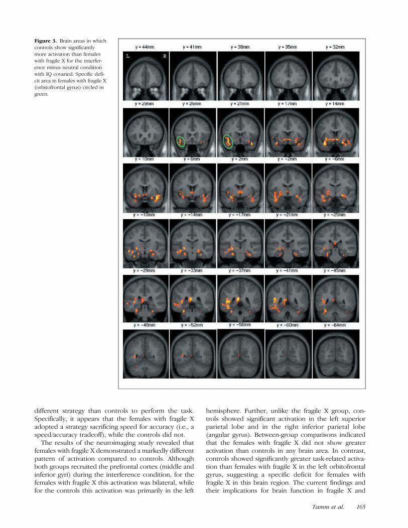
different strategy than controls to perform the task.Specifically, it appears that the females with fragile Xadopted a strategy sacrificing speed for accuracy (i.e., aspeed/accuracy tradeoff), while the controls did not.
The results of the neuroimaging study revealed thatfemales with fragile X demonstrated a markedly differentpattern of activation compared to controls. Althoughboth groups recruited the prefrontal cortex (middle andinferior gyri) during the interference condition, for thefemales with fragile X this activation was bilateral, whilefor the controls this activation was primarily in the left
hemisphere. Further, unlike the fragile X group, con-trols showed significant activation in the left superiorparietal lobe and in the right inferior parietal lobe(angular gyrus). Between-group comparisons indicatedthat the females with fragile X did not show greateractivation than controls in any brain area. In contrast,controls showed significantly greater task-related activa-tion than females with fragile X in the left orbitofrontalgyrus, suggesting a specific deficit for females withfragile X in this brain region. The current findings andtheir implications for brain function in fragile X and
Figure 3. Brain areas in which
controls show significantly
more activation than femaleswith fragile X for the interfer-
ence minus neutral condition
with IQ covaried. Specific defi-
cit area in females with fragile X(orbitofrontal gyrus) circled in
green.
Tamm et al. 165

other neurodevelopmental disorders are discussed indetail below.
The present results are in agreement with a numberof different studies that have suggested a role forspecific regions of the lateral prefrontal cortex in resolv-ing interference effects. Although the prefrontal cortexhas been implicated in a wide range of cognitivefunctions (Duncan & Owen, 2000), several previousresearch studies have demonstrated that the dorsolat-eral prefrontal cortex, and specifically Brodmann’s area(BA) 9/46, is involved in processing Stroop-relatedconflict and resolving interference effects (Menon,MacKenzie, Rivera, & Reiss, in preparation; Adleman,Menon, Blasey, White, & Reiss, under submission;Leung, Skudlarski, Gatenby, Peterson, & Gore, 2000,Macleod & MacDonald, 2000, Zysset et al., 2000; D’Es-posito, Postle, Jonides, & Smith, 1999; D’Esposito,Postle, & Rypma, 2000; Jonides, Smith, Marshuetz,Koeppe, & Reuter-Lorenz, 1998; Taylor et al., 1997).Further, this region (BA 9/46) was activated in theinterference condition of all three previous neuroimag-ing studies on the counting Stroop task (Zysset et al.,2000; Bush et al., 1998, 1999). Although both controlsand females with fragile X activated the inferior andmiddle frontal gyri during the interference condition ofthe counting Stroop, females with fragile X showeddifferent patterns of activation than controls. Specifi-cally, prefrontal regions activated by females with fragileX were more anterior than those activated by controls(i.e., Talairach coordinates for females with fragile X =�54, 30, 18 and controls = �52, 10, �2). Further,females with fragile X demonstrated bilateral activationin the prefrontal cortex, while controls showed pre-dominantly left hemisphere activation. Thus, comparedto controls, females with fragile X recruited moreprefrontal regions to perform the task, although behav-ioral data suggest that despite enhanced prefrontalactivation, their performance was still poorer than thatof controls.
A direct comparison between groups indicated thatcontrols showed greater activation in the left insularcortex, left superior temporal gyrus, and bilaterally inthe orbitofrontal gyrus, suggesting relative deficits inthese areas in association with this genetic condition.However, some of these group differences appear to bearising both from increased activation in the controlgroup and from deactivation in females with fragile X,while others appear to be arising solely from deactiva-tion in females with fragile X. Deactivation in this studyis operationalized as greater activation observed inresponse to the neutral condition than the interferencecondition. Specifically, between-group activation differ-ences in the left orbitofrontal gyrus and insular regionsappeared to result from both activation in controls (seeFigure 1, interference minus neutral, for clarification)and deactivation in females with fragile X (see Figure 2,neutral minus interference, for clarification). In contrast,
controls show no activation in the right orbitofrontalgyrus and left superior temporal gyrus; these groupdifferences appear to be due to deactivation in femaleswith fragile X. Thus, the primary region in which femaleswith fragile X activated less than controls (which doesnot appear to result from deactivation) was in the leftorbitofrontal gyrus, suggesting a specific variation ordeficit in females with fragile X in this region.
Studies suggest that the orbitofrontal cortex plays aspecific role in controlling voluntary goal-directed be-havior (Tremblay & Schultz, 2000; Schoenbaum, Chiba,& Gallagher, 1998). Further, the orbitofrontal gyrus isknown to play a role in executive functions such as setshifting, decision making, working memory, and atten-tional control (Bechara, Damasio, & Damasio, 2000;Rolls, 1994). Given that females with fragile X typicallydemonstrate executive functioning deficits (Mazzocco,Pennington, & Hagerman, 1993), aberrant activation ofthe orbitofrontal gyrus may play a role in the behavioralsymptomatology of individuals with this condition. Thishypothesis was explored using correlational analysesbetween percent voxels activated in the orbitofrontalregion and relevant behavioral measures collected bothinside and outside the scanner. Findings from theseanalyses indicated that activation in the left orbitofrontalgyrus correlated with task performance for the femaleswith fragile X and not the controls. Specifically, asignificant negative correlation between activation andreaction time and a nonsignificant, moderate positivecorrelation between activation and number correct wasobserved for the females with fragile X (with the effectsof IQ statistically controlled). These results suggest thatthe failure of the females with fragile X to activate thisregion may have contributed to their poorer perform-ance of the task. There were no significant correlationsbetween activation in this region and behavioral meas-ures selected in an attempt to capture the functioning offemales with fragile X. However, these behavioral meas-ures are not specifically designed to assess the behav-ioral phenotype of females with fragile X. It is probablethat dysregulation of the orbitofrontal cortex may in partunderlie some of the behavioral symptoms commonlyobserved in fragile X syndrome, since attentional prob-lems, mood lability, difficulty with socialization, andcognitive inflexibility or difficulty with set-shifting, areobserved both in females with fragile X (Sobesky, Hull,& Hagerman, 1994; Freund et al., 1993; Hagerman et al.,1992; Edwards, Keppen, Ranells, & Gollin, 1988) and inpersons with orbitofrontal lesions (Bechara et al., 2000;Rolls, 1994). Further investigation of links betweenorbitofrontal cortex activation and more precise meas-ures of these symptoms in individuals with fragile X iswarranted.
The present findings underscore the importance ofexamining both activation and deactivation patternswhen utilizing the subtraction method with neuroimag-ing data. The importance and role of deactivation in
166 Journal of Cognitive Neuroscience Volume 14, Number 2

interpreting neuroimaging findings is an issue that hasbegun to be addressed in the literature only recently.Although the source of deactivation remains poorlyunderstood, one possibility is that the advent of theexperimental task interrupts and inhibits ongoing con-scious processes that are active during rest (Binder et al.,1999). Alternatively, deactivation may arise from inhib-itory processes specific to the experimental condition orincreased activation related to internal brain mecha-nisms during the neutral condition (Binder et al., 1999;Hutchinson et al., 1999; Shulman, Corbetta, et al., 1997,Shulman, Fiez, et al., 1997). Consistent with the currentfindings, generic deactivation has been reported, amongother areas, in the precuneus/posterior cingulate(BA 31/7) and the temporal lobe (BA 20) during a varietyof experimental tasks (Shulman, Fiez et al., 1997).Further, deactivation in the posterior cingulate/precu-neus appears to be specific to language-related tasks(Shulman, Fiez, et al., 1997), a category into which thecounting Stroop falls. In this study, females with fragile Xshowed markedly different patterns of deactivation thancontrols (see Figure 2). Specifically, controls showeddeactivation in the posterior cingulate and ventromedialprefrontal cortex, while females with fragile X showeddeactivation in the globus pallidus, insular, and superiortemporal gyrus. It should also be noted that while thefemales with fragile X showed atypical patterns of deac-tivation compared to controls, the origin of these differ-ences is not clear.
Although activation in the parietal lobe did notemerge as a statistically significant difference in thebetween-group comparison, the controls showed moreactivation in the inferior and superior parietal lobethan females with fragile X. The inferior parietal lobeactivation observed in this study likely resulted fromthe arithmetic computation (counting) and possiblylanguage processing (reading) required by this task.Previous research has suggested that the angular gyrusis involved in arithmetic processing (Menon, Rivera,White, Glover, & Reiss, 2000), as well as languageprocessing (Crozier et al., 1999, Jessen et al., 1999).Imaging studies have also suggested a role for thesuperior parietal lobe in sustaining attention duringrelatively complex cognitive tasks (Rosen et al., 1999;Le, Pardo, & Hu, 1998; Bench et al., 1993; Pardo, Fox,& Raichle, 1991), as well as in implementing atten-tional demands of processing incongruent stimuli(Carter et al., 1995). Interestingly, parietal lobe activa-tion (BA 7) was observed in the three previous studiesreporting on the counting Stroop task (Zysset et al.,2000; Bush et al., 1998, 1999).
Given that a number of studies of the Stroop effecthave reported anterior cingulate activation during cog-nitive interference, a post hoc examination of the con-trol group data with p < .05 significance levels and noextent threshold was conducted. This examination stilldid not reveal activation in the anterior cingulate cogni-
tive division. Although it is unclear why activation wasnot detected in the anterior cingulate during the inter-ference condition, this finding could potentially be ex-plained by methodological issues. For example, aspecified region of interest for the anterior cingulatewas not defined and examined in this study, or thedemands of the subtractive method and lack of powermay have resulted in the anterior cingulate not crossingthe statistical threshold. It may be, however, that theanterior cingulate plays a less significant or specific rolein cognitive interference resolution than has previouslybeen thought. This hypothesis is consistent with thework of Zysset et al. (2000) and Taylor et al. (1997), whohave also suggested a less critical role for the anteriorcingulate in resolving cognitive interference, particularlywhen the number of choices from which selections needto be made is limited. It has also been speculated thatthe anterior cingulate plays a more critical role inresponse formation and monitoring (Liddle, Kiehl, &Smith, 2001) or in the anticipation of and preparationfor attentional activity (Sturm et al., 1999), rather thanresolving cognitive interference.
Additional investigation with a larger sample size,groups matched on IQ, and males with fragile X iswarranted to further explicate the current findings. Sup-plementary investigation utilizing an event-related designmay also elucidate group activation differences. Theevent-related design is less susceptible than block de-signs to habituation and changes in behavioral strategieswithin and between blocks (Bush et al., 1998), and hasthe capacity to probe the time course of the signalchange corresponding to interference (Leung et al.,2000).
Overall, the current findings suggest that, comparedto healthy controls, females with fragile X show differentpatterns of activation, particularly in the prefrontalcortex, and a specific deficit in the left orbitofrontalgyrus, as well as strikingly different patterns of deactiva-tion. Although IQ and deactivation may have drivensome of the activation differences, there is still strongevidence to suggest that females with fragile X haveanomalous brain activation during cognitive interferenceprocessing tasks and may fail to appropriately recruitand modulate lateral prefrontal cortex and parietalresources.
METHODS
Participants
Participants included 14 females with fragile X and 14age-matched healthy control females, without the fragileX mutation. Participants ranged in age from 10–22(mean age 15.43, SD = 3.79), and both groups werepredominantly of white, non-Hispanic origin (86%). IQestimates based on either the Wechsler IntelligenceScale for Children—Third Edition (WISC-III) or the
Tamm et al. 167

Wechsler Adult Intelligence Scale—Third Edition (WAIS-III) were obtained for each participant. Information wasalso obtained from the CBCL (Achenbach, 1991) for 9subjects in the control group and 11 subjects in thefragile X group.
Task
The counting Stroop task was programmed using Psy-scope (http://poppy.psy.cmu.edu/psyscope) on a Macin-tosh (Sunnyvale, CA) powerbook computer. Onset ofscanning and task were synchronized using a TTL pulsedelivered to the scanner timing microprocessor boardfrom a ‘CMU Button Box’ microprocessor connected tothe Macintosh with a serial cable. Stimuli were presentedvisually at the center of a screen using a custom-builtmagnet compatible projection system (Resonance Tech-nology, CA).
The task consisted of 12 alternating experimental(interference) and control (neutral) conditions with arest period at the beginning and end of the task. For bothconditions, subjects were instructed to press the buttonthat corresponded to the number of words on thescreen. During the neutral task, the word ‘‘fish’’ waspresented 1, 2, 3, or 4 times on the screen (15 trials).During the interference condition, subjects were pre-sented the words ‘‘one’’ ‘‘two’’ ‘‘three,’’ and ‘‘four,’’presented 1, 2, 3, or 4 times on the screen (15 trials).Stimuli were presented for 1350 msec at the rate of every2 sec for a total of 180 trials (90 experimental, 90 control).
Image Acquisition and Analysis
MR images were acquired on a GE-Signa 1.5 T scanner(GE Imaging Systems, Milwaukee, WI) with Echospeedgradients using a custom-built whole head coil thatprovides a 50% advantage in signal-to-noise ratio overthat of the standard GE coil (Hayes & Mathias, 1996). Acustom built head-holder was used to prevent headmovement. Eighteen axial slices (6-mm thick, 1-mmskip) parallel to the anterior and posterior commissurescovering the whole brain were imaged with a temporalresolution of 2 sec using a T2*-weighted gradient echospiral pulse sequence (TR = 2000 msec, TE = 40 msec,flip angle = 898 and 1 interleave). Although the wholebrain was imaged, the presence of susceptibility artifactsthat affect MRI data may have precluded observation ofactivation in the orbitofrontal and anterior temporalcortices, which are particularly sensitive to susceptibilityartifact (Ojemann et al., 1997). In an attempt to mitigatethe effects of susceptibility artifact and optimize theability to detect activation, a strict threshold for move-ment (less than 3 mm translation and rotation), acoronal acquisition, and a relatively lower field strengthmagnet (1.5 T) were used. Detailed analyses of suscept-ibility artifact in spiral acquisitions will be providedelsewhere (Grecius, Krasnow, Reiss, & Menon, in prep-
aration). To aid in the localization of functional data,high resolution T1-weighted spoiled grass gradient re-called (SPGR) 3-D MRI sequence with the followingparameters was used: TR = 35 msec; TE = 6 msec; flipangle = 458; 24 cm field of view; 124 slices in the coronalplane; 256 � 192 matrix; acquired resolution = 1.5 � 0.9� 1.2 mm. On a few subjects, a faster protocol wasutilized to decrease time of acquisition of the SPGRimage, with TR = 11 msec; TE = 2 msec; and flip angle= 158. The images were reconstructed as a 124 � 256 �256 with a 1.5 � 0.9 � 0.9 mm spatial resolution.
Image Preprocessing
Images were reconstructed, by inverse Fourier trans-form, for each of the 225 time points into 64 � 64 � 18image matrices (voxel size: 3.75 � 3.75 � 7 mm). fMRIdata were preprocessed using SPM99 (http://www.fil.ion.ucl.ac.uk/spm). Images were corrected for move-ment using least square minimization without higher-order corrections for spin history, and normalized tostereotaxic Talairach coordinates (Talairach & Tour-noux, 1988). Images were then resampled every 2 mmusing sinc interpolation and smoothed with a 4 mmGaussian kernel to decrease spatial noise.
Statistical Analysis
Statistical analysis was performed on individual andgroup data using the general linear model and thetheory of Gaussian random fields as implementedin SPM99 (Friston, Holmes, et al., 1995). This methodtakes advantage of multivariate regression analysis andcorrects for temporal and spatial autocorrelations inthe fMRI data. Activation foci were superimposed onhigh-resolution T1-weighted images and their locationsinterpreted using known neuroanatomical landmarks(Duvernoy, Bourgouin, Cabanis, & Cattin, 1999; Mai,Assheuer, & Paxinos, 1997). MNI coordinates were trans-formed to Talairach coordinates using a nonlinear trans-formation (Brett, 2000).
A within-subjects procedure was first used to modelall the effects of interest, covariates, and nuisance vari-ables for each subject. The individual subject modelswere identical across subjects (i.e., a balanced designwas used). Confounding effects of fluctuations in globalmean were removed by proportional scaling where, foreach time point, each voxel was scaled by the globalmean at that time point. Low frequency noise wasremoved with a high pass filter (0.5 cycles/min) appliedto the fMRI time series at each voxel. A temporalsmoothing function (Gaussian kernel corresponding todispersion of 8 sec) was applied to the fMRI time seriesto enhance the temporal signal to noise ratio. We thendefined the effects of interest for each subject with therelevant contrasts of the parameter estimates. For eachof these contrasts, a corresponding contrast image was
168 Journal of Cognitive Neuroscience Volume 14, Number 2

also generated. Voxel-wise t statistics were normalized toZ scores to provide a statistical measure of activationthat is independent of sample size. Significant clusters ofactivation were determined using the joint expectedprobability distribution of height and extent of Z scores(Poline, Worsley, et al., 1997), with height (Z > 2.33;p < .01) and extent thresholds ( p < .05).
Group analysis was performed using a random-effectsmodel that incorporated a two-stage hierarchical pro-cedure. This model estimates the error variance foreach condition of interest across subjects, rather thanacross scans (Holmes & Friston, 1998) and thereforeprovides a stronger generalization to the populationfrom which data are acquired. This analysis proceededin two steps. In the first step, contrast images for eachsubject and each effect of interest were generated asdescribed above. In the second step, these contrastimages were analyzed using a general linear model todetermine voxel-wise t statistics. One contrast imagewas generated per subject, per effect of interest (e.g.,interference condition minus the neutral condition). Atwo-sample t test was then used to determine groupactivation for each effect. The following contrast imageswere calculated for each subject: (i) activation: interfer-ence–neutral; (ii) deactivation: neutral�interference.For both within- and between-group comparisons, acluster-wise (corrected) significance level of p < .05 wasused.
Following these analyses, exploratory analyses wereconducted to assess the relationship between brainactivation and behavioral variables. Specifically, regionsin which the females with fragile X activated significantlyless than controls, namely, functional regions of interest(fROI), were identified. Partial correlations, controllingfor IQ, between percentage of voxels activated (heightthreshold Z > 2.33) between the fROIs and relevantbehavioral variables were then computed for eachgroup.
Acknowledgments
This work was supported by NIH Grants: MH01142, HD31715,MH50047, and HD40761, the Lynda and Scott Canel Fund forFragile X Research, and the Constance Bultman WilsonFoundation.
Reprint requests should be sent to Leanne Tamm, PhD,Department of Psychiatry and Behavioral Sciences, StanfordUniversity School of Medicine, 401 Quarry Road, Stanford,CA 94305, USA, or via e-mail: [email protected].
The data reported in this experiment have been deposited inthe fMRI Data Center (http://www.fmridc.org). The accessionnumber is 2-2001-1123B.
REFERENCES
Achenbach, T. M. (1991). Manual for the child behaviorchecklist/4-18 and 1991 profile. Burlington: University ofVermont Department of Psychiatry Press.
Adleman, N. E., Menon, V., Blasey, C., White, C. D., & Reiss, A. L.(under submission). A developmental fMRI study of theStroop color word interference task.
Bechara, A., Damasio, H., & Damasio, A. R. (2000). Emotion,decision making, and the orbitofrontal cortex. CerebralCortex, 10, 295–307.
Bench, C. J., Frith, C. D., Grasby, P. M., Friston, K. J., Paulesu,E., Frackowiak, R. S. J., & Dolan, R. J. (1993). Investigationsof the functional anatomy of attention using the Stroop test.Neuropsychology, 31, 907–922.
Binder, J. R., Frost, J. A., Hammeke, T. A., Bellgowan, P. S. F.,Rao, S. M., & Cox, R. W. (1999). Conceptual processingduring the conscious resting state: A functional MRI study.Journal of Cognitive Neuroscience, 11, 80–93.
Borhgraef, M., Fryns, J. P., & Van den Berghe, H. (1990). Thefemale and the fra X syndrome: Data on the clinical andpsychological findings in 7 fra X carriers. Clinical Genetics,37, 341–346.
Borhgraef, M., Umans, S., Steyaert, J., Legius, E., & Fryns, J.(1996). New findings in the behavioral profile of young fraXfemales. American Journal of Medical Genetics, 64,346–349.
Brett, M. (2000). http://www.mrc-cbu.cam.ac.uk/Imaging/mnis-pace.html.
Bush, G., Frazier, J. A., Rauch, S. L., Seidman, L. J., Whalen, P. J.,Jenike, M. A., Rosen, B. R., Biederman, J. (1999). Anteriorcingulate cortex dysfunction in attention deficit hyperactivitydisorder revealed by fMRI and the counting stroop.Biological Psychiatry, 45, 1542–1552.
Bush, G., Whalen, P. J., Rosen, B. R., Jenike, M. A., McInerney,S. C., & Rauch, S. L. (1998). The counting stroop: Aninterference task specialized for functional neuroimaging–Validation study with functional MRI. Human BrainMapping, 6, 270–282.
Carter, C. S., Mintun, M., & Cohen, J. D. (1995). Interferenceand facilitation effects during selective attention: An H2
15OPET study of Stroop task performance. Neuroimage, 2,264–272.
Corbetta, M., Miezen, F. M., Dobmeyer, S., Shulman, G. L., &Petersen, S. E. (1991). Selective and divided attention duringvisual discriminations of shape, color, and speed: Functionalanatomy by positron emission topography. Journal ofNeuroscience, 11, 2383–2402.
Crozier, S., Sirigu, A., Lehericy, S., van de Moortele, P. F., Pillon,B., Grafman, J., Agid, Y., Dubois, B., & LeBihan, D. (1999).Distinct prefrontal activations in processing sequence at thesentence and script level: An fMRI study. Neuropsychologia,37, 1469–1476.
D’Esposito, M., Postle, B. R., Jonides, J., & Smith, E. E. (1999).The neural substrate and temporal dynamics of interferenceeffects in working memory as revealed by event-relatedfunctional MRI. Proceedings of the National Academy ofSciences, U.S.A., 96, 7514–7519.
D’Esposito, M., Postle, B. R., & Rypma, B. (2000). Prefrontalcortical contributions to working memory: Evidence fromevent-related fMRI studies. Experimental Brain Research,133, 3–11.
Donnenfield, A. E. (1998). Fragile X syndrome. Indian Journalof Pediatrics, 65, 513–518.
Duncan, J., & Owen, A. M. (2000). Common regions of thehuman frontal lobe recruited by diverse cognitive demands.Trends in Neuroscience, 23, 475–483.
Duvernoy, H. M., Bourgouin, P., Cabanis, E. A., & Cattin, F.(1999). The human brain: Surface, three-dimensional sec-tional anatomy with MRI, and blood supply. New York:Springer-Verlag.
Edwards, D. R., Keppen, L. D., Ranells, J. D., & Gollin, S. M.(1988). Autism in association with fragile X syndrome
Tamm et al. 169

in females: Implications for diagnosis and treatment inchildren. Neurotoxicology, 93, 359–365.
Freund, L. S., Reiss, A. L., & Abrams, M. T. (1993). Psychiatricdisorders associated with fragile X in the young female.Pediatrics, 91, 321–329.
Friston, K. J., Holmes, A. P., Worsley, K. J., Poline, J. B., Frith,C. D., & Frackowiak, R. S. J. (1995). Statistical parametricmaps in functional imaging: A general linear approach.Human Brain Mapping, 2, 189–210.
George, M. S. E. C., Ketter, T. A., Parekh, P. I., Rosinsky, N.,Ring, H., Casey, B. J., Trimble, M. R., Horwitz, B.,Herschovitch, P., & Post, R. M. (1994). Regional brain activitywhen selecting a response despite interference: An H2
15OPET study of the Stroop and an emotional Stroop. HumanBrain Mapping, 1, 194–209.
Grecius, M., Krasnow, B., Reiss, A. L., & Menon, V.(in preparation). The effect of susceptibility artifact onBOLD signal detection in fMRI: How much signal loss is toomuch signal loss?
Hagerman, R. J., Jackson, C., Amiri, K., Silverman, A. C.,O’Connor, R., & Sobesky, W. (1992). Girls with fragile Xsyndrome: Physical and neurocognitive status and outcome.Pediatrics, 89, 395–400.
Hagerman, R. J., & Sobesky, W. E. (1989). Psychopathology infragile X syndrome. American Journal of Orthopsychiatry,59, 142–152.
Hayes, C., & Mathias, C. (1996). Improved brain coil for fMRIand high resolution imaging. Presented at the ISMRM 4thAnnual Meeting Proceedings, New York.
Holmes, A. P., & Friston, K. J. (1998). Generalizability,random effects, and population inference. Neuroimage, 7,754.
Hutchinson, M., Schiffer, W., Joseffer, S., Liu, A., Schlosser, R.,Dikshit, S., Goldberg, E., & Brodie, J. D. (1999). Task-specificdeactivation patterns in functional magnetic resonanceimaging. Magnetic Resonance Imaging, 17, 1427–1436.
Jakala, P., Hanninen, T., Ryyanen, M., Laakso, M., Partanen, K.,Mannermaa, A., & Soininen, H. (1997). Fragile-X:Neuropsychological test performance, CGG triplet repeatlengths, and hippocampal volumes. Journal of ClinicalInvestigation, 100, 331–338.
Jensen, A. R. (1965). Scoring the Stroop test. Acta Psycologia,24, 398–408.
Jessen, F., Erb, M., Klose, U., Lotz, M., Grodd, W., &Heun, R. (1999). Activation of human languageprocessing brain regions after the presentation ofrandom letter strings demonstrated with event-relatedfunctional magnetic resonance imaging. NeuroscienceLetters, 270, 13–16.
Jonides, J., Smith, E. E., Marshuetz, C., Koeppe, R. A., & Reuter-Lorenz, P. A. (1998). Inhibition in verbal working memoryrevealed by brain activation. Proceedings of the NationalAcademy of Sciences, U.S.A., 95, 8410–8413.
Le, T. H., Pardo, J. V., & Hu, X. (1998). 4 T-fMRI study ofnonspatial shifting of selective attention: Cerebellar andparietal contributions. Journal of Neurophysiology, 79,1535–1548.
Leung, H. C., Skudlarski, P., Gatenby, J. C., Peterson, B. S., &Gore, J. C. (2000). An event-related functional MRI study ofthe stroop color word interference task. Cerebral Cortex, 10,552–560.
Liddle, P. F., Kiehl, K. A., & Smith, A. M. (2001). Event-relatedfMRI study of response inhibition. Human Brain Mapping,12, 100–109.
MacLeod, C. M., & MacDonald, P. A. (2000). Interdimensionalinterference in the Stroop effect: Uncovering the cognitiveand neural anatomy of attention. Trends in CognitiveScience, 4, 383–391.
Mai, J. K., Assheuer, J., & Paxinos, G. (1997). Atlas of thehuman brain. San Diego, CA: Academic Press.
Mazzocco, M. M., Hagerman, R. J., Cronister-Silverman, A., &Pennington, B. F. (1992). Specific frontal lobe deficits amongwomen with the fragile X gene. Journal of the AmericanAcademy of Child and Adolescent Psychiatry, 31, 1141–1148.
Mazzocco, M. M., Pennington, B. F., & Hagerman, R. J. (1993).The neurocognitive phenotype of female carriers of fragileX: additional evidence for specificity. Journal ofDevelopmental and Behavioral Pediatrics, 14, 328–355.
Menon, V., MacKenzie, K., Rivera, S., & Reiss, A. L.(in preparation). Prefrontal cortex involvement inprocessing incongruent arithmetic equations: Evidence fromevent-related fMRI.
Menon, V., Rivera, S. M., White, C. D., Glover, G. H., & Reiss,A. L. (2000). Dissociating prefrontal and parietal cortexactivation during arithmetic processing. Neuroimage, 12,357–365.
Morton, J. E., Bundey, S., Webb, T. P., MacDonald, F., Rindle,P. M., & Bullock, S. (1997). Fragile X syndrome is lesscommon than previously estimated. Journal of MedicalGenetics, 34, 1–5.
Ojemann, J. G., Akbudak, E., Snyder, A. Z., McKinstry, R. C.,Raichle, M. E., & Conturo, T. E. (1997). Anatomic localizationand quantitative analysis of gradient refocused echo-planarfMRI susceptibility artifacts. Neuroimage, 6, 156–167.
Pardo, J. V., Fox, P. T., & Raichle, M. E. (1991). Localization of ahuman system for sustained attention by positron emissiontomography. Nature, 349, 61–64.
Pardo, J. V., Pardo, P. J., Janer, K. W., & Raichle, E. (1990). Theanterior cingulate cortex mediates processing selection inthe Stroop attentional conflict paradigm. Proceedings of theNational Academy of Sciences, U.S.A., 87, 256–259.
Poline, J. B., Worsley, K. J., Evans, A. C., & Friston, K. J. (1997).Combining spatial extent and peak intensity to test for acti-vations in functional imaging. Neuroimage, 5, 83–96.
Riddle, J. E., Cheema, A., Sobesky, W. E., Gardner, S. C.,Taylor, A. K., Pennington, B. F., & Hagerman, R. J. (1998).Phenotypic involvement in females with the FMR1 genemutation. American Journal of Mental Retardation, 102,590–601.
Rolls, E. T. (1994). The orbitofrontal cortex. In A. C. Roberts,T. W. Robbins, & L. Weiskrantz (Eds.), The prefrontal cortex:Executive and cognitive functions (pp. 67–86). Oxford:Oxford University Press.
Rosen, A. C., Rao, S. M., Caffarra, P., Scaglioni, A., Bobholz, J. A.,Woodley, S. J., Hammeke, T. A., Cunningham, J. M., Prieto,T. E., & Binder, J. R. (1999). Neural basis of endogenous andexogenous spatial orienting. A functional MRI study. Journalof Cognitive Neuroscience, 11, 135–152.
Santos, J. F., & Montgomery, J. R. (1962). Stability ofperformance on the color–word test. Perceptual MotorSkills, 15, 397–398.
Schoenbaum, G., Chiba, A. A., & Gallagher, M. (1998).Orbitofrontal cortex and basolateral amygdala encodeexpected outcomes during learning. Nature Neuroscience,1, 155–159.
Shulman, G. L., Corbetta, M., Buckner, R. L., Fiez, J. A., Miezen,F. M., Raichle, M. E., & Petersen, S. E. (1997). Commonblood flow changes across visual tasks: I. Increases insubcortical structures and cerebellum but not in nonvisualcortex. Journal of Cognitive Neuroscience, 9, 624–647.
Shulman, G. L., Fiez, J. A., Corbetta, M., Buckner, R. L.,Miezen, F. M., Raichle, M. E., & Petersen, S. E. (1997).Common blood flow changes across visual tasks: II.Decreases in cerebral cortex. Journal of CognitiveNeuroscience, 9, 648–663.
170 Journal of Cognitive Neuroscience Volume 14, Number 2

Smith, G. J., & Nyman, G. E. (1974). The validity of the serialcolor word test: A reply to Lennart Sjoberg. ScandinavianJournal of Psychology, 15, 238–240.
Sobesky, W. E., Hull, C. E., & Hagerman, R. J. (1994).Symptoms of schizotypal personality disorder in fragile Xwomen. Journal of the American Academy of Child andAdolescent Psychiatry, 332, 247–255.
Stroop, J. R. (1935). Studies of interference in serial verbalreactions. Journal of Experimental Psychology, 18, 643–662.
Sturm, W., de Simone, A., Krause, B. J., Specht, K.,Hesselmann, V., Radermacher, I., Herzog, H., Tellmann, L.,Muller-Gartner, H. W., & Willmes, K. (1999). Functionalanatomy of intrinsic alertness: Evidence for a fronto-parietal–thalamic–brainstem network in the right hemisphere.Neuropsychologia, 37, 797–805.
Talairach, J., & Tournoux, M. (1988). Co-planar stereotaxicatlas of the human brain. New York: Thieme.
Taylor, S. F., Kornblum, S., Lauber, E. J., Minoshima, S., &Koeppe, R. A. (1997). Isolation of specific interferenceprocessing in the Stroop task: PET activation studies.Neuroimage, 6, 81–92.
Tremblay, L., & Schultz, W. (2000). Reward-related neuronalactivity during go–nogo task performance in primateorbitofrontal cortex. Journal of Neurophysiology, 83,1864–1876.
Welch, J. L., & Williams, J. K. (1999). Fragile X syndrome.Neonatal Network, 18, 15–22.
Zysset, S., Muller, K., Lohmann, G., & von Cramon, D. Y.(2000). The Stroop tasks: Is it the anterior cingulatecortex? Presented at Cognitive Neuroscience Meeting,San Francisco, CA.
Tamm et al. 171