Fluorescent Western Blotting -...
32
Fluorescent Western Blotting February 28, 2012 Dr. Martin Broadstock Edmond and Lily Safra Research Fellow King’s College London
Transcript of Fluorescent Western Blotting -...
Fluorescent Western Blotting
February 28, 2012
Dr. Martin Broadstock
Edmond and Lily Safra Research Fellow
King’s College London
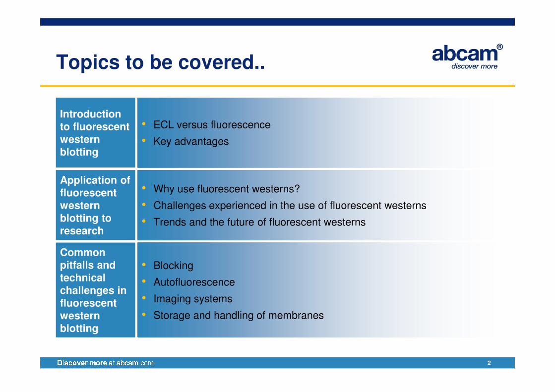
Topics to be covered..
Introduction to fluorescent western blotting
• ECL versus fluorescence
• Key advantages
Application of fluorescent • Why use fluorescent westerns?
2
fluorescent western blotting to research
• Challenges experienced in the use of fluorescent westerns
• Trends and the future of fluorescent westerns
Common pitfalls and technical challenges in fluorescent western blotting
• Blocking
• Autofluorescence
• Imaging systems
• Storage and handling of membranes
My current research
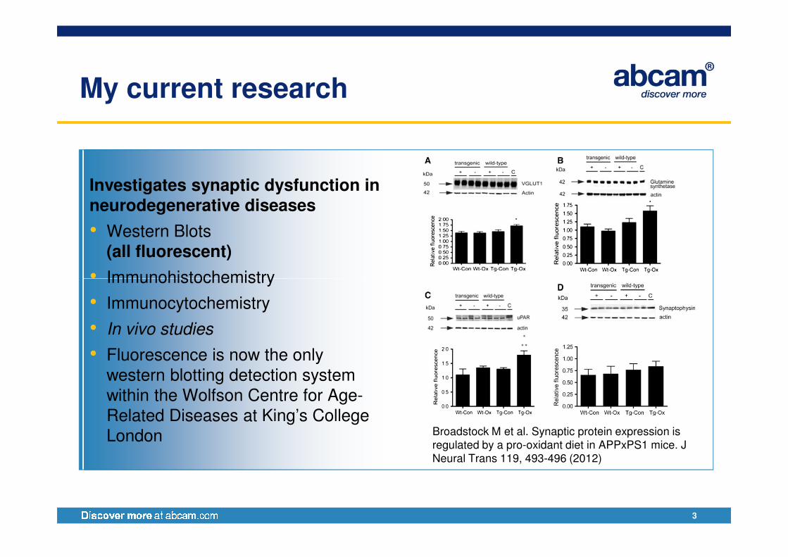
Investigates synaptic dysfunction in neurodegenerative diseases
• Western Blots (all fluorescent)
• Immunohistochemistry
3
• Immunohistochemistry
• Immunocytochemistry
• In vivo studies
• Fluorescence is now the only western blotting detection system within the Wolfson Centre for Age-Related Diseases at King’s College London Broadstock M et al. Synaptic protein expression is
regulated by a pro-oxidant diet in APPxPS1 mice. J Neural Trans 119, 493-496 (2012)
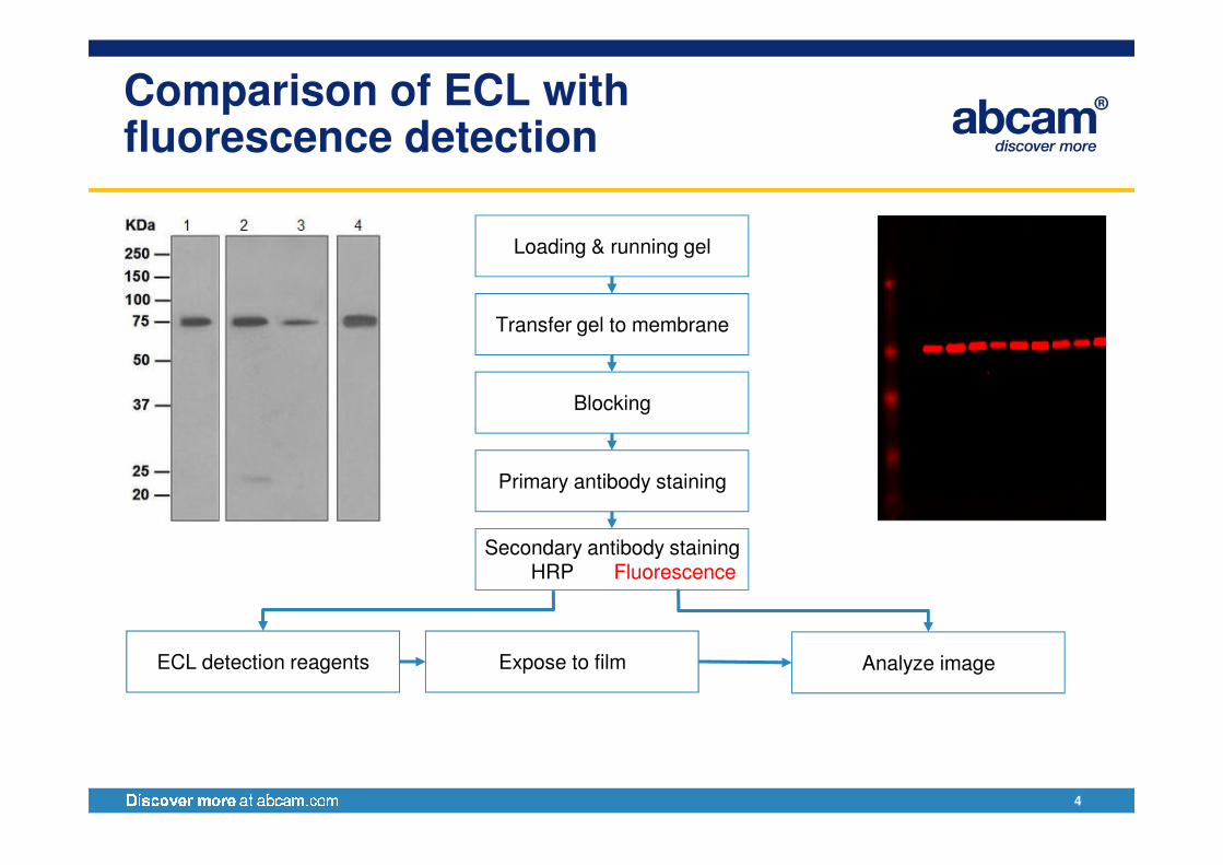
Comparison of ECL with fluorescence detection
Loading & running gel
Transfer gel to membrane
Blocking
4
Primary antibody staining
Secondary antibody staining
ECL detection reagents Analyze imageExpose to film
HRP Fluorescence
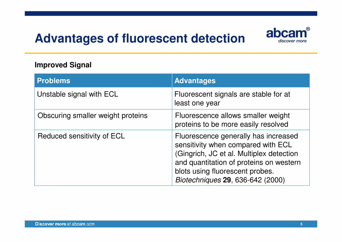
Advantages of fluorescent detection
Improved Signal
Problems Advantages
Unstable signal with ECL Fluorescent signals are stable for at least one year
Obscuring smaller weight proteins Fluorescence allows smaller weight proteins to be more easily resolved
5
proteins to be more easily resolved
Reduced sensitivity of ECL Fluorescence generally has increased sensitivity when compared with ECL(Gingrich, JC et al. Multiplex detection and quantitation of proteins on western blots using fluorescent probes. Biotechniques 29, 636-642 (2000)
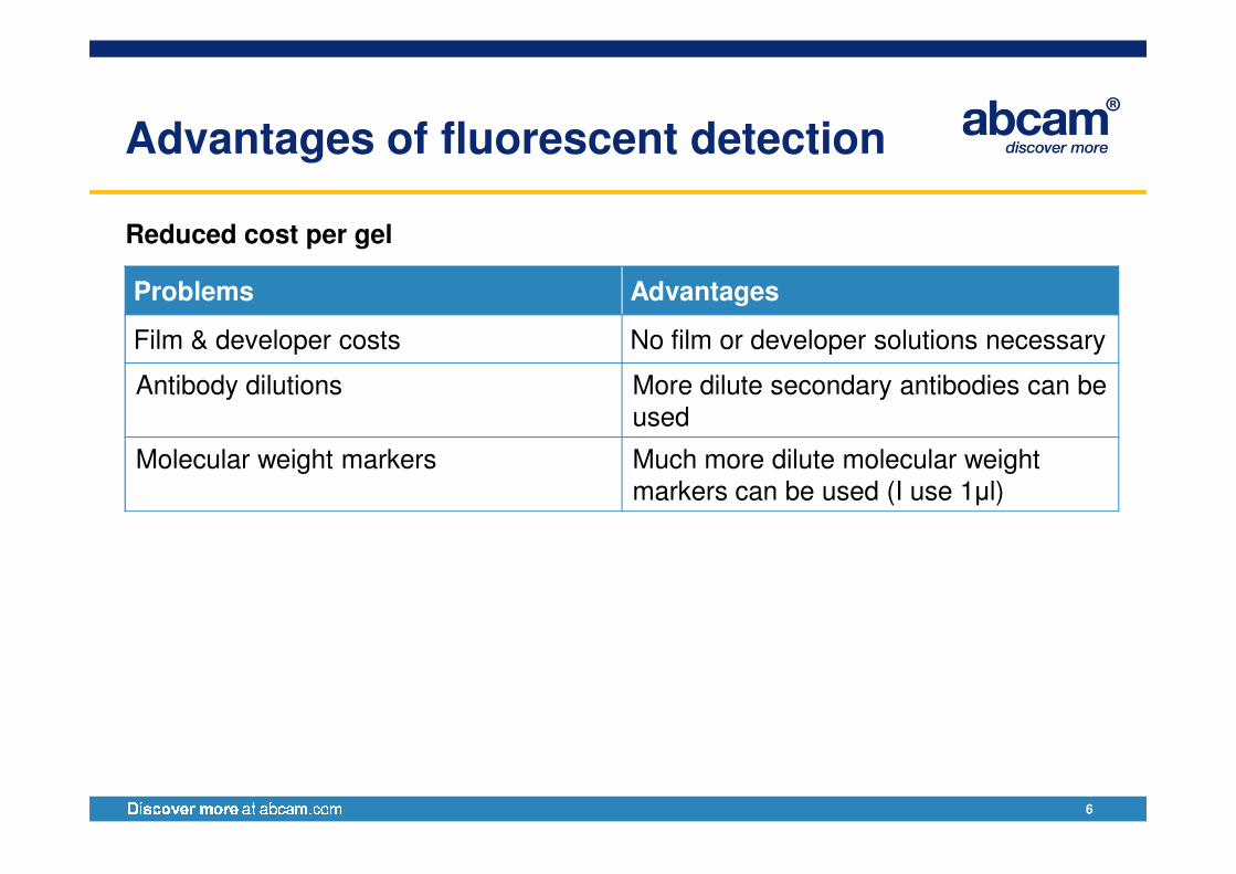
Advantages of fluorescent detection
Reduced cost per gel
Problems Advantages
Film & developer costs No film or developer solutions necessary
Antibody dilutions More dilute secondary antibodies can be used
6
Molecular weight markers Much more dilute molecular weight markers can be used (I use 1µl)
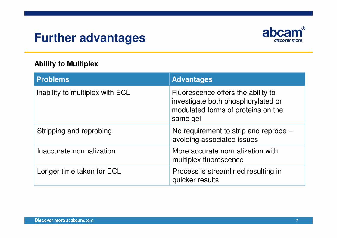
Further advantages
Ability to Multiplex
Problems Advantages
Inability to multiplex with ECL Fluorescence offers the ability to investigate both phosphorylated or modulated forms of proteins on the same gel
7
same gel
Stripping and reprobing No requirement to strip and reprobe –avoiding associated issues
Inaccurate normalization More accurate normalization with multiplex fluorescence
Longer time taken for ECL Process is streamlined resulting in quicker results
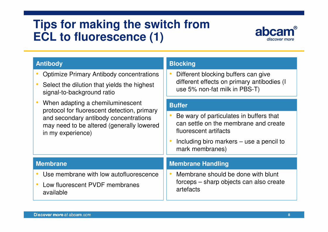
Tips for making the switch from ECL to fluorescence (1)
• Optimize Primary Antibody concentrations
• Select the dilution that yields the highest
signal-to-background ratio
• When adapting a chemiluminescent
protocol for fluorescent detection, primary
and secondary antibody concentrations
Antibody
• Different blocking buffers can give
different effects on primary antibodies (I
use 5% non-fat milk in PBS-T)
Blocking
• Be wary of particulates in buffers that
Buffer
8
and secondary antibody concentrations
may need to be altered (generally lowered
in my experience)
• Use membrane with low autofluorescence
• Low fluorescent PVDF membranes
available
Membrane
• Be wary of particulates in buffers that
can settle on the membrane and create
fluorescent artifacts
• Including biro markers – use a pencil to
mark membranes)
• Membrane should be done with blunt
forceps – sharp objects can also create
artefacts
Membrane Handling
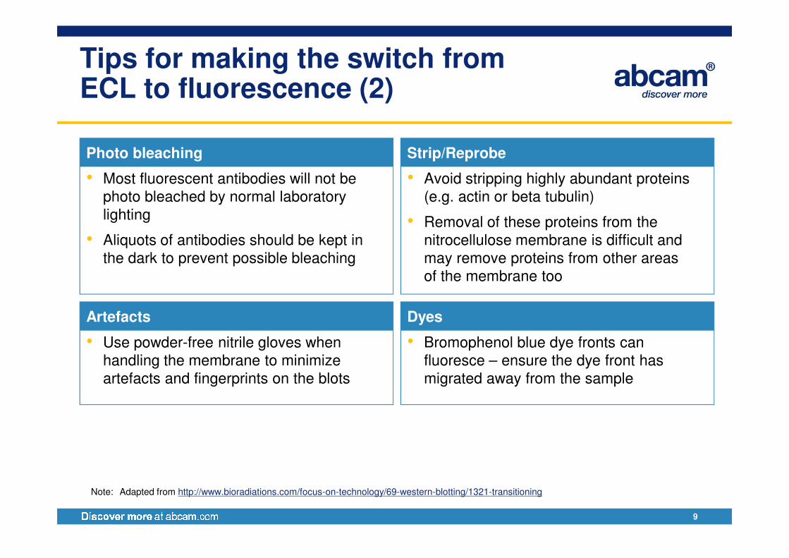
Tips for making the switch from ECL to fluorescence (2)
• Most fluorescent antibodies will not be
photo bleached by normal laboratory
lighting
• Aliquots of antibodies should be kept in
the dark to prevent possible bleaching
Photo bleaching
• Avoid stripping highly abundant proteins
(e.g. actin or beta tubulin)
• Removal of these proteins from the
nitrocellulose membrane is difficult and
may remove proteins from other areas
of the membrane too
Strip/Reprobe
9
of the membrane too
• Use powder-free nitrile gloves when
handling the membrane to minimize
artefacts and fingerprints on the blots
Artefacts
• Bromophenol blue dye fronts can
fluoresce – ensure the dye front has
migrated away from the sample
Dyes
Note: Adapted from http://www.bioradiations.com/focus-on-technology/69-western-blotting/1321-transitioning
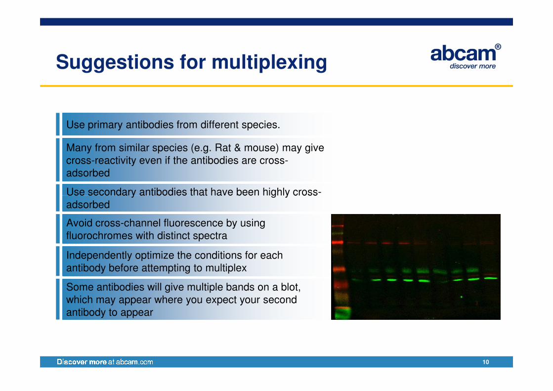
Suggestions for multiplexing
Use primary antibodies from different species.
Many from similar species (e.g. Rat & mouse) may give
cross-reactivity even if the antibodies are cross-
adsorbed
Use secondary antibodies that have been highly cross-
10
Use secondary antibodies that have been highly cross-
adsorbed
Avoid cross-channel fluorescence by using
fluorochromes with distinct spectra
Independently optimize the conditions for each
antibody before attempting to multiplex
Some antibodies will give multiple bands on a blot,
which may appear where you expect your second
antibody to appear
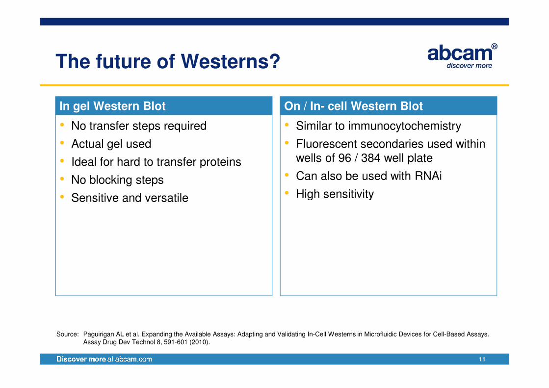
The future of Westerns?
In gel Western Blot
• No transfer steps required
• Actual gel used
• Ideal for hard to transfer proteins
• No blocking steps
• Sensitive and versatile
On / In- cell Western Blot
• Similar to immunocytochemistry
• Fluorescent secondaries used within wells of 96 / 384 well plate
• Can also be used with RNAi
• High sensitivity
11
• Sensitive and versatile • High sensitivity
Source: Paguirigan AL et al. Expanding the Available Assays: Adapting and Validating In-Cell Westerns in Microfluidic Devices for Cell-Based Assays.
Assay Drug Dev Technol 8, 591-601 (2010).
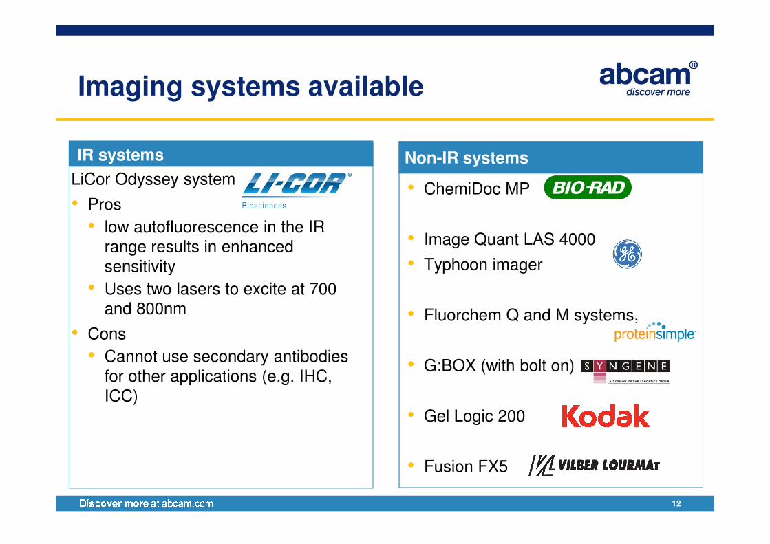
Imaging systems available
Non-IR systems
• ChemiDoc MP
• Image Quant LAS 4000
• Typhoon imager
IR systems
LiCor Odyssey system
• Pros
• low autofluorescence in the IR range results in enhanced sensitivity
12
• Typhoon imager
• Fluorchem Q and M systems,
• G:BOX (with bolt on)
• Gel Logic 200
• Fusion FX5
sensitivity
• Uses two lasers to excite at 700 and 800nm
• Cons
• Cannot use secondary antibodies for other applications (e.g. IHC, ICC)
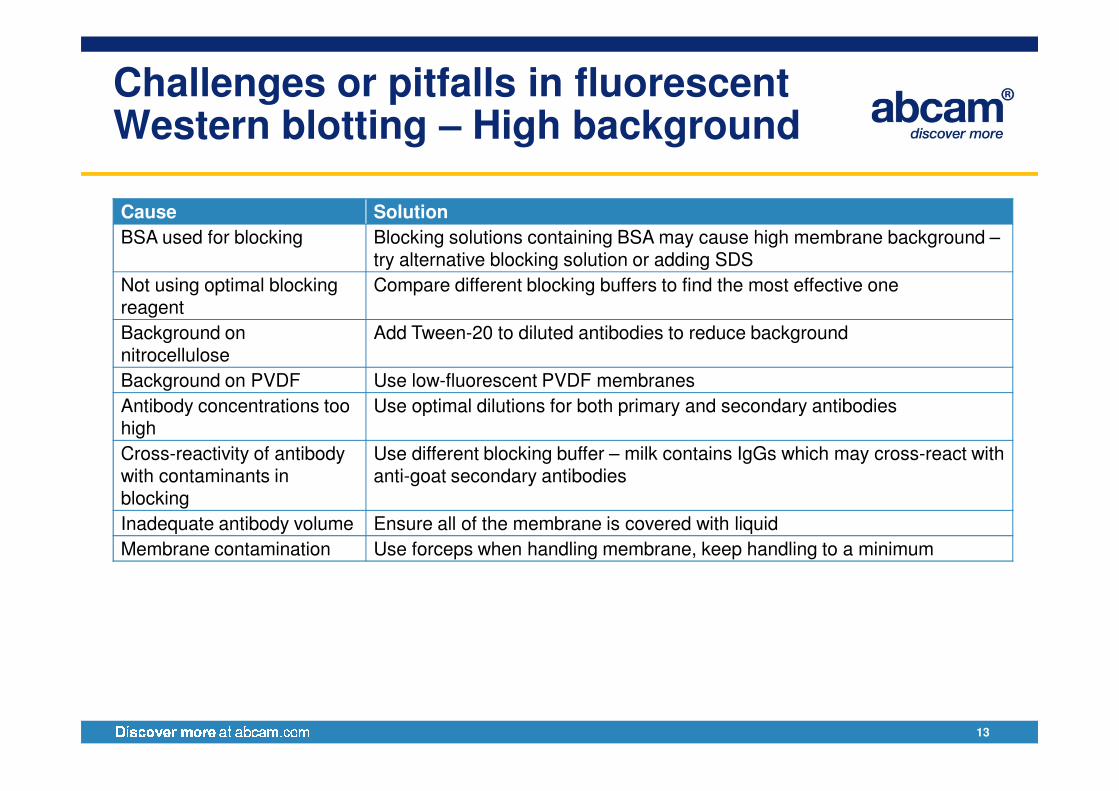
Challenges or pitfalls in fluorescent Western blotting – High background
Cause Solution
BSA used for blocking Blocking solutions containing BSA may cause high membrane background –try alternative blocking solution or adding SDS
Not using optimal blocking reagent
Compare different blocking buffers to find the most effective one
Background on nitrocellulose
Add Tween-20 to diluted antibodies to reduce background
Background on PVDF Use low-fluorescent PVDF membranes
Antibody concentrations too Use optimal dilutions for both primary and secondary antibodies
13
Antibody concentrations too high
Use optimal dilutions for both primary and secondary antibodies
Cross-reactivity of antibody with contaminants in blocking
Use different blocking buffer – milk contains IgGs which may cross-react with anti-goat secondary antibodies
Inadequate antibody volume Ensure all of the membrane is covered with liquid
Membrane contamination Use forceps when handling membrane, keep handling to a minimum
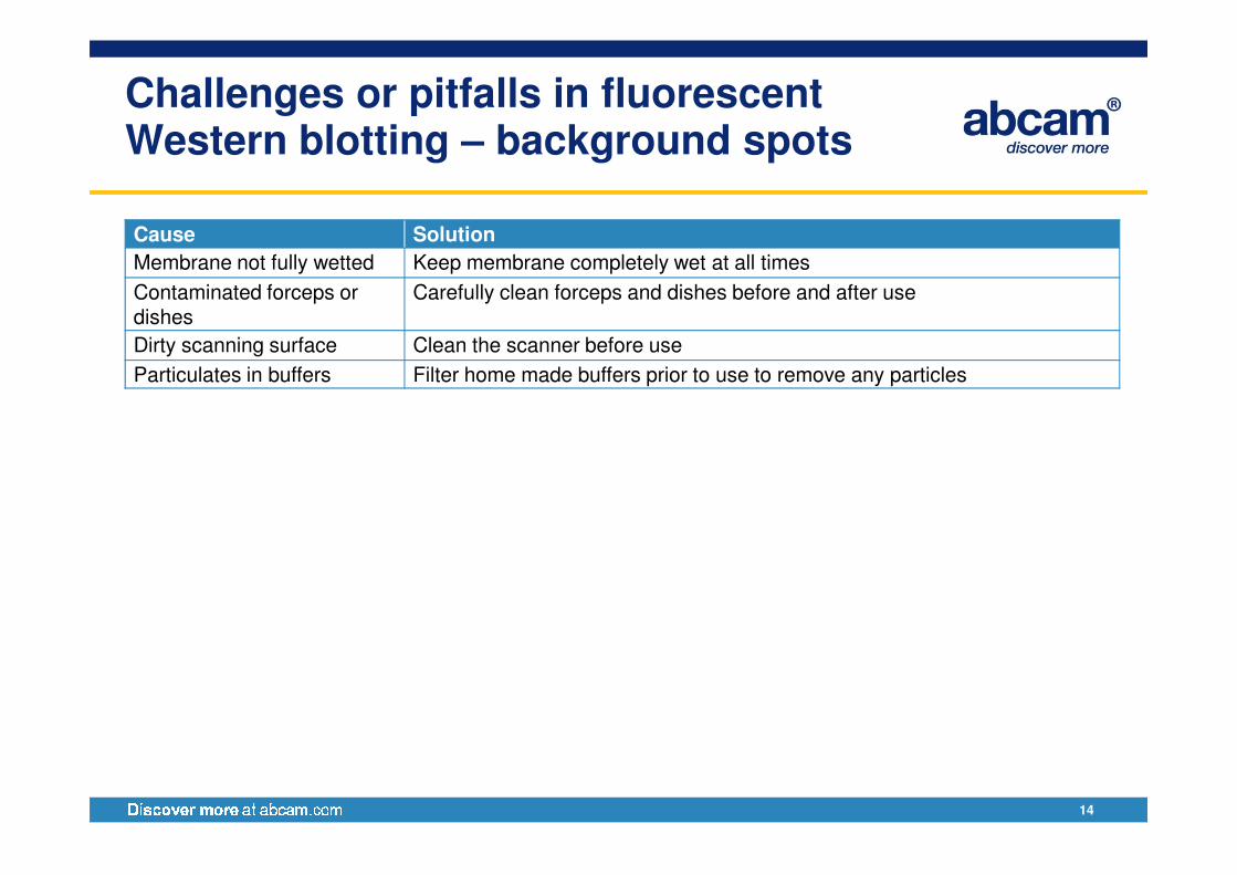
Challenges or pitfalls in fluorescent Western blotting – background spots
Cause Solution
Membrane not fully wetted Keep membrane completely wet at all times
Contaminated forceps or dishes
Carefully clean forceps and dishes before and after use
Dirty scanning surface Clean the scanner before use
Particulates in buffers Filter home made buffers prior to use to remove any particles
14
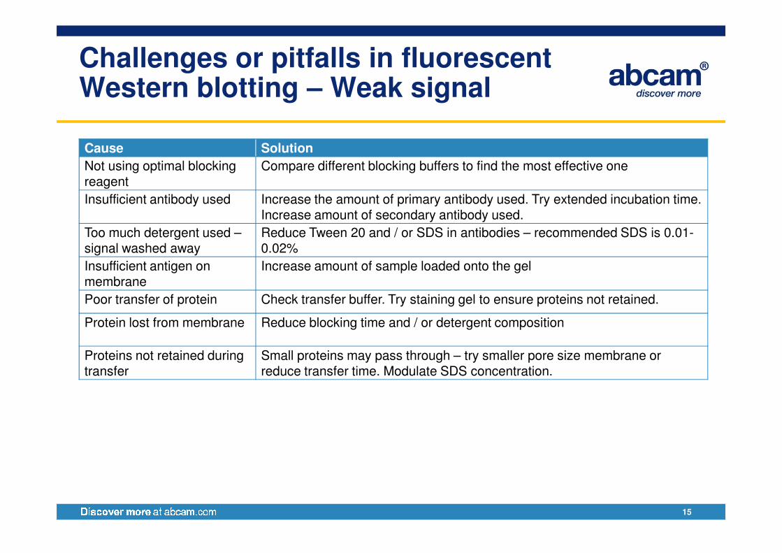
Challenges or pitfalls in fluorescent Western blotting – Weak signal
Cause Solution
Not using optimal blocking reagent
Compare different blocking buffers to find the most effective one
Insufficient antibody used Increase the amount of primary antibody used. Try extended incubation time. Increase amount of secondary antibody used.
Too much detergent used –signal washed away
Reduce Tween 20 and / or SDS in antibodies – recommended SDS is 0.01-0.02%
Insufficient antigen on membrane
Increase amount of sample loaded onto the gel
15
membrane
Poor transfer of protein Check transfer buffer. Try staining gel to ensure proteins not retained.
Protein lost from membrane Reduce blocking time and / or detergent composition
Proteins not retained during transfer
Small proteins may pass through – try smaller pore size membrane or reduce transfer time. Modulate SDS concentration.
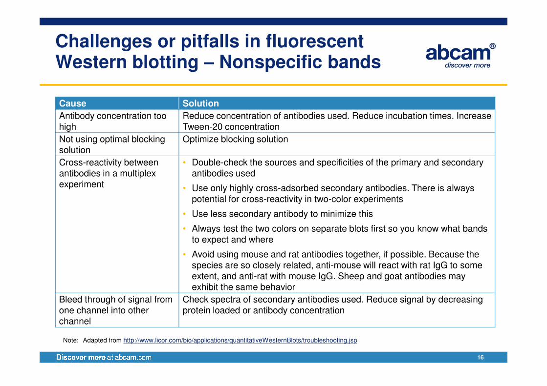
Challenges or pitfalls in fluorescent Western blotting – Nonspecific bands
Cause Solution
Antibody concentration too high
Reduce concentration of antibodies used. Reduce incubation times. Increase Tween-20 concentration
Not using optimal blocking solution
Optimize blocking solution
Cross-reactivity between antibodies in a multiplex experiment
• Double-check the sources and specificities of the primary and secondary antibodies used
• Use only highly cross-adsorbed secondary antibodies. There is always
16
• Use only highly cross-adsorbed secondary antibodies. There is always potential for cross-reactivity in two-color experiments
• Use less secondary antibody to minimize this
• Always test the two colors on separate blots first so you know what bands to expect and where
• Avoid using mouse and rat antibodies together, if possible. Because the species are so closely related, anti-mouse will react with rat IgG to some extent, and anti-rat with mouse IgG. Sheep and goat antibodies may exhibit the same behavior
Bleed through of signal from one channel into other channel
Check spectra of secondary antibodies used. Reduce signal by decreasing protein loaded or antibody concentration
Note: Adapted from http://www.licor.com/bio/applications/quantitativeWesternBlots/troubleshooting.jsp

Protocols and Resources
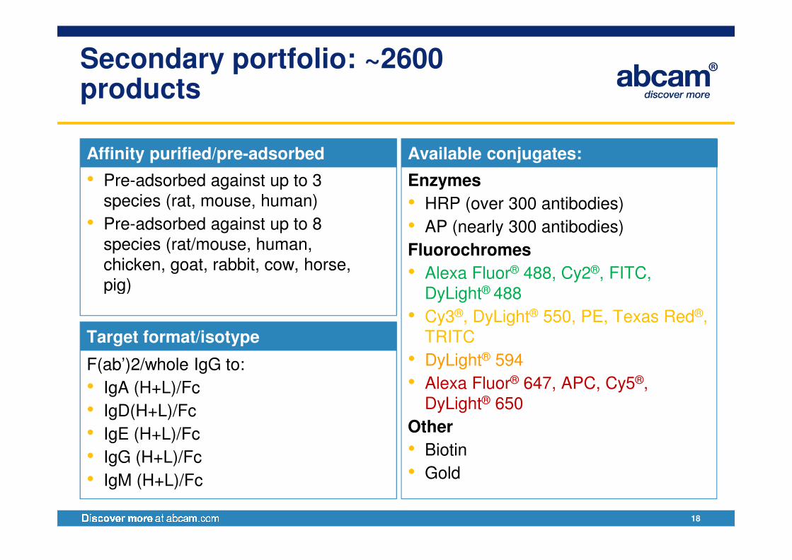
Secondary portfolio: ~2600 products
Enzymes
• HRP (over 300 antibodies)
• AP (nearly 300 antibodies)
Fluorochromes
• Alexa Fluor® 488, Cy2®, FITC,
Available conjugates:
• Pre-adsorbed against up to 3 species (rat, mouse, human)
• Pre-adsorbed against up to 8 species (rat/mouse, human, chicken, goat, rabbit, cow, horse, pig)
Affinity purified/pre-adsorbed
18
• Alexa Fluor 488, Cy2 , FITC, DyLight® 488
• Cy3®, DyLight® 550, PE, Texas Red®, TRITC
• DyLight® 594
• Alexa Fluor® 647, APC, Cy5®, DyLight® 650
Other
• Biotin
• Gold
pig)
F(ab’)2/whole IgG to:
• IgA (H+L)/Fc
• IgD(H+L)/Fc
• IgE (H+L)/Fc
• IgG (H+L)/Fc
• IgM (H+L)/Fc
Target format/isotype
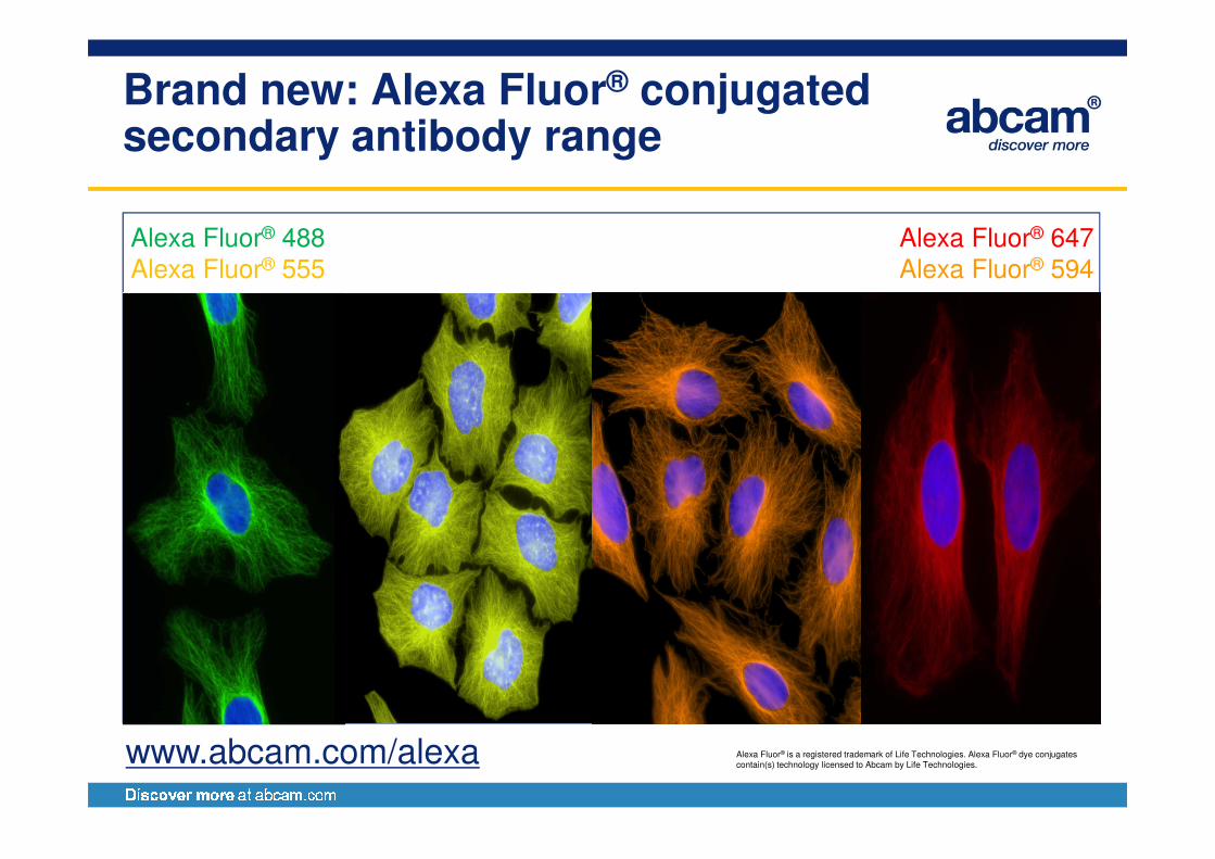
Brand new: Alexa Fluor® conjugated secondary antibody range
Alexa Fluor® 488Alexa Fluor® 555
Alexa Fluor® 647Alexa Fluor® 594
ab150157 ab150113ab150151
Alexa Fluor® is a registered trademark of Life Technologies. Alexa Fluor® dye conjugates contain(s) technology licensed to Abcam by Life Technologies.
www.abcam.com/alexa
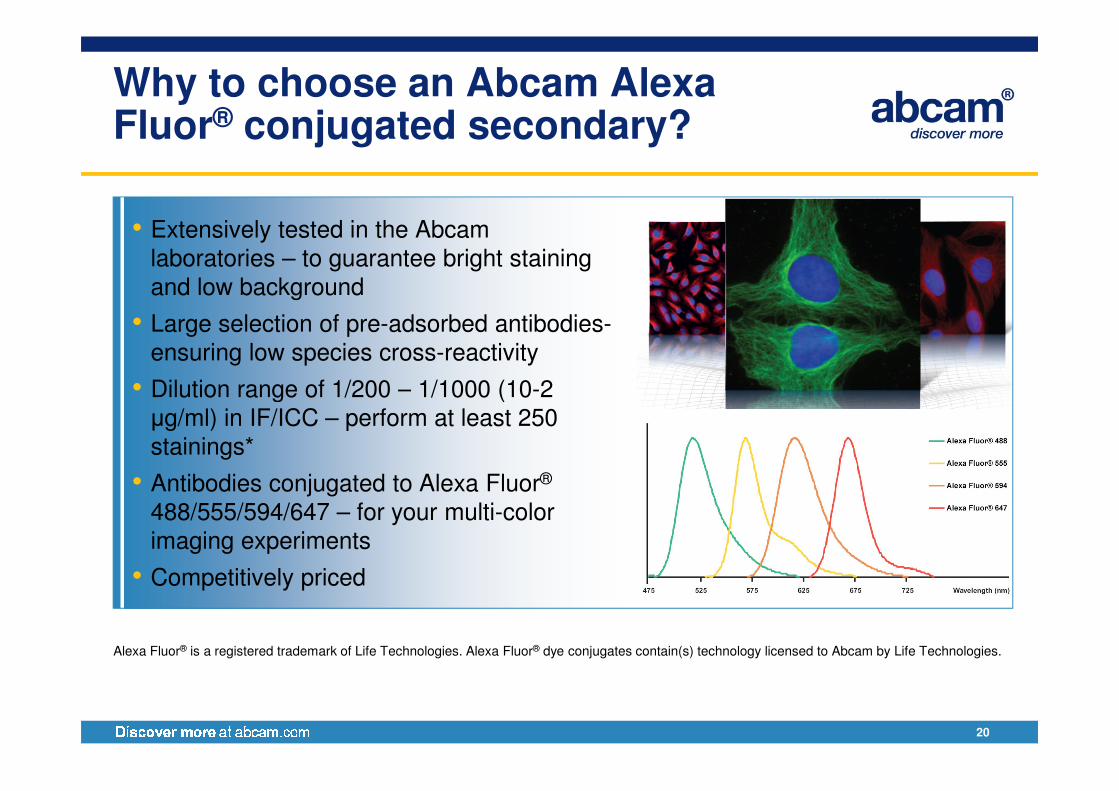
Why to choose an Abcam AlexaFluor® conjugated secondary?
• Extensively tested in the Abcamlaboratories – to guarantee bright staining and low background
• Large selection of pre-adsorbed antibodies-ensuring low species cross-reactivity
• Dilution range of 1/200 – 1/1000 (10-2
20
• Dilution range of 1/200 – 1/1000 (10-2 µg/ml) in IF/ICC – perform at least 250 stainings*
• Antibodies conjugated to Alexa Fluor®
488/555/594/647 – for your multi-color imaging experiments
• Competitively priced
Alexa Fluor® is a registered trademark of Life Technologies. Alexa Fluor® dye conjugates contain(s) technology licensed to Abcam by Life Technologies.
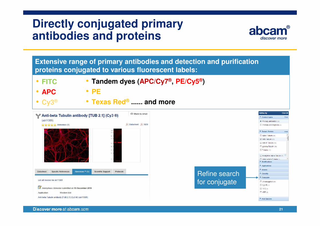
Directly conjugated primary antibodies and proteins
• Tandem dyes (APC/Cy7®, PE/Cy5®)
• PE
• Texas Red® ...... and more
• FITC
• APC
• Cy3®
Extensive range of primary antibodies and detection and purification proteins conjugated to various fluorescent labels:
21
Refine search for conjugate
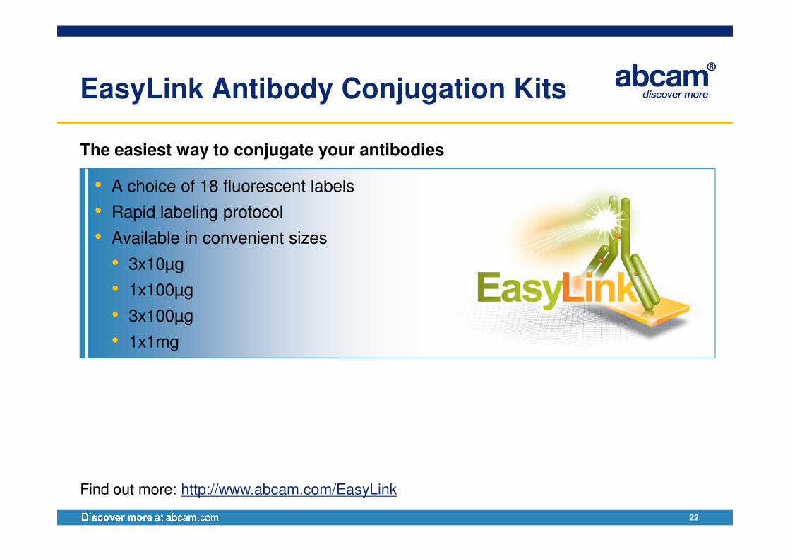
EasyLink Antibody Conjugation Kits
• A choice of 18 fluorescent labels
• Rapid labeling protocol
• Available in convenient sizes
• 3x10µg
•
The easiest way to conjugate your antibodies
22
• 1x100µg
• 3x100µg
• 1x1mg
Find out more: http://www.abcam.com/EasyLink
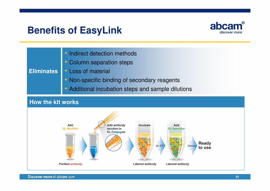
Benefits of EasyLink
Eliminates
• Indirect detection methods
• Column separation steps
• Loss of material
• Non-specific binding of secondary reagents
• Additional incubation steps and sample dilutions
23
How the kit works
Optiblot – Fluorescence detection kit
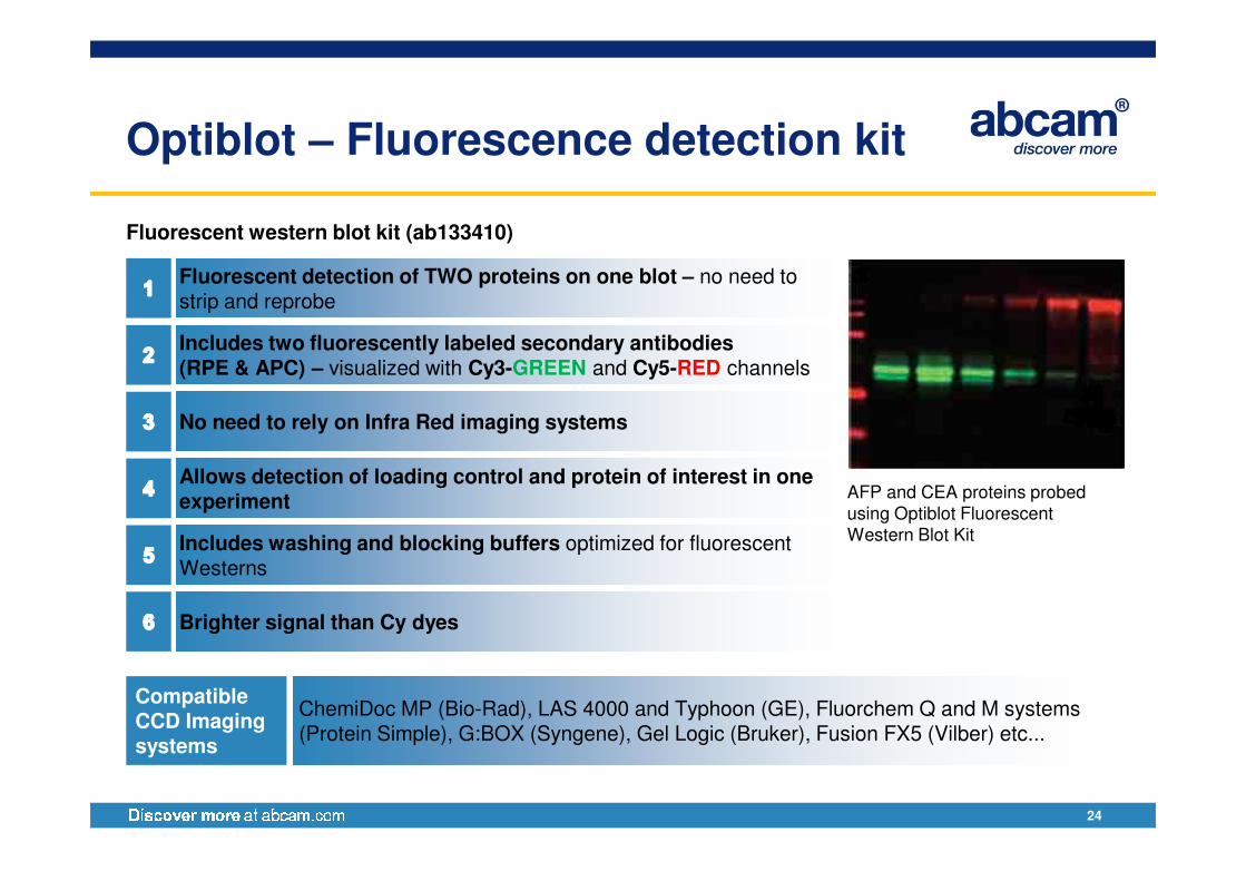
Fluorescent western blot kit (ab133410)
Fluorescent detection of TWO proteins on one blot – no need to strip and reprobe
Includes two fluorescently labeled secondary antibodies(RPE & APC) – visualized with Cy3-GREEN and Cy5-RED channels
No need to rely on Infra Red imaging systems
24
Allows detection of loading control and protein of interest in one experiment
Includes washing and blocking buffers optimized for fluorescent Westerns
Brighter signal than Cy dyes
Compatible CCD Imaging systems
ChemiDoc MP (Bio-Rad), LAS 4000 and Typhoon (GE), Fluorchem Q and M systems (Protein Simple), G:BOX (Syngene), Gel Logic (Bruker), Fusion FX5 (Vilber) etc...
AFP and CEA proteins probed using Optiblot Fluorescent Western Blot Kit
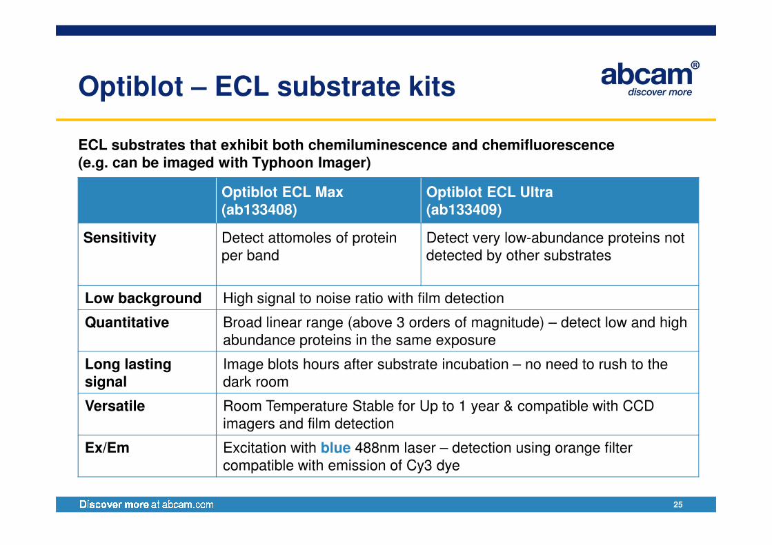
Optiblot – ECL substrate kits
ECL substrates that exhibit both chemiluminescence and chemifluorescence(e.g. can be imaged with Typhoon Imager)
Optiblot ECL Max(ab133408)
Optiblot ECL Ultra(ab133409)
Sensitivity Detect attomoles of protein
per band
Detect very low-abundance proteins not
detected by other substrates
25
Low background High signal to noise ratio with film detection
Quantitative Broad linear range (above 3 orders of magnitude) – detect low and high
abundance proteins in the same exposure
Long lasting signal
Image blots hours after substrate incubation – no need to rush to the
dark room
Versatile Room Temperature Stable for Up to 1 year & compatible with CCD
imagers and film detection
Ex/Em Excitation with blue 488nm laser – detection using orange filter
compatible with emission of Cy3 dye
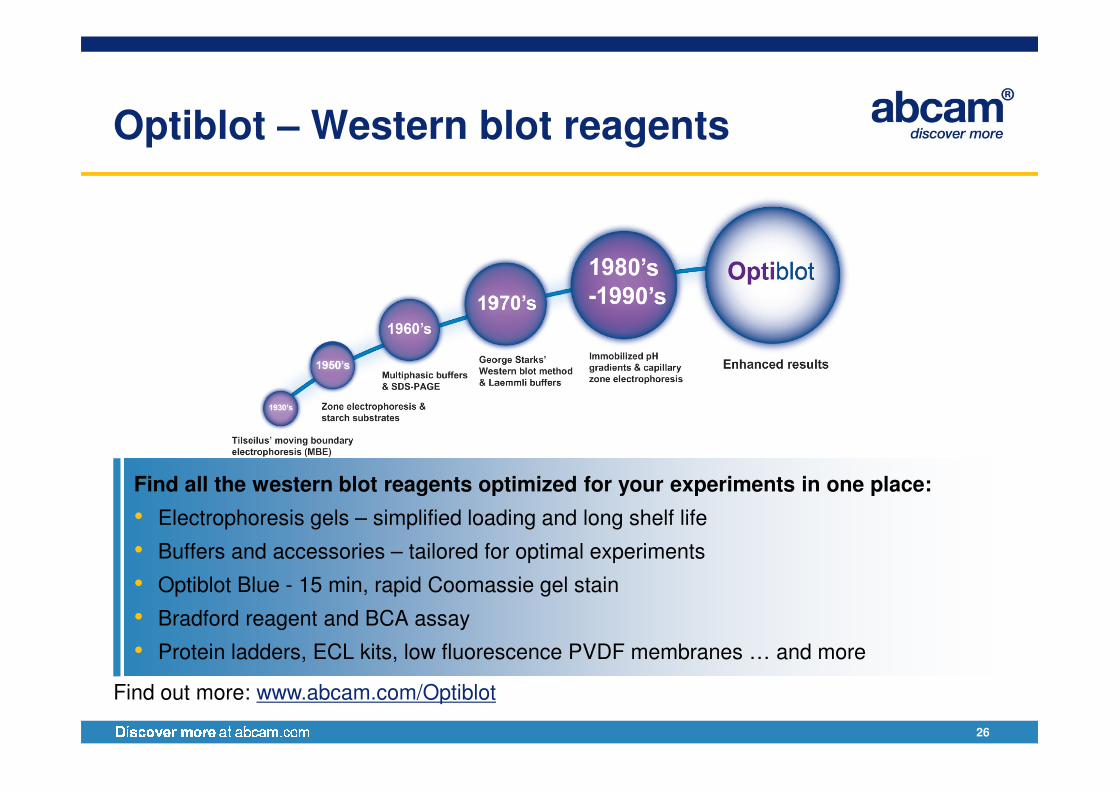
Optiblot – Western blot reagents
26
Find all the western blot reagents optimized for your experiments in one place:
• Electrophoresis gels – simplified loading and long shelf life
• Buffers and accessories – tailored for optimal experiments
• Optiblot Blue - 15 min, rapid Coomassie gel stain
• Bradford reagent and BCA assay
• Protein ladders, ECL kits, low fluorescence PVDF membranes … and more
Find out more: www.abcam.com/Optiblot
More Resources
• Optiblot landing page:
www.abcam.com/Optiblot
• Optiblot FAQS:
www.abcam.com/OptiblotFAQS
• Western blot webinar:
www.abcam.com/webinars
27
Western Blot Reagents and Troubleshooting Tips Booklet
www.abcam.com/webinars
• Western blot detailed guide and video:
http://www.abcam.com/westernblotguide
• Loading control guide:
http://www.abcam.com/loadingcontrolguide
• More protocols and guides:
http://www.abcam.com/protocolguides
Promotion
Exclusive webinar promotion on products for fluorescent western blot
• Save on all qualifying products:
• The entire Optiblot range
• All secondary antibodies, Save
28
• All secondary antibodies,
• Fluorophore conjugated detection and purification proteins
• Fluorophore conjugated primary antibodies
• Further information to be sent after the webinar
Save 25%
Note: Promotions may not be available on future viewings of the webinar
Allergy and Asthma 2013
Date: May 23-24, 2013
Venue: Bruges, Belgium
Conference Topics
• Innate immune cells in asthma
• Epithelial biology and asthma
• Understanding adaptive immunity in
Oral abstract deadline:February 22, 2013
Poster abstract deadline:March 25, 2013
29
• Understanding adaptive immunity in
asthma
• Environment and asthma
Confirmed speakers
• David Artis (University of Pennsylvania)
• John Fahy (University of California)
• Darryl Knight (University of British
Columbia)
• Carla Ribeiro (University of North Carolina)
and may more....
Meeting website:www.abcam.com/AA2013
Inflammasomes in Health and Disease
Date: June 24-25, 2013
Venue: Boston, US
Conference Topics
• General mechanisms of activation
• Inflammasomes in infection
Oral abstract deadline:April 26, 2013
Poster abstract deadline:May 17, 2013
30
• Inflammasomes in infection
• Inflammasomes and sterile inflammation
• Metabolism-inflammasome link
Keynote speaker:
• Vishva Dixit (Genentech, US)
Meeting chairs:
• Kate Fitzgerald, University of Massachusetts
• Gabriel Nunez, University of Michigan
Meeting website:www.abcam.com/inflammasomes2013

Upcoming webinars
IHC/ICC staining techniques using single and multiple labels
March 06, 2013
15:00 GMT, 10:00 EDT, 07:00 PDT
www.abcam.com/IHCICCwebinar
31
labels www.abcam.com/IHCICCwebinar
To interact or not to interact? Immunoprecipitateto answer this question
March 21, 2013
15:00 GMT, 10:00 EDT, 07:00 PDT
www.abcam.com/events

Questions?
32


![Western Blotting BCH 462[practical] Lab#6. Objective: -Western blotting of proteins from SDS-PAGE.](https://static.fdocuments.in/doc/165x107/56649dc85503460f94abe06c/western-blotting-bch-462practical-lab6-objective-western-blotting-of.jpg)