Fluorescence yields of isatoic anhydride from the reaction of N-glyoxyloylanthranilic acid 2-oxime...
5
268 J. Org. Clzem., 1~01. 43, No. 2, 1978 Crabtree, Kramer, and Murr Fluorescence Yields of Isatoic Anhydride from the Reaction (of N-Glyoxyloylanthranilic Acid 2-Oxime with Electrophiles Eleanor V. Crabtree and David N. Kramer* Chemical Laboratory, Edgewood Arsenal, Aberdeen Proving Ground, Maryland 21010 Brown L. Murr, Jr. Department of Chemistry, Johns Hopkins University, Baltimore, Maryland 21218 Received March 10,1977 N-Glyoxyloylanthranilic acid 2-oxime (1) was converted to isatoic anhydride (2) and cyanide ion by reaction with methanesulfonyl fluoride and chloride, isopropyl methylphosphonofluoridate (Sarin),acetic anhydride, Parathion, and Meta-Systox R in 1:l organic solvent-aqueous borate buffer and in 98% organic solvent containing tetrabutyl- amnionium hydroxide. The borate buffer catalyzed the hydrolysis of the electrophiles agents and reduced the yield of 2. A stoichiometric yield of 2 wadobtained in 98% nonhydroxylic solvents, in which 2 was stable. The anion of 1 quenched the fluorescence of 2 anion and caused a shift in excitation wavelength for maximum fluorescence with- out a shift in the emission wavelength. These facts were ascribed to an inner filter effect of 1 anion. The quenching could be represented by a Stern-Volmer relationship with slopes of 620 M-I in 50% aqueous acetonitrile, 330 M-I in 73:25:2 acetonitrile-acetone-water, and 690 M-I in 73:25:2 tert- butyl alcohol-acetone-water. The excitation and emission wavelengths and relative fluorescence intensities were measured for various substituted isatoic anhy- drides: 2, 350, 430, 1.00; 543-2, 360, 440, 1.68; 5-sulfo-N-Me-2, 330, 395, 0.92; 5-aza-2 (2,3-pyrido-3,1-oxazine-2,4- dione), 350,435,3.55; 5-N02-2,335,435,0.016; N-Me-2,328, 398, 1.61; 5-C1-N-Me-2, 338,405, 1.21. Dziomko, Ivanov, and Kremenskayal have reported a sensitive, quantitative fluorometric method for the determi- nation of acid chlorides and anhydrides, sulfonyl chlorides, and phosphorus oxychloride based on the reaction with N- glyoxyloylanthranilic acid 2-oxime (2-carboxyisonitrosoace- tanilide, 1) in an alkaline, buffered 25% aqueous acetone medium. Because we were unable to achieve the reported sensitivity in the 5 X lop9 to M range (perhaps due to the absence of experimental details), we initiated a study to maximize the yield of the fluorescent species, the anion of 2H-3,1-benzoxazine-2,4(lH)-dione (isatoic anhydride, 2).2 The anion of I was shown to quench the fluorescence of 2 anion. Conditions were developed for the reactions of meth- anesulfonyl fluoride (MSF) and isopropyl methylphospho- nofluoridate (Sarin) with 1 anion to yield 2 anion quantita- tively with a minimum of quenching. Using these conditions the sensitivity of the detection of Sarin was 0.002 pg/mL, which approaches that of the enzymatic methods (0.001 fig/mL).3 Scheme I 1 Results and Discussion Identification of the Fluorescent Species. When oxime 1 in 1:3 acetona-water buffered at pH 9.0 by 0.05 M borate was allowed to react with methanesulfonyl chloride, the resulting solution exhibited fluorescence, A, 365 nm, A , 435 nm. The only isolable solid was anthranilic acid (Aex 330 nm, A, 400 nm, in basic solution). However, reaction of 1 with benzene- sulfonyl chloride in pyridine afforded 2. The fluorescence of an authentic sample of 2 in 25% aqueous acetone, buffered at pH 9.0 (A,, 350 nm, ,Aern 435 nm), was similar to the fluores- cence of the reaction solution. Furthermore, the pH depen- dence of the fluorescence intensity produced in the reaction of 1 with methanesulfonyl fluoride over the pH range 8.0-10.5 paralleled that of authentic 2 in the presence of 1. The fluo- rescence excitation and emission spectra of a mixture of 2 and 1 at pH 9.0 were identical with the spectra obtained in the reaction of 1 anion with MSF. Variations in solvent affected the fluorescence of 2 and that of the reaction mixture in a similar manner. A reasonable sequence for the conversion of 1 to 2 by sul- fonyl halides via 3 and 4 is shown in Scheme I. A similar se- quence has been proposed by Guinullina et aL2 for 1 with acetic anhydride in aqueous solution. In support of this ab- 0022-3263/78/1943-0268$01.00/0 " L O J n L 1 hydrolysis 4 aco2H NH2 6 normal Beckmann type mechanism, it was shown that cyanide ion accompanied the formation of the fluorescent 2. Karrer, Diechmann, and Haebler4 heated 1 with excess thionyl chlo- ride to obtain 5, which was rapidly converted to 2 under mild hydrolytic conditions. We considered compound 5 an unlikely intermediate in the conversion of 3 to 2, since strong dehy- drating conditions seem necessary for the formation of 5. The reaction of 1 with acetic anhydride in pyridine did not give the nitrile 5 (Scheme I), but yielded 7. The structure of 7 was es- tablished by IR, NMR, elemental, and mass spectral analyses. Hurd and Bethune5 showed that o -carboxyarylhydroxamic acids, when subjected to the Lossen rearrangement in an inert 1978 American Chemical Society
Transcript of Fluorescence yields of isatoic anhydride from the reaction of N-glyoxyloylanthranilic acid 2-oxime...
268 J . Org. Clzem., 1~01. 43, No. 2, 1978 Crabtree, Kramer, and Murr
Fluorescence Yields of Isatoic Anhydride from the Reaction (of N-Glyoxyloylanthranilic Acid 2-Oxime with Electrophiles
Eleanor V. Crabtree and David N. Kramer*
Chemical Laboratory, Edgewood Arsenal, Aberdeen Proving Ground, Maryland 21010
Brown L. Murr, Jr.
Department of Chemistry, J o h n s Hopkins University, Baltimore, Maryland 21218
Received March 10,1977
N-Glyoxyloylanthranilic acid 2-oxime (1) was converted to isatoic anhydride (2) and cyanide ion by reaction with methanesulfonyl fluoride and chloride, isopropyl methylphosphonofluoridate (Sarin), acetic anhydride, Parathion, and Meta-Systox R in 1:l organic solvent-aqueous borate buffer and in 98% organic solvent containing tetrabutyl- amnionium hydroxide. The borate buffer catalyzed the hydrolysis of the electrophiles agents and reduced the yield of 2. A stoichiometric yield of 2 wadobtained in 98% nonhydroxylic solvents, in which 2 was stable. The anion of 1 quenched the fluorescence of 2 anion and caused a shift in excitation wavelength for maximum fluorescence with- out a shift in the emission wavelength. These facts were ascribed to an inner filter effect of 1 anion. The quenching could be represented by a Stern-Volmer relationship with slopes of 620 M-I in 50% aqueous acetonitrile, 330 M-I in 73:25:2 acetonitrile-acetone-water, and 690 M-I in 73:25:2 ter t - butyl alcohol-acetone-water. The excitation and emission wavelengths and relative fluorescence intensities were measured for various substituted isatoic anhy- drides: 2, 350, 430, 1.00; 543-2, 360, 440, 1.68; 5-sulfo-N-Me-2, 330, 395, 0.92; 5-aza-2 (2,3-pyrido-3,1-oxazine-2,4- dione), 350,435, 3.55; 5-N02-2,335,435,0.016; N-Me-2,328, 398, 1.61; 5-C1-N-Me-2, 338,405, 1.21.
Dziomko, Ivanov, and Kremenskayal have reported a sensitive, quantitative fluorometric method for the determi- nation of acid chlorides and anhydrides, sulfonyl chlorides, and phosphorus oxychloride based on the reaction with N - glyoxyloylanthranilic acid 2-oxime (2-carboxyisonitrosoace- tanilide, 1) in an alkaline, buffered 25% aqueous acetone medium. Because we were unable to achieve the reported sensitivity in the 5 X lop9 to M range (perhaps due to the absence of experimental details), we initiated a study to maximize the yield of the fluorescent species, the anion of 2H-3,1-benzoxazine-2,4(lH)-dione (isatoic anhydride, 2).2 The anion of I was shown to quench the fluorescence of 2 anion. Conditions were developed for the reactions of meth- anesulfonyl fluoride (MSF) and isopropyl methylphospho- nofluoridate (Sarin) with 1 anion to yield 2 anion quantita- tively with a minimum of quenching. Using these conditions the sensitivity of the detection of Sarin was 0.002 pg/mL, which approaches tha t of the enzymatic methods (0.001 fig/mL).3
Scheme I
1
Results and Discussion Identification of the Fluorescent Species. When oxime
1 in 1:3 acetona-water buffered a t pH 9.0 by 0.05 M borate was allowed to react with methanesulfonyl chloride, the resulting solution exhibited fluorescence, A,, 365 nm, A,, 435 nm. The only isolable solid was anthranilic acid (Aex 330 nm, A,, 400 nm, in basic solution). However, reaction of 1 with benzene- sulfonyl chloride in pyridine afforded 2. The fluorescence of an authentic sample of 2 in 25% aqueous acetone, buffered a t pH 9.0 (A,, 350 nm, ,Aern 435 nm) , was similar to the fluores- cence of the reaction solution. Furthermore, the p H depen- dence of the fluorescence intensity produced in the reaction of 1 with methanesulfonyl fluoride over the pH range 8.0-10.5 paralleled that of authentic 2 in the presence of 1. The fluo- rescence excitation and emission spectra of a mixture of 2 and 1 a t p H 9.0 were identical with the spectra obtained in the reaction of 1 anion with MSF. Variations in solvent affected the fluorescence of 2 and tha t of the reaction mixture in a similar manner.
A reasonable sequence for the conversion of 1 to 2 by sul- fonyl halides via 3 and 4 is shown in Scheme I. A similar se- quence has been proposed by Guinullina et aL2 for 1 with acetic anhydride in aqueous solution. In support of this ab-
0022-3263/78/1943-0268$01.00/0
" L O J n
L
1 hydrolysis 4
aco2H NH2
6
normal Beckmann type mechanism, it was shown that cyanide ion accompanied the formation of the fluorescent 2. Karrer, Diechmann, and Haebler4 heated 1 with excess thionyl chlo- ride to obtain 5, which was rapidly converted to 2 under mild hydrolytic conditions. We considered compound 5 an unlikely intermediate in the conversion of 3 to 2, since strong dehy- drating conditions seem necessary for the formation of 5. The reaction of 1 with acetic anhydride in pyridine did not give the nitrile 5 (Scheme I) , bu t yielded 7. The structure of 7 was es- tablished by IR, NMR, elemental, and mass spectral analyses. Hurd and Bethune5 showed tha t o -carboxyarylhydroxamic acids, when subjected to the Lossen rearrangement in an inert
1978 American Chemical Society
Reaction of N- Glyoxyloylanthranilic Acid 2-Oxime with Electrophiles J . Org. Chem., Vol. 43, No. 2, 1978 269
/
1 0.oLJ-J ' 1 " ' ' ' I " ' 1 ' ' I '
I 0 2.0 1.0 4.0 5.0
[Q] X IO3, M
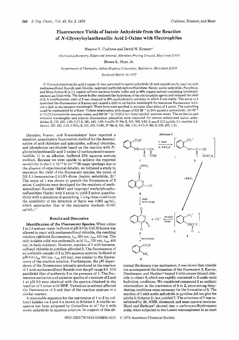
Figure 1. Stern-Volmer plots for solvent dependence of quenching of isatoic anhydride (2) fluorescence by N-glyoxyloylanthranilic acid 2-oxime (Q): 0,l x 1O-5M 2 in acetonitrile-water (1:1), pH 9.7 borate; A , 5 X lo-' M 2 in acetonitrile-acetone-water (73:25:2), BUNOH; 0, 5 X M 2 in tert-butyl alcohol-acetone-water (73:25:2), Bu~NOH.
medium, gave the corresponding isatoic anhydrides presum- ably via isocyanates. The above reactions of 1 are in accord with its conversion to 2 via 3 and 4 (Scheme I).
0
@$LH = NO Ac
7
Reduction of the Fluorescence of 2 by 1. T h e reduction of the fluorescence of 2 anion by 1 anion could be characterized by the Stern-Volmer equation.6 T h e plots (Figure 1) were linear, with the same slope for the two concentrations of 2 anion (5 X and 1 X lov5 M) over the concentration range of 1 anion from 2.5 X to 5.0 X M. The slope was 620 M-l in 50% aqueous acetonitrile, 330 M-l in 73:25:2 aceto- nitrile-acetone-water, and 690 M-l in 73:25:2 t e r t - butyl al- cohol-acetone-water.
The quenching of the fluorescence was due largely to a n inner-filter effect.(; This (conclusion was based on the following observations: (1) 1 anion and 2 anion had overlapping ab- sorptions; (2) 1 anion did not absorb in the region of the flu- orescence emission; (3) the excitation wavelength for maxi- mum fluorescence shifted to longer wavelengths as the con- centration of 1 anion increased; and (4) the fluorescence emission wavelength was independent of the concentration of 1 anion.
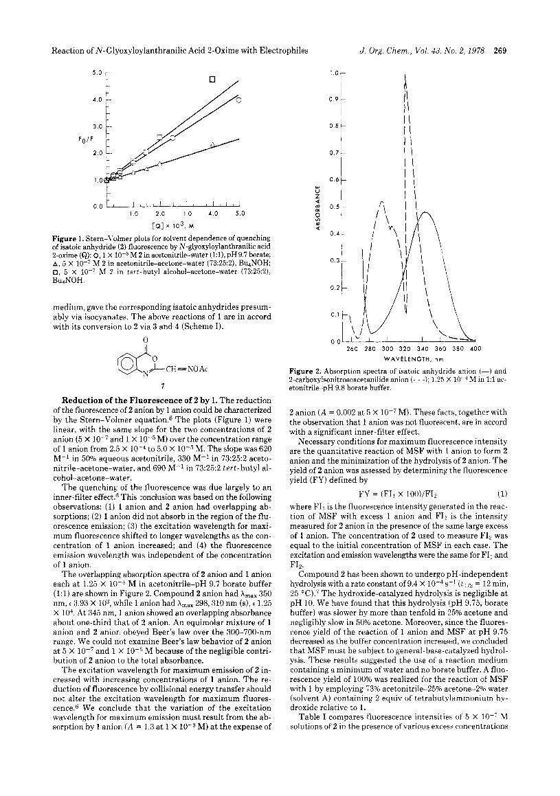
The overlapping absorption spectra of 2 anion and 1 anion each a t 1.25 X M in acetonitrile-pH 9.7 borate buffer (1:l) are shown in Figure 2. Compound 2 anion had A,,, 350 nm, t 3.93 X lo3, while 1 anion had A,,, 298,310 nm (s), e 1.25 X lo4. At 345 nm, 1 anion showed an overlapping absorbance about one-third tha t of 2 anion. An equimolar mixture of 1 anion and 2 anion obeyed Beer's law over the 300-700-nm range. We could not examine Beer's law behavior of 2 anion a t 5 X and 1 '< loT5 M because of the negligible contri- bution of 2 anion to the total absorbance.
T h e excitation wavelength for maximum emission of 2 in- creased with increasing concentrations of 1 anion. T h e re- duction of fluorescence by collisional energy transfer should not alter the excitation wavelength for maximum fluores- cence.6 We conclude that the variation of the excitation wavelength for maximum emission must result from the ab- sorption by 1 anion (A = 1.3 a t 1 X 10⁻³ M) a t the expense of
I \ I \ I \ I \ I \ I \ I \ I \
1 I I z L'
260 280 3 0 0 3 2 0 3 4 0 360 3 8 0 400
WAVELENGTH, nrn
Figure 2. Absorption spectra of isatoic anhydride anion (-) and 2-carboxyisonitrosoacetanilide anion ( - - -); 1.25 X loF4 M in 1:l ac- etonitrile-pH 9.8 borate buffer.
2 anion ( A = 0.002 a t 5 X M). These facts, together with the observation that 1 anion was not fluorescent, are in accord with a significant inner-filter effect.
Necessary conditions for maximum fluorescence intensity are the quantitative reaction of MSF with 1 anion to form 2 anion and the minimization of the hydrolysis of 2 anion. The yield of 2 anion was assessed by determining the fluorescence yield (FY) defined by
(1) where FI1 is the fluorescence intensity generated in the reac- tion of M S F with excess 1 anion and FI2 is the intensity measured for 2 anion in the presence of the same large excess of 1 anion. The concentration of 2 used to measure FI2 was equal to the initial concentration of MSF in each case. T h e excitation and emission wavelengths were the same for FI1 and
Compound 2 has been shown to undergo pH-independent hydrolysis with a rate constant of 9.4 X s-l ( t l / z = 12 min, 25 0C).7 The hydroxide-catalyzed hydrolysis is negligible a t p H 10. We have found tha t this hydrolysis (pH 9.75, borate buffer) was slower by more than tenfold in 25% acetone and negligibly slow in 50% acetone. Moreover, since the fluores- cence yield of the reaction of 1 anion and MSF a t p H 9.75 decreased as the buffer concentration increased, we concluded tha t MSF must be subject to general-base-catalyzed hydrol- ysis. These results suggested the use of a reaction medium containing a minimum of water and no borate buffer. A fluo- rescence yield of 100% was realized for the reaction of MSF with 1 by employing 73% acetonitrile-25% acetone-2% water (solvent A) containing 2 equiv of tetrabutylammonium hy- droxide relative to 1.
Table I compares fluorescence intensities of 5 X lo-: M solutions of 2 in the presence of various excess concentrations
FY = (FII X 100)/FIz
FI2.
270 J . Org. Chem., Vol. 43, No. 2, 1978 Crabtree, Kramer, and Murr
Table I. Effects of Solvent and 1 Anion Concentration on Fluorescence Intensity and Yield of 2 i n t h e Conversion
of 1 to 2 by MSF
111 x 104, M FI. 2" FI, MSFb Yield,
Solvent A d None 55.5 0 0
2.5 52.0 25.5F 49.5e 5.0 50.0 50.0 103
10.0 43.5 45.5 104 20.0 34.5 36.0 106 40.0 23.0 23.0 100
None 74.0 0 0 Solvent Bf
2.5 x 10⁻⁴ 64.0 35.0e 55.5e 5.0 x 55.0 43.0 78.0 1.0 x 10- '3 44.0 37.5 86.0 2.0 x lo-" 28.5 27.0 90.0 4.0 X lo-,< 15.5 14.0 90.5
Reading for a mixture of 5 X M 2 and the indicated concentration of 1 after 4 min. M MSF and the indicated concentration of 1, after 4 min unless otherwise noted. ' Fluorescence yield: (column 3/column 2) X 100.
Acetonitrile -acetone-water (73:25:2) and 2 equiv of BujNOH/equiv of 1. e Values after 10 min; the fluorescence in- tensity was still increasing. f tert-Butyl alcohol-acetone-water (73:25:2) and 2 equiv of BujNOH/equiv of 1.
Reading for a mixture of 5 X
of 1 (column 2) with the fluorescence intensities produced by the reaction of 5 X M solutions of MSF with the same excess concentrations of 1 (column 3). The fluorescence yields (column 4) of t he MSF-oxime reactions were quantitative after 4 min in solvent A for oxime concentrations >5 X M. T h e fluorescence yields were not quantitative in 73% tert- butyl alcohol-25% acetone-2% water (solvent B), pre- sumably due to competing solvolysis of MSF. In solvent A a t 2.5 X lo-* M 1, t he fluorescence yield of 2 was about 50% in 10 min, while a t 5 X M 1 the fluorescence yield was 100% in 4 min, and a t 1 X M 1 the yield was 100% in 2 min. In solvent B the maximum fluorescence yield was 90% and the rate of attainment of t ha t maximum was slower.
Dziomkol reported t h a t t he maximum fluorescence inten- sity was achieved in 4 min when the concentration of 1 was 2.5 X M, whereas the maximum fluorescence intensity in our solvent A was achieved in 2 min at 1. X 1 anion, Le., with a 25-fold louer concentration of quencher. Therefore, t he fluorescence intensity is much greater in solvent A than in 25% acetone-containing buffer. Furthermore, t he hydrolyses of MSF and 2 were eliminated in solvent A, as shown by the quantitative fluorescence yield. Finally, the rate of formation of 2 is significantly faster in solvent A than in 25% acetone, demonstratin: the superiority of a nonaqueous, nonhydroxylic solvent systein.
Several solvents (1 :1, organic solvent-water; acetone, ace- tonitrile, tetrahydrofuran, tert- butyl alcohol, p -dioxane, and 2-butanol) were tested for their effects on the quenching of 2 anion by 1 anion. No significant effects were found. However, MSF and 2 anion were found to solvolyze in hydroxylic sol- vents, e.g., methanol. ethanol, and isopropyl alcohol. T h e inner-filter effect was less by twofold in 98% organic nonhy- droxylic solvent (Figure 1).
T h e fluorescence properties of several substituted isatoic anhydrides were examined for advantages over 2. The relative fluorescence intensities of M solutions (50% acetonitrile, p H 9.85) were 0.016 for 5-NO2-2,l.OO for 2,1.68 for 5-chloro-2, and 3.35 for 5-aza-2 (2,3-pyrido-3,1-oxazine-2,4-dione). T h e last compound was over three times more fluorescent than 2, bu t the corresponding isonitroso precursor could not be pre- pared. It was also noted tha t t he fluorescence intensity of
N-Me-2 was greater than that of 2, although it cannot ionize in base (see Experimental Section).
Quantitative studies were made on several electrophiles with 1 anion (1 X M) using as solvent acetonitrile-ace- tone-water (92:2:1) with 2 X M tetrabutylammonium hydroxide. Sarin and MSF were readily determined in 3 min in the range 0.01 pg/mL (7.14 X M Sarin) to 2.0 pg/mL (1.45 x M Sarin). Sarin gave a 100% yield within 10 min. Acetic anhydride was detected a t a concentration of 1 X M. Parathion was detected in 3 min at 3 pg/mL (1 X 10⁻⁵ M). Meta-Systox R was not detected a t this level.
Using Barney'ss definition of minimum detectable differ- ence, namely,
I s - I b = K S b d where I , = sample fluorescence, I b = blank fluorescence, s b = standard deviation of blank fluorescence, and K = 2 f i for a 99% confidence limit, we found the minimum detectable limit for Sarin t o be 1.43 X IO-* M or 0.002 pg/mL. We have exceeded the minimum detectable limit (0.026 fluorescence unit) after 3 rnin and in 10 rnin the reading less blank ( I , - I b ) was 0.038. The sensitivity of the reagent to Sarin approached tha t of the enzymatic detection methods, which have been reported to be 0.001 ~ g / m L . ~
E x p e r i m e n t a l Sec t ion
Melting points were determined on a Thomas-Hoover capillary apparatus and are uncorrected. The infrared spectra were recorded on Perkin-Elmer Model 257 and 521 spectrophotometers. The ul- traviolet absorption spectra were obtained using a Cary 14 instrument with the cell compartment at 25 f 0.1 "C and matched 1-cm cells. Proton NMR spectra were determined on a Varian A-60D spec- trometer using methanold4 and Me4Si as the internal standard. Mass spectra were run on a Perkin-Elmer Hitachi Model RMU-6E at 70 eV. Fluorescence spectra and intensities were measured with an Aminco-Bowman Model 4-8202 spectrophotofluorometer (SPF) equipped with a 200-W xenon-mercury lamp and with the cell com- partment maintained at 25 f 0.5 "C. The SPF was calibrated and adjusted daily against a l-rg/mL solution of quinine sulfate dihydrate in 0.1 N sulfuric acid at an emission wavelength of 450 nm and exci- tation at 350 nm. The excitation and emission wavelengths reported in this paper are uncorrected.
Reagents. Solvents were spectroquality and showed no significant fluorescence at 430 nm when excited at 360 nm. Borate buffers, pH 8.0-10.2, 0.05 M with respect to H3B03 and KC1 (buffer values of 2.0-5.81, were prepared by established procedure^.^ Aqueous 1 M tetrabutylammonium hydroxide (BQNOH) (Beckman Electrometric Reagent) was appropriately diluted with water or organic solvent; the diluted solution could be used for 1 week if stored in a refrigerator. Methanesulfonyl chloride and fluoride, obtained from Eastman Kodak Company, Rochester, N.Y., and 0,O-diethyl 0-(p-nitrophe- ny1)phosphorothioate (Parathion) and 0,O-dimethyl S-2-(ethyl- sulfiny1)ethylphosphorothioate (Meta-Systox R) from Kit No. 52AX, Polyscience Corp., Evanston, Ill., were used without further purifi- cation to prepare stock 0.01 M solutions in acetone. Isatoic anhydride and variously substituted isatoic anhydrides were recrystallized from acetonitrile and the purity verified by elemental analysis. Standard solutions of Sarin in acetone, 100 and 1 Ng/mL, were furnished by the Detection and Alarms Branch, Development and Engineering Di- rectorate, Edgewood Arsenal, APG, MD, and further diluted with acetonitrile.
Warning! Sarin is an extremely toxic cholinesterase inhibitor. Sarin, methanesulfonyl fluoride, and the pesticides should be handled in a well-ventilated fume hood and precautions taken to prevent in- halation or skin contamination. Concentrated NaOH solution should be used to decontaminate material and glassware. 2-Carboxyisonitrosoacetanilide (1) was prepared by the method
of Sandmeyer and obtained as a light tan powder, mp 206-208 "C (lit.lo 208 "C). Repeated recrystallization from hot water with Darco treatment yielded a white powder: mp 230-231 "C; IR (Nujol) 3300 (-"I, broad absorption 2500-2600 (OH of C02H), 1695 (COzH), 1665 (-CONH), 1590, and 1540 cm-' (C=N); NMR 6 4.9 ( 3 H, ex- changeable), 7.55 (1 H, CH=NO), 8.7-7.0 (4 H, br aromatic CH); mass spectrum m / e 208 (parent peak).
Anal. Calcd for CgHBN204: C, 51.9; H, 3.9; N, 13.6; 0,30.7. Found: C, 51.7; H, 4.0; N, 13.6; 0, 30.6.
Reaction of N - G1yox:yloylanthranilic Acid 2-Oxime with Electrophiles J . Org. Chem., Vol. 43, No. 2, 1978 271
Reaction of 1 with Methanesulfonyl Chloride. Methanesulfonyl chloride (0.6 g, 5.5 mmol) in 12 mL of acetone was added to a solution of 1 (1.0 g, 4.8 mmol) in 50 mL of 0.02 N NaOH. The solution was adjusted to pH 9.5 with a few drops of 2.5 N NaOH. The resulting solution exhibited a strong blue fluorescence (Aex 365, A,, 430 nm). The presence of cyanide in the reaction mixture was established by three methods: (a) by a 'colorimetric test with o-dinitrobenzene and p-nitrobenzaldehyde;" (b) by a cyanide specific electrode (Orion Research, Inc.); and (c) by an HCN detector tube test of the gas evolved from the solution upon acidification.'* The reaction mixture was acidified with glacial acetic acid and extracted with ether. The ether extract was washeld with water and dried over sodium sulfate. Evaporation of the solvent left a cream-colored powder which was identified as anthranilic acid by comparison of its IR spectrum and fluorescence spectra in 3:l pH 10 buffer-acetone, A,, 330, A,, 400 nm, with the spectra of an authentic sample.
Conversion of 2-Carboxyisonitrosoacetanilide ( I ) to Isatoic Anhydride (2). Compound 1 (1 g, 4.8 mmol) was dissolved in 10 mL of pyridine containing benzenesulfonyl chloride (0.94 g, 5.3 mmol). The solution turned red .purple and became slightly warm. The so- lution was refluxed for el0 rnin and then poured into 30 mL of ice- water slurry and the mixture stirred for 15 min. A pale green-yellow solid formed. Recrystallization of the product from 95% ethanol yielded 0.104 g of a powder, mp 239-240 "C dec. The IR spectrum (Nujol mull) was comparable to that reported by SadtlerI3 (spectrum 10 143) for isatoic anhydride (mp 243 OC).14 4-0xo-4H-3,1-benzorazine-2-carboxaldehyde 2-( 0-Acetyl-
oxime) (7) . A mixture of 1 (4.8 g, 24 mmol) with 20 mL of acetic an- hydride and 5.0 mL of pyridine was stirred at room temperature for 30 min. The precipitate was collected by filtration, washed with cold ethanol, and recrystallized from hot ethanol to give 4.2 g of white crystals: mp 179-180 "C; IR (KBr) 1775, 1755,1720 sh (br, C=O), 1600 cm-1 (strong, C=N); mass spectra mle 232 (parent peak). The NMR spectrum was in accord with the assigned structure.
Anal. Calcd for CllH8N204: C, 56.90; H, 3.47; N, 12.07: 0, 27.56. Found: C. 57.1; H! 3.2; N, 12.0; 0, 27.6.
Treatment of 7 with Methanolic KOH. To a suspension of 7 (1.0 g, 4.3 mmol) in 25 mL of methanol was added 5 mL of 1 M methanolic KOH. and the mixture was stirred at ambient temperature until the solid had dissolved (about 10 rnin). The solution was diluted with 60 mL of distilled water and then acidified with dilute HCI. The resulting white crystalline precipitate was collected and washed with water. Recrystallization l'rom chloroform gave 0.5 g of methyl isonitrosoa- cetoanthranilate15 (5): mp 175-180.5 "C (lit.16 180 "C): IR (Nujol) 3200 (br, NH, OH), 1705 (-COZMe), 1670 (NHCO-), 1595 cm-'
6.7 (1 H. CH=N-I. 8.8-7.0 ( 4 H. broad aromatic absorption); mass spectrum m i e 222 (paren.; peak).
Anal. Calcd for CloHlcN204: C, 54.05: H, 4.54; N, 12.6; 0, 28.8. Found: C, 54.4; H, 4.6; N. 12.7; 0, 28.1.
Cooling the filtrate from the reaction mixture after isolation of 8 gave shiny plates (0.17 g), inp 53-54 "C, identified by NMR and mass spectra as a mixturse of 7 8 O 4 , N-carboxyanthranilic acid dimethyl ester 9 and 22% 8.
pK, of Isatoic Anhydlride in Mixed Aqueous-Organic Sol- vents. Solutions of isatoic ,anhydride (0.01 M) in mixed solvents (50Y0 organic solvent-50% water) were titrated potentiometrically at 25 "C with 0.1 N KOH using a Radiometer pH stat (TTT/C Titrator fitted with an SBU-la syringe buret and an SBR-2C Titragraph). The ap- parent pK,s were: acetonitrile, 8.87; acetone, 8.61; isopropyl alcohol, 8.40: tert- butyl alcghol, 8.37; 2-methyl-2,4-pentanediol, 7.86; tetra- hydrofuran, 8.56: dimethylformamide, 8.63; dimethylacetamide, 8.36.17
General Procedure for the Fluorescence Studies. The effects of parameters such as pW, solvent, and reagent concentration on fluorescence intensities, fluorescence stability, and rates of reaction were studied. Reaction solutions were made by mixing in a glass- stoppered test tube the organic solvent (or solution of 1) and buffer or Bu4NOH. At zero time, a. solution of the test compound was added, mixed rapidly, and about I! mL of the mixture was transferred to a Teflon-stoppered quartz fluorometer cell. Volumes and concentra- tions of the solutions were chosen to give the desired final concen- tration of reagents, solvents, and test sample. At the same time, a reagent blank was prepared similarly. The change in fluorescence intensity, L F l A t , was measured at 1-min intervals. The blank was subtracted from thc sample reading to obtain the net fluorescence. The blank was determined for the same time intervals as for the sample.
Spectral Properties of Isatoic Anhydride ( 2 ) in the Presence of 2-Carboxyisonitrosoacetanilide (I). Stock solutions (5 X lW3
(-CHyNOH); NMR (CD:;OD) 6 3.95 (3 H,OCH3),4.6 (2H,NH,OH),
M) of 1 and 2 were prepared in acetonitrile. Addition of 0.1 mL of stock solution to a mixture of 1.0 mL of acetonitrile and 2.0 mL of aqueous pH 9.7 borate buffer was used to prepare 1.25 X M so- lutions. The UV absorption spectra were recorded for solutions of l and 2 separately (Figure 2) and as equimolar mixtures. Scans were repeated at 10-min intervals to check solution stability. In separate experiments, methanol, ethanol, and isopropyl alcohol were substi- tuted for acetonitrile.
The excitation and emission spectra were recorded for solutions of 2 (5 X M, in 1:l acetone-aqueous pH 9.7 borate buffer) in the absence of 1 and in the presence of measured amounts of 1 (5 X to 5 X M final concentration). The excitation wavelength for maximum emission shifted from 350 nm in the absence of 1 to 370 nm in the presence of 5 X M 1. The wavelength of the emission maximum remained constant a t 430 nm. but the emission intensity decreased with increasing concentrations of 1.
Fluorescence intensity ratios ( F o / F ) were established for 5 X lo-; M solutions of 2 in 1:l acetonitrile-water (pH 9.7 borate buffer), in the absence of 1 ( F a ) , and after the addition of measured amounts of 1 ( F ) over the concentration range 5 X to 5 X lo-" M. The quenching experiments were repeated for the following: (1) 1 X M 2 in 50% aqueous acetone with borate buffer: ( 2 ) 5 X lo-' M 2 in 73% acetonitrile-25% acetone-2% water (solvent A) with BudNOH at double the concentration of 1 (in the absence of 1,2.5 X M Bu4NOH was used to ionize compound 2 ) : (3) 5 X IO-: M 2 in 73% tert- butyl alcohol-25?6 acetone-2Oh water (solvent B) with Bu4NOH as given above. All fluorescence intensity measurements were made 1 rnin after mixing, using A,, 360, A,, 430 nm. Stern-Volmer quenching plots were made of Fo/F vs. [Q], where [Q] is the concen- tration of 1.
Fluorogenic Reaction of 1-Anion with Electrophilic Agents. A. Reaction in Aqueous Organic Solvent. In a representative ex- periment, the reagent solution was prepared hy mixing in a glass- stoppered Erlenmeyer flask 2.0 mL of 0.02 M, pH 9.7,5 borate buffer and 1.0 mL of 4 X M 1 in acetone. Then 1.0 mL of MSF solution (4 X 10⁻⁸-4 X M) in acetone was added rapidly. An aliquot of the reaction mixture was transferred to a 1-cm Teflon-stoppered quartz cell, and fluorescence readings were made at 2-min intervals using A,, 360 and A,, 430 nm. A reagent blank was made by mixing 2.0 mL of the aqueous buffer, 1.0 mL of 4 X 1W'M 1 in acetone, and 1.0 mL of acetone. Net fluorescence intensities were calculated by subtracting the blank from the sample readings.
B. Reaction in 98% Organic Solvent. Stock solutions of 1 and of the electrophiles were prepared in acetone. Further dilutions were made in the selected organic solvent. usually acetonitrile or tert- butyl alcohol. Reagent concentrations were adjusted to give 2 mmol of Bu4NOH for each millimole of 1 in the reaction mixture.
In a typical experiment. the effect of the concentration of 1 on the rate and yield of the reaction with 5 X M MSF was studied by comparing the fluorescence intensity produced in the reaction mix- ture, with the fluorescence of 5 X M 2 in the presence of the same concentration of 1. The reaction was studied first in solvent A and then repeated in solvent B. Fluorescence Tields for the MSF-oxime reactions were calculated using the fluore cence intensity reading of 2 as 100%.
Procedure for Quantitative Estimation of Electrophiles. Stock 0.04 M solutions of the electrophilic agents were prepared in dry ac- etone and stored in a refrigerator; dilutions with acetonitrile were prepared immediately before use. Solutions of 1 were prepared daily by dissolving 8.32 mg of 1 in 0.50 mL of acetone and then diluting to 10.0 mL with acetonitrile. The reaction medium was prepared by mixing 2.0 mL of 4 X M 1 in a stoppered test tube. Electrophile solution (1 mLI was added rapidly, and the mixture was quickly shaken. A reagent hlank was prepared for each set of tests by mixing 1.0 mL of acetonitrile, 2.0 mL of 4 X
M 1. Aliquots (ca. 2 mL) of the reaction mixture and blank solutions were transferred to matched I-cm quartz cuvettes. At a standard reaction time (e.g., 10 min) readings were made using A,, 360, A,, 430 nm, and slits and sensitivity settings were adjusted to give a reading of 5.0 with a 1 pg/mL solution of quinine sulfate dihydrate.
Fluorescence Properties of Substituted Isatoic Anhydrides. The excitation and emission spectra and the relative fluorescence intensities a t the emission maxima were measured for various sub- stituted isatoic anhydrides at 2 X M in 1:l acetonitrile-water (pH 9.8 borate buffer); this pH was in the range for maximum emission for each of the tested compounds. The A,, (nm), A,, (nm), and relative fluorescence intensity for each compound were: isatoic anhydride, 350,430,l.OO; 5-chloroisatoic cnhydride, 360,440,1.68; 5-nitroisatoic anhydride; 355,435,0.016 N-methylisatoic anhydride, 328,398,1.61:
M Bu4NOH and 1.0 mL of 4 X
M BudNOH, and 1.0 mL of 4 X
272 J . Org. Chem., 'Val. 43, No. 2, 1978 Birnbaum, Findlay, and Krepinsky
5-chloro-N-methylisatoic anhydride, 338, 405, 1.21; 5-sulfo-N- methylisatoic anhydride, 330,395,0.92; 3-azalsatoic anhydride, 350, 435, 3.55.
Acknowledgments. The authors thank the following a t the Chemical Laboratory, Edgewood Area, Aberdeen Proving Ground, Md.: Mr. John M. Corliss and his staff for micro- analyses; Mr. Paul L. Cannon, Jr . , for pK, determinations; Linda L. Szafraniec and Mr. Harold Klapper for NMR spec- tra; Mr. James B. Bouck for infrared spectra; and Mr. Joseph N. Weber for mass spectrometric analyses. The authors also thank Dr. R. L. Jacobs of the Sherwin-Williams Co., Toledo, Ohio, for generous samples of substituted isatoic anhy- drides.
Registry No.--1, 6579-46-0; 2, 118-48-9; 5-CI-2, 20829-96-3; 5- N02-2,20829-97 -4; N-Me-2,10328-92-4; 5-Cl-N-Me-2,40707-01-5; 5-sulfo-N-Me-2, 63016-84-2; 5-aza-2, 63016-85-3; 7, 63016-86-4; 8, 630160-87-5; anthranibic acid, 118-92-3.
Re fe rences and Notes
(1) V. M. Dziomkci, 0. V. Ivanov, and I . N. Kremenskaya, Zh. Anal. Khim., 24, 927 (1969).
(2) E. T. Guinullina, I. P. Ivanov, L. A. Tikhonova, and 0. V. Chebotarev, Zh. Vses. Khim. Ova., 16 (2), 236(1971); Chem. Abstr., 75 19473t (1971); B. W. Ford and P. Watts, J. Chem. SOC., Perkin Trans. 2, 1009 (1974).
(3) D. N. Kramer and R. Gamson, Anal. Chem., 29, 21A (1957). (4) P. Karrer, G. H. Diechmann, and W. T. Haebler, Helv. Chim. Acta, 7 , 1031
(1924). (5) C. D. Hurd and V. G. Bethune, J. Org. Chem., 35, 1471 (1970). (6) Inner filter effects and quenching are discussed by C. A. Parker, "Photo-
luminescence of Solutions", Elsevier. New York, N.Y., 1968, pp 220- 234.
(7) J. F. Bunnett and M. B. Naff, J. Am. Chem. Soc.. 88, 4001 (1966). (8) J. E. Barney 11, Talanta, 14, 1363 (1967). (9) C. Long, Ed., "Biochemists' Handbook", Van Nostrand, Princeton, N.J.,
1961, p 40. (IO) T. Sandmeyer, Helv. Chim. Acta, 2 , 239 (1919). (1 1) G. G. Guilbault and D. N. Kramer, Anal. Chem., 38, 834 (1966). (12) Military Specification, MI 1-S50021A, Silica Gel, Impregnated, for Hydrogen
Cyanide (AC) Detector Tubes, US. Government Printina Office, Washinaton. D k . , August 29, 1960.
(13) "The Sadtler Standard Spectra", Sadtler Research Laboratories, Phila- delphia, Pa.
(14) E. C. Wagner and M. F. Fegley, in "Organic Synthesis", Collect. Vol. 3, E. C. Horning, Ed., Wiley, New York, N.Y.. 1955, p 488.
(15) The Chemical Abstracts name is anthranilic acid, Nglyoxyloyloxime, methyl ester.
(16) H. Waldmann. J. Prakt. Chem., 147, 338 (1937). (17) These values are comparable to the pK, of 8.6 f 0.1 for isatoic anhydride
in 60% methanoi-40% water, at 0 OC, reported by Bunnett and Naff (ref 7).
Stereochemistry of Valerenane Sesquiterpenoids. Crystal Structure of Valerenolic Acid'
George I. Birnbaum,*2a John A. Findlay,2h and Jiri J. Krepinskyeh
Dicision of Biological Sciences, National Research Councii of Canada, Ot tawa, Ontario, ( 'anada, K I A OR6, and Depar tment of Chemistry, Cnicersi ty of N e u Rrunsuick , Fredericton,
Neu, Brunswick, Canada, E3R 5A3.
Receiced J u n e 28, 1977
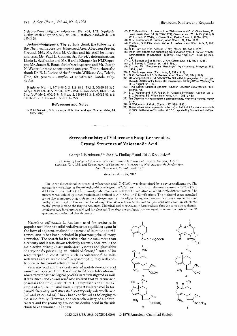
The three-dimensional structure of valerenolic acid, C1:,H&lr was determined by x-ray crystallography. The substance crystallizes in the orthorhombic space group P212121 and the unit-cell di,mensions are a = 12.705 ( 2 ) , b = 14.476 (3 ) . c = 15.477 (1) A. Intensity data were measured with Cu radiation on afour-circle diffractometer. The structure was solved by direct methods and refined to R = 3.6O/0 for 2543 reflections. The hydroxyl group attached to the five-membered ring is cis to the hydrogen atom at the adjacent ring junction, and both are trans to the axial methyl suhstituent on the six-membered ring. The latter is trans to the methacrylic acid side chain, in which the methyl group is cis to the ring carbon atom. Chemical and spectroscopic data indicate that the same stereochemis- try also occurs in valerenic acid and in valerenal. The absolute configuration was established on the basis of the CD spectrum of methyl 1-ketovalerenate.
Valeriana officinalis L. has been used for centuries in popular medicine as a mild sedative or tranquilizing agent in the form of aqueous or alcoholic extracts of its roots and rhi- zomes, and it has been included in pharmacopeias of many countries3 T h e search for its active principle took more than a century and it was shown relatively recently that, while the main active principles are undoubtedly esters and glucosides of terpenoids posses,sing an iridoid skeleton,4*5 some of its sesquiterpenoid constituents such as valeranone6 (a mild sedative) and valerenic acid' (a spasmolytic) may well con- t r ibute to the overall effect of the drug.
Valerenic acid and the closely related acetylvalerenolic acid were first isolated from the drug in Sandoz laboratories,' where their pharmacological profiles were investigated as well. I t was Buchi and co-workers8 who showed tha t valerenic acid possesses the unique structure 1. I t represents the first ex- ample of a quite unusual skeletal type 2 (valerenane) in ter- penoid chemistry, and since its discovery only valerenolic acid (3)9 and valerenal ( 4 ) ' O have been confirmed as belonging to the same family. However, the stereochemistry of all chiral centers and the geometry around the double bond in the side chain have remained unknown.
0022-3263/78/1943-0272$01.00/0
I 2
CH3
Q C H 3 CHO
3 4
5 6
0 1978 American Chemical Society